The New TEM facility at LCI, KSU. Min Gao Liquid Crystal Institute Kent State University
|
|
|
- Jewel Nash
- 6 years ago
- Views:
Transcription

1 The New TEM facility at LCI, KSU Min Gao Liquid Crystal Institute Kent State University 1


2 Questions to be answered How does TEM work? Why thin specimen? How to prepare one? What is the TEM at LCI like? Functions available and principles? How is the facility running so far? Any exciting results? How to use the TEM lab? 2
3 Facts about the TEM facility at LCI Instruments Specifications Applications 3

 Gatan imaging filter (incl.")
4 What we have at LCI FEI Tecnai F20ST-STEM Schottky field mission gun 40 kv-200 kv 0.24 nm point-to-point resolution ±70 tilting angle 0.2 nm spot size Attachments Low-dose mode & tomography EDAX energy dispersive x-ray spectrometer (EDS) Gatan imaging filter (incl. EELS) 4k*4K slow scan CCD Cryo-holder & anti-contaminator Assisting equipments Vitrobot Plasma cleaner Vacuum station 4

 1800 1840 1880 1920 1960 2000 2040 Year Borisevich, 2004 Batson et al.")
5 How good is this microscope? It would be very easy to make an analysis of any complicated chemical substance; all one would have to do would be to look at it and see where the atoms are. I put this out as a challenge: Is there no way to make the electron microscope more powerful? Richard P. Feynman, 1959, There s Plenty of Room at the Bottom 1 Corrected EM Haider (200keV) La in γ-al 2 O 3 in situ -1 Resolution (Ang. ) Electron Microscope Light Microscope Amici Ross Abbe Marton Ruska Dietrich (200keV) Year Borisevich, 2004 Batson et al. Nature 2002 Second best thing after corrected TEM It is the best thing considering Cost of TEM and maintenance Easy to use 85% applications do not need Cs corrector 5
6 General description of the TEM facility at LCI Things making this facility standing out: the optimum integration of some most advanced TEM techniques, for example: cryoem An application-oriented versatile microscope Excellent for materials science/life science/solid state physics/chemistry Current setup: nanomaterials/soft-materials requiring simple sample preparation A heavily computerized TEM with some bugseasy to operate 6


7 Philips and FEI microscopes EM420 CM200 7


8 I've gotta see some atoms, excuse me, --Obama Titan 8
9 How much a TEM cost? $4-5 per ev 200 kev: 1M$ + accessories (easily up to 0.5-1M) extremely stable high-voltage supplies, extremely stable currents to each electromagnetic coil/lens, continuously-pumped high- or ultra-high-vacuum systems, and a cooling water supply circulation through the lenses and pumps. As they are very sensitive to vibration and external magnetic fields, microscopes designed to achieve high resolutions must be housed in stable buildings (sometimes underground) with special services such as magnetic field cancelling systems. There must be some solid reasons that we need such an expensive machine! 9

10 How does TEM work? Importance of thin specimens Electron-material interaction Techniques and Information 10
11 Advantage of thin samples 50 nm Cu, 25 kv, 5 nm probe, 1000 electrons 50 nm Cu, 200 kv, 1 nm probe, 1000 electrons Thin specimen in SEM (Thin specimen in) TEM Where is my surface? Surface damage 11
12 What thin samples do you need? Represent the material you are studying: sometimes the thicker the better Electron transparent: dependent on the accelerating voltage, thickness of the specimen, and atomic number of the specimen. Uniformly thin Stable under the electron beam Conducing and nonmagnetic in the laboratory environment Generally speaking: nm for conventional TEM; 15 nm for high resolution TEM 12


13 How to put the thin samples into the TEM? The general form of a TEM specimen is a 3mm thin disk Supporting grids Put the thin disk into a holder which goes to the TEM sample stage 13


14 How to prepare a thin sample? Bulk materials: Agate Mortar and Pestle Bulk & thin films: Wheel Saw Polishing machine Dimpler Ion milling A lot of techniques have been developed to make TEM specimens 14


15 Focused ion beam: TEM samples Au+Pd coating first Deposition of Pt protection layer View from 52 Start of cutting Under cut

16 Other materials Nanomaterials: Biomaterials Very easy Quite difficult Dispersed on supporting carbon film Vitrobot Ultramicrotomy 16
17 Electron-thin specimen interaction Backscattered Electrons (Rutherford) Incident electron beam (E) Characteristic X- rays EDS Secondary electrons Auger electrons Cathodoluminescence Light E f E f E f E f E E e-h pair EBIC Specimen Large-angle Incoherent elastically scattered electrons (Rutherford) Z-contrast STEM Coherent elastically scattered electrons Diffraction Imaging HRTEM E- E Inelastically scattered electrons Other signals from interaction with many nuclei or electrons: Bremsstrahlung x-ray (EDS), Plasma excitation (EELS) EELS EFTEM 17
18 Instruments (TEM & STEM) Reimer, TEM,


19 Information from a modern TEM Gao et al. PRB, 62, 5413 (2000) In situ Gao et al. APL, 72, 2544 (1998) Gao et al. Unpublished Why TEM? CBED Gao et al. JAP, 80, 4767 (1996) EELS /EDS Gao et al. Z. Metallkd. 93, 438 (2002) O-K 1s 2p O Si Mo Mo/SiO x Gao et al. PRB, 62, 5413 (2000) Gao & Duan, Mater. Trans. JIM, 39, 883, 1998 Si 19
 Electron beam irradiation and contamination (diffraction imaging, low temperature, low dose, cryoem) Sample preparation (low temperature,")
20 Knowing the limitations of TEM Artifacts due to 2-D projections (3D TEM) Interpretation of images Poor Sampling: all TEMs have only examined <1 mm 3 of materials! (First use low resolution but better sampling tools) Electron beam irradiation and contamination (diffraction imaging, low temperature, low dose, cryoem) Sample preparation (low temperature, low-energy low angle ion milling, FIB-site specific, freeze-fracture, vitreous ethane ice) Vacuum environment (environmental EM, in vitro) Not so much about properties (in situ, combination) 20
21 Au+Si+O Si_L C_K O_K What have been done in the test running period at LCI? Service summary Available techniques Representative results 21
22 TEM Startup Holiday Cryo-trial Min in China Service hours FEI Cryo Training Number of samples Weekly service hours and number of samples Total service hours: 186 Cryo: 83 hrs Week Total number of samples: 113
23 Service hours vs groups Soumitra Basu Teaching Laurie Broadwater 6%1% 4% 20% Peter Palffy-Muhoray Songping Huang Mieteck Jaroniec 20% L.C. Chen 4% 8% 30% 4% 3% Quan Li Oleg Lavrentovich Tony Jakli

 Diffraction")
 Spectrometry")
24 Available TEM techniques Imaging (diffraction contract, high resolution, Z-contrast, energy-filtered) Diffraction (select-area, convergent beam, large angle CBED, nano-area) Spectrometry (EDS, EELS) CryoEM 3D tomography 24

25 Imaging (contrast) Mass and thickness contrast Diffraction contrast Phase contrast (e.g., HRTEM) Z-contrast 25
")


26 Imaging (LCI)


27 TEM bright field image and STEM Z-contrast (dark field) image Sample from Prof. Peter Palffy-Muhoray s group


28 HRTEM Sample from Prof. Peter Palffy- Muhoray s group

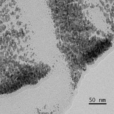
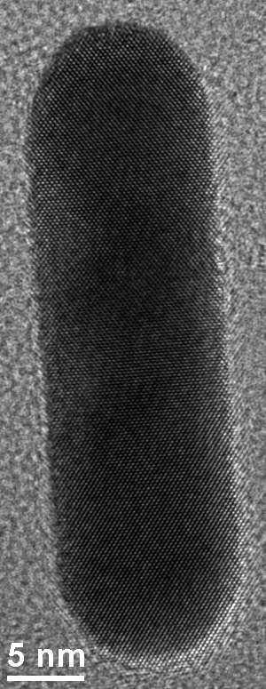
29 HRTEM of quantum dots Sample from Prof. LC Chien s group


30 Diffraction Sample from Dr. Quan Li s group 30

31 An nteresting sample C SiO x Au Sample from Prof. Peter Palffy-Muhoray s group


32 EDS spectral imaging Au O Si Au+Si+O Si_L C_K O_K Sample from Prof. Peter Palffy-Muhoray s group
 designed for bio and liquid A small droplet of liquid is applied to pre-treated carbon film Two filter papers are used to blot the liquid and leave a thin layer of liquid (e.g., 100 nm thick, adjustable blot force, time and repetition).")
33 Cryo-Vitrobot Controlled environment: temperature (4-60 C) and humidity (room cond. 100%) designed for bio and liquid A small droplet of liquid is applied to pre-treated carbon film Two filter papers are used to blot the liquid and leave a thin layer of liquid (e.g., 100 nm thick, adjustable blot force, time and repetition). Shoot the sample into coolant (ethane) and freeze it at very high speed.


34 First bio-sample Laurie Broadwater, Chemistry

35 A thermotropic sample Sample from Prof. LC Chien s group


36 A lyotropic sample Sample from Prof. Oleg Lavrentovich s group


37 Resolution beyond freeze fracture Very challenging: <10 e/nm 2 Sample from Prof. Jakli s group


38 Diffraction in Liquid Crystals Larger area Sample from Prof. Jakli s group
 during the cryoem")
39 3D tomography A video taken by Dr. Lee Pullan (FEI) during the cryoem training 39
40 Plan for the next step Soon be running regularly Rules and operation procedures Usage rates (machine use, staff service) and billing Self-user Training: Single operator MG + several self-users Right now: 1-on-1 based on research needs Later: a TEM course including lab training if allowed Web page of the facility 40
41 How to use the facility more efficiently Highly efficient, professional, yet friendly service The TEM will be aligned by the staff routinely, so you do not need to be an expert in TEM Know your sample as well as possible If you don t know TEM so well, have an in-depth discussion with the staff before scheduling your experiment Have a student with good experimental skills and a lot of patience Analyze your data immediately Introduce more outside users to keep the rates low Acknowledge the facility properly Pay your bill in time 41
42 Questions being answered How does TEM work? Why thin specimen? How to prepare one? What is the TEM at LCI like? Functions available and principles? How is the facility running so far? Any exciting results? How to use the TEM lab? 42
Chemical Analysis in TEM: XEDS, EELS and EFTEM. HRTEM PhD course Lecture 5
 Chemical Analysis in TEM: XEDS, EELS and EFTEM HRTEM PhD course Lecture 5 1 Part IV Subject Chapter Prio x-ray spectrometry 32 1 Spectra and mapping 33 2 Qualitative XEDS 34 1 Quantitative XEDS 35.1-35.4
Gaetano L Episcopo. Scanning Electron Microscopy Focus Ion Beam and. Pulsed Plasma Deposition
 Gaetano L Episcopo Scanning Electron Microscopy Focus Ion Beam and Pulsed Plasma Deposition Hystorical background Scientific discoveries 1897: J. Thomson discovers the electron. 1924: L. de Broglie propose
Chapter 9. Electron mean free path Microscopy principles of SEM, TEM, LEEM
 Chapter 9 Electron mean free path Microscopy principles of SEM, TEM, LEEM 9.1 Electron Mean Free Path 9. Scanning Electron Microscopy (SEM) -SEM design; Secondary electron imaging; Backscattered electron
Transmission Electron Microscopy
 L. Reimer H. Kohl Transmission Electron Microscopy Physics of Image Formation Fifth Edition el Springer Contents 1 Introduction... 1 1.1 Transmission Electron Microscopy... 1 1.1.1 Conventional Transmission
MT Electron microscopy Scanning electron microscopy and electron probe microanalysis
 MT-0.6026 Electron microscopy Scanning electron microscopy and electron probe microanalysis Eero Haimi Research Manager Outline 1. Introduction Basics of scanning electron microscopy (SEM) and electron
PHYS-E0541:Special Course in Physics Gas phase synthesis of carbon nanotubes for thin film application. Electron Microscopy. for
 PHYS-E0541:Special Course in Physics Gas phase synthesis of carbon nanotubes for thin film application Electron Microscopy for Introduction to Electron Microscopy Carbon Nanomaterials (nanotubes) Dr. Hua
ELECTRON MICROSCOPE UNIT
 ELECTRON MICROSCOPE Director: Prof Richard Tilley Associate Director: Dr Nicholas Ariotti Lab Manager: Ms Katie Levick Administrative Assistant: Mr Michael Zhi UNIT Basement Level: F10 Chemical Sciences
EPIC: Keck-II: SPID:
 The Northwestern University Atomic and Nanoscale Characterization Experimental Center (NUANCE) was established during 2001-02 to integrate complementary analytical instruments and characterization capabilities
HOW TO APPROACH SCANNING ELECTRON MICROSCOPY AND ENERGY DISPERSIVE SPECTROSCOPY ANALYSIS. SCSAM Short Course Amir Avishai
 HOW TO APPROACH SCANNING ELECTRON MICROSCOPY AND ENERGY DISPERSIVE SPECTROSCOPY ANALYSIS SCSAM Short Course Amir Avishai RESEARCH QUESTIONS Sea Shell Cast Iron EDS+SE Fe Cr C Objective Ability to ask the
In-Situ Combination of TOF-SIMS and EDS Analysis During FIB Sectioning
 In-Situ Combination of TOF-SIMS and EDS Analysis During FIB Sectioning V. Ray 1, E. Principe 2, T. Piper 2 vray@umd.edu 1. AIM Lab University of Maryland Nanocenter, College Park, MD 2. Tescan USA, Warrendale
MSE 321 Structural Characterization
 Auger Spectroscopy Auger Electron Spectroscopy (AES) Scanning Auger Microscopy (SAM) Incident Electron Ejected Electron Auger Electron Initial State Intermediate State Final State Physical Electronics
3D and Atomic-resolution Imaging with Coherent Electron Nanobeams - Opportunities and Challenges for X-rays
 3D and Atomic-resolution Imaging with Coherent Electron Nanobeams - Opportunities and Challenges for X-rays David A. Muller Lena Fitting Kourkoutis, Megan Holtz, Robert Hovden, Qingyun Mao, Julia Mundy,
User Fees for the 4D LABS Characterization Facility
 User Fees for the 4D LABS Characterization Facility 1. Imaging Fees Service SEM 1 - Helios: SEM/EDS mode SEM/EDS mode with FIB functions Cryo mode setup SEM 2 Nova NanoSEM SEM/EDS mode SEM/EDS mode with
= 6 (1/ nm) So what is probability of finding electron tunneled into a barrier 3 ev high?
 So what is probability of finding electron tunneled into a barrier 3 ev high?") STM STM With a scanning tunneling microscope, images of surfaces with atomic resolution can be readily obtained. An STM uses quantum tunneling of electrons to map the density of electrons on the surface
STM STM With a scanning tunneling microscope, images of surfaces with atomic resolution can be readily obtained. An STM uses quantum tunneling of electrons to map the density of electrons on the surface
AP5301/ Name the major parts of an optical microscope and state their functions.
 Review Problems on Optical Microscopy AP5301/8301-2015 1. Name the major parts of an optical microscope and state their functions. 2. Compare the focal lengths of two glass converging lenses, one with
CHEM 681 Seminar Mingqi Zhao April 20, 1998 Room 2104, 4:00 p.m. High Resolution Transmission Electron Microscopy: theories and applications
 CHEM 681 Seminar Mingqi Zhao April 20, 1998 Room 2104, 4:00 p.m. High Resolution Transmission Electron Microscopy: theories and applications In materials science, people are always interested in viewing
MSE 321 Structural Characterization
 Auger Spectroscopy Auger Electron Spectroscopy (AES) Scanning Auger Microscopy (SAM) Incident Electron Ejected Electron Auger Electron Initial State Intermediate State Final State Physical Electronics
Scanning Electron Microscopy
 Scanning Electron Microscopy Field emitting tip Grid 2kV 100kV Anode ZEISS SUPRA Variable Pressure FESEM Dr Heath Bagshaw CMA bagshawh@tcd.ie Why use an SEM? Fig 1. Examples of features resolvable using
Model : JEOL JSM 7610f
 Name Field Emission Electron Scanning Microscopy (FESEM) Make: JEOL India Pvt Ltd. Model : JEOL JSM 7610f Specification Electron gun : Schottky type field emission (T-FE) gun Electron Beam resolution (secondary
Dual Beam Helios Nanolab 600 and 650
 Dual Beam Helios Nanolab 600 and 650 In the Clean Room facilities of the INA LMA, several lithography facilities permit to pattern structures at the micro and nano meter scale and to create devices. In
Electron Microscopy I
 Characterization of Catalysts and Surfaces Characterization Techniques in Heterogeneous Catalysis Electron Microscopy I Introduction Properties of electrons Electron-matter interactions and their applications
Techniques EDX, EELS et HAADF en TEM: possibilités d analyse et applications
 Techniques EDX, EELS et HAADF en TEM: possibilités d analyse et applications Thomas Neisius Université Paul Cézanne Plan Imaging modes HAADF Example: supported Pt nanoparticles Electron sample interaction
Chapter 10. Nanometrology. Oxford University Press All rights reserved.
 Chapter 10 Nanometrology Oxford University Press 2013. All rights reserved. 1 Introduction Nanometrology is the science of measurement at the nanoscale level. Figure illustrates where nanoscale stands
2012/01/20. Thierry EPICIER. MATEIS-CLYM, umr CNRS5510, Director METSA (FR 3507 CNRS) M. AOUINE, JP MILLET et al.
 M. AOUINE, JP MILLET et al.") 2012/01/20 Thierry EPICIER MATEIS-CLYM, umr CNRS5510, Director METSA (FR 3507 CNRS) thierry.epicier@insa-lyon.fr M. AOUINE, JP MILLET et al. MATERIALS SCIENCES & NANOTECHNOLOGIES HIGH RESOLUTION MICROSCOPY
2012/01/20 Thierry EPICIER MATEIS-CLYM, umr CNRS5510, Director METSA (FR 3507 CNRS) thierry.epicier@insa-lyon.fr M. AOUINE, JP MILLET et al. MATERIALS SCIENCES & NANOTECHNOLOGIES HIGH RESOLUTION MICROSCOPY
CBE Science of Engineering Materials. Scanning Electron Microscopy (SEM)
") CBE 30361 Science of Engineering Materials Scanning Electron Microscopy (SEM) Scale of Structure Organization Units: micrometer = 10-6 m = 1µm nanometer= 10-9 m = 1nm Angstrom = 10-10 m = 1Å A hair is
CBE 30361 Science of Engineering Materials Scanning Electron Microscopy (SEM) Scale of Structure Organization Units: micrometer = 10-6 m = 1µm nanometer= 10-9 m = 1nm Angstrom = 10-10 m = 1Å A hair is
Elastic and Inelastic Scattering in Electron Diffraction and Imaging
 Elastic and Inelastic Scattering in Electron Diffraction and Imaging Contents Introduction Symbols and definitions Part A Diffraction and imaging of elastically scattered electrons Chapter 1. Basic kinematical
Everhart-Thornley detector
 SEI Detector Everhart-Thornley detector Microscope chamber wall Faraday cage Scintillator Electrons in Light pipe Photomultiplier Electrical signal out Screen Quartz window +200 V +10 kv Always contains
MSE 321 Structural Characterization
 Optical Microscope Plan Lenses In an "ideal" single-element lens system all planar wave fronts are focused to a point at distance f from the lens; therefore: Image near the optical axis will be in perfect
Chapter 2 Instrumentation for Analytical Electron Microscopy Lecture 5. Chapter 2 CHEM 793, 2011 Fall 1
 Chater Instrumentation for Analytical Electron Microscoy Lecture 5 Chater CHEM 793, 011 Fall 1 Outline Electron Sources (Electron Guns) Thermionic: LaB 6 or W Field emission gun: cold or Schottky Lenses
4. Inelastic Scattering
 1 4. Inelastic Scattering Some inelastic scattering processes A vast range of inelastic scattering processes can occur during illumination of a specimen with a highenergy electron beam. In principle, many
Chapter 12. Nanometrology. Oxford University Press All rights reserved.
 Chapter 12 Nanometrology Introduction Nanometrology is the science of measurement at the nanoscale level. Figure illustrates where nanoscale stands in relation to a meter and sub divisions of meter. Nanometrology
Nova 600 NanoLab Dual beam Focused Ion Beam IITKanpur
 Nova 600 NanoLab Dual beam Focused Ion Beam system @ IITKanpur Dual Beam Nova 600 Nano Lab From FEI company (Dual Beam = SEM + FIB) SEM: The Electron Beam for SEM Field Emission Electron Gun Energy : 500
Part II: Thin Film Characterization
 Part II: Thin Film Characterization General details of thin film characterization instruments 1. Introduction to Thin Film Characterization Techniques 2. Structural characterization: SEM, TEM, AFM, STM
Characterisation of Catalysts Using Secondary and Backscattered Electron In-lens Detectors
 Platinum Metals Rev., 2014, 58, (2), 106 110 FINAL ANALYSIS Characterisation of Catalysts Using Secondary and Backscattered Electron In-lens Detectors Heterogeneous catalysis often involves the use of
Praktikum zur. Materialanalytik
 Praktikum zur Materialanalytik Energy Dispersive X-ray Spectroscopy B513 Stand: 19.10.2016 Contents 1 Introduction... 2 2. Fundamental Physics and Notation... 3 2.1. Alignments of the microscope... 3 2.2.
Transmission Electron Microscopy. Part #2 High Resolution Imaging XEDS EELS spectroscopies Aberration corrected TEM
 Transmission Electron Microscopy Part #2 High Resolution Imaging XEDS EELS spectroscopies Aberration corrected TEM Nicolas Menguy Institut de Minéralogie, de Physique des Matériaux et de Cosmochimie Part
Electron-Matter Interactions
 Electron-Matter Interactions examples of typical EM studies properties of electrons elastic electron-matter interactions scattering processes; coherent and incoherent image formation; chemical contrast;
MEMS Metrology. Prof. Tianhong Cui ME 8254
 MEMS Metrology Prof. Tianhong Cui ME 8254 What is metrology? Metrology It is the science of weights and measures Refers primarily to the measurements of length, weight, time, etc. Mensuration- A branch
Combinatorial RF Magnetron Sputtering for Rapid Materials Discovery: Methodology and Applications
 Combinatorial RF Magnetron Sputtering for Rapid Materials Discovery: Methodology and Applications Philip D. Rack,, Jason D. Fowlkes,, and Yuepeng Deng Department of Materials Science and Engineering University
History of 3D Electron Microscopy and Helical Reconstruction
 T H E U N I V E R S I T Y of T E X A S S C H O O L O F H E A L T H I N F O R M A T I O N S C I E N C E S A T H O U S T O N History of 3D Electron Microscopy and Helical Reconstruction For students of HI
Ecole Franco-Roumaine : Magnétisme des systèmes nanoscopiques et structures hybrides - Brasov, Modern Analytical Microscopic Tools
 1. Introduction Solid Surfaces Analysis Group, Institute of Physics, Chemnitz University of Technology, Germany 2. Limitations of Conventional Optical Microscopy 3. Electron Microscopies Transmission Electron
Energy-Filtering. Transmission. Electron Microscopy
 Part 3 Energy-Filtering Transmission Electron Microscopy 92 Energy-Filtering TEM Principle of EFTEM expose specimen to mono-energetic electron radiation inelastic scattering in the specimen poly-energetic
Transmission Electron Microscopy
 Transmission Electron Microscopy Fu-Rong Chen Transmission Electron Microscopy David B. Williams C. Barry Carter Background:Solid State Physics Materials Science 1.1 Why Electron Microscope? 1.1 Why Electron
Instantaneous reduction of graphene oxide at room temperature
 Instantaneous reduction of graphene oxide at room temperature Barun Kuma Burman, Pitamber Mahanandia and Karuna Kar Nanda Materials Research Centre, Indian Institute of Science, Bangalore-560012, India
MS482 Materials Characterization ( 재료분석 ) Lecture Note 5: RBS
 Lecture Note 5: RBS") 2016 Fall Semester MS482 Materials Characterization ( 재료분석 ) Lecture Note 5: RBS Byungha Shin Dept. of MSE, KAIST 1 Course Information Syllabus 1. Overview of various characterization techniques (1 lecture)
2016 Fall Semester MS482 Materials Characterization ( 재료분석 ) Lecture Note 5: RBS Byungha Shin Dept. of MSE, KAIST 1 Course Information Syllabus 1. Overview of various characterization techniques (1 lecture)
The Basic of Transmission Electron Microscope. Text book: Transmission electron microscopy by David B Williams & C. Barry Carter.
 The Basic of Transmission Electron Microscope Text book: Transmission electron microscopy by David B Williams & C. Barry Carter. 2009, Springer Background survey http://presemo.aalto.fi/tem1 Microscopy
h p λ = mν Back to de Broglie and the electron as a wave you will learn more about this Equation in CHEM* 2060
 Back to de Broglie and the electron as a wave λ = mν h = h p you will learn more about this Equation in CHEM* 2060 We will soon see that the energies (speed for now if you like) of the electrons in the
SUPPLEMENTARY MATERIALS FOR PHONON TRANSMISSION COEFFICIENTS AT SOLID INTERFACES
 148 A p p e n d i x D SUPPLEMENTARY MATERIALS FOR PHONON TRANSMISSION COEFFICIENTS AT SOLID INTERFACES D.1 Overview The supplementary information contains additional information on our computational approach
Characterization of zeolites by advanced SEM/STEM techniques
 SCIENTIFIC INSTRUMENT NEWS 2016 Vol. 7 SEPTEMBER Technical magazine of Electron Microscope and Analytical Instruments. Article Characterization of zeolites by advanced SEM/STEM techniques Toshiyuki Yokoi
IMAGING DIFFRACTION SPECTROSCOPY
 TEM Techniques TEM/STEM IMAGING DIFFRACTION SPECTROSCOPY Amplitude contrast (diffracion contrast) Phase contrast (highresolution imaging) Selected area diffraction Energy dispersive X-ray spectroscopy
TESCAN S New generation of FIB-SEM microscope
 TESCAN S New generation of FIB-SEM microscope rising standards in sample preparation Key Features SEM COLUMN Versatile system for unlimited applications: resolution imaging (0.9 nm at 15 kev, 1.4 nm at
Electron Microprobe Analysis and Scanning Electron Microscopy
 Electron Microprobe Analysis and Scanning Electron Microscopy Electron microprobe analysis (EMPA) Analytical technique in which a beam of electrons is focused on a sample surface, producing X-rays from
Methods of surface analysis
 Methods of surface analysis Nanomaterials characterisation I RNDr. Věra Vodičková, PhD. Surface of solid matter: last monoatomic layer + absorbed monolayer physical properties are effected (crystal lattice
Analysis of Cadmium (Cd) in Plastic Using X-ray Fluorescence Spectroscopy
 in Plastic Using X-ray Fluorescence Spectroscopy") Analysis of Cadmium (Cd) in Plastic Using X-ray Fluorescence Spectroscopy Hiroshi Onodera Application & Research Center, JEOL Ltd. Introduction um, PBB and PBDE) are subject to usage restrictions in Europe.
Analysis of Cadmium (Cd) in Plastic Using X-ray Fluorescence Spectroscopy Hiroshi Onodera Application & Research Center, JEOL Ltd. Introduction um, PBB and PBDE) are subject to usage restrictions in Europe.
April 10th-12th, 2017
 Thomas LaGrange, Ph.D. Faculty Lecturer and Senior Staff Scientist Introduction: Basics of Transmission Electron Microscopy (TEM) TEM Doctoral Course MS-637 April 10th-12th, 2017 Outline 1. What is microcopy?
CHARACTERIZATION of NANOMATERIALS KHP
 CHARACTERIZATION of NANOMATERIALS Overview of the most common nanocharacterization techniques MAIN CHARACTERIZATION TECHNIQUES: 1.Transmission Electron Microscope (TEM) 2. Scanning Electron Microscope
CHEM-E5225 :Electron Microscopy X-Ray Spectrometry
 CHEM-E5225 :Electron Microscopy X-Ray Spectrometry 2016.11 Yanling Ge Outline X-ray Spectrometry X-ray Spectra and Images Qualitative and Quantitative X-ray Analysis and Imaging Discussion of homework
SEM Doctoral Course MS-636. April 11-13, 2016
 Thomas LaGrange, Ph.D. Faculty Lecturer and Senior Staff Scientist Electron Sources, Optics and Detectors SEM Doctoral Course MS-636 April 11-13, 2016 Summary Electron propagation is only possible through
Preamble: Emphasis: Material = Device? MTSE 719 PHYSICAL PRINCIPLES OF CHARACTERIZATION OF SOLIDS
 MTSE 719 PHYSICAL PRINCIPLES OF CHARACTERIZATION OF SOLIDS MTSE 719 - PHYSCL PRIN CHARACTIZTN SOLIDS Section # Call # Days / Times 001 96175 -View Book Info - F:100PM - 355PM - TIER114 Preamble: Core course
M2 TP. Low-Energy Electron Diffraction (LEED)
") M2 TP Low-Energy Electron Diffraction (LEED) Guide for report preparation I. Introduction: Elastic scattering or diffraction of electrons is the standard technique in surface science for obtaining structural
M2 TP Low-Energy Electron Diffraction (LEED) Guide for report preparation I. Introduction: Elastic scattering or diffraction of electrons is the standard technique in surface science for obtaining structural
Surface Analysis. Dr. Lynn Fuller Dr. Fuller s Webpage:
 ROCHESTER INSTITUTE OF TECHNOLOGY MICROELECTRONIC ENGINEERING Surface Analysis Dr. Lynn Fuller Dr. Fuller s Webpage: http://people.rit.edu/lffeee 82 Lomb Memorial Drive Rochester, NY 14623-5604 Tel (585)
Supporting Information
 Supporting Information High Performance Electrocatalyst: Pt-Cu Hollow Nanocrystals Xiaofei Yu, a Dingsheng, a Qing Peng a and Yadong Li* a a Department of Chemistry, Tsinghua University, Beijing, 100084
ABNORMAL X-RAY EMISSION FROM INSULATORS BOMBARDED WITH LOW ENERGY IONS
 302 ABNORMAL X-RAY EMISSION FROM INSULATORS BOMBARDED WITH LOW ENERGY IONS M. Song 1, K. Mitsuishi 1, M. Takeguchi 1, K. Furuya 1, R. C. Birtcher 2 1 High Voltage Electron Microscopy Station, National
Nanoscale voxel spectroscopy by simultaneous EELS and EDS tomography
 Electronic Supplementary Material (ESI) for Nanoscale. This journal is The Royal Society of Chemistry 2014 Supplementary Information Nanoscale voxel spectroscopy by simultaneous EELS and EDS tomography
Weak-Beam Dark-Field Technique
 Basic Idea recall bright-field contrast of dislocations: specimen close to Bragg condition, s î 0 Weak-Beam Dark-Field Technique near the dislocation core, some planes curved to s = 0 ) strong Bragg reflection
ECE Semiconductor Device and Material Characterization
 ECE 4813 Semiconductor Device and Material Characterization Dr. Alan Doolittle School of Electrical and Computer Engineering Georgia Institute of Technology As with all of these lecture slides, I am indebted
Cryo-TEM. Subtitle. Fanny Caputo François Saint-Antonin Dora Mehn Matthias Roesslein
 Project: Cryo-TEM Subtitle AUTHORED BY: DATE: Fanny Caputo 18-01-2016 REVIEWED BY: DATE: François Saint-Antonin 20-01-2016 Dora Mehn 21-01-2016 Matthias Roesslein 21-01-2016 APPROVED BY: DATE: Matthias
Final exam: take-home part
 Final exam: take-home part! List five things that can be done to improve this class. Be specific; give much detail.! (You will be penalized only for insulting comments made for no benefit; you will not
Introduction to EDX. Energy Dispersive X-ray Microanalysis (EDS, Energy dispersive Spectroscopy) Basics of EDX
 Basics of EDX") Introduction to EDX Energy Dispersive X-ray Microanalysis (EDS, Energy dispersive Spectroscopy) EDX Marco Cantoni 1 Basics of EDX a) Generation of X-rays b) Detection Si(Li) Detector, SDD Detector, EDS
Introduction to EDX Energy Dispersive X-ray Microanalysis (EDS, Energy dispersive Spectroscopy) EDX Marco Cantoni 1 Basics of EDX a) Generation of X-rays b) Detection Si(Li) Detector, SDD Detector, EDS
Modern Optical Spectroscopy
 Modern Optical Spectroscopy X-Ray Microanalysis Shu-Ping Lin, Ph.D. Institute of Biomedical Engineering E-mail: splin@dragon.nchu.edu.tw Website: http://web.nchu.edu.tw/pweb/users/splin/ Backscattered
Electron Microprobe Analysis 1 Nilanjan Chatterjee, Ph.D. Principal Research Scientist
 12.141 Electron Microprobe Analysis 1 Nilanjan Chatterjee, Ph.D. Principal Research Scientist Massachusetts Institute of Technology Electron Microprobe Facility Department of Earth, Atmospheric and Planetary
MS482 Materials Characterization ( 재료분석 ) Lecture Note 5: RBS. Byungha Shin Dept. of MSE, KAIST
 Lecture Note 5: RBS. Byungha Shin Dept. of MSE, KAIST") 2015 Fall Semester MS482 Materials Characterization ( 재료분석 ) Lecture Note 5: RBS Byungha Shin Dept. of MSE, KAIST 1 Course Information Syllabus 1. Overview of various characterization techniques (1 lecture)
2015 Fall Semester MS482 Materials Characterization ( 재료분석 ) Lecture Note 5: RBS Byungha Shin Dept. of MSE, KAIST 1 Course Information Syllabus 1. Overview of various characterization techniques (1 lecture)
Electron Microprobe Analysis 1 Nilanjan Chatterjee, Ph.D. Principal Research Scientist
 12.141 Electron Microprobe Analysis 1 Nilanjan Chatterjee, Ph.D. Principal Research Scientist Massachusetts Institute of Technology Electron Microprobe Facility Department of Earth, Atmospheric and Planetary
Electron Rutherford Backscattering, a versatile tool for the study of thin films
 Electron Rutherford Backscattering, a versatile tool for the study of thin films Maarten Vos Research School of Physics and Engineering Australian National University Canberra Australia Acknowledgements:
MT Electron microscopy Scanning electron microscopy and electron probe microanalysis
 MT-0.6026 Electron microscopy Scanning electron microscopy and electron probe microanalysis Eero Haimi Research Manager Outline 1. Introduction Basics of scanning electron microscopy (SEM) and electron
Auger Electron Spectroscopy Overview
 Auger Electron Spectroscopy Overview Also known as: AES, Auger, SAM 1 Auger Electron Spectroscopy E KLL = E K - E L - E L AES Spectra of Cu EdN(E)/dE Auger Electron E N(E) x 5 E KLL Cu MNN Cu LMM E f E
Massachusetts Institute of Technology. Dr. Nilanjan Chatterjee
 Massachusetts Institute of Technology Dr. Nilanjan Chatterjee Electron Probe Micro-Analysis (EPMA) Imaging and micrometer-scale chemical compositional analysis of solids Signals produced in The Electron
Electron and electromagnetic radiation
 Electron and electromagnetic radiation Generation and interactions with matter Stimuli Interaction with sample Response Stimuli Waves and energy The energy is propotional to 1/λ and 1/λ 2 λ λ 1 Electromagnetic
Analytical Methods for Materials
 Analytical Methods for Materials Lesson 21 Electron Microscopy and X-ray Spectroscopy Suggested Reading Leng, Chapter 3, pp. 83-126; Chapter 4, pp. 127-160; Chapter 6, pp. 191-219 P.J. Goodhew, J. Humphreys
How to Use the Devices in the Analytical Instrument Laboratory
 How to Use the Devices in the Analytical Instrument Laboratory 1. Eligible users To be eligible, the user must be a member of the faculty or staff, a researcher, or a Waseda University student. If the
Scanning Electron Microscopy & Ancillary Techniques
 Scanning Electron Microscopy & Ancillary Techniques By Pablo G. Caceres-Valencia The prototype of the first Stereoscan supplied by the Cambridge Instrument Company to the dupont Company, U.S.A. (1965)
Supporting Information s for
 Supporting Information s for # Self-assembling of DNA-templated Au Nanoparticles into Nanowires and their enhanced SERS and Catalytic Applications Subrata Kundu* and M. Jayachandran Electrochemical Materials
Nanoelectronics 09. Atsufumi Hirohata Department of Electronics. Quick Review over the Last Lecture
 Nanoelectronics 09 Atsufumi Hirohata Department of Electronics 13:00 Monday, 12/February/2018 (P/T 006) Quick Review over the Last Lecture ( Field effect transistor (FET) ): ( Drain ) current increases
XPS & Scanning Auger Principles & Examples
 XPS & Scanning Auger Principles & Examples Shared Research Facilities Lunch Talk Contact info: dhu Pujari & Han Zuilhof Lab of rganic Chemistry Wageningen University E-mail: dharam.pujari@wur.nl Han.Zuilhof@wur.nl
Auger Electron Spectroscopy
 Auger Electron Spectroscopy Auger Electron Spectroscopy is an analytical technique that provides compositional information on the top few monolayers of material. Detect all elements above He Detection
Basic structure of SEM
 Table of contents Basis structure of SEM SEM imaging modes Comparison of ordinary SEM and FESEM Electron behavior Electron matter interaction o Elastic interaction o Inelastic interaction o Interaction
Depth Distribution Functions of Secondary Electron Production and Emission
 Depth Distribution Functions of Secondary Electron Production and Emission Z.J. Ding*, Y.G. Li, R.G. Zeng, S.F. Mao, P. Zhang and Z.M. Zhang Hefei National Laboratory for Physical Sciences at Microscale
Photoemission Spectroscopy
 FY13 Experimental Physics - Auger Electron Spectroscopy Photoemission Spectroscopy Supervisor: Per Morgen SDU, Institute of Physics Campusvej 55 DK - 5250 Odense S Ulrik Robenhagen,
SOLID STATE PHYSICS PHY F341. Dr. Manjuladevi.V Associate Professor Department of Physics BITS Pilani
 SOLID STATE PHYSICS PHY F341 Dr. Manjuladevi.V Associate Professor Department of Physics BITS Pilani 333031 manjula@bits-pilani.ac.in Characterization techniques SEM AFM STM BAM Outline What can we use
stands for Transmission Electron (Microscope/Microscopy) Q: Why use electrons instead of light for imaging nanomaterials?
 Q: Why use electrons instead of light for imaging nanomaterials?") What is TEM? stands for Transmission Electron (Microscope/Microscopy) Q: Why use electrons instead of light for imaging nanomaterials? A: 1) Shorter wavelength () Higher resolution ) Wavelength determined
What is TEM? stands for Transmission Electron (Microscope/Microscopy) Q: Why use electrons instead of light for imaging nanomaterials? A: 1) Shorter wavelength () Higher resolution ) Wavelength determined
Electron Microscopy. SEM = Scanning Electron Microscopy TEM = Transmission Electron Microscopy. E. coli, William E. Bentley, Maryland, USA
 Electron Microscopy Transmission Electron Microscopy SEM = Scanning Electron Microscopy TEM = Transmission Electron Microscopy Sara Henriksson, UCEM 2017-02-16 E. coli, William E. Bentley, Maryland, USA
Practical course in scanning electron microscopy
 Practical course in scanning electron microscopy Fortgeschrittenen Praktikum an der Technischen Universität München Wintersemester 2017/2018 Table of contents 1. Introduction 3 2. Formation of an electron
Imaging Methods: Scanning Force Microscopy (SFM / AFM)
") Imaging Methods: Scanning Force Microscopy (SFM / AFM) The atomic force microscope (AFM) probes the surface of a sample with a sharp tip, a couple of microns long and often less than 100 Å in diameter.
Imaging Methods: Scanning Force Microscopy (SFM / AFM) The atomic force microscope (AFM) probes the surface of a sample with a sharp tip, a couple of microns long and often less than 100 Å in diameter.
SCANNING ELECTRON MICROSCOPE
 21.05.2010 Hacettepe University SCANNING ELECTRON MICROSCOPE Berrak BOYBEK Tuğba ÖZTÜRK Vicdan PINARBAŞI Cahit YAYAN OUTLINE Definition of scanning electron microscope History Applications of SEM Components
Microscopy: Principles
 Low Voltage Electron Microscopy: Principles and Applications Edited by David C. Bell Harvard University, USA and Natasha Erdman JEOL USA Inc., USA Published in association with the Royal Microscopical
Aberration-corrected TEM studies on interface of multilayered-perovskite systems
 Aberration-corrected TEM studies on interface of multilayered-perovskite systems By Lina Gunawan (0326114) Supervisor: Dr. Gianluigi Botton November 1, 2006 MSE 702(1) Presentation Outline Literature Review
EE 527 MICROFABRICATION. Lecture 5 Tai-Chang Chen University of Washington
 EE 527 MICROFABRICATION Lecture 5 Tai-Chang Chen University of Washington MICROSCOPY AND VISUALIZATION Electron microscope, transmission electron microscope Resolution: atomic imaging Use: lattice spacing.
Supporting Information
 Supporting Information Identification of the nearby hydroxyls role in promoting HCHO oxidation over a Pt catalyst Ying Huo #, Xuyu Wang #, Zebao Rui *, Xiaoqing Yang, Hongbing Ji * School of Chemical Engineering
sin" =1.22 # D "l =1.22 f# D I: In introduction to molecular electron microscopy - Imaging macromolecular assemblies
 I: In introduction to molecular electron microscopy - Imaging macromolecular assemblies Yifan Cheng Department of Biochemistry & Biophysics office: GH-S472D; email: ycheng@ucsf.edu 2/20/2015 - Introduction
Chapter 37 Early Quantum Theory and Models of the Atom
 Chapter 37 Early Quantum Theory and Models of the Atom Units of Chapter 37 37-7 Wave Nature of Matter 37-8 Electron Microscopes 37-9 Early Models of the Atom 37-10 Atomic Spectra: Key to the Structure