Transmission Electron Microscopy
|
|
|
- Brett Griffin
- 5 years ago
- Views:
Transcription
1 Transmission Electron Microscopy Fu-Rong Chen Transmission Electron Microscopy David B. Williams C. Barry Carter Background:Solid State Physics Materials Science
2 1.1 Why Electron Microscope?
3 1.1 Why Electron Microscope? Optical Microscope Electron Microscope source visible light electron lens glass electro-magnetic lens


4 Optical Microscope Electron Microscope visible light glass lens magnetic lens camera Liquid TEM image
5 Optical Microscope Electron Microscope visible light glass lens magnetic lens 衣藻 camera Liquid TEM image Chlamydomonas Reinhardtii

6 Ernst Ruska Nobel Prize in Physics 1986

7 Ernst Ruska Nobel Prize in Physics 1986

8 Better Electron Microscope... If we can see where the atoms are,then the analysis of chemical substance become very easy... The most difficulty is the power of electron microscope must be improved 100 times.. -Richard F. Feynman American Physics Society, CIT
9 A. Resolution NTHU
10 A. Resolution NTHU
11 A. Resolution NTHU visible light : λ~ 6000Å Rayleigh resolution (1.1) δ:rayleigh resolution λ: wavelength µ: refractive index β: semi-angle of lens µsinβ:numerical Aperature~1
12 A. Resolution NTHU visible light : λ~ 6000Å Rayleigh resolution (1.1) δ:rayleigh resolution λ: wavelength µ: refractive index β: semi-angle of lens µsinβ:numerical Aperature~1 resolution of optical microscope is about half of wavelength ~3000Å(1000 atoms)
 δ:rayleigh resolution λ: wavelength µ: refractive index β: semi-angle of lens µsinβ:numerical Aperature~1 resolution of optical microscope is about half of")
13 A. Resolution visible light : λ~ 6000Å Rayleigh resolution NTHU A the resolution of an optical microscope is limited by the wavelength (1.1) δ:rayleigh resolution λ: wavelength µ: refractive index β: semi-angle of lens µsinβ:numerical Aperature~1 resolution of optical microscope is about half of wavelength ~3000Å(1000 atoms)
 λ(å) 100 0.037 200 0.025 300 0.0196 400 0.")
14 Particle/ Wave Duality NTHU de Broglie s matter/ wave theory E=1/2 m o υ 2 Louis de Broglie Nobel laureate in 1929 E (kev) λ(å)
15 Evolution of resolution in EM
16 High and Median Voltage TEM 200keV

17 High and Median Voltage TEM 200keV 1MeV
18 B. MIcroanalysis,(X-Ray),(EELS)
19 B. MIcroanalysis,(X-Ray),(EELS) Imaging
20 B. MIcroanalysis,(X-Ray),(EELS) SEM SEM Imaging
21 B. MIcroanalysis,(X-Ray),(EELS) SEM SEM EDX EDX Imaging
,(EELS)")
22 B. MIcroanalysis,(X-Ray),(EELS) SEM SEM EDX EDX EELS Imaging
23 Typical TEM+EDX+EELS projector lenses EDX EELS
24 Typical TEM+EDX+EELS
25 Typical TEM+EDX+EELS
26 Typical TEM+EDX+EELS e-gun
27 Typical TEM+EDX+EELS e-gun condenser lenses
28 Typical TEM+EDX+EELS e-gun condenser lenses objective lenses
29 Typical TEM+EDX+EELS e-gun condenser lenses objective lenses projector lenses


30 Typical TEM+EDX+EELS e-gun condenser lenses objective lenses projector lenses
31 Typical TEM+EDX+EELS
32 Typical TEM+EDX+EELS

33 Typical TEM+EDX+EELS


34 Typical TEM+EDX+EELS
 Ψ (γ) Image (real")
35 C. Diffraction No lens for X-ray: Only diffraction can be seen Electron microscope utilizes magnetic lens to see both image and difffraction incident electron sample C s Δf 0 g g' g" Diffraction (reciprocal space) Ψ (γ) Image (real space)


36 Imaging, Spectroscopy and Diffraction 1.4Å Atomic Resolution
37 Imaging, Spectroscopy and Diffraction
")
38 Imaging, Spectroscopy and Diffraction Atomic resolution of EELS of La0.7Sr0.3MnO3/SrTiO3 multilayer - D. A. Muller, et al, SCIENCE (2008)

39 Imaging, Spectroscopy and Diffraction La M edge Ti L edge Mn L edge Atomic resolution compositional and bonding maps
40 Imaging, Spectroscopy and Diffraction Atomic-resolution phase recovery with dynamic support MgO 10 nm
41 Imaging, Spectroscopy and Diffraction Atomic-resolution phase recovery with dynamic support Electron Diffrac.ve Tomography MgO 10 nm
42 Imaging, Spectroscopy and Diffraction Atomic-resolution phase recovery with dynamic support Electron Diffrac.ve Tomography reconstructed wave MgO 10 nm
43 Imaging, Spectroscopy and Diffraction Atomic-resolution phase recovery with dynamic support Electron Diffrac.ve Tomography reconstructed wave MgO 10 nm


44 Imaging, Spectroscopy and Diffraction Atomic-resolution phase recovery with dynamic support Electron Diffrac.ve Tomography reconstructed wave MgO 10 nm reconstructed phase 0.21 nm
45 1.2 limitation of TEM A 2D projection: Averaged from the thickn sample > tomography B Radiation Damage--polymer, bio-sample and ceramics C thin sample is very difficult to be made t <50nm ~ 100nm D only dry sample can be observed E. NO time resolved capability
46 1.2 limitation of TEM A 2D projection: Averaged from the thickn sample > tomography B Radiation Damage--polymer, bio-sample and ceramics C thin sample is very difficult to be made t <50nm ~ 100nm D only dry sample can be observed E. NO time resolved capability The Future of TEM: 3D atomic resolution in wet environment with time resolved
47 Why 3D?
48 Why 3D?
49 Why 3D?


50 Why 3D?
51 Atomic Resolution Tomography Graphene 0.14nm
52 Atomic Resolution Tomography Graphene 0.14nm


53 Atomic Resolution Tomography Graphene 0.14nm Big-Bang Tomography (NATURE, June 2012)

54 A1HJP Protein on Graphene


55 A1HJP Protein on Graphene


56 Ferritin on Carbon Film Cd S O C
57 Self Aligned Wet (SAW) Cell
58 Self Aligned Wet (SAW) Cell


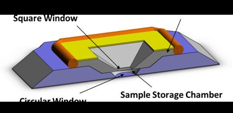
59 Self Aligned Wet (SAW) Cell
60 Self Aligned Wet (SAW) Cell Self-Aligned Wet-Cell for Hydrated Microbiology Observation in TEM, Lab on a Chip, 12(2), pp , 2012.
 Aurous acid H2O")
61 dissolution of Au nano-particles Au + e - Au(I) Aurous acid H2O unstable
62 Dynamics of NW Growth from GNPs
63 Dynamics of NW Growth from GNPs Au + e - H2O stable Au(III) auric acid


64 Dynamics of NW Growth from GNPs 15sec 30 sec 45 sec 60 sec
65 Magic Bullet: Targeting Drug ~ proposed by Nobel Prize winner (1908) Paul Ehrlich 50~200 nm Fe2O3 Easy Storage High Drug Content Stimuli Responsive Large Surface Area Modification Superparamagnetic iron oxide (SPIO)
66 Magic Bullet: Targeting Drug ~ proposed by Nobel Prize winner (1908) Paul Ehrlich Chitosan 50~200 nm Fe2O3 Easy Storage High Drug Content Stimuli Responsive Large Surface Area Modification Superparamagnetic iron oxide (SPIO)
67 Magic Bullet: Targeting Drug ~ proposed by Nobel Prize winner (1908) Paul Ehrlich Chitosan 50~200 nm Fe2O3 Easy Storage High Drug Content Stimuli Responsive Large Surface Area Modification Superparamagnetic iron oxide (SPIO)

68 Magic Bullet: Targeting Drug ~ proposed by Nobel Prize winner (1908) Paul Ehrlich Chitosan-SPIO: Targeting drug for MRI contrast agent Chitosan 50~200 nm Fe2O3 Easy Storage High Drug Content Stimuli Responsive Large Surface Area Modification Superparamagnetic iron oxide (SPIO)
69 Mechanisms for core-(single crystal shells) nanospheres under HFMF Single crsytal Poly-crsytal Rupture


70 Mechanisms for core-(single crystal shells) nanospheres under HFMF Single crsytal Poly-crsytal Rupture

71 27 Reduction of Fe2O3 NPs


72 27 Reduction of Fe2O3 NPs 10 sec 20 sec 30 sec 40 sec 50 sec 60 sec 70 sec 80 sec


73 Nano-drug Tumor Targeting MRI contrast agent Endocytosis Phagocytosis
74 Nano- Bubbles for Cancer Therapy Protein Liquid Au NPs in Protein Liquid P=2γ/R 29
75 Nano- Bubbles for Cancer Therapy Protein Liquid Au NPs in Protein Liquid P=2γ/R H 2 Knudsen gas (classical ideal gas) Bose gas (quantum gas: Bose Einstein condensate 29
76 Nano- Bubbles for Cancer Therapy Protein Liquid Au NPs in Protein Liquid P=2γ/R H 2 Knudsen gas (classical ideal gas) Bose gas (quantum gas: Bose Einstein condensate 29
77 Nano- Bubbles for Cancer Therapy Au NPs with anrbody cancer cell Protein Liquid Au NPs in Protein Liquid P=2γ/R H 2 Knudsen gas (classical ideal gas) Bose gas (quantum gas: Bose Einstein condensate 29
78 Nano- Bubbles for Cancer Therapy Au NPs with anrbody cancer cell Protein Liquid Au NPs in Protein Liquid P=2γ/R H 2 Knudsen gas (classical ideal gas) Bose gas (quantum gas: Bose Einstein condensate 29
79 Nano- Bubbles for Cancer Therapy Au NPs with anrbody cancer cell Protein Liquid Au NPs in Protein Liquid P=2γ/R H 2 Knudsen gas (classical ideal gas) Bose gas (quantum gas: Bose Einstein condensate 29
80 Nano- Bubbles for Cancer Therapy Au NPs with anrbody cancer drug cancer cell Protein Liquid Au NPs in Protein Liquid P=2γ/R H 2 Knudsen gas (classical ideal gas) Bose gas (quantum gas: Bose Einstein condensate 29
81 Nano- Bubbles for Cancer Therapy Au NPs with anrbody cancer drug cancer cell Laser Protein Liquid Au NPs in Protein Liquid P=2γ/R H 2 Knudsen gas (classical ideal gas) Bose gas (quantum gas: Bose Einstein condensate 29
82 Nano- Bubbles for Cancer Therapy Au NPs with anrbody cancer drug cancer cell Laser Protein Liquid P=2γ/R Au NPs in Protein Liquid Plasmonic Nanobubble H 2 Knudsen gas (classical ideal gas) Bose gas (quantum gas: Bose Einstein condensate 29
83 Nano- Bubbles for Cancer Therapy Au NPs with anrbody cancer drug cancer cell Protein Liquid P=2γ/R Au NPs in Protein Liquid Plasmonic Nanobubble H 2 Knudsen gas (classical ideal gas) Bose gas (quantum gas: Bose Einstein condensate 29
84 Nano- Bubbles for Cancer Therapy Au NPs with anrbody cancer drug cancer cell Protein Liquid Au NPs in Protein Liquid P=2γ/R H 2 Knudsen gas (classical ideal gas) Bose gas (quantum gas: Bose Einstein condensate 29
85 Nano- Bubbles for Cancer Therapy Au NPs with anrbody cancer drug cancer cell Protein Liquid Au NPs in Protein Liquid P=2γ/R H 2 Knudsen gas (classical ideal gas) Bose gas (quantum gas: Bose Einstein condensate 29


")


86 Nano- Bubbles for Cancer Therapy Au NPs with anrbody cancer drug cancer cell Protein Liquid Au NPs in Protein Liquid P=2γ/R CSPIO (or PLGA+Fe 3 O 4 ) upuptake kinetics and 4hrs 24hrs structural evolution48hrs 29
87 Ultra-fast Electron Microscope High Speed Camera 30
88 Ultra-fast Electron Microscope High Speed Camera 30


89 Ultra-fast Electron Microscope High Speed Camera A falling apple photographed by stroboscopic illumination at intervals of 1/25 s. The acceleration due to gravity is clear. 30


 Laser (probe)")
90 Time-resolved TEM was developed at TU-Berlin beginning in the late 1970's Oleg Bostanjoglo photocathode H. Dömer and O. Bostanjoglo, Rev. Sci. Inst. 74, 4369 (2003) Laser (probe) Laser (pump) sample


91 Time-resolved TEM ablation of Ni film by ultrashort laser pulse 2.5 ns 5 ns 10 ns 20 ns 40 ns + H. Domer and O. Bostanjoglo, Journal of Applied Physics 91, (2002).
92 Time-resolved electron diffraction α-ti (hcp)---> β-ti(bcc) martensiic transformation Bryan W. Reed, NLLB, UC, 2008
93 Time-resolved electron diffraction α-ti (hcp)---> β-ti(bcc) martensiic transformation Melting transition hcp Ti, α-phase Melt structure Bryan W. Reed, NLLB, UC, 2008
--->")


94 Time-resolved electron diffraction α-ti (hcp)---> β-ti(bcc) martensiic transformation Melting transition hcp Ti, α-phase Melt structure Phase transformation (α to β) Bryan W. Reed, NLLB, UC, 2008 hcp Ti, α-phase bcc Ti, β-phase
95 The END
AP5301/ Name the major parts of an optical microscope and state their functions.
 Review Problems on Optical Microscopy AP5301/8301-2015 1. Name the major parts of an optical microscope and state their functions. 2. Compare the focal lengths of two glass converging lenses, one with
CHEM 681 Seminar Mingqi Zhao April 20, 1998 Room 2104, 4:00 p.m. High Resolution Transmission Electron Microscopy: theories and applications
 CHEM 681 Seminar Mingqi Zhao April 20, 1998 Room 2104, 4:00 p.m. High Resolution Transmission Electron Microscopy: theories and applications In materials science, people are always interested in viewing
Chapter 10: Wave Properties of Particles
 Chapter 10: Wave Properties of Particles Particles such as electrons may demonstrate wave properties under certain conditions. The electron microscope uses these properties to produce magnified images
tip conducting surface
 PhysicsAndMathsTutor.com 1 1. The diagram shows the tip of a scanning tunnelling microscope (STM) above a conducting surface. The tip is at a potential of 1.0 V relative to the surface. If the tip is sufficiently
Scanning Tunneling Microscopy Transmission Electron Microscopy
 Scanning Tunneling Microscopy Transmission Electron Microscopy Speakers Burcu Başar Semih Gezgin Yavuz Selim Telis Place Hacettepe University Department of Chemical Engineering It s a small world after
Single-shot Ultrafast Electron Microscopy
 Single-shot Ultrafast Electron Microscopy Renkai Li and Pietro Musumeci Department of Physics and Astronomy, UCLA 25 th North American Particle Accelerator Conference Sep 30 - Oct 4, 2013, Pasadena, CA,
CHARACTERIZATION of NANOMATERIALS KHP
 CHARACTERIZATION of NANOMATERIALS Overview of the most common nanocharacterization techniques MAIN CHARACTERIZATION TECHNIQUES: 1.Transmission Electron Microscope (TEM) 2. Scanning Electron Microscope
Transmission Electron Microscopy
 L. Reimer H. Kohl Transmission Electron Microscopy Physics of Image Formation Fifth Edition el Springer Contents 1 Introduction... 1 1.1 Transmission Electron Microscopy... 1 1.1.1 Conventional Transmission
Ecole Franco-Roumaine : Magnétisme des systèmes nanoscopiques et structures hybrides - Brasov, Modern Analytical Microscopic Tools
 1. Introduction Solid Surfaces Analysis Group, Institute of Physics, Chemnitz University of Technology, Germany 2. Limitations of Conventional Optical Microscopy 3. Electron Microscopies Transmission Electron
X-Ray Photoelectron Spectroscopy (XPS) Prof. Paul K. Chu
 Prof. Paul K. Chu") X-Ray Photoelectron Spectroscopy (XPS) Prof. Paul K. Chu X-ray Photoelectron Spectroscopy Introduction Qualitative analysis Quantitative analysis Charging compensation Small area analysis and XPS imaging
X-Ray Photoelectron Spectroscopy (XPS) Prof. Paul K. Chu X-ray Photoelectron Spectroscopy Introduction Qualitative analysis Quantitative analysis Charging compensation Small area analysis and XPS imaging
3D and Atomic-resolution Imaging with Coherent Electron Nanobeams - Opportunities and Challenges for X-rays
 3D and Atomic-resolution Imaging with Coherent Electron Nanobeams - Opportunities and Challenges for X-rays David A. Muller Lena Fitting Kourkoutis, Megan Holtz, Robert Hovden, Qingyun Mao, Julia Mundy,
stands for Transmission Electron (Microscope/Microscopy) Q: Why use electrons instead of light for imaging nanomaterials?
 Q: Why use electrons instead of light for imaging nanomaterials?") What is TEM? stands for Transmission Electron (Microscope/Microscopy) Q: Why use electrons instead of light for imaging nanomaterials? A: 1) Shorter wavelength () Higher resolution ) Wavelength determined
What is TEM? stands for Transmission Electron (Microscope/Microscopy) Q: Why use electrons instead of light for imaging nanomaterials? A: 1) Shorter wavelength () Higher resolution ) Wavelength determined
MSE 321 Structural Characterization
 Optical Microscope Plan Lenses In an "ideal" single-element lens system all planar wave fronts are focused to a point at distance f from the lens; therefore: Image near the optical axis will be in perfect
Seminars in Nanosystems - I
 Seminars in Nanosystems - I Winter Semester 2011/2012 Dr. Emanuela Margapoti Emanuela.Margapoti@wsi.tum.de Dr. Gregor Koblmüller Gregor.Koblmueller@wsi.tum.de Seminar Room at ZNN 1 floor Topics of the
Chapter 12. Nanometrology. Oxford University Press All rights reserved.
 Chapter 12 Nanometrology Introduction Nanometrology is the science of measurement at the nanoscale level. Figure illustrates where nanoscale stands in relation to a meter and sub divisions of meter. Nanometrology
Gaetano L Episcopo. Scanning Electron Microscopy Focus Ion Beam and. Pulsed Plasma Deposition
 Gaetano L Episcopo Scanning Electron Microscopy Focus Ion Beam and Pulsed Plasma Deposition Hystorical background Scientific discoveries 1897: J. Thomson discovers the electron. 1924: L. de Broglie propose
Why microscopy?
 Electron Microscopy Why microscopy? http://www.cellsalive.com/howbig.htm 2 Microscopes are used as magnifying tools (although not exclusively as will see later on). The resolution of the human eye is limited
Chapter 10. Nanometrology. Oxford University Press All rights reserved.
 Chapter 10 Nanometrology Oxford University Press 2013. All rights reserved. 1 Introduction Nanometrology is the science of measurement at the nanoscale level. Figure illustrates where nanoscale stands
High-Resolution. Transmission. Electron Microscopy
 Part 4 High-Resolution Transmission Electron Microscopy 186 Significance high-resolution transmission electron microscopy (HRTEM): resolve object details smaller than 1nm (10 9 m) image the interior of
sin" =1.22 # D "l =1.22 f# D I: In introduction to molecular electron microscopy - Imaging macromolecular assemblies
 I: In introduction to molecular electron microscopy - Imaging macromolecular assemblies Yifan Cheng Department of Biochemistry & Biophysics office: GH-S472D; email: ycheng@ucsf.edu 2/20/2015 - Introduction
MSE 321 Structural Characterization
 Auger Spectroscopy Auger Electron Spectroscopy (AES) Scanning Auger Microscopy (SAM) Incident Electron Ejected Electron Auger Electron Initial State Intermediate State Final State Physical Electronics
Nanoelectronics 09. Atsufumi Hirohata Department of Electronics. Quick Review over the Last Lecture
 Nanoelectronics 09 Atsufumi Hirohata Department of Electronics 13:00 Monday, 12/February/2018 (P/T 006) Quick Review over the Last Lecture ( Field effect transistor (FET) ): ( Drain ) current increases
Spectroscopy of Nanostructures. Angle-resolved Photoemission (ARPES, UPS)
") Spectroscopy of Nanostructures Angle-resolved Photoemission (ARPES, UPS) Measures all quantum numbers of an electron in a solid. E, k x,y, z, point group, spin E kin, ϑ,ϕ, hν, polarization, spin Electron
Spectroscopy of Nanostructures Angle-resolved Photoemission (ARPES, UPS) Measures all quantum numbers of an electron in a solid. E, k x,y, z, point group, spin E kin, ϑ,ϕ, hν, polarization, spin Electron
INDIAN INSTITUTE OF TECHNOLOGY ROORKEE NPTEL NPTEL ONLINE CERTIFICATION COURSE. Biomedical Nanotechnology. Lec-05 Characterisation of Nanoparticles
 INDIAN INSTITUTE OF TECHNOLOGY ROORKEE NPTEL NPTEL ONLINE CERTIFICATION COURSE Biomedical Nanotechnology Lec-05 Characterisation of Nanoparticles Dr. P. Gopinath Department of Biotechnology Indian Institute
Wave Nature of Matter
 Wave Nature of Matter Wave-Particle Duality de Broglie proposed that particles with momentum could have an associated wavelength (converse of photons having momentum) de Broglie wavelength h λ = p or p
MSE 321 Structural Characterization
 Auger Spectroscopy Auger Electron Spectroscopy (AES) Scanning Auger Microscopy (SAM) Incident Electron Ejected Electron Auger Electron Initial State Intermediate State Final State Physical Electronics
object objective lens eyepiece lens
 Advancing Physics G495 June 2015 SET #1 ANSWERS Field and Particle Pictures Seeing with electrons The compound optical microscope Q1. Before attempting this question it may be helpful to review ray diagram
QUANTUM PHYSICS. Limitation: This law holds well only for the short wavelength and not for the longer wavelength. Raleigh Jean s Law:
 Black body: A perfect black body is one which absorbs all the radiation of heat falling on it and emits all the radiation when heated in an isothermal enclosure. The heat radiation emitted by the black
Transmission Electron Microscopy: A Textbook For Materials Science (4-Vol Set) By C. Barry Carter, David B. Williams
 By C. Barry Carter, David B. Williams") Transmission Electron Microscopy: A Textbook For Materials Science (4-Vol Set) By C. Barry Carter, David B. Williams If you are searched for the ebook Transmission Electron Microscopy: A Textbook for Materials
Transmission Electron Microscopy: A Textbook For Materials Science (4-Vol Set) By C. Barry Carter, David B. Williams If you are searched for the ebook Transmission Electron Microscopy: A Textbook for Materials
Lecture 5: Characterization methods
 Lecture 5: Characterization methods X-Ray techniques Single crystal X-Ray Diffration (XRD) Powder XRD Thin film X-Ray Reflection (XRR) Microscopic methods Optical microscopy Electron microscopies (SEM,
A) n L < 1.0 B) n L > 1.1 C) n L > 1.3 D) n L < 1.1 E) n L < 1.3
 n L < 1.0 B) n L > 1.1 C) n L > 1.3 D) n L < 1.1 E) n L < 1.3") 1. A beam of light passes from air into water. Which is necessarily true? A) The frequency is unchanged and the wavelength increases. B) The frequency is unchanged and the wavelength decreases. C) The
1. A beam of light passes from air into water. Which is necessarily true? A) The frequency is unchanged and the wavelength increases. B) The frequency is unchanged and the wavelength decreases. C) The
Scanning Electron Microscopy & Ancillary Techniques
 Scanning Electron Microscopy & Ancillary Techniques By Pablo G. Caceres-Valencia The prototype of the first Stereoscan supplied by the Cambridge Instrument Company to the dupont Company, U.S.A. (1965)
X-Rays From Laser Plasmas
 X-Rays From Laser Plasmas Generation and Applications I. C. E. TURCU CLRC Rutherford Appleton Laboratory, UK and J. B. DANCE JOHN WILEY & SONS Chichester New York Weinheim Brisbane Singapore Toronto Contents
29: Nanotechnology. What is Nanotechnology? Properties Control and Understanding. Nanomaterials
 29: Nanotechnology What is Nanotechnology? Properties Control and Understanding Nanomaterials Making nanomaterials Seeing at the nanoscale Quantum Dots Carbon Nanotubes Biology at the Nanoscale Some Applications
JRE Group of Institutions ASSIGNMENT # 1 Special Theory of Relativity
 ASSIGNMENT # 1 Special Theory of Relativity 1. What was the objective of conducting the Michelson-Morley experiment? Describe the experiment. How is the negative result of the experiment interpreted? 2.
h p λ = mν Back to de Broglie and the electron as a wave you will learn more about this Equation in CHEM* 2060
 Back to de Broglie and the electron as a wave λ = mν h = h p you will learn more about this Equation in CHEM* 2060 We will soon see that the energies (speed for now if you like) of the electrons in the
Structure analysis: Electron diffraction LEED TEM RHEED
 Structure analysis: Electron diffraction LEED: Low Energy Electron Diffraction SPA-LEED: Spot Profile Analysis Low Energy Electron diffraction RHEED: Reflection High Energy Electron Diffraction TEM: Transmission
Particles and Waves Particles Waves
 Particles and Waves Particles Discrete and occupy space Exist in only one location at a time Position and velocity can be determined with infinite accuracy Interact by collisions, scattering. Waves Extended,
Molecular electron microscopy
 Molecular electron microscopy - Imaging macromolecular assemblies Yifan Cheng Department of Biochemistry & Biophysics office: GH-S427B; email: ycheng@ucsf.edu 2/22/2013 - Introduction of Molecular Microscopy:
The Basic of Transmission Electron Microscope. Text book: Transmission electron microscopy by David B Williams & C. Barry Carter.
 The Basic of Transmission Electron Microscope Text book: Transmission electron microscopy by David B Williams & C. Barry Carter. 2009, Springer Background survey http://presemo.aalto.fi/tem1 Microscopy
An Introduction to: Light
 An Introduction to: Light Created by Anna Opitz July 2007 Why is light important? Light allows us to see. Light carries information from our surroundings to our eyes and brain. Light enables us to communicate
Lecture CIMST Winter School. 1. What can you see by TEM?
 Lecture CIMST Winter School Cryo-electron microscopy and tomography of biological macromolecules 20.1.2011 9:00-9:45 in Y03G91 Dr. Takashi Ishikawa OFLB/005 Tel: 056 310 4217 e-mail: takashi.ishikawa@psi.ch
1) Introduction 2) Photo electric effect 3) Dual nature of matter 4) Bohr s atom model 5) LASERS
 Introduction 2) Photo electric effect 3) Dual nature of matter 4) Bohr s atom model 5) LASERS") 1) Introduction 2) Photo electric effect 3) Dual nature of matter 4) Bohr s atom model 5) LASERS 1. Introduction Types of electron emission, Dunnington s method, different types of spectra, Fraunhoffer
1) Introduction 2) Photo electric effect 3) Dual nature of matter 4) Bohr s atom model 5) LASERS 1. Introduction Types of electron emission, Dunnington s method, different types of spectra, Fraunhoffer
11/10/2014. Chapter 1: Introduction to Medical Imaging. Projection (Transmission) vs. Emission Imaging. Emission Imaging
 vs. Emission Imaging. Emission Imaging") Chapter 1: Introduction to Medical Imaging Overview of Modalities Properties of an Image: Limitations on Information Content Contrast (both object & image): Brightness difference Sharpness (blur): Smallest
Chapter 1: Introduction to Medical Imaging Overview of Modalities Properties of an Image: Limitations on Information Content Contrast (both object & image): Brightness difference Sharpness (blur): Smallest
WAVES AND PARTICLES. (c)
") WAVES AND PARTICLES 1. An electron and a proton are accelerated through the same potential difference. The ration of their De Broglie wave length will be -- (a) (b) (c) (d) 1 2. What potential must be
WAVES AND PARTICLES 1. An electron and a proton are accelerated through the same potential difference. The ration of their De Broglie wave length will be -- (a) (b) (c) (d) 1 2. What potential must be
TEST BANK FOR PRESCOTTS MICROBIOLOGY 9TH EDITION BY WILLEY SHERWOOD WOOLVERTON
 TEST BANK FOR PRESCOTTS MICROBIOLOGY 9TH EDITION BY WILLEY SHERWOOD WOOLVERTON Link download full: https://testbankservice.com/download/test-bank-for-prescottsmicrobiology-9th-edition-by-willey-sherwood-woolverton/
Electron beam scanning
 Electron beam scanning The Electron beam scanning operates through an electro-optical system which has the task of deflecting the beam Synchronously with cathode ray tube which create the image, beam moves
KMÜ 396 MATERIALS SCIENCE AND TECH. I PRESENTATION ELECTRON ENERGY LOSS SPECTROSCOPY (EELS) TUĞÇE SEZGİN
 TUĞÇE SEZGİN") KMÜ 396 MATERIALS SCIENCE AND TECH. I PRESENTATION ELECTRON ENERGY LOSS SPECTROSCOPY (EELS) TUĞÇE SEZGİN 20970725 HACETTEPE UNIVERSITY DEPARTMENT OF CHEMICAL ENGINEERING, SPRING 2011,APRIL,ANKARA CONTENTS
KMÜ 396 MATERIALS SCIENCE AND TECH. I PRESENTATION ELECTRON ENERGY LOSS SPECTROSCOPY (EELS) TUĞÇE SEZGİN 20970725 HACETTEPE UNIVERSITY DEPARTMENT OF CHEMICAL ENGINEERING, SPRING 2011,APRIL,ANKARA CONTENTS
The Wave Nature of Matter *
 OpenStax-CNX module: m42576 1 The Wave Nature of Matter * OpenStax This work is produced by OpenStax-CNX and licensed under the Creative Commons Attribution License 3.0 Abstract Describe the Davisson-Germer
Transmission Electron Microscopy. Part #1 Diffraction Conventional Imaging
 Transmission Electron Microscopy Part #1 Diffraction Conventional Imaging Nicolas Menguy Institut de Minéralogie, de Physique des Matériaux et de Cosmochimie Outline Part 1 : Conventional TEM - Transmission
David Martin Challenges in High Precision Beamline Alignment at the ESRF FIG Working Week Christchurch New Zealand 2016
 Presented at the FIG Working Week 2016, May 2-6, 2016 in Christchurch, New Zealand David Martin Challenges in High Precision Beamline Alignment at the ESRF FIG Working Week Christchurch New Zealand 2016
Unknown X -Rays:high penetration
 The nobel prize in physics 1901 Wilhelm Conrad Röntgen Germany in recognition of the extraordinary services he has rendered by the discovery of the remarkable rays subsequently named after him" Discovery
Neutron Imaging at Spallation Neutron Sources
 Neutron Imaging at Spallation Neutron Sources E.H. LEHMANN, A. KAESTNER Paul Scherrer Institut, Deptm. Spallation Neutron Source, Switzerland OUTLINE 1. Introduction: Motivation for Neutron Imaging 2.
Practical course in scanning electron microscopy
 Practical course in scanning electron microscopy Fortgeschrittenen Praktikum an der Technischen Universität München Wintersemester 2017/2018 Table of contents 1. Introduction 3 2. Formation of an electron
Nano-Microscopy. Lecture 2. Scanning and Transmission Electron Microscopies: Principles. Pavel Zinin HIGP, University of Hawaii, Honolulu, USA
 GG 711: Advanced Techniques in Geophysics and Materials Science Nano-Microscopy. Lecture 2 Scanning and Transmission Electron Microscopies: Principles Pavel Zinin HIGP, University of Hawaii, Honolulu,
Quantum Condensed Matter Physics Lecture 12
 Quantum Condensed Matter Physics Lecture 12 David Ritchie QCMP Lent/Easter 2016 http://www.sp.phy.cam.ac.uk/drp2/home 12.1 QCMP Course Contents 1. Classical models for electrons in solids 2. Sommerfeld
Chapter 37 Early Quantum Theory and Models of the Atom
 Chapter 37 Early Quantum Theory and Models of the Atom Units of Chapter 37 37-7 Wave Nature of Matter 37-8 Electron Microscopes 37-9 Early Models of the Atom 37-10 Atomic Spectra: Key to the Structure
Conventional Transmission Electron Microscopy. Introduction. Text Books. Text Books. EMSE-509 CWRU Frank Ernst
 Text Books Conventional Transmission Electron Microscopy EMSE-509 CWRU Frank Ernst D. B. Williams and C. B. Carter: Transmission Electron Microscopy, New York: Plenum Press (1996). L. Reimer: Transmission
Lecture 2: Quantum Mechanics and Relativity
 Lecture 2: Quantum Mechanics and Relativity Atom Atomic number A Number of protons Z Number of neutrons A-Z Number of electrons Z Charge of electron = charge of proton ~1.6 10-19 C Size of the atom ~10-10
Lecture 6: Individual nanoparticles, nanocrystals and quantum dots
 Lecture 6: Individual nanoparticles, nanocrystals and quantum dots Definition of nanoparticle: Size definition arbitrary More interesting: definition based on change in physical properties. Size smaller
HOW TO APPROACH SCANNING ELECTRON MICROSCOPY AND ENERGY DISPERSIVE SPECTROSCOPY ANALYSIS. SCSAM Short Course Amir Avishai
 HOW TO APPROACH SCANNING ELECTRON MICROSCOPY AND ENERGY DISPERSIVE SPECTROSCOPY ANALYSIS SCSAM Short Course Amir Avishai RESEARCH QUESTIONS Sea Shell Cast Iron EDS+SE Fe Cr C Objective Ability to ask the
Chapter 2 Instrumentation for Analytical Electron Microscopy Lecture 5. Chapter 2 CHEM 793, 2011 Fall 1
 Chater Instrumentation for Analytical Electron Microscoy Lecture 5 Chater CHEM 793, 011 Fall 1 Outline Electron Sources (Electron Guns) Thermionic: LaB 6 or W Field emission gun: cold or Schottky Lenses
Entering the 2009 Raab Contest Steve Brehmer
 Entering the 2009 Raab Contest Steve Brehmer stbrehmer70@gmail.com Mayo High School Rochester, Minnesota The Bakken Museum Minneapolis, Minnesota Enjoy the Day Absorb as much as you can from the lectures
Soft X-ray Spectromicroscopy
 Soft X-ray Spectromicroscopy oncept of x-ray spectromicroscopy Instrumentation in spectromicroscopy Transmission spectromicroscopy examples Polymers and polymer composites Wet cell studies of bio-inorganic
Ion Acceleration from the Interaction of Ultra-Intense Laser Pulse with a Thin Foil
 Ion Acceleration from the Interaction of Ultra-Intense Laser Pulse with a Thin Foil Matthew Allen Department of Nuclear Engineering UC Berkeley mallen@nuc.berkeley.edu March 15, 2004 8th Nuclear Energy
Nova 600 NanoLab Dual beam Focused Ion Beam IITKanpur
 Nova 600 NanoLab Dual beam Focused Ion Beam system @ IITKanpur Dual Beam Nova 600 Nano Lab From FEI company (Dual Beam = SEM + FIB) SEM: The Electron Beam for SEM Field Emission Electron Gun Energy : 500
Nanotechnology. Gavin Lawes Department of Physics and Astronomy
 Nanotechnology Gavin Lawes Department of Physics and Astronomy Earth-Moon distance 4x10 8 m (courtesy NASA) Length scales (Part I) Person 2m Magnetic nanoparticle 5x10⁻⁹ m 10 10 m 10 5 m 1 m 10-5 m 10-10
Question 1. (Marks 16)
") 5 Question 1. (Marks 16) Consider the circuit shown in the figure, where C 1 = 6.00µF, C 2 = 3.00µF, and V = 20.0V. Capacitor C 1 is first charged by closing switch S 1. Switch S 1 is then opened, and
5 Question 1. (Marks 16) Consider the circuit shown in the figure, where C 1 = 6.00µF, C 2 = 3.00µF, and V = 20.0V. Capacitor C 1 is first charged by closing switch S 1. Switch S 1 is then opened, and
Electron Microscopy I
 Characterization of Catalysts and Surfaces Characterization Techniques in Heterogeneous Catalysis Electron Microscopy I Introduction Properties of electrons Electron-matter interactions and their applications
The Use of Synchrotron Radiation in Modern Research
 The Use of Synchrotron Radiation in Modern Research Physics Chemistry Structural Biology Materials Science Geochemical and Environmental Science Atoms, molecules, liquids, solids. Electronic and geometric
Supporting Information s for
 Supporting Information s for # Self-assembling of DNA-templated Au Nanoparticles into Nanowires and their enhanced SERS and Catalytic Applications Subrata Kundu* and M. Jayachandran Electrochemical Materials
Small Quantum Systems Scientific Instrument
 Small Quantum Systems Scientific Instrument WP-85 A. De Fanis, T. Mazza, H. Zhang, M. Meyer European XFEL GmbH TDR_2012: http://www.xfel.eu/documents/technical_documents XFEL Users Meeting 2014, January
Wave properties of matter & Quantum mechanics I. Chapter 5
 Wave properties of matter & Quantum mechanics I Chapter 5 X-ray diffraction Max von Laue suggested that if x-rays were a form of electromagnetic radiation, interference effects should be observed. Crystals
Continuous Production of Nanoparticles using Laser Radiation
 Continuous Production of Nanoparticles using Laser Radiation Niko Bärsch Stephan Barcikowski Boris Chichkov NanoDay, October 6th, 2005 Laser Zentrum Hannover e.v. Founded in 1986 Staff: approx. 215 people
bio-molecular studies Physical methods in Semmelweis University Osváth Szabolcs
 Physical methods in bio-molecular studies Osváth Szabolcs Semmelweis University szabolcs.osvath@eok.sote.hu Light emission and absorption spectra Stokes shift is the difference (in wavelength or frequency
Nanomaterials and their Optical Applications
 Nanomaterials and their Optical Applications Winter Semester 2013 Lecture 02 rachel.grange@uni-jena.de http://www.iap.uni-jena.de/multiphoton Lecture 2: outline 2 Introduction to Nanophotonics Theoretical
Matter Waves. Chapter 5
 Matter Waves Chapter 5 De Broglie pilot waves Electromagnetic waves are associated with quanta - particles called photons. Turning this fact on its head, Louis de Broglie guessed : Matter particles have
Chapter 27. Quantum Physics
 Chapter 27 Quantum Physics Need for Quantum Physics Problems remained from classical mechanics that relativity didn t explain Blackbody Radiation The electromagnetic radiation emitted by a heated object
Set-up for ultrafast time-resolved x-ray diffraction using a femtosecond laser-plasma kev x-ray-source
 Set-up for ultrafast time-resolved x-ray diffraction using a femtosecond laser-plasma kev x-ray-source C. Blome, K. Sokolowski-Tinten *, C. Dietrich, A. Tarasevitch, D. von der Linde Inst. for Laser- and
Optical Spectroscopy of Advanced Materials
 Phys 590B Condensed Matter Physics: Experimental Methods Optical Spectroscopy of Advanced Materials Basic optics, nonlinear and ultrafast optics Jigang Wang Department of Physics, Iowa State University
Radioactivity and Ionizing Radiation
 Radioactivity and Ionizing Radiation QuarkNet summer workshop June 24-28, 2013 1 Recent History Most natural phenomena can be explained by a small number of simple rules. You can determine what these rules
Electron and electromagnetic radiation
 Electron and electromagnetic radiation Generation and interactions with matter Stimuli Interaction with sample Response Stimuli Waves and energy The energy is propotional to 1/λ and 1/λ 2 λ λ 1 Electromagnetic
MeV electron diffraction and microscopy
 UESDM, UCLA, Dec. 12 14, 2012 MeV electron diffraction and microscopy in Osaka University Jinfeng Yang Osaka University, Japan Collaborators: (RIKEN) Yoshie Murooka (Osaka Univ.) Y. Naruse, K. Kan, K.
National 3 Waves and Radiation
 What is a wave? National 3 Waves and Radiation 1. Wave Properties The basic definition Waves are a way of transporting energy from one place to another. They do this through some form of vibration. We
X-rays. X-ray Radiography - absorption is a function of Z and density. X-ray crystallography. X-ray spectrometry
 X-rays Wilhelm K. Roentgen (1845-1923) NP in Physics 1901 X-ray Radiography - absorption is a function of Z and density X-ray crystallography X-ray spectrometry X-rays Cu K α E = 8.05 kev λ = 1.541 Å Interaction
EP118 Optics. Content TOPIC 1 LIGHT. Department of Engineering Physics University of Gaziantep
 EP11 Optics TOPIC 1 LIGHT Department of Engineering Physics University of Gaziantep July 2011 Sayfa 1 Content 1. History of Light 2. Wave Nature of Light 3. Quantum Theory of Light 4. Elecromagnetic Wave
From nanophysics research labs to cell phones. Dr. András Halbritter Department of Physics associate professor
 From nanophysics research labs to cell phones Dr. András Halbritter Department of Physics associate professor Curriculum Vitae Birth: 1976. High-school graduation: 1994. Master degree: 1999. PhD: 2003.
Ch 2 Part 2. The Microscope
 Ch 2 Part 2 The Microscope SLOs for Microscopic Analysis of Microorganisms Convert among the different units of the metric system. List and describe three elements of good microscopy. Differentiate between
Administrative details:
 Administrative details: Anything from your side? www.photonics.ethz.ch 1 Where do we stand? Optical imaging: Focusing by a lens Angular spectrum Paraxial approximation Gaussian beams Method of stationary
Chapter 9. Electron mean free path Microscopy principles of SEM, TEM, LEEM
 Chapter 9 Electron mean free path Microscopy principles of SEM, TEM, LEEM 9.1 Electron Mean Free Path 9. Scanning Electron Microscopy (SEM) -SEM design; Secondary electron imaging; Backscattered electron
Modern Optical Spectroscopy
 Modern Optical Spectroscopy X-Ray Microanalysis Shu-Ping Lin, Ph.D. Institute of Biomedical Engineering E-mail: splin@dragon.nchu.edu.tw Website: http://web.nchu.edu.tw/pweb/users/splin/ Backscattered
= 6 (1/ nm) So what is probability of finding electron tunneled into a barrier 3 ev high?
 So what is probability of finding electron tunneled into a barrier 3 ev high?") STM STM With a scanning tunneling microscope, images of surfaces with atomic resolution can be readily obtained. An STM uses quantum tunneling of electrons to map the density of electrons on the surface
STM STM With a scanning tunneling microscope, images of surfaces with atomic resolution can be readily obtained. An STM uses quantum tunneling of electrons to map the density of electrons on the surface
Femtosecond laser microfabrication in. Prof. Dr. Cleber R. Mendonca
 Femtosecond laser microfabrication in polymers Prof. Dr. Cleber R. Mendonca laser microfabrication focus laser beam on material s surface laser microfabrication laser microfabrication laser microfabrication
PRINCIPLES OF PHYSICAL OPTICS
 PRINCIPLES OF PHYSICAL OPTICS C. A. Bennett University of North Carolina At Asheville WILEY- INTERSCIENCE A JOHN WILEY & SONS, INC., PUBLICATION CONTENTS Preface 1 The Physics of Waves 1 1.1 Introduction
Physics 2D Lecture Slides Lecture 11: Jan. 27 th Sunil Sinha UCSD Physics
 Physics 2D Lecture Slides Lecture 11: Jan. 27 th 2010 Sunil Sinha UCSD Physics Einstein s Explanation of PhotoElectric Effect What Maxwell Saw of EM Waves What Einstein Saw of EM Waves Light as bullets
Chap. 3. Elementary Quantum Physics
 Chap. 3. Elementary Quantum Physics 3.1 Photons - Light: e.m "waves" - interference, diffraction, refraction, reflection with y E y Velocity = c Direction of Propagation z B z Fig. 3.1: The classical view
Lecture 5. X-ray Photoemission Spectroscopy (XPS)
") Lecture 5 X-ray Photoemission Spectroscopy (XPS) 5. Photoemission Spectroscopy (XPS) 5. Principles 5.2 Interpretation 5.3 Instrumentation 5.4 XPS vs UV Photoelectron Spectroscopy (UPS) 5.5 Auger Electron
Lecture 5 X-ray Photoemission Spectroscopy (XPS) 5. Photoemission Spectroscopy (XPS) 5. Principles 5.2 Interpretation 5.3 Instrumentation 5.4 XPS vs UV Photoelectron Spectroscopy (UPS) 5.5 Auger Electron
Question 11.1: Find the
 Question 11.1: Find the (a) maximum frequency, and (b) minimum wavelength of X-rays produced by 30 kv electrons. Potential of the electrons, V = 30 kv = 3 10 4 V Hence, energy of the electrons, E = 3 10
Technology, Techniques and Applications. Ric Allott Business Development Manager
 Technology, Techniques and Applications Ric Allott Business Development Manager 1 Central Laser Facility ASTRA GEMINI VULCAN ARTEMIS ULTRA OCTOPUS High power, ultrashort pulse dual beams of 15 J, 30 fs
Microscopy: Principles
 Low Voltage Electron Microscopy: Principles and Applications Edited by David C. Bell Harvard University, USA and Natasha Erdman JEOL USA Inc., USA Published in association with the Royal Microscopical