Lecture CIMST Winter School. 1. What can you see by TEM?
|
|
|
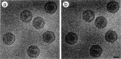
- Janice Nash
- 5 years ago
- Views:
Transcription
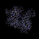
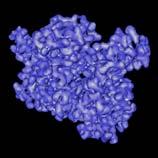
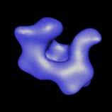
1 Lecture CIMST Winter School Cryo-electron microscopy and tomography of biological macromolecules :00-9:45 in Y03G91 Dr. Takashi Ishikawa OFLB/005 Tel: Lab webpage: 1. What can you see by TEM? Transmission electron microscopy (TEM) is an imaging technique. It can visualize biological, organic and inorganic materials as far as the specimen is thin enough for electrons to go through. Here in this lecture, we will focus on TEM technology to observe 3D structure of biological macromolecules. 2. Hardware Diagram of the transmission electron microscope is similar to that of a light microscope (LM). Both of them have a gun. Electrons or photons are emitted from the gun, penetrate the specimen, focused and enlarged by lenses (TEM uses electromagnetic lenses) and projected on a screen or a camera. Inside of the electron microscope is vacuum. 3. Image formation by TEM In principle, micrographs of TEM are 2D projections of the 3D objects. However, there are significant differences due to weak interaction between the electron beam and the specimen (i.e. TEM specimen is transparent against electrons). Therefore, instead of amplitude contrast LM utilizes, image formation of TEM depends on phase contrast which utilizes interference of electron waves. For phase contrast image must not be perfectly focused; there must be defocus. 4. Specimen preparation Since the contrast between biological materials (density ~1.4) and water (1.0) is poor, previously templates of heavy metal were made to enhance the contrast. With these methodologies (shadowing and negative stain) only the surface of the molecules is visualized. In 1980 s cryo-em was developed. There ice-embedded biological macromolecules are observed directly at the liquid nitrogen temperature. 5. Resolution and radiation damage
2 Available discussion on the molecular structure depends on the level of resolution. Electron beams damage the specimen and limit the resolution. However, if the illumination is too dark, enough amount of contrast for image analysis cannot be obtained. The most essential and serious problem of 3D EM of biological molecules is this dilemma. 6. Various ways of three-dimensional reconstruction technique Here we will discuss four different approaches to reconstruct 3D structure of biological macromolecules from cryo-electron micrographs D crystal In the 2D crystal molecules array two-dimensionally. The Fourier transform of the image is a lattice. Thus, in the Fourier space, you can earn higher signal-to-noise ratio. Which enable the high quality data from lower illumination (i.e. lower radiation damage), resulting higher resolution (more than ten structures have been solved at the atomic resolution). However, this methodology demands, as X-ray crystallography, crystallization. Advantage, comparing to X-ray crystallography, is that we do not need heavy atoms, because direct images are recorded. For 3D information, tilted 2D crystals are inserted into the microscope with special holder. Since you cannot tilt 2D crystal at 90 degrees in the microscope, you will miss some information in the 3D Fourier space (missing cone). Example: bacteriorhodopsin, water channel Tubular crystal Molecules are arrayed as spirals. Since the signal will be spread along one dimensional lines (layer lines), the resolution is not so high as 2D crystals. All the necessary view angles for 3D reconstruction can be obtained from tubular crystal. Therefore, there is no missing information and tilt is not needed. Example: acetylcholine receptor, actin, microtubule 6-3. Single particle analysis This is a technique to calculate the 3D structure from many single particles (>5,000), determining their angles by computation. Since the molecules are dispersed in the solution, you can set solution condition freely, which allows you to see different stages of reaction, for example. However, the angle determination is complicated. Initially, you do not know either the final 3D structure or the view angles of individual particles. You must determine these two unknown factors at the same time. One approach is shown here: projection matching, which starts from a certain template and find the best-fitted view angle for each raw data particle. To generate initial templates, there are a few options. The resolution is evaluated using Fourier shell correlation. Example: Clp protease, ribosome 6-4. Electron tomography In single particle analysis, although you do not need crystals any more, you still average different particles, assuming they share the same structure. If not, the structural
3 variability and flexibility are averaged out. Single particle analysis does not work for highly heterogeneous systems like whole cells or organelles, in which molecules are stacked vertically. Electron tomography is used for such specimens. You record the micrographs of the same specimen from various view angles by tilting the stage of the microscope. Since you control the tilt angle, you have no angle-determination problem, although you still have to align the origin of images. However, the resolution is limited because of multi radiation. Example: Eukaryotic flagella, whole cell tomography
4 References Many examples of structural biology by EM can be seen in Alberts et al. (2002) Molecular biology of the cell, forth edition, Garland Science. p (techniques), p (molecular motor research combining various methodologies including EM). There are some (but, not many) descriptions in Branden, C. and Tooze, J. (1999) Introduction to structural biology, second edition Garland. A good review on single particle analysis and electron tomography Baumeister, W. and Steven, A.C. (2000) Macromolecular electron microscopy in the era of structural genomics Trends in Biochemical Sciences 25, Mathematical discussion on optics (including phase contrast, spherical aberration) Born, M. and Wolf, E. (1980) Principles of optics Pergamon Press Text books on single particle analysis Frank, J. (2006) Three-dimensional electron microscopy of macromolecular assemblies Oxford University Press.
 Transmission")


5 Our target: Biological macromolecules (Protein, Nucleic Acids) Transmission Electron Microscope From the material of Virginia Tech. From Molecular Biology of the Cell Fourth Edition TEM and light microscopy share the similar optical system Differences 1. LM uses a photon as a probe, whereas EM uses an electron 2. LM uses glass lenses, whereas EM uses electromagnetic lenses 1

, and different pumps are used.")
 From the material of Virginia")


6 Inside of the electron microscope must be vacuum. The level of the vacuum varies, depending on the components (gun, column, camera etc.), and different pumps are used. Material Science You can see atoms directly Transmission Electron Microscopy (TEM) From the material of Virginia Tech. Biological Science 1. Low Contrast 2. High Sensitivity to the Electron Beam Why can t we see the atom directly in biological TEM? Wave particle duality of electrons Interference of waves Screen L de Brogli proposed and G. P. Thomson proved that electron has wave-particle duality. 2

7 Amplitude contrast (very weak with cryo-em) Phase contrast Defocus: interference generates contrast In focus: no interference, no phase constrast 3D reconstruction is equivalent to filling 3D Fourier space with many sections, determining the angles Various specimen preparations for biological macromolecules We would like to observe molecules as close to the physiological conditions as possible. Mica platinum Mica Carbon Carbon stain Shadowing Negative Staining Cryo (ice embedding) Baumeister, W. and Steven, A.C. (2000) 3


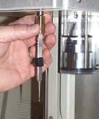

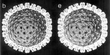

8 Cryo-EM: Ice-Embedding Cryo-holder Holey carbon grid Resolution 3 Å 8 Å Biological specimen (especially ice-embedded) is very sensitive to the radiation damage 6 e - /Å 2 30 e - /Å 2 15 Å 20 Å 6e - /Å 2 30 e - /Å 2 Conway et al. (1993) 4
 J. Struct. Biol.")
 3D view angles can be")


9 2D crystal Gyobu et al., (2006) J. Struct. Biol. 146, 325. Stahlberg, H. et al. (2001) 3D view angles can be determined by diffraction pattern 2D crystal: electron microscopy and electron diffraction Missing wedge, missing pyramid and missing cone Gonen et al. (2005) Nature 438, 633. Molecular Biology of the Cell Lucic et al. (2005) Annu. Rev. Biochem. 5

 Diffraction of")

")


10 Fourier Tubular crystal Kikkawa, T. et al. (1995) Diffraction of the tubular crystal is layer lines Acetylcholine receptor Unwin (1993) J. Mol. Biol. 229, 1101 Example of dynamic structural change: Actin / Myosin complex Tubular crystal has no missing information. Miyazawa et al. (2003) Nature 423, 949. Whittaker et al. (1995) Nature, 378,


 ATP hydrolysis Spahn et")
")
 20 nm Single Particle Analysis:")


11 Single particle analysis: 2D averaging Advantage to reconstruct without crystallization: Free solution condition Example of huge complexes: Ribosome Clp enzyme w/o substrate with substrate (before reaction) ATP hydrolysis Spahn et al. (2001) 107, 373. A P A with substrate (after reaction) Ishikawa et al. (2001) 20 nm Single Particle Analysis: Projection Matching Substrates ClpA 3D Reconstruction Initial Model Project Reproject Iteration ClpP Classification based on Cross Correlation Subaverage for each Euler angle 3D Reconstruction (New Reference) Data Final Reconstruction 5 nm 7


12 Electron Tomography of Chlamydomonas Flagella Gold clusters as marker for translational alignment -60 deg. 30 deg. 0 deg. 60 deg. In electron tomography, you have already known the view angle of images. But, images must be aligned translationally. 8

 Radial spoke Inner dynein arm (regulator) Microtubule Bui et al. (2008) J. Cell Biol.")
13 3D alignment and averaging improve S/N 3D Reconstruction of Chlamydomonas Flagella by electron cryo-tomography 3D Reconstruction of Chlamydomonas Flagella by electron cryo-tomography Averaging with 96nm periodicity reveals the IDA/ODA/RS/MT structure 96nm Outer dynein arm (accelerator, force generator) Radial spoke Inner dynein arm (regulator) Microtubule Bui et al. (2008) J. Cell Biol. 183, 923 9


14 Tail and AAA-ring arrangement of inner and outer arm dyneins Missing wedge, missing pyramid and missing cone Proxymal(-) Distal(+) Tails extend from the rings toward the distal end (tip of the flagellum, + end) Bui et al. (2008) J. Cell Biol. 183, 923 Baumeister, W. and Steven, A.C. (2000) Example: Whole cell tomography Missing wedge problem can be avoided by averaging In the particular case of flagella, structural information will be recovered for the whole Fourier space by averaging nine microtubule doublets. 10


 Science 288, 877.")
 Nat.")


 Resolution of various")
15 Combination of high-resolution single particle analysis and medium-resolution electron tomography: Herpes simplex virus Single particle analysis with multirefrence for 8.5A reconstruction of procapsid by single particle analysis Zhou et al. (2000) Science 288, 877. coexisting structures: maturation of herpes simplex virus Heymann et al. (2000) Nat. Struct. Biol. 10, 334. More intact structure Lower dose Higher resolution tomography single particle analysis tubular crystal 2D crystal Electron tomography to see the whole virus (capsid, tegument, membrane) Resolution of various methodology of 3D cryo-tem Various Methods to reconstruct 3D structure of biological Molecules by EM Grunewald et al. (2003) Science 302,
CIMST Summer School Cryo-electron microscopy for structural biology 11 September, 2014 Dr. Takashi Ishikawa
 CIMST Summer School Cryo-electron microscopy for structural biology 11 September, 2014 Dr. Takashi Ishikawa Tel: 056 310 4217 e-mail: takashi.ishikawa@psi.ch Lab webpage: http://www.psi.ch/lbr/takashi-ishikawa
sin" =1.22 # D "l =1.22 f# D I: In introduction to molecular electron microscopy - Imaging macromolecular assemblies
 I: In introduction to molecular electron microscopy - Imaging macromolecular assemblies Yifan Cheng Department of Biochemistry & Biophysics office: GH-S472D; email: ycheng@ucsf.edu 2/20/2015 - Introduction
Molecular electron microscopy
 Molecular electron microscopy - Imaging macromolecular assemblies Yifan Cheng Department of Biochemistry & Biophysics office: GH-S427B; email: ycheng@ucsf.edu 2/22/2013 - Introduction of Molecular Microscopy:
Electron microscopy in molecular cell biology II
 Electron microscopy in molecular cell biology II Cryo-EM and image processing Werner Kühlbrandt Max Planck Institute of Biophysics Sample preparation for cryo-em Preparation laboratory Specimen preparation
History of 3D Electron Microscopy and Helical Reconstruction
 T H E U N I V E R S I T Y of T E X A S S C H O O L O F H E A L T H I N F O R M A T I O N S C I E N C E S A T H O U S T O N History of 3D Electron Microscopy and Helical Reconstruction For students of HI
There and back again A short trip to Fourier Space. Janet Vonck 23 April 2014
 There and back again A short trip to Fourier Space Janet Vonck 23 April 2014 Where can I find a Fourier Transform? Fourier Transforms are ubiquitous in structural biology: X-ray diffraction Spectroscopy
Three-Dimensional Electron Microscopy of Macromolecular Assemblies
 Three-Dimensional Electron Microscopy of Macromolecular Assemblies Joachim Frank Wadsworth Center for Laboratories and Research State of New York Department of Health The Governor Nelson A. Rockefeller
Transmission Electron Microscopy
 L. Reimer H. Kohl Transmission Electron Microscopy Physics of Image Formation Fifth Edition el Springer Contents 1 Introduction... 1 1.1 Transmission Electron Microscopy... 1 1.1.1 Conventional Transmission
AP5301/ Name the major parts of an optical microscope and state their functions.
 Review Problems on Optical Microscopy AP5301/8301-2015 1. Name the major parts of an optical microscope and state their functions. 2. Compare the focal lengths of two glass converging lenses, one with
What can electron microscopy tell us about chaperoned protein folding? Helen R Saibil
 Review R45 What can electron microscopy tell us about chaperoned protein folding? Helen R Saibil Chaperonins form large, cage-like structures that act as protein folding machines. Recent developments in
Structure, Dynamics and Function of Membrane Proteins using 4D Electron Microscopy
 Structure, Dynamics and Function of Membrane Proteins using 4D Electron Microscopy Anthony W. P. Fitzpatrick Department of Chemistry, Lensfield Road, Cambridge, CB2 1EW My research aims to understand the
ParM filament images were extracted and from the electron micrographs and
 Supplemental methods Outline of the EM reconstruction: ParM filament images were extracted and from the electron micrographs and straightened. The digitized images were corrected for the phase of the Contrast
Electron Microscopy (TEM and SEM)
") 7 Electron Microscopy (TEM and SEM) Paul Verkade Wolfson Bioimaging Facility, Physiology & Pharmacology and Biochemistry, University of Bristol, UK 7.1 Basic how-to-do and why-do section 7.1.1 Electron
7 Electron Microscopy (TEM and SEM) Paul Verkade Wolfson Bioimaging Facility, Physiology & Pharmacology and Biochemistry, University of Bristol, UK 7.1 Basic how-to-do and why-do section 7.1.1 Electron
Scattering Lecture. February 24, 2014
 Scattering Lecture February 24, 2014 Structure Determination by Scattering Waves of radiation scattered by different objects interfere to give rise to an observable pattern! The wavelength needs to close
Chapter 4 A Tour of the Cell. The human body is made up of trillions of cells many of which are specialized - Muscle cells
 Chapter 4 A Tour of the Cell State Standards Standard 1.c. Standard 1.e. Introduction to Cells Organisms are either - Single-celled, such as - Multicelled, such as The human body is made up of trillions
Slide 1. Slide 2. Slide 3. Chapter 4 A Tour of the Cell. State Standards. Introduction to Cells. Standard 1.c. Standard 1.e.
 Slide 1 Chapter 4 A Tour of the Cell Slide 2 State Standards Standard 1.c. Standard 1.e. Slide 3 Introduction to Cells Organisms are either - Single-celled, such as - Multicelled, such as The human body
Transmission Electron Microscopy: A Textbook For Materials Science (4-Vol Set) By C. Barry Carter, David B. Williams
 By C. Barry Carter, David B. Williams") Transmission Electron Microscopy: A Textbook For Materials Science (4-Vol Set) By C. Barry Carter, David B. Williams If you are searched for the ebook Transmission Electron Microscopy: A Textbook for Materials
Transmission Electron Microscopy: A Textbook For Materials Science (4-Vol Set) By C. Barry Carter, David B. Williams If you are searched for the ebook Transmission Electron Microscopy: A Textbook for Materials
Tutorial 1 Geometry, Topology, and Biology Patrice Koehl and Joel Hass
 Tutorial 1 Geometry, Topology, and Biology Patrice Koehl and Joel Hass University of California, Davis, USA http://www.cs.ucdavis.edu/~koehl/ims2017/ Biology = Multiscale. 10 6 m 10 3 m m mm µm nm Å ps
Electron Microscopy. SEM = Scanning Electron Microscopy TEM = Transmission Electron Microscopy. E. coli, William E. Bentley, Maryland, USA
 Electron Microscopy Transmission Electron Microscopy SEM = Scanning Electron Microscopy TEM = Transmission Electron Microscopy Sara Henriksson, UCEM 2017-02-16 E. coli, William E. Bentley, Maryland, USA
Machines with Moving Parts See How They Run.
 NRAMM Workshop, CIMBio, La Jolla CA November 8-13, 2009 Machines with Moving Parts See How They Run. Alasdair C. Steven Laboratory of Structural Biology, NIAMS-NIH Sources of Variability in Electron Micrographs
Protein Structure Determination 9/25/2007
 One-dimensional NMR spectra Ethanol Cellulase (36 a.a.) Branden & Tooze, Fig. 18.16 1D and 2D NMR spectra of inhibitor K (57 a.a.) K. Wuthrich, NMR of Proteins and Nucleic Acids. (Wiley, 1986.) p. 54-55.
Wave Nature of Matter
 Wave Nature of Matter Wave-Particle Duality de Broglie proposed that particles with momentum could have an associated wavelength (converse of photons having momentum) de Broglie wavelength h λ = p or p
Conventional Transmission Electron Microscopy. Introduction. Text Books. Text Books. EMSE-509 CWRU Frank Ernst
 Text Books Conventional Transmission Electron Microscopy EMSE-509 CWRU Frank Ernst D. B. Williams and C. B. Carter: Transmission Electron Microscopy, New York: Plenum Press (1996). L. Reimer: Transmission
tip conducting surface
 PhysicsAndMathsTutor.com 1 1. The diagram shows the tip of a scanning tunnelling microscope (STM) above a conducting surface. The tip is at a potential of 1.0 V relative to the surface. If the tip is sufficiently
Laboratory-Based Cryogenic Soft X-ray Tomography and Correlative Microscopy: 3D Visualization Inside the Cell
 Laboratory-Based Cryogenic Soft X-ray Tomography and Correlative Microscopy: 3D Visualization Inside the Cell David Carlson, Jeff Gelb, Vadim Palshin and James Evans Pacific Northwest National Laboratory
Opportunity and Challenge: the new era of cryo-em. Ning Gao School of Life Sciences Tsinghua University
 Opportunity and Challenge: the new era of cryo-em Ning Gao School of Life Sciences Tsinghua University 2015-10-20 Education and Research Experience Principal Investigator 2008.11-present Postdoc, Department
Praktikum zur. Materialanalytik
 Praktikum zur Materialanalytik Energy Dispersive X-ray Spectroscopy B513 Stand: 19.10.2016 Contents 1 Introduction... 2 2. Fundamental Physics and Notation... 3 2.1. Alignments of the microscope... 3 2.2.
object objective lens eyepiece lens
 Advancing Physics G495 June 2015 SET #1 ANSWERS Field and Particle Pictures Seeing with electrons The compound optical microscope Q1. Before attempting this question it may be helpful to review ray diagram
Topic 3: Cells Ch. 6. Microscopes pp Microscopes. Microscopes. Microscopes. Microscopes
 Topic 3: Cells Ch. 6 -All life is composed of cells and all cells have a plasma membrane, cytoplasm, and DNA. pp.105-107 - The development of the microscope was the key to understanding that all living
CHEM 681 Seminar Mingqi Zhao April 20, 1998 Room 2104, 4:00 p.m. High Resolution Transmission Electron Microscopy: theories and applications
 CHEM 681 Seminar Mingqi Zhao April 20, 1998 Room 2104, 4:00 p.m. High Resolution Transmission Electron Microscopy: theories and applications In materials science, people are always interested in viewing
Conformationally Variable Single Particles Heterogeneity in the real world
 Conformationally Variable Single Particles Heterogeneity in the real world Stan Burgess University of Leeds, UK Workshop on Advanced Topics in EM Structure Determination: Challenging Molecules November
This is a topic I encountered a number of years ago while at LBNL, when I came across the following statement [1]:
![This is a topic I encountered a number of years ago while at LBNL, when I came across the following statement [1]:](/thumbs/88/117124552.jpg "This is a topic I encountered a number of years ago while at LBNL, when I came across the following statement [1]:") Equivalent signal to noise in diffraction vs. bright-field experiments Charles Sindelar January 2010 SUMMARY This is a topic I encountered a number of years ago while at LBNL, when I came across the following
Equivalent signal to noise in diffraction vs. bright-field experiments Charles Sindelar January 2010 SUMMARY This is a topic I encountered a number of years ago while at LBNL, when I came across the following
Image Assessment San Diego, November 2005
 Image Assessment San Diego, November 005 Pawel A. Penczek The University of Texas Houston Medical School Department of Biochemistry and Molecular Biology 6431 Fannin, MSB6.18, Houston, TX 77030, USA phone:
High-Resolution. Transmission. Electron Microscopy
 Part 4 High-Resolution Transmission Electron Microscopy 186 Significance high-resolution transmission electron microscopy (HRTEM): resolve object details smaller than 1nm (10 9 m) image the interior of
Supplementary Information
 SSZ-52, a zeolite with an 18-layer aluminosilicate framework structure related to that of the DeNOx catalyst Cu-SSZ-13 Dan Xie 1,2, Lynne B. McCusker 2 *, Christian Baerlocher 2, Stacey I. Zones 1 *, Wei
Structure analysis: Electron diffraction LEED TEM RHEED
 Structure analysis: Electron diffraction LEED: Low Energy Electron Diffraction SPA-LEED: Spot Profile Analysis Low Energy Electron diffraction RHEED: Reflection High Energy Electron Diffraction TEM: Transmission
Overview of scattering, diffraction & imaging in the TEM
 Overview of scattering, diffraction & imaging in the TEM Eric A. Stach Purdue University Scattering Electrons, photons, neutrons Radiation Elastic Mean Free Path (Å)( Absorption Length (Å)( Minimum Probe
Cells Under the Microscope Measuring Cell Structures
 Copy into Note Packet and Return to Teacher Chapter 3 Cell Structure Section 1: Looking at Cells Objectives Describe how scientists measure the length of objects. Relate magnification and resolution in
= 6 (1/ nm) So what is probability of finding electron tunneled into a barrier 3 ev high?
 So what is probability of finding electron tunneled into a barrier 3 ev high?") STM STM With a scanning tunneling microscope, images of surfaces with atomic resolution can be readily obtained. An STM uses quantum tunneling of electrons to map the density of electrons on the surface
STM STM With a scanning tunneling microscope, images of surfaces with atomic resolution can be readily obtained. An STM uses quantum tunneling of electrons to map the density of electrons on the surface
DIFFRACTION PHYSICS THIRD REVISED EDITION JOHN M. COWLEY. Regents' Professor enzeritus Arizona State University
 DIFFRACTION PHYSICS THIRD REVISED EDITION JOHN M. COWLEY Regents' Professor enzeritus Arizona State University 1995 ELSEVIER Amsterdam Lausanne New York Oxford Shannon Tokyo CONTENTS Preface to the first
CHEM-E5225 :Electron Microscopy Imaging
 CHEM-E5225 :Electron Microscopy Imaging 2016.10 Yanling Ge Outline Planar Defects Image strain field WBDF microscopy HRTEM information theory Discuss of question homework? Planar Defects - Internal Interface
Part 1 X-ray Crystallography
 Part 1 X-ray Crystallography What happens to electron when it is hit by x-rays? 1. The electron starts vibrating with the same frequency as the x-ray beam 2. As a result, secondary beams will be scattered
Guided Reading Activities
 Name Period Chapter 4: A Tour of the Cell Guided Reading Activities Big Idea: Introduction to the Cell Answer the following questions as you read Modules 4.1 4.4: 1. A(n) uses a beam of light to illuminate
Biochemistry 9001 Protein structure determination by 3D Electron Microscopy
 Biochemistry 9001 Protein structure determination by 3D Electron Microscopy Tommi A. White, Ph.D. Associate Director, Electron Microscopy Core Facility Assistant Research Professor, Biochemistry Goals
Application examples of single particle 3D reconstruction. Ning Gao Tsinghua University
 Application examples of single particle 3D reconstruction Ning Gao Tsinghua University ninggao@tsinghua.edu.cn Electron Microscopes First electron microscope constructed by Ernst Ruska in 1930 s (1986
Microscopy, Staining, and Classification
 PowerPoint Lecture Presentations prepared by Mindy Miller-Kittrell, North Carolina State University C H A P T E R 4 Microscopy, Staining, and Classification Microscopy Light Microscopy 1) Bright-field
What can we see with a transmission electron microscope?
 What can we see with a transmission electron microscope? β-galactosidase Subramaniam Lab o.lambert@cbmn.u-bordeaux.fr Renafobis 2017 1 Film or camera Renafobis 2017 2 1897 J.J. Thomson Electron discovery
HIGH RESOLUTION ELECTRON MICROSCOPY
 HIGH RESOLUTION ELECTRON MICROSCOPY BC530 Fall Quarter 2014 Wim G. J. Hol http://www.bmsc.washington.edu/wimhol/ Structure Determination by ELECTRON MICROSCOPY STRENGTHS: - Small amounts of material usually
Index. Born approximation, 128 Bremsstrahlung, 41, 42, 53 angular distribution, 42 energy distribution, 42
 Index Aberrations aberration-free holography, 123 chromatic, in confocal imaging, 195 off-axis, 2, 211 optical correction methods, 122 reduction of, 2 Acetylcholine receptors, 30 Acoustic impedance, 200
Introduction to Electron Microscopy Andres Kaech. Instrumentation
 Center for Microscopy and Image Analysis Introduction to Electron Microscopy Andres Kaech Instrumentation The types of electron microscopes Transmission electron microscope (TEM) Scanning electron microscope
Solution set for EXAM IN TFY4265/FY8906 Biophysical microtechniques
 ENGLISH NORWEGIAN UNIVERSITY OF SCIENCE AND TECHNOLOGY DEPARTMENT OF PHYSICS Contact during exam: Magnus Borstad Lilledahl Telefon: 73591873 (office) 92851014 (mobile) Solution set for EXAM IN TFY4265/FY8906
Imaging Methods: Scanning Force Microscopy (SFM / AFM)
") Imaging Methods: Scanning Force Microscopy (SFM / AFM) The atomic force microscope (AFM) probes the surface of a sample with a sharp tip, a couple of microns long and often less than 100 Å in diameter.
Imaging Methods: Scanning Force Microscopy (SFM / AFM) The atomic force microscope (AFM) probes the surface of a sample with a sharp tip, a couple of microns long and often less than 100 Å in diameter.
Basic Principles Brief history of EM
 SIR WILLIAM DUNN SCHOOL OF PATHOLOGY Basic Principles of Electron Microscopy (EM) Dr Errin Johnson Head of the Dunn School EM Facility Basic Principles Brief history of EM 1873 Hermann von Helmholtz &
Introduction to Cryo Microscopy. Nikolaus Grigorieff
 Introduction to Cryo Microscopy Nikolaus Grigorieff Early TEM and Biology 1906-1988 Bacterial culture in negative stain. Friedrich Krause, 1937 Nobel Prize in Physics 1986 Early design, 1931 Decades of
Chapter 16. Cellular Movement: Motility and Contractility. Lectures by Kathleen Fitzpatrick Simon Fraser University Pearson Education, Inc.
 Chapter 16 Cellular Movement: Motility and Contractility Lectures by Kathleen Fitzpatrick Simon Fraser University Two eukaryotic motility systems 1. Interactions between motor proteins and microtubules
Modern Optical Spectroscopy
 Modern Optical Spectroscopy X-Ray Microanalysis Shu-Ping Lin, Ph.D. Institute of Biomedical Engineering E-mail: splin@dragon.nchu.edu.tw Website: http://web.nchu.edu.tw/pweb/users/splin/ Backscattered
Transmission Electron Microscopy
 Transmission Electron Microscopy Fu-Rong Chen Transmission Electron Microscopy David B. Williams C. Barry Carter Background:Solid State Physics Materials Science 1.1 Why Electron Microscope? 1.1 Why Electron
A short overview of electron tomography
 A short overview of electron tomography Ozan Öktem ozan.oktem@sidec.com Sidec Technologies January 17, 2006 IMA Workshop: New Mathematics and Algorithms for 3-D Image Analysis, January 9-12, 2006 Outline
Chapter 4 Active Reading Guide A Tour of the Cell
 Name: AP Biology Mr. Croft Chapter 4 Active Reading Guide A Tour of the Cell Section 1 1. The study of cells has been limited by their small size, and so they were not seen and described until 1665, when
From electron crystallography to single particle cryoem
 From electron crystallography to single particle cryoem Nobel Lectures in Chemistry 8 th December 2017 Richard Henderson From Baker & Henderson (2001) Int.Tab.Cryst.Vol.F, on-line (2006), revised (2011)
TEST BANK FOR PRESCOTTS MICROBIOLOGY 9TH EDITION BY WILLEY SHERWOOD WOOLVERTON
 TEST BANK FOR PRESCOTTS MICROBIOLOGY 9TH EDITION BY WILLEY SHERWOOD WOOLVERTON Link download full: https://testbankservice.com/download/test-bank-for-prescottsmicrobiology-9th-edition-by-willey-sherwood-woolverton/
Why microscopy?
 Electron Microscopy Why microscopy? http://www.cellsalive.com/howbig.htm 2 Microscopes are used as magnifying tools (although not exclusively as will see later on). The resolution of the human eye is limited
Spliceosome and Localization of Its Catalytic Core
 Molecular Cell, Volume 40 Supplemental Information 3D Cryo-EM Structure of an Active Step I Spliceosome and Localization of Its Catalytic Core Monika M. Golas, Bjoern Sander, Sergey Bessonov, Michael Grote,
JRE Group of Institutions ASSIGNMENT # 1 Special Theory of Relativity
 ASSIGNMENT # 1 Special Theory of Relativity 1. What was the objective of conducting the Michelson-Morley experiment? Describe the experiment. How is the negative result of the experiment interpreted? 2.
LECTURE 11 ELECTROMAGNETIC WAVES & POLARIZATION. Instructor: Kazumi Tolich
 LECTURE 11 ELECTROMAGNETIC WAVES & POLARIZATION Instructor: Kazumi Tolich Lecture 11 2 25.5 Electromagnetic waves Induced fields Properties of electromagnetic waves Polarization Energy of electromagnetic
LECTURE 11 ELECTROMAGNETIC WAVES & POLARIZATION. Instructor: Kazumi Tolich
 LECTURE 11 ELECTROMAGNETIC WAVES & POLARIZATION Instructor: Kazumi Tolich Lecture 11 2 25.5 Electromagnetic waves Induced fields Properties of electromagnetic waves Polarization Energy of electromagnetic
MODULE 2 : FOUNDATIONS IN BIOLOGY
 OCR A LEVEL BIOLOGY MODULE 2 : FOUNDATIONS IN BIOLOGY REVISION NOTES For 2015 onwards specification Miss T Banda All living things are primarily made from 4 key elements: Carbon (C) Hydrogen (H) Oxygen
QUANTUM PHYSICS. Limitation: This law holds well only for the short wavelength and not for the longer wavelength. Raleigh Jean s Law:
 Black body: A perfect black body is one which absorbs all the radiation of heat falling on it and emits all the radiation when heated in an isothermal enclosure. The heat radiation emitted by the black
INDIAN INSTITUTE OF TECHNOLOGY ROORKEE NPTEL NPTEL ONLINE CERTIFICATION COURSE. Biomedical Nanotechnology. Lec-05 Characterisation of Nanoparticles
 INDIAN INSTITUTE OF TECHNOLOGY ROORKEE NPTEL NPTEL ONLINE CERTIFICATION COURSE Biomedical Nanotechnology Lec-05 Characterisation of Nanoparticles Dr. P. Gopinath Department of Biotechnology Indian Institute
New algoritms for electron diffraction of 3D protein crystals. JP Abrahams, D Georgieva, L Jiang, I Sikhuralidze, NS Pannu
 New algoritms for electron diffraction of 3D protein crystals JP Abrahams, D Georgieva, L Jiang, I Sikhuralidze, NS Pannu Why new algorithms? New research questions New experimental techniques Better insight
Crystal Structure and Electron Diffraction
 Crystal Structure and Electron Diffraction References: Kittel C.: Introduction to Solid State Physics, 8 th ed. Wiley 005 University of Michigan, PHY441-44 (Advanced Physics Laboratory Experiments, Electron
Weak-Beam Dark-Field Technique
 Basic Idea recall bright-field contrast of dislocations: specimen close to Bragg condition, s î 0 Weak-Beam Dark-Field Technique near the dislocation core, some planes curved to s = 0 ) strong Bragg reflection
The basics of structural biology. And Why we use synchrotron sources Sean McSweeney ESRF Structural Biology Group
 The basics of structural biology And Why we use synchrotron sources Sean McSweeney ESRF Structural Biology Group The rise and rise of structural biology. 2 The aim of the game 3 What information does structure
Improving cryo-em maps: focused 2D & 3D classifications and focused refinements
 Improving cryo-em maps: focused 2D & 3D classifications and focused refinements 3rd International Symposium on Cryo-3D Image Analysis 23.3.2018, Tahoe Bruno Klaholz Centre for Integrative Biology, IGBMC,
Experimental methods in Physics. Electron Microscopy. Basic Techniques (MEP-I) SEM, TEM
 SEM, TEM") Experimental methods in Physics Electron Microscopy Basic Techniques (MEP-I) SEM, TEM Advanced Techniques (MEP-II) HR-TEM, STEM Analytical-TEM 3D-Microscopy Spring 2012 Experimental Methods in Physics
Experimental methods in Physics Electron Microscopy Basic Techniques (MEP-I) SEM, TEM Advanced Techniques (MEP-II) HR-TEM, STEM Analytical-TEM 3D-Microscopy Spring 2012 Experimental Methods in Physics
Spatial Frequency and Transfer Function. columns of atoms, where the electrostatic potential is higher than in vacuum
 Image Formation Spatial Frequency and Transfer Function consider thin TEM specimen columns of atoms, where the electrostatic potential is higher than in vacuum electrons accelerate when entering the specimen
Electron microscopy in molecular cell biology I
 Electron microscopy in molecular cell biology I Electron optics and image formation Werner Kühlbrandt Max Planck Institute of Biophysics chemistry biology Objects of interest Galaxy 10 6 light years 10
BIO 311C Spring 2010
 BIO 311C Spring 2010 Prokaryotic cells contain structures that are very similar to structures of the eukaryotic cytoskeleton. Prokaryotic cytoskeletal elements are required for cell division, maintaining
Gaetano L Episcopo. Scanning Electron Microscopy Focus Ion Beam and. Pulsed Plasma Deposition
 Gaetano L Episcopo Scanning Electron Microscopy Focus Ion Beam and Pulsed Plasma Deposition Hystorical background Scientific discoveries 1897: J. Thomson discovers the electron. 1924: L. de Broglie propose
General theory of diffraction
 General theory of diffraction X-rays scatter off the charge density (r), neutrons scatter off the spin density. Coherent scattering (diffraction) creates the Fourier transform of (r) from real to reciprocal
Electron Microscopy of Tobacco Mosaic Virus Prepared with the Aid of Negative Staining-Carbon Film Techniques
 J. gen. Virol. (1976), :3I, 265-269 Printed in Great Britain 265 Electron Microscopy of Tobacco Mosaic Virus Prepared with the Aid of Negative Staining-Carbon Film Techniques (Accepted 13 January I976)
2.1 CELL STRUCTURE. The cell is the smallest unit of living organisms that shows the characteristics of life.
 2.1.1 Microscopy The cell is the smallest unit of living organisms that shows the characteristics of life. A general introduction to the microscope. The light microscope All cells are microscopic which
Supporting Online Material for
 www.sciencemag.org/cgi/content/full/310/5753/1513/dc1 Supporting Online Material for Structural Roles for Human Translation Factor eif3 in Initiation of Protein Synthesis Bunpote Siridechadilok, Christopher
CHARACTERIZATION of NANOMATERIALS KHP
 CHARACTERIZATION of NANOMATERIALS Overview of the most common nanocharacterization techniques MAIN CHARACTERIZATION TECHNIQUES: 1.Transmission Electron Microscope (TEM) 2. Scanning Electron Microscope
Chapter 6: A Tour of the Cell
 AP Biology Reading Guide Fred and Theresa Holtzclaw Chapter 6: A Tour of the Cell Name Period Chapter 6: A Tour of the Cell Concept 6.1 To study cells, biologists use microscopes and the tools of biochemistry
Ecole Franco-Roumaine : Magnétisme des systèmes nanoscopiques et structures hybrides - Brasov, Modern Analytical Microscopic Tools
 1. Introduction Solid Surfaces Analysis Group, Institute of Physics, Chemnitz University of Technology, Germany 2. Limitations of Conventional Optical Microscopy 3. Electron Microscopies Transmission Electron
Chapter 6: A Tour of the Cell
 Chapter 6: A Tour of the Cell 1. The study of cells has been limited by their small size, and so they were not seen and described until 1665, when Robert Hooke first looked at dead cells from an oak tree.
Electron beam scanning
 Electron beam scanning The Electron beam scanning operates through an electro-optical system which has the task of deflecting the beam Synchronously with cathode ray tube which create the image, beam moves
Nuclear import of DNA: genetic modification of plants
 Nuclear import of DNA: genetic modification of plants gene delivery by Agrobacterium tumefaciens T. Tzfira & V. Citovsky. 2001. Trends in Cell Biol. 12: 121-129 VirE2 binds ssdna in vitro forms helical
From x-ray crystallography to electron microscopy and back -- how best to exploit the continuum of structure-determination methods now available
 From x-ray crystallography to electron microscopy and back -- how best to exploit the continuum of structure-determination methods now available Scripps EM course, November 14, 2007 What aspects of contemporary
Microscopy, Staining, and Classification. ~10 um. Red Blood Cells = mm 1500 um. Width of penny
 PowerPoint Lecture Presentations prepared by Mindy Miller-Kittrell, North Carolina State University C H A P T E R 4 Microscopy, Staining, and Classification Figure 3.4 Approximate size of various types
Atomic and Nuclear Physics
 Atomic and Nuclear Physics Introductory experiments ualism of wave and particle L Physics Leaflets P6.1.5.1 iffraction of electrons in a polycrystalline lattice (ebye-scherrer diffraction) Objects of the
Electron energy filtering significantly improves amplitude contrast of frozen-hydrated protein at 300 kv
 Journal of Structural Biology xxx (2006) xxx xxx Journal of Structural Biology www.elsevier.com/locate/yjsbi Electron energy filtering significantly improves amplitude contrast of frozen-hydrated protein
ITERATIVE METHODS IN LARGE FIELD ELECTRON MICROSCOPE TOMOGRAPHY. Xiaohua Wan
 ITERATIVE METHODS IN LARGE FIELD ELECTRON MICROSCOPE TOMOGRAPHY Xiaohua Wan wanxiaohua@ict.ac.cn Institute of Computing Technology, Chinese Academy of Sciences Outline Background Problem Our work Future
Wave properties of matter & Quantum mechanics I. Chapter 5
 Wave properties of matter & Quantum mechanics I Chapter 5 X-ray diffraction Max von Laue suggested that if x-rays were a form of electromagnetic radiation, interference effects should be observed. Crystals
Introduction to Three Dimensional Structure Determination of Macromolecules by Cryo-Electron Microscopy
 Introduction to Three Dimensional Structure Determination of Macromolecules by Cryo-Electron Microscopy Amit Singer Princeton University, Department of Mathematics and PACM July 23, 2014 Amit Singer (Princeton
Atomic and molecular interactions. Scanning probe microscopy.
 Atomic and molecular interactions. Scanning probe microscopy. Balázs Kiss Nanobiotechnology and Single Molecule Research Group, Department of Biophysics and Radiation Biology 27. November 2013. 2 Atomic
Microscopy, Staining, and Classification
 PowerPoint Lecture Presentations prepared by Mindy Miller-Kittrell, North Carolina State University C H A P T E R 4 Microscopy, Staining, and Classification 4. Discuss how microscopy has revealed the structure
channel/ryanodine receptor from sarcoplasmic reticulum
 Cryo-EM of the native structure of the calcium release channel/ryanodine receptor from sarcoplasmic reticulum M. Radermacher, T. Wagenknecht, R. Grassucci, J. Frank, M. Inui,* C. Chadwick,* and S. Fleischer*
Warm-Up Pairs Discuss the diagram What Where Which Why
 Warm-Up In Pairs Discuss the diagram What is it? Where does it come from? Which parts can you label? (in pencil) Why do you think you will learn about it? 5 m Eukaryote: Organelles, Structure and Function