From electron crystallography to single particle cryoem
|
|
|
- Violet Hensley
- 6 years ago
- Views:
Transcription
1 From electron crystallography to single particle cryoem Nobel Lectures in Chemistry 8 th December 2017 Richard Henderson

2

3 From Baker & Henderson (2001) Int.Tab.Cryst.Vol.F, on-line (2006), revised (2011) What is electron cryomicroscopy? - CryoEM comes in several flavours 70S ribosome 11 Å hepatitis B cores 7 Å decorated actin 30 Å LHC II 3.4 Å Gabashvili (2000) Boettcher (1997) Milligan (~1995) Kuhlbrandt (1994)

4
 for")
5 Growth in cryoem community PDB cryoem annual coordinates deposition 3dem mailing list Started in 1995 by Ross Smith at NYU transferred to NCMIR at UCSD in 2000 Gina Sosinsky, now Guy Perkins Current list has 3202 subscribers Gordon Research Conference on 3dem started by Wah Chiu & Nigel Unwin in 1985, held every year rotating between USA, Europe & Asia EMDB (Electron Microscopy Data Bank) for deposition of cryoem maps (5400) PDB (Protein Data Bank) for coordinate depositions (less)
6 Outline 1. X-ray diffraction, electron crystallography (diffraction, microscopy) 2. Bacteriorhodopsin at 7Å, then 3.5Å, refinement & kinetics 3. Single particle cryoem - blobology then resolution revolution

7 Halobacterium salinarum Walther Stoeckenius, Rockefeller

8 Purified purple membranes on carbon

9 X-ray powder diffraction pattern of oriented pellet of purple membrane 1973

10


11 bacteriorhodopsin projection glucose embedding, no cryoem Unwin & Henderson JMB, 1975 electron diffraction (amplitudes) optical diffraction of electron micrograph (FFT --> phases) 4,3 4,3


12

13 7Å 3D map, 1975

14 7Å 3D model, 1975
15 Outline 1. X-ray diffraction, electron crystallography (diffraction, microscopy) 2. Bacteriorhodopsin at 7Å, then 3.5Å, refinement & kinetics 3. Single particle cryoem - blobology then resolution revolution

16 Faruqi CCD detectors

17 Jeng, Chiu, Zemlin & Zeitler, 1984 JMB Crotoxin thin 3D xtals


18 Solving bacteriorhodopsin structure at high resolution High-resolution cryoem imaging heavy atom derivatives molecular replacement
19 The path from 7 Å resolution to 3.5 Å and atomic model Cooling specimen to liq. N 2 or liq. He temperature reduces the effects of radiation damage and gives 4- to 5-fold increase in diffraction Very few electron microscopes were stable enough in 1980s to achieve imaging with 3.5 Å resolution using cold stages Collaborations with and travelling to three different labs were essential: Lepault/Dubochet at EMBL Zemlin/Beckmann/Zeitler at Fritz-Haber-Institute in Berlin Downing/Glaeser at Berkeley Beam tilt was a key feature that required computational correction Correcting for defocus gradient when tilting specimen was another technical challenge Finally, 70 images allowed a map to be calculated, adequate to build an atomic model Refinement by Niko Grigorieff + increase to 100 images with 30 more from Ken Downing Yoshi Fujiyoshi independently determined the structure with an improved map All subsequent X-ray structures used the cryoem coordinates for molecular replacement

20 Progress at last!

21 Bacteriorhodopsin 3.5 Å 1990

22 Bacteriorhodopsin 3.5 Å 1990
23 Outline 1. X-ray diffraction, electron crystallography (diffraction, microscopy) 2. Bacteriorhodopsin at 7Å, then 3.5Å, refinement & kinetics 3. Single particle cryoem - blobology then resolution revolution


24 From Dubochet et al, QRB (1988)


25 Preparing a cryoem grid using the method of Adrian, McDowell & Dubochet Controlled environment plunge-freeze apparatus of Ishi Talmon (1988)

26 5-fold 2-fold ~3-fold

 first")


 30 Å")


27 3.5Å ptcles hepb virus cores (1997) first sub-nm single particle structure hepb most recent structure Yu, Jin, Jih, Shih & Zhou (2013) 30 Å (1994) 20 ptcles T=3 T=4 7.4Å 6600 ptcles Boettcher, Wynne & Crowther (1997)
28 Comparison of 300keV DQE of direct electron detectors versus film McMullan et al (2014) Ultramicroscopy
29


30 Å Å Å Bartesaghi et al & Subramaniam Science (2015), 2.2Å 2014, Relion (Scheres) 3.8Å


31 Zhu, Vinothkumar (2016) Vinothkumar, Zhu & Hirst, Nature (2014)
32 slope = 6Å 2 per electron dose of 1 el/å 2 Russo & Henderson unpublished


33 Nigel Unwin bacteriorhodopsin 7Å Colleagues in rough chronological order bacteriorhodopsin 3.5Å using cryoem Joyce Baldwin Tom Ceska David Agard Bob Glaeser Jean Lepault Fritz Zemlin Erich Beckmann Ken Downing detector & microscope technical developments Wasi Faruqi Greg McMullan Renato Turchetta Nicola Guerrini Shaoxia Chen single particle cryoem Sriram Subramaniam Niko Grigorieff Peter Rosenthal Chris Russo Jacqueline Milne John Rubinstein Vinothkumar
34 With many thanks to current and previous colleagues Electron crystallography Nigel Unwin, Tom Ceska, Joyce Baldwin, David Agard, Jean Lepault, Fritz Zemlin, Erich Beckmann, Ken Downing, Bob Glaeser, Niko Grigorieff, Sriram Subramaniam Single particle cryoem Niko Grigorieff, Sriram Subramaniam, Jacqueline Milne, Peter Rosenthal, John Rubinstein, Vinothkumar Detector development Wasi Faruqi, Greg McMullan, Renato Turchetta, Nicola Guerrini, Shaoxia Chen Grand scheme Chris Russo
Introduction to Cryo Microscopy. Nikolaus Grigorieff
 Introduction to Cryo Microscopy Nikolaus Grigorieff Early TEM and Biology 1906-1988 Bacterial culture in negative stain. Friedrich Krause, 1937 Nobel Prize in Physics 1986 Early design, 1931 Decades of
Application examples of single particle 3D reconstruction. Ning Gao Tsinghua University
 Application examples of single particle 3D reconstruction Ning Gao Tsinghua University ninggao@tsinghua.edu.cn Electron Microscopes First electron microscope constructed by Ernst Ruska in 1930 s (1986
Molecular electron microscopy
 Molecular electron microscopy - Imaging macromolecular assemblies Yifan Cheng Department of Biochemistry & Biophysics office: GH-S427B; email: ycheng@ucsf.edu 2/22/2013 - Introduction of Molecular Microscopy:
Electron microscopy in molecular cell biology II
 Electron microscopy in molecular cell biology II Cryo-EM and image processing Werner Kühlbrandt Max Planck Institute of Biophysics Sample preparation for cryo-em Preparation laboratory Specimen preparation
sin" =1.22 # D "l =1.22 f# D I: In introduction to molecular electron microscopy - Imaging macromolecular assemblies
 I: In introduction to molecular electron microscopy - Imaging macromolecular assemblies Yifan Cheng Department of Biochemistry & Biophysics office: GH-S472D; email: ycheng@ucsf.edu 2/20/2015 - Introduction
Recent Developments in Direct Electron Detectors for Electron Cryo-Microscopy
 Recent Developments in for Electron Cryo-Microscopy A.R.Faruqi*, R.Henderson and G.McMullan MRC Laboratory of Molecular Biology, Francis Crick Avenue, Cambridge Biomedical Campus, Cambridge CB2 0QH E-mail:
What can electron microscopy tell us about chaperoned protein folding? Helen R Saibil
 Review R45 What can electron microscopy tell us about chaperoned protein folding? Helen R Saibil Chaperonins form large, cage-like structures that act as protein folding machines. Recent developments in
Halorhodopsin (hr) is a light-driven chloride pump in the
 is a light-driven chloride pump in the") The three-dimensional structure of halorhodopsin to 5 Å by electron crystallography: A new unbending procedure for two-dimensional crystals by using a global reference structure Edmund R. S. Kunji*, Susanne
The three-dimensional structure of halorhodopsin to 5 Å by electron crystallography: A new unbending procedure for two-dimensional crystals by using a global reference structure Edmund R. S. Kunji*, Susanne
Lecture CIMST Winter School. 1. What can you see by TEM?
 Lecture CIMST Winter School Cryo-electron microscopy and tomography of biological macromolecules 20.1.2011 9:00-9:45 in Y03G91 Dr. Takashi Ishikawa OFLB/005 Tel: 056 310 4217 e-mail: takashi.ishikawa@psi.ch
What can we see with a transmission electron microscope?
 What can we see with a transmission electron microscope? β-galactosidase Subramaniam Lab o.lambert@cbmn.u-bordeaux.fr Renafobis 2017 1 Film or camera Renafobis 2017 2 1897 J.J. Thomson Electron discovery
History of 3D Electron Microscopy and Helical Reconstruction
 T H E U N I V E R S I T Y of T E X A S S C H O O L O F H E A L T H I N F O R M A T I O N S C I E N C E S A T H O U S T O N History of 3D Electron Microscopy and Helical Reconstruction For students of HI
Electron microscopy in molecular cell biology I
 Electron microscopy in molecular cell biology I Electron optics and image formation Werner Kühlbrandt Max Planck Institute of Biophysics chemistry biology Objects of interest Galaxy 10 6 light years 10
Critical Review. Viruses and the Development of Quantitative Biological Electron Microscopy*
 IUBMB Life, 56(5): 239 248, May 2004 Critical Review Viruses and the Development of Quantitative Biological Electron Microscopy* R. A. Crowther MRC Laboratory of Molecular Biology, Hills Road, Cambridge
CIMST Summer School Cryo-electron microscopy for structural biology 11 September, 2014 Dr. Takashi Ishikawa
 CIMST Summer School Cryo-electron microscopy for structural biology 11 September, 2014 Dr. Takashi Ishikawa Tel: 056 310 4217 e-mail: takashi.ishikawa@psi.ch Lab webpage: http://www.psi.ch/lbr/takashi-ishikawa
Reaching the Information Limit in Cryo-EM of Biological Macromolecules: Experimental Aspects
 Biophysical Journal Volume 100 May 2011 2331 2337 2331 Reaching the Information Limit in Cryo-EM of Biological Macromolecules: Experimental Aspects Robert M. Glaeser * and Richard J. Hall Life Sciences
Single-Particle Cryo-EM at Crystallographic Resolution
 Leading Edge Review Single-Particle Cryo-EM at Crystallographic Resolution Yifan Cheng 1, * 1 Department of Biochemistry and Biophysics, University of California San Francisco, 600 16th Street, San Francisco,
arxiv: v1 [cond-mat.mtrl-sci] 3 Apr 2015
![arxiv: v1 [cond-mat.mtrl-sci] 3 Apr 2015](/thumbs/84/89384737.jpg "arxiv: v1 [cond-mat.mtrl-sci] 3 Apr 2015") Thon rings from amorphous ice and implications of beam-induced Brownian motion in single particle electron cryo-microscopy G. McMullan a,, K.R. Vinothkumar a, R. Henderson a a MRC Laboratory of Molecular
Thon rings from amorphous ice and implications of beam-induced Brownian motion in single particle electron cryo-microscopy G. McMullan a,, K.R. Vinothkumar a, R. Henderson a a MRC Laboratory of Molecular
The Nobel Prize in Chemistry 2017: High-Resolution Cryo-Electron Microscopy
 pissn 2287-5123 eissn 2287-4445 https://doi.org/10.9729/am.2017.47.4.218 Review Article The Nobel Prize in Chemistry 2017: High-Resolution Cryo-Electron Microscopy Jae-Hee Chung, Ho Min Kim 1, * Department
Biochemistry 9001 Protein structure determination by 3D Electron Microscopy
 Biochemistry 9001 Protein structure determination by 3D Electron Microscopy Tommi A. White, Ph.D. Associate Director, Electron Microscopy Core Facility Assistant Research Professor, Biochemistry Goals
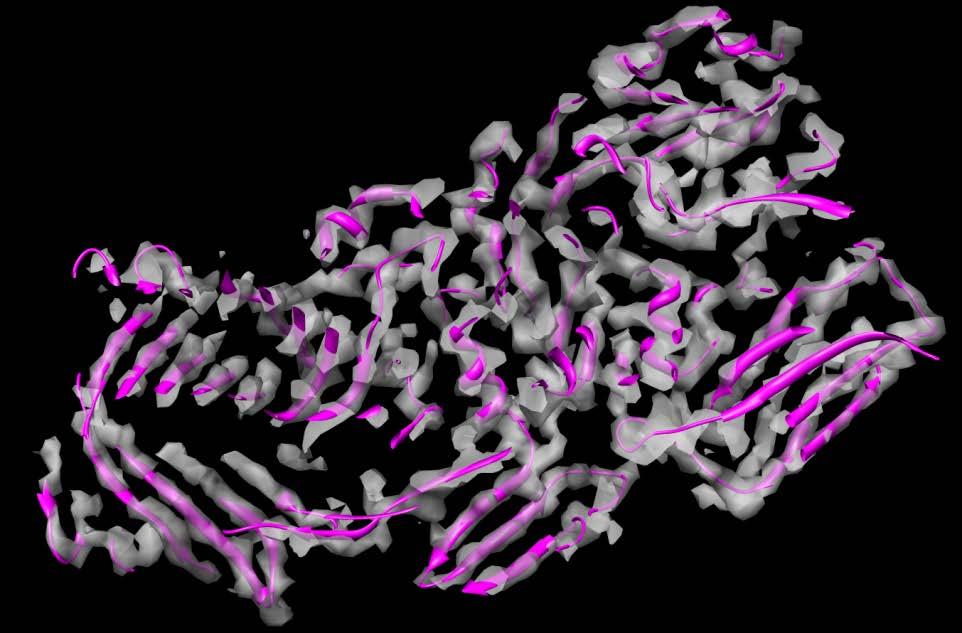
Nature Methods: doi: /nmeth Supplementary Figure 1. Atomic models in cryo-em maps.
 Supplementary Figure 1 Atomic models in cryo-em maps. (a) Two alpha (blue) and two beta (green) subunits of the T20S proteasome are shown as cartoon tubes fitted in a 3.2 Å potential map at isolevel 0.25
Part 1 X-ray Crystallography
 Part 1 X-ray Crystallography What happens to electron when it is hit by x-rays? 1. The electron starts vibrating with the same frequency as the x-ray beam 2. As a result, secondary beams will be scattered
Near-atomic resolution using electron cryomicroscopy and single-particle reconstruction
 Near-atomic resolution using electron cryomicroscopy and single-particle reconstruction Xing Zhang*, Ethan Settembre, Chen Xu, Philip R. Dormitzer, Richard Bellamy, Stephen C. Harrison **, and Nikolaus
Protein structure determination by 3D Electron Microscopy
 Biochemistry 9001 Protein structure determination by 3D Electron Microscopy Tommi A. White, Ph.D. Director, Electron Microscopy Core Facility Assistant Research Professor, Biochemistry Goals of this course
Chapter Electron cryomicroscopy of biological macromolecules
 International Tables for Crystallography (2012). Vol. F, Chapter 19.6, pp. 593 614. Chapter 19.6. Electron cryomicroscopy of biological macromolecules T. S. Baker and R. Henderson 0D 1D 2D 3D EM CryoTEM
THE DEVELOPMENT OF CRYO-ELECTRON MICROSCOPY
 4 OCTOBER 2017 Scientific Background on the Nobel Prize in Chemistry 2017 THE DEVELOPMENT OF CRYO-ELECTRON MICROSCOPY THE ROYAL SWEDISH ACADEMY OF SCIENCES has as its aim to promote the sciences and strengthen
Appl. Phys. Lett. 99, (2011) DOI: /
 DOI: /") Appl. Phys. Lett. 99, 133701 (2011) DOI: 10.1063/1.3645010 Single-walled carbon nanotubes and nanocrystalline graphene reduce beam-induced movements in high-resolution electron cryo-microscopy of ice-embedded
Appl. Phys. Lett. 99, 133701 (2011) DOI: 10.1063/1.3645010 Single-walled carbon nanotubes and nanocrystalline graphene reduce beam-induced movements in high-resolution electron cryo-microscopy of ice-embedded
From x-ray crystallography to electron microscopy and back -- how best to exploit the continuum of structure-determination methods now available
 From x-ray crystallography to electron microscopy and back -- how best to exploit the continuum of structure-determination methods now available Scripps EM course, November 14, 2007 What aspects of contemporary
Hommage à Jean Baptiste Joseph Fourier ( )
") 61.16D It Microsc. MicroanaL Microstruct. 1 (1990) OCTOBER/DECEMBER 1990, PAGE 423 423 Classification Physics Abstracts 61.00 - - 42.30 Structure analysis by crystallographic image processing Hommage à
61.16D It Microsc. MicroanaL Microstruct. 1 (1990) OCTOBER/DECEMBER 1990, PAGE 423 423 Classification Physics Abstracts 61.00 - - 42.30 Structure analysis by crystallographic image processing Hommage à
Review: Electron Crystallography: Present Excitement, a Nod to the Past, Anticipating the Future
 Journal of Structural Biology 128, 3 14 (1999) Article ID jsbi.1999.4172, available online at http://www.idealibrary.com on Review: Electron Crystallography: Present Excitement, a Nod to the Past, Anticipating
Single Particle Reconstruction with EMAN. GroEL. GroEL 2000 (15 Å) GroEL 2001 (11.5 Å) GroEL 2003 (6 Å)
 GroEL 2001 (11.5 Å) GroEL 2003 (6 Å)") Single Reconstruction with EMAN Methods for High Resolution Refinement in Single Processing Steve Ludtke Asst. Professor, Biochemistry, BCM Co-director,NCMI Donghua Chen Joanita Jakana Wah Chiu GroEL Jiu-Li
Single Reconstruction with EMAN Methods for High Resolution Refinement in Single Processing Steve Ludtke Asst. Professor, Biochemistry, BCM Co-director,NCMI Donghua Chen Joanita Jakana Wah Chiu GroEL Jiu-Li
Iterative Data Refinement for Soft X-ray Microscopy
 Iterative Data Refinement for Soft X-ray Microscopy Presented by Joanna Klukowska August 2, 2013 Outline Iterative Data Refinement Transmission X-ray Microscopy Numerical Tests Joanna Klukowska IDR for
Structure, Dynamics and Function of Membrane Proteins using 4D Electron Microscopy
 Structure, Dynamics and Function of Membrane Proteins using 4D Electron Microscopy Anthony W. P. Fitzpatrick Department of Chemistry, Lensfield Road, Cambridge, CB2 1EW My research aims to understand the
Visualization of Biological Nano-Machines at Subnanometer Resolutions
 Visualization of Biological Nano-Machines at Subnanometer Resolutions Wah Chiu,, Donghua Chen, Joanita Jakana, Juan Chang,, Wen Jiang,, Steven J Ludtke and Matthew L Baker National Center for Macromolecular
Realizing the potential of electron cryo-microscopy
 Quarterly Reviews of Biophysics 37, 1 (2004), pp. 3 13. f 2004 Cambridge University Press 3 DOI: 10.1017/S0033583504003920 Printed in the United Kingdom ESSAY Realizing the potential of electron cryo-microscopy
Determining Protein Structure BIBC 100
 Determining Protein Structure BIBC 100 Determining Protein Structure X-Ray Diffraction Interactions of x-rays with electrons in molecules in a crystal NMR- Nuclear Magnetic Resonance Interactions of magnetic
channel/ryanodine receptor from sarcoplasmic reticulum
 Cryo-EM of the native structure of the calcium release channel/ryanodine receptor from sarcoplasmic reticulum M. Radermacher, T. Wagenknecht, R. Grassucci, J. Frank, M. Inui,* C. Chadwick,* and S. Fleischer*
History of electron microscopy
 History of electron microscopy C9940 3-Dimensional Transmission Electron Microscopy S1007 Doing structural biology with the electron microscope February 16, 2015 Syllabus Week Date Instructor Topic 1 02/16
Atomic Resolution Cryo-EM Structure of
 Article Atomic Resolution Cryo-EM Structure of b-galactosidase Graphical Abstract Authors Alberto Bartesaghi, Cecilia Aguerrebere, Veronica Falconieri,..., Guillermo Sapiro, Xiongwu Wu, Sriram Subramaniam
HIGH RESOLUTION ELECTRON MICROSCOPY
 HIGH RESOLUTION ELECTRON MICROSCOPY BC530 Fall Quarter 2014 Wim G. J. Hol http://www.bmsc.washington.edu/wimhol/ Structure Determination by ELECTRON MICROSCOPY STRENGTHS: - Small amounts of material usually
Low-dose cryo electron ptychography via non-convex Bayesian optimization
 Low-dose cryo electron ptychography via non-convex Bayesian optimization Philipp Michael Pelz 1,,3,*, Wen Xuan Qiu 4, Robert Buecker 1,, Guenther Kassier 1,, and R.J. Dwayne Miller 1,,3,4 1 Max Planck
Fan, Hai-fu Institute of Physics, Chinese Academy of Sciences, Beijing , China
 Direct Methods in Crystallography Fan, Hai-fu Institute of Physics, Chinese Academy of Sciences, Beijing 100080, China An important branch of crystallography is the X-ray diffraction analysis of crystal
Three-Dimensional Electron Microscopy of Macromolecular Assemblies
 Three-Dimensional Electron Microscopy of Macromolecular Assemblies Joachim Frank Wadsworth Center for Laboratories and Research State of New York Department of Health The Governor Nelson A. Rockefeller
Nobel Prize in Physiology or Medicine 2017
 Nobel Prize in Physiology or Medicine 2017 Left to right: Michael Rosbash, Michael W. Young and Jeffrey C. Hall, have been awarded the Nobel Prize in Physiology/ Medicine, 2017. Who has won the Nobel Prize
Nanotechnology. CryoEM from Molecular Machines to Cells. electron, cryomicroscopy, cryotomography, nanomachine, cell
 Journal: CryoEM from Molecular Machines to Cells Manuscript ID: Wiley - Manuscript type: Date Submitted by the Author: Complete List of Authors: Keywords: NANO-31723-013.R1 Standard Article n/a Chiu, Wah
Electron-crystallographic Refinement of the Structure of Bacteriorhodopsin
 J. Mol. Biol. (1996) 259, 393 421 Electron-crystallographic Refinement of the Structure of Bacteriorhodopsin N. Grigorieff 1, T. A. Ceska 1, K. H. Downing 2, J. M. Baldwin 1 and R. Henderson 1 1 MRC Laboratory
Supplementary Information
 Supplementary Information Direct observation of crystal defects in an organic molecular crystals of copper hexachlorophthalocyanine by STEM-EELS Mitsutaka Haruta*, Hiroki Kurata Institute for hemical Research,
Electron Microscopy of Tobacco Mosaic Virus Prepared with the Aid of Negative Staining-Carbon Film Techniques
 J. gen. Virol. (1976), :3I, 265-269 Printed in Great Britain 265 Electron Microscopy of Tobacco Mosaic Virus Prepared with the Aid of Negative Staining-Carbon Film Techniques (Accepted 13 January I976)
This is a topic I encountered a number of years ago while at LBNL, when I came across the following statement [1]:
![This is a topic I encountered a number of years ago while at LBNL, when I came across the following statement [1]:](/thumbs/88/117124552.jpg "This is a topic I encountered a number of years ago while at LBNL, when I came across the following statement [1]:") Equivalent signal to noise in diffraction vs. bright-field experiments Charles Sindelar January 2010 SUMMARY This is a topic I encountered a number of years ago while at LBNL, when I came across the following
Equivalent signal to noise in diffraction vs. bright-field experiments Charles Sindelar January 2010 SUMMARY This is a topic I encountered a number of years ago while at LBNL, when I came across the following
Protein Crystallography
 Protein Crystallography Part II Tim Grüne Dept. of Structural Chemistry Prof. G. Sheldrick University of Göttingen http://shelx.uni-ac.gwdg.de tg@shelx.uni-ac.gwdg.de Overview The Reciprocal Lattice The
The Use of Synchrotron Radiation in Modern Research
 The Use of Synchrotron Radiation in Modern Research Physics Chemistry Structural Biology Materials Science Geochemical and Environmental Science Atoms, molecules, liquids, solids. Electronic and geometric
New algoritms for electron diffraction of 3D protein crystals. JP Abrahams, D Georgieva, L Jiang, I Sikhuralidze, NS Pannu
 New algoritms for electron diffraction of 3D protein crystals JP Abrahams, D Georgieva, L Jiang, I Sikhuralidze, NS Pannu Why new algorithms? New research questions New experimental techniques Better insight
Introduction to Three Dimensional Structure Determination of Macromolecules by Cryo-Electron Microscopy
 Introduction to Three Dimensional Structure Determination of Macromolecules by Cryo-Electron Microscopy Amit Singer Princeton University, Department of Mathematics and PACM July 23, 2014 Amit Singer (Princeton
On the interpretation of electron microscopic maps of biological macromolecules
 On the interpretation of electron microscopic maps of biological macromolecules Jimin Wang 1 * and Peter B. Moore 1,2 1 Department of Molecular Biophysics and Biochemistry, Yale University, New Haven,
3D and Atomic-resolution Imaging with Coherent Electron Nanobeams - Opportunities and Challenges for X-rays
 3D and Atomic-resolution Imaging with Coherent Electron Nanobeams - Opportunities and Challenges for X-rays David A. Muller Lena Fitting Kourkoutis, Megan Holtz, Robert Hovden, Qingyun Mao, Julia Mundy,
Measurement of ice thickness on vitreous ice embedded cryo-em grids: investigation of optimizing condition for visualizing macromolecules
 Cho et al. Journal of Analytical Science and Technology 2013, 4:7 TECHNICAL NOTE Measurement of ice thickness on vitreous ice embedded cryo-em grids: investigation of optimizing condition for visualizing
with cryo-electron microscopy
 2016MacmilanPublishersLimited,partofSpringerNature.Unravelling biological macromolecules with cryo-electron microscopy Rafael Fernandez-Leiro 1 * & Sjors H. W. Scheres 1 * Knowledge of the three-dimensional
Structure of the N intermediate of bacteriorhodopsin revealed by x-ray diffraction
 Proc. Natl. Acad. Sci. USA Vol. 93, pp. 1386-1390, February 1996 Biophysics Structure of the N intermediate of bacteriorhodopsin revealed by x-ray diffraction HIRONARI KAMIKUBO*, MIKIo KATAOKAtt, GYORGY
X-Ray structure analysis
 X-Ray structure analysis Kay Diederichs kay.diederichs@uni-konstanz.de Analysis of what? Proteins ( /ˈproʊˌtiːnz/ or /ˈproʊti.ɨnz/) are biochemical compounds consisting of one or more polypeptides typically
ParM filament images were extracted and from the electron micrographs and
 Supplemental methods Outline of the EM reconstruction: ParM filament images were extracted and from the electron micrographs and straightened. The digitized images were corrected for the phase of the Contrast
Research Article 289. *Corresponding author.
 Research Article 289 The three-dimensional structure of a DNA translocating machine at 10 Å resolution José María Valpuesta 1, José Jesús Fernández 2, José María Carazo 1 and José L Carrascosa 1 * Background:
 Supplemental Figure and Movie Legends Figure S1. Time course experiments on the thermal stability of apo AT cpn-α by native PAGE (related to Figure 2B). The samples were heated, respectively, to (A) 45
Molecular Mechanisms of self-assembly and motion of the bacterial flagellum
 Molecular Mechanisms of self-assembly and motion of the bacterial flagellum Mexico-Japan Workshop on Pharmacobiology and Nanobiology Universidad Nacional Autonoma de México (UNAM) Mexico City, Mexico 2009.02.25-26
Near-atomic resolution reconstructions of icosahedral viruses from electron cryo-microscopy Nikolaus Grigorieff 1,3 and Stephen C Harrison 2,3
 Available online at www.sciencedirect.com Near-atomic resolution reconstructions of icosahedral viruses from electron cryo-microscopy Nikolaus Grigorieff 1,3 and Stephen C Harrison 2,3 Nine different near-atomic
Electron Crystallography of CaMoO 4 Using High Voltage Electron Microscopy
 Electron Crystallography of CaMoO 4 Using High Voltage Electron Microscopy Bull. Korean Chem. Soc. 2007, Vol. 28, No. 3 391 Electron Crystallography of CaMoO 4 Using High Voltage Electron Microscopy Jin-Gyu
Electron energy filtering significantly improves amplitude contrast of frozen-hydrated protein at 300 kv
 Journal of Structural Biology xxx (2006) xxx xxx Journal of Structural Biology www.elsevier.com/locate/yjsbi Electron energy filtering significantly improves amplitude contrast of frozen-hydrated protein
Supporting Online Material for
 www.sciencemag.org/cgi/content/full/310/5753/1513/dc1 Supporting Online Material for Structural Roles for Human Translation Factor eif3 in Initiation of Protein Synthesis Bunpote Siridechadilok, Christopher
European Molecular Biology Laboratory Postfach , D-6900 Heidelberg, Fed. Rep. of Germany
 Ultramicroscopy 10 (1982) 55-62 North-Holland Publishing Company 55 FROZEN AQUEOUS SUSPENSIONS J. DUBOCHET, J.-J. CHANG, R. FREEMAN, J. LEPAULT and A.W. McDOWALL European Molecular Biology Laboratory Postfach
Scattering Lecture. February 24, 2014
 Scattering Lecture February 24, 2014 Structure Determination by Scattering Waves of radiation scattered by different objects interfere to give rise to an observable pattern! The wavelength needs to close
X-ray Crystallography
 2009/11/25 [ 1 ] X-ray Crystallography Andrew Torda, wintersemester 2009 / 2010 X-ray numerically most important more than 4/5 structures Goal a set of x, y, z coordinates different properties to NMR History
Comparison of single-particle analysis and electron tomography approaches: an overview
 Journal of Microscopy, Vol. 232, Pt 3 2008, pp. 562 579 Received 4 July 2007; accepted 2 March 2008 Comparison of single-particle analysis and electron tomography approaches: an overview S. JONIĆ, C.O.S.
Controlling protein adsorption on graphene for cryo-em using lowenergy hydrogen plasmas
 BRIEF COMMUNICATIONS 214 Nature America, Inc. All rights reserved. Controlling protein adsorption on graphene for cryo-em using lowenergy hydrogen plasmas Christopher J Russo & Lori A Passmore Despite
HOW TO ANALYZE SYNCHROTRON DATA
 HOW TO ANALYZE SYNCHROTRON DATA 1 SYNCHROTRON APPLICATIONS - WHAT Diffraction data are collected on diffractometer lines at the world s synchrotron sources. Most synchrotrons have one or more user facilities
MSE 321 Structural Characterization
 Auger Spectroscopy Auger Electron Spectroscopy (AES) Scanning Auger Microscopy (SAM) Incident Electron Ejected Electron Auger Electron Initial State Intermediate State Final State Physical Electronics
Designing of metallic nanocrystals embedded in non-stoichiometric perovskite nanomaterial and its surface-electronic characteristics
 Designing of metallic nanocrystals embedded in non-stoichiometric perovskite nanomaterial and its surface-electronic characteristics Jagadeesh Suriyaprakash 1,2, Y. B. Xu 1, Y. L. Zhu 1, L. X. Yang 1,
Improving cryo-em maps: focused 2D & 3D classifications and focused refinements
 Improving cryo-em maps: focused 2D & 3D classifications and focused refinements 3rd International Symposium on Cryo-3D Image Analysis 23.3.2018, Tahoe Bruno Klaholz Centre for Integrative Biology, IGBMC,
arxiv:physics/ v1 [physics.optics] 11 Feb 2005
![arxiv:physics/ v1 [physics.optics] 11 Feb 2005](/thumbs/87/97204936.jpg "arxiv:physics/ v1 [physics.optics] 11 Feb 2005") An assessment of the resolution limitation due to radiation-damage in x-ray diffraction microscopy arxiv:physics/0502059v1 [physics.optics] 11 Feb 2005 M. R. Howells, 1, T. Beetz, 1 H. N. Chapman, 2 C.
An assessment of the resolution limitation due to radiation-damage in x-ray diffraction microscopy arxiv:physics/0502059v1 [physics.optics] 11 Feb 2005 M. R. Howells, 1, T. Beetz, 1 H. N. Chapman, 2 C.
Transmission Electron Microscopy
 Transmission Electron Microscopy Fu-Rong Chen Transmission Electron Microscopy David B. Williams C. Barry Carter Background:Solid State Physics Materials Science 1.1 Why Electron Microscope? 1.1 Why Electron
Physical resolution limits of single particle 3D imaging with X-rays and electrons
 Physical resolution limits of single particle 3D imaging with X-rays and electrons Coherence 2005 Dirk Van Dyck and Sandra Van Aert June 15, 2005 Ultimate goal: imaging of atoms! Understanding properties
Technology for Micro- and Nanostructures Micro- and Nanotechnology
 Lecture 5: Electron-Beam Lithography, Part 1 Technology for Micro- and Nanostructures Micro- and Nanotechnology Peter Unger mailto: peter.unger @ uni-ulm.de Institute of Optoelectronics University of Ulm
Focused-ion-beam BRIEF COMMUNICATIONS. Michael Marko 1, Chyongere Hsieh 1, Richard Schalek 2, Joachim Frank 1,3 & Carmen Mannella 1
 BRIEF COMMUNICATIONS 2007 Nature Publishing Group http://www.nature.com/naturemethods Focused-ion-beam thinning of frozenhydrated biological specimens for cryoelectron microscopy Michael Marko 1, Chyongere
Determination of the high- pressure phases (II and IV ) ) of. techniques
 ) of. techniques") Determination of the high- pressure phases (II and IV ) ) of CuGeO 3 using single-crystal techniques Lauren A. Borkowski, 1,2 Barbara Lavina, 1,2 Przemyslaw Dera, 2 and Hanns-Peter Liermann 3 1 High Pressure
Determination of the high- pressure phases (II and IV ) ) of CuGeO 3 using single-crystal techniques Lauren A. Borkowski, 1,2 Barbara Lavina, 1,2 Przemyslaw Dera, 2 and Hanns-Peter Liermann 3 1 High Pressure
The Basic of Transmission Electron Microscope. Text book: Transmission electron microscopy by David B Williams & C. Barry Carter.
 The Basic of Transmission Electron Microscope Text book: Transmission electron microscopy by David B Williams & C. Barry Carter. 2009, Springer Background survey http://presemo.aalto.fi/tem1 Microscopy
Acta Cryst. (2017). D73, , doi: /s
. D73, , doi: /s") Supporting information Volume 73 (2017) Supporting information for article: 2017 publication guidelines for structural modelling of small-angle scattering data from biomolecules in solution: an update
Supporting information Volume 73 (2017) Supporting information for article: 2017 publication guidelines for structural modelling of small-angle scattering data from biomolecules in solution: an update
Measurement of Dynamic trna Movement and Visualizing Ribosome Biogenesis Applications of Time resolved Electron Cryo microscopy
 2011 TIGP CBMB Seminar Measurement of Dynamic trna Movement and Visualizing Ribosome Biogenesis Applications of Time resolved Electron Cryo microscopy Ribosome dynamics and trna movement by time resolved
object objective lens eyepiece lens
 Advancing Physics G495 June 2015 SET #1 ANSWERS Field and Particle Pictures Seeing with electrons The compound optical microscope Q1. Before attempting this question it may be helpful to review ray diagram
Synchrotron Methods in Nanomaterials Research
 Synchrotron Methods in Nanomaterials Research Marcel MiGLiERiNi Slovak University of Technology in Bratislava and Centre for Nanomaterials Research, Olomouc marcel.miglierini@stuba.sk www.nuc.elf.stuba.sk/bruno
David Martin Challenges in High Precision Beamline Alignment at the ESRF FIG Working Week Christchurch New Zealand 2016
 Presented at the FIG Working Week 2016, May 2-6, 2016 in Christchurch, New Zealand David Martin Challenges in High Precision Beamline Alignment at the ESRF FIG Working Week Christchurch New Zealand 2016
Studying Metal to Insulator Transitions in Solids using Synchrotron Radiation-based Spectroscopies.
 PY482 Lecture. February 28 th, 2013 Studying Metal to Insulator Transitions in Solids using Synchrotron Radiation-based Spectroscopies. Kevin E. Smith Department of Physics Department of Chemistry Division
AP5301/ Name the major parts of an optical microscope and state their functions.
 Review Problems on Optical Microscopy AP5301/8301-2015 1. Name the major parts of an optical microscope and state their functions. 2. Compare the focal lengths of two glass converging lenses, one with
Supplementary Information
 SSZ-52, a zeolite with an 18-layer aluminosilicate framework structure related to that of the DeNOx catalyst Cu-SSZ-13 Dan Xie 1,2, Lynne B. McCusker 2 *, Christian Baerlocher 2, Stacey I. Zones 1 *, Wei
X-Ray Damage to Biological Crystalline Samples
 X-Ray Damage to Biological Crystalline Samples Gerd Rosenbaum Structural Biology Center, ANL and Dept. of Biochemistry, UGA ACA Summer School IIT, 19 July 2007 A U.S. Department of Energy laboratory managed
Index. Born approximation, 128 Bremsstrahlung, 41, 42, 53 angular distribution, 42 energy distribution, 42
 Index Aberrations aberration-free holography, 123 chromatic, in confocal imaging, 195 off-axis, 2, 211 optical correction methods, 122 reduction of, 2 Acetylcholine receptors, 30 Acoustic impedance, 200
Chapter 10. Nanometrology. Oxford University Press All rights reserved.
 Chapter 10 Nanometrology Oxford University Press 2013. All rights reserved. 1 Introduction Nanometrology is the science of measurement at the nanoscale level. Figure illustrates where nanoscale stands
On the appearance of carboxylates in electrostatic potential maps
 ACCELERATED COMMUNICATIONS On the appearance of carboxylates in electrostatic potential maps Jimin Wang* Department of Molecular Biophysics and Biochemistry, Yale University, New Haven, Connecticut 06520
Opportunity and Challenge: the new era of cryo-em. Ning Gao School of Life Sciences Tsinghua University
 Opportunity and Challenge: the new era of cryo-em Ning Gao School of Life Sciences Tsinghua University 2015-10-20 Education and Research Experience Principal Investigator 2008.11-present Postdoc, Department
Cell Structure. Lab Exercise 6. Contents. Objectives. Introduction
 Lab Exercise Cell Structure Contents Objectives 1 Introduction 1 Activity.1 Cellular Structures 2 Activity.2 Matching Exercise 2 Activity.3 Identify Organelles 2 Resutls Section 3 Objectives - Identify
HHS Public Access Author manuscript Mol Cell. Author manuscript; available in PMC 2016 May 21.
 Cryo-EM: A Unique Tool for the Visualization of Macromolecular Complexity Eva Nogales 1 and Sjors H.W. Scheres 2 1 Molecular and Cell Biology Department, UC Berkeley, Berkeley, CA 94720-3220 Howard Hughes
Applications of scattering theory! From the structure of the proton! to protein structure!
 Applications of scattering theory From the structure of the proton to protein structure Nicuşor Tîmneanu 2016 Contents and goals What is scattering and why study it? How is the structure of matter determined?
15 Physics of Biological Systems
 15 Physics of Biological Systems Conrad Escher, Hans-Werner Fink, Tatiana Latychevskaia, Jean-Nicolas Longchamp, Marianna Lorenzo, Jonas Verges and Flavio Wicki in collaboration with: Dr. Roger Morin,
Ion Optics Simulations
 Ion Optics Simulations What it is. How it s useful. The SIMION ion optics software. How it works. Limitations and cautions Demonstrations and examples A little hands on exploring 1 Ion Optics Simulations