X-Ray structure analysis
|
|
|
- Gabriel Newman
- 6 years ago
- Views:
Transcription
1 X-Ray structure analysis Kay Diederichs
 are biochemical compounds consisting of one or more polypeptides typically folded into a globular or fibrous form, facilitating a")

2 Analysis of what? Proteins ( /ˈproʊˌtiːnz/ or /ˈproʊti.ɨnz/) are biochemical compounds consisting of one or more polypeptides typically folded into a globular or fibrous form, facilitating a biological function. A polypeptide is a single linear polymer chain of amino acids bonded together by peptide bonds between the carboxyl and amino groups of adjacent amino acid residues. The sequence of amino acids in a protein is defined by the sequence of a gene, which is encoded in the genetic code. In general, the genetic code specifies 20 standard amino acids. (Wikipedia) 2
3 What is Macromolecular Crystallography? The art of getting a protein to sit still then taking a 3D picture What are protein crystals? Static, well-ordered arrays of protein molecules 3D-pictures are stitched together from 2D ones. How are the 2D-pictures made? By irradiating the ordered protein array with X-rays, collecting the constructively diffracted X-rays, and reconstructing a likely model of the protein s 3D structure using a computer 3 3
4 Overview History and current status Practical aspects Theory Comparison with other techniques 4


")
5 Absorption of X-rays Wilhelm Conrad Röntgen ( ) January 23,
6 Diffraction of X-rays Max von Laue ( ) Paul Peter Ewald ( ) X-ray diffraction of crystals (1912) theoretical explanation (1912) William Henry Bragg ( ) William Lawrence Bragg ( ) Bragg's equation: nλ = 2d sin Θ (Nobel Prize, 1915) 6
 indicate that the bases are stacked perpendicular to the axis of the molecule. http://www.pbs.")
7 X-ray diffraction patterns of DNA Rosalind Franklin and Maurice Wilkins (1953) The central cross shaped pattern as indicative of a helical structure. The heavy dark patterns (left and right) indicate that the bases are stacked perpendicular to the axis of the molecule. 7

8 DNA Structure: History 8
 sausage Balsa")

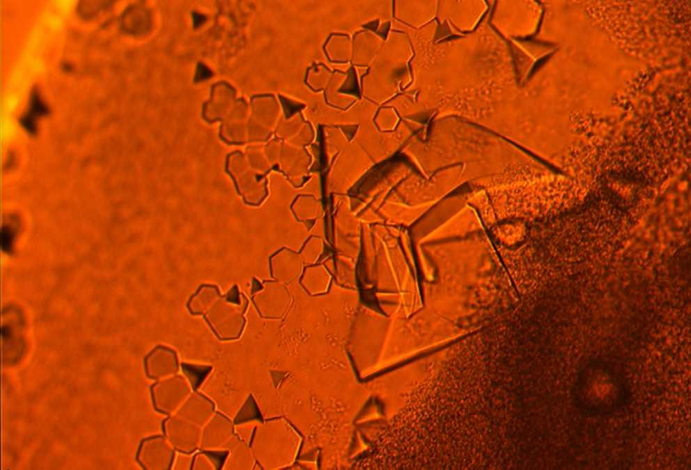
9 Myo- and haemoglobin models at 5.5 Å resolution (1959) sausage Balsa wood 9
, 1960 10 www.umass.")
10 2 Å Myoglobin model built by A. A. Barker, Model Maker in Cambridge (UK),

11 11
12 So far, 29 Nobel Prizes are associated with crystallography For a list, see Either for physical basis or mathematical treatment ( Physics ) or important chemical compounds ( Chemistry ) or Physiology and Medicine (DNA; Crick, Watson, Wilkins 1962) Most recently: (2013 Karplus, Levitt & Warshel); 2012 Lefkowitz & Kubilka; 2011 Shechtman: Quasicrystals; 2009 Ramakrishnan, Steitz, Yonath: Studies of the structure and function of the ribosome 14 of the 29 were awarded in Structural Biology (starting in 1946) See 12


13 Examples of high-profile structures Protein translocation through the SecA SecY complex Ribosome with mrna Structure of Ebola virus 13


14 14

15 From Protein Data Bank (PDB) file Crystal Structure at 1.9 Å Resolution of HIV II Protease J.Biol.Chem. v269 pp ,
16 HEADER COMPND COMPND SOURCE SOURCE EXPDTA REMARK REMARK REMARK REMARK REMARK SEQRES HYDROLASE (ACID PROTEINASE) 31-MAR-95 2 MOLECULE: HIV-1 PROTEASE; 3 CHAIN: A, B; 2 ORGANISM_SCIENTIFIC: HUMAN IMMUNODEFICIENCY VIRUS TYPE 1; 3 GENE: HIV-1 PROTEASE FROM THE NY5 ISOLATE; X-RAY DIFFRACTION 2 2 RESOLUTION. 2.0 ANGSTROMS. 3 R VALUE RMSD BOND DISTANCES ANGSTROMS 3 RMSD BOND ANGLES 1.9 DEGREES 1 A 99 PRO GLN ILE THR LEU TRP GLN ARG PRO LEU VAL THR ILE 1 N PRO A CA PRO A C PRO A O PRO A CB PRO A CG PRO A CD PRO A N GLN A CA GLN A C GLN A O GLN A CB GLN A CG GLN A CD GLN A OE1 GLN A NE2 GLN A N ILE A CA ILE A C ILE A O ILE A

17 Thermus thermophilus 70S ribosome PDB id 2WDI: 32 chains; atoms reflections, 3.3 Å resolution Voorhees et al (2009) Nature Structural Molecular Biology 16,

 (x,y,z)")
18 Structure Determination Phases (hkl) Crystal h, k, l, I, (I) (x,y,z) Structure 18


19 First steps of X-ray structure analysis: Choice of protein/organism/expression system Expression and purification Crystallization ( 19


20 Crystals R32 & R3 P321 C2 20

21 Synchrotron Radiation Synchrotron Radiation occurs when a charge moves at relativistic speed following a curved trajectory. 1. high brilliance 1. large spectral range 2. time structure 21
, Villigen")
22 Data collection: Swiss Light Source Paul-Scherrer-Institut (PSI), Villigen (CH) 22
23 Diffraction Data Collection The data are 3-dimensional the crystal has to be rotated through a large angular range, and for each orientation a diffraction image is recorded on the detector. The symmetry of the diffraction pattern means that depending on the space group, e.g. 90 rotation suffice. 23

 std dev of")
24 Diffraction Data Collection 2 pieces of information h, k, l Miller indices I(h,k,l) intensity I(hkl) std dev of I(h,k,l) 24

25 The measured intensity (and the accuracy of its measurement) are influenced by: - Crystal quality - Poisson (counting) statistics - Beam strength and quality; exposure time - Radiation damage - Beamline setup and qualitymurakami et al., Nature (2002) 25
26 Ewald sphere: Bragg's eqn in 3D 26
27 27
28 28
29 Theory: the electromagnetic wave... can be mathematically described by Maxwell s equations (1864): 29
30 What does this mean? Visualization is possible with Radiation2D see T. Shintake, New Mathematical Method for Radiation Field of Moving Charge, Proc. EPAC (2002) download of binary from for Linux, Mac, Windows 30
31 Diffraction maths Superposition of all waves emanating from all electrons of an object results in a diffraction image Mathematical description of wave from x,y,z is f*e-2 i(hx+ky+lz) Mathematically, addition of waves is a Fourier transform (array of complex numbers that is 1:1 related to the electrons of the object) The amplitude of the Fourier transform can be measured by a detector Its phase cannot be measured ( Phase Problem ) but is required to calculate the electron density A regularly ordered (i.e. crystalline) sample has a diffraction image consisting of regularly spaced reflections that are characterized by their position and intensity on the detector All electrons of the object contribute to all reflections! 31
32 The Structure Factor Equation F(hkl) = F(hkl) ei (hkl) = j fj e2 i(hxj+kyj+lzj) Structure factor amplitude F(hkl) I(hkl)1/2 Atomic form factor fj Phase (hkl) Complex plane The calculation of F(hkl) from a structure (xj,yj,zj) is just a summation of the waves originating from each atom (j) in the direction defined by (hkl). 32
33 The Electron Density Equation (x,y,z) = 1/V hkl F(hkl) ei (hkl) e-2 i(hx+ky+lz) Structure factor amplitude F(hkl) I(hkl)1/2 Phase (hkl) Mathematically inclined people will notice: this is just the Fourier back transform! 33
34 The Electron Density Equation The electron density (x,y,z) is a three-dimensional function (with the unit e/å3), which describes where in the unit cell of the crystal the electrons (and therefore the atoms) are. It is basically the image of the structure we want to determine. (x,y,z) = 1/V hkl F(hkl) ei (hkl) e-2 i(hx+ky+lz) It is important to note that every reflection (hkl) of the diffraction pattern contributes to the electron density at each and every position (xyz) in the unit cell of the crystal. 34
35 Interactive tutorials
36 Detectors do not measure amplitudes! they measure deposited energy the energy is ~ amplitude 2 thus, detectors don't measure phase because amplitude * ei (hkl) = amplitude 36
37 Data processing Indexing Integration (=summation) Space group determination Scaling => alle h,k,l,i(hkl),σ(ihkl) PDB depositions XDS DENZO or HKL MOSFLM
38 The Phase Problem From the diffraction pattern, we can only obtain the intensities I(hkl) of the reflections (hkl). Intensities are the squares of the (complex) amplitudes: I(hkl) ~ F(hkl) F*(hkl) = F(hkl) ei (hkl) F(hkl) e-i (hkl) = F(hkl) 2 The phase (hkl) cannot be measured. 38
39 How to solve the Phase problem / an X-ray structure - Direct Methods: suitable for highest resolution data and few atoms, usually not applicable for macromolecules - Molecular Replacement: obtain a related/similar (= approximately correct) structure from the PDB, orient it correctly in the crystal lattice, identify and remove errors until the atomic model agrees with the experimental data. Not applicable to new/unknown structures. 2/3 of X-ray entries of PDB. - Experimental Phase Determination (MIR/MAD/SAD): modify the scattering of the object, measure intensities again, work out phase from change in intensities. Requires highly accurate measurement of intensities. Always applicable. Other 1/3 of X-ray entries of PDB. 39
Protein Crystallography. Mitchell Guss University of Sydney Australia
 Protein Crystallography Mitchell Guss University of Sydney Australia Outline of the talk Recap some basic crystallography and history Highlight the special requirements for protein (macromolecular) structure
Fundamentals of X-ray diffraction
 Fundamentals of X-ray diffraction Elena Willinger Lecture series: Modern Methods in Heterogeneous Catalysis Research Outline History of X-ray Sources of X-ray radiation Physics of X-ray scattering Fundamentals
Lecture 1. Introduction to X-ray Crystallography. Tuesday, February 1, 2011
 Lecture 1 Introduction to X-ray Crystallography Tuesday, February 1, 2011 Protein Crystallography Crystal Structure Determination in Principle: From Crystal to Structure Dr. Susan Yates Contact Information
The ideal fiber pattern exhibits 4-quadrant symmetry. In the ideal pattern the fiber axis is called the meridian, the perpendicular direction is
 Fiber diffraction is a method used to determine the structural information of a molecule by using scattering data from X-rays. Rosalind Franklin used this technique in discovering structural information
X-ray Crystallography
 2009/11/25 [ 1 ] X-ray Crystallography Andrew Torda, wintersemester 2009 / 2010 X-ray numerically most important more than 4/5 structures Goal a set of x, y, z coordinates different properties to NMR History
Drug targets, Protein Structures and Crystallography
 Drug targets, Protein Structures and Crystallography NS5B viral RNA polymerase (RNA dep) Hepa88s C drug Sofosbuvir (Sovaldi) FDA 2013 Epclusa - combina8on with Velpatasvir approved in in 2016) Prodrug
Scattering Lecture. February 24, 2014
 Scattering Lecture February 24, 2014 Structure Determination by Scattering Waves of radiation scattered by different objects interfere to give rise to an observable pattern! The wavelength needs to close
Proteins. Central Dogma : DNA RNA protein Amino acid polymers - defined composition & order. Perform nearly all cellular functions Drug Targets
 Proteins Central Dogma : DNA RNA protein Amino acid polymers - defined composition & order Perform nearly all cellular functions Drug Targets Fold into discrete shapes. Proteins - cont. Specific shapes
Scattering by two Electrons
 Scattering by two Electrons p = -r k in k in p r e 2 q k in /λ θ θ k out /λ S q = r k out p + q = r (k out - k in ) e 1 Phase difference of wave 2 with respect to wave 1: 2π λ (k out - k in ) r= 2π S r
Methods in Chemistry III Part 1 Modul M.Che.1101 WS 2010/11 1 Modern Methods of Inorganic Chemistry Mi 10:15-12:00, Hörsaal II George Sheldrick
 Methods in Chemistry III Part 1 Modul M.Che.1101 WS 2010/11 1 Modern Methods of Inorganic Chemistry Mi 10:15-12:00, Hörsaal II George Sheldrick gsheldr@shelx.uni-ac.gwdg.de Part 1. Crystal structure determination
Data File Formats. There are dozens of file formats for chemical data.
 1 Introduction There are dozens of file formats for chemical data. We will do an overview of a few that are often used in structural bioinformatics. 2 1 PDB File Format (1) The PDB file format specification
Protein Crystallography
 Protein Crystallography Part II Tim Grüne Dept. of Structural Chemistry Prof. G. Sheldrick University of Göttingen http://shelx.uni-ac.gwdg.de tg@shelx.uni-ac.gwdg.de Overview The Reciprocal Lattice The
3.012 Fund of Mat Sci: Structure Lecture 18
 3.012 Fund of Mat Sci: Structure Lecture 18 X-RAYS AT WORK An X-ray diffraction image for the protein myoglobin. Source: Wikipedia. Model of helical domains in myoglobin. Image courtesy of Magnus Manske
X-ray Data Collection. Bio5325 Spring 2006
 X-ray Data Collection Bio535 Spring 006 Obtaining I hkl and α (Ihkl) from Frame Images Braggs Law -predicts conditions for in-phase scattering by equivalent atoms lying in planes that transect a crystal.
Crystals, X-rays and Proteins
 Crystals, X-rays and Proteins Comprehensive Protein Crystallography Dennis Sherwood MA (Hons), MPhil, PhD Jon Cooper BA (Hons), PhD OXFORD UNIVERSITY PRESS Contents List of symbols xiv PART I FUNDAMENTALS
Protein crystallography. Garry Taylor
 Protein crystallography Garry Taylor X-ray Crystallography - the Basics Grow crystals Collect X-ray data Determine phases Calculate ρ-map Interpret map Refine coordinates Do the biology. Nitrogen at -180
Principles of Physical Biochemistry
 Principles of Physical Biochemistry Kensal E. van Hold e W. Curtis Johnso n P. Shing Ho Preface x i PART 1 MACROMOLECULAR STRUCTURE AND DYNAMICS 1 1 Biological Macromolecules 2 1.1 General Principles
Determining Protein Structure BIBC 100
 Determining Protein Structure BIBC 100 Determining Protein Structure X-Ray Diffraction Interactions of x-rays with electrons in molecules in a crystal NMR- Nuclear Magnetic Resonance Interactions of magnetic
X-ray Crystallography. Kalyan Das
 X-ray Crystallography Kalyan Das Electromagnetic Spectrum NMR 10 um - 10 mm 700 to 10 4 nm 400 to 700 nm 10 to 400 nm 10-1 to 10 nm 10-4 to 10-1 nm X-ray radiation was discovered by Roentgen in 1895. X-rays
Rajesh Prasad Department of Applied Mechanics Indian Institute of Technology New Delhi
 TEQIP WORKSHOP ON HIGH RESOLUTION X-RAY AND ELECTRON DIFFRACTION, FEB 01, 2016, IIT-K. Introduction to x-ray diffraction Peak Positions and Intensities Rajesh Prasad Department of Applied Mechanics Indian
Computational Molecular Modeling
 Computational Molecular Modeling Lecture 1: Structure Models, Properties Chandrajit Bajaj Today s Outline Intro to atoms, bonds, structure, biomolecules, Geometry of Proteins, Nucleic Acids, Ribosomes,
PROBING CRYSTAL STRUCTURE
 PROBING CRYSTAL STRUCTURE Andrew Baczewski PHY 491, October 10th, 2011 OVERVIEW First - we ll briefly discuss Friday s quiz. Today, we will answer the following questions: How do we experimentally probe
3.012 Structure An Introduction to X-ray Diffraction
 3.012 Structure An Introduction to X-ray Diffraction This handout summarizes some topics that are important for understanding x-ray diffraction. The following references provide a thorough explanation
Molecular Biology Course 2006 Protein Crystallography Part I
 Molecular Biology Course 2006 Protein Crystallography Part I Tim Grüne University of Göttingen Dept. of Structural Chemistry November 2006 http://shelx.uni-ac.gwdg.de tg@shelx.uni-ac.gwdg.de Overview Overview
X-ray Crystallography BMB/Bi/Ch173 02/06/2017
 1. Purify your protein 2. Crystallize protein 3. Collect diffraction data 4. Get experimental phases 5. Generate an electron density map 6. Build a model 7. Refine the model 8. Publish X-ray Crystallography
 Helpful resources for all X ray lectures Crystallization http://www.hamptonresearch.com under tech support: crystal growth 101 literature Spacegroup tables http://img.chem.ucl.ac.uk/sgp/mainmenu.htm Crystallography
X-Ray Scattering Studies of Thin Polymer Films
 X-Ray Scattering Studies of Thin Polymer Films Introduction to Neutron and X-Ray Scattering Sunil K. Sinha UCSD/LANL Acknowledgements: Prof. R.Pynn( Indiana U.) Prof. M.Tolan (U. Dortmund) Wilhelm Conrad
Introduction. Chem 6850/8850 X-ray Crystallography The University of Toledo.
 Introduction Chem 6850/8850 X-ray Crystallography The University of Toledo cora.lind@utoledo.edu Course Goals To develop an understanding of basic crystallographic concepts - Helpful if you ever need to
David Martin Challenges in High Precision Beamline Alignment at the ESRF FIG Working Week Christchurch New Zealand 2016
 Presented at the FIG Working Week 2016, May 2-6, 2016 in Christchurch, New Zealand David Martin Challenges in High Precision Beamline Alignment at the ESRF FIG Working Week Christchurch New Zealand 2016
Handout 7 Reciprocal Space
 Handout 7 Reciprocal Space Useful concepts for the analysis of diffraction data http://homepages.utoledo.edu/clind/ Concepts versus reality Reflection from lattice planes is just a concept that helps us
Protein Data Bank Contents Guide: Atomic Coordinate Entry Format Description. Version Document Published by the wwpdb
 Protein Data Bank Contents Guide: Atomic Coordinate Entry Format Description Version 3.30 Document Published by the wwpdb This format complies with the PDB Exchange Dictionary (PDBx) http://mmcif.pdb.org/dictionaries/mmcif_pdbx.dic/index/index.html.
Working with protein structures. Benjamin Jack
 Working with protein structures Benjamin Jack Structure of Triosephosphate Isomerase PDB ID: 1HTI loop beta sheet alpha helix Different perspectives of the same structure Structure of Truncated Hemoglobin
Analytical Methods for Materials
 Analytical Methods for Materials Lesson 15 Reciprocal Lattices and Their Roles in Diffraction Studies Suggested Reading Chs. 2 and 6 in Tilley, Crystals and Crystal Structures, Wiley (2006) Ch. 6 M. DeGraef
Crystallography past, present and future
 Crystallography past, present and future Jenny P. Glusker Philadelphia, PA, U. S. A. International Year of Crystallography UNESCO, Paris, France 20 January 2014 QUARTZ CRYSTALS Quartz crystals found growing
SOLID STATE 18. Reciprocal Space
 SOLID STATE 8 Reciprocal Space Wave vectors and the concept of K-space can simplify the explanation of several properties of the solid state. They will be introduced to provide more information on diffraction
3D Visualization of Drugs-Protein Complexes
 3D Visualization of Drugs-Protein Complexes Goal: Develop better understanding of Protein Database and its entries Plan Introductory information about protein structure database Learn Molsoft-browser for
Chapter 2 Structures. 2.1 Introduction Storing Protein Structures The PDB File Format
 Chapter 2 Structures 2.1 Introduction The three-dimensional (3D) structure of a protein contains a lot of information on its function, and can be used for devising ways of modifying it (propose mutants,
Protein Structure Determination. Why Bother With Structure? Protein Sequences Far Outnumber Structures. Growth of Structural Data
 Protein Structure Determination Why Bother With Structure? The amino acid sequence of a protein contains interesting information. A protein sequence can be compared to other protein sequences to establish
Two Lectures in X-ray Crystallography
 Biochemistry 503 Michael Wiener (mwiener@virginia.edu, 3-2731, Snyder 360) Two Lectures in X-ray Crystallography Outline 1. Justification & introductory remarks 2. Experimental setup 3. Protein crystals
Crystal planes. Neutrons: magnetic moment - interacts with magnetic materials or nuclei of non-magnetic materials. (in Å)
") Crystallography: neutron, electron, and X-ray scattering from periodic lattice, scattering of waves by periodic structures, Miller indices, reciprocal space, Ewald construction. Diffraction: Specular,
Crystallography: neutron, electron, and X-ray scattering from periodic lattice, scattering of waves by periodic structures, Miller indices, reciprocal space, Ewald construction. Diffraction: Specular,
X-ray, Neutron and e-beam scattering
 X-ray, Neutron and e-beam scattering Introduction Why scattering? Diffraction basics Neutrons and x-rays Techniques Direct and reciprocal space Single crystals Powders CaFe 2 As 2 an example What is the
Basics of protein structure
 Today: 1. Projects a. Requirements: i. Critical review of one paper ii. At least one computational result b. Noon, Dec. 3 rd written report and oral presentation are due; submit via email to bphys101@fas.harvard.edu
Resolution: maximum limit of diffraction (asymmetric)
") Resolution: maximum limit of diffraction (asymmetric) crystal Y X-ray source 2θ X direct beam tan 2θ = Y X d = resolution 2d sinθ = λ detector 1 Unit Cell: two vectors in plane of image c* Observe: b*
Resolution: maximum limit of diffraction (asymmetric) crystal Y X-ray source 2θ X direct beam tan 2θ = Y X d = resolution 2d sinθ = λ detector 1 Unit Cell: two vectors in plane of image c* Observe: b*
PSD '18 -- Xray lecture 4. Laue conditions Fourier Transform The reciprocal lattice data collection
 PSD '18 -- Xray lecture 4 Laue conditions Fourier Transform The reciprocal lattice data collection 1 Fourier Transform The Fourier Transform is a conversion of one space into another space with reciprocal
Details of Protein Structure
 Details of Protein Structure Function, evolution & experimental methods Thomas Blicher, Center for Biological Sequence Analysis Anne Mølgaard, Kemisk Institut, Københavns Universitet Learning Objectives
Roger Johnson Structure and Dynamics: X-ray Diffraction Lecture 6
 6.1. Summary In this Lecture we cover the theory of x-ray diffraction, which gives direct information about the atomic structure of crystals. In these experiments, the wavelength of the incident beam must
Crystal lattice Real Space. Reflections Reciprocal Space. I. Solving Phases II. Model Building for CHEM 645. Purified Protein. Build model.
 I. Solving Phases II. Model Building for CHEM 645 Purified Protein Solve Phase Build model and refine Crystal lattice Real Space Reflections Reciprocal Space ρ (x, y, z) pronounced rho F hkl 2 I F (h,
Molecular Modeling lecture 2
 Molecular Modeling 2018 -- lecture 2 Topics 1. Secondary structure 3. Sequence similarity and homology 2. Secondary structure prediction 4. Where do protein structures come from? X-ray crystallography
Basic Crystallography Part 1. Theory and Practice of X-ray Crystal Structure Determination
 Basic Crystallography Part 1 Theory and Practice of X-ray Crystal Structure Determination We have a crystal How do we get there? we want a structure! The Unit Cell Concept Ralph Krätzner Unit Cell Description
X-ray analysis. 1. Basic crystallography 2. Basic diffraction physics 3. Experimental methods
 X-ray analysis 1. Basic crystallography 2. Basic diffraction physics 3. Experimental methods Introduction Noble prizes associated with X-ray diffraction 1901 W. C. Roentgen (Physics) for the discovery
1/40. Cellular mechanics I nd term
 1/40 Cellular mechanics I 2018. 2 nd term Various Science 2/40 Biology Physics Chemistry Biology is the science of life.... Biologists study the structure, function, growth, origin, evolution and distribution
CRYSTALLOGRAPHY AND STORYTELLING WITH DATA. President, Association of Women in Science, Bethesda Chapter STEM Consultant
 CRYSTALLOGRAPHY AND STORYTELLING WITH DATA President, Association of Women in Science, Bethesda Chapter STEM Consultant MY STORY Passion for Science BS Biology Major MS Biotechnology & Project in Bioinformatics
Biological Small Angle X-ray Scattering (SAXS) Dec 2, 2013
 Dec 2, 2013") Biological Small Angle X-ray Scattering (SAXS) Dec 2, 2013 Structural Biology Shape Dynamic Light Scattering Electron Microscopy Small Angle X-ray Scattering Cryo-Electron Microscopy Wide Angle X- ray
Biological Small Angle X-ray Scattering (SAXS) Dec 2, 2013 Structural Biology Shape Dynamic Light Scattering Electron Microscopy Small Angle X-ray Scattering Cryo-Electron Microscopy Wide Angle X- ray
Contents. xiii. Preface v
 Contents Preface Chapter 1 Biological Macromolecules 1.1 General PrincipIes 1.1.1 Macrornolecules 1.2 1.1.2 Configuration and Conformation Molecular lnteractions in Macromolecular Structures 1.2.1 Weak
Supplementary Figure 3 a. Structural comparison between the two determined structures for the IL 23:MA12 complex. The overall RMSD between the two
 Supplementary Figure 1. Biopanningg and clone enrichment of Alphabody binders against human IL 23. Positive clones in i phage ELISA with optical density (OD) 3 times higher than background are shown for
X-ray crystallography NMR Cryoelectron microscopy
 Molecular Graphics with PyMOL Overview of: Protein Data Bank Coordinates Jean-Yves Sgro PyMOL interface Hands-on! Experimental Methods 3 Main: X-ray crystallography NMR Cryoelectron microscopy X-ray source
Protein Structure and Visualisation. Introduction to PDB and PyMOL
 Protein Structure and Visualisation Introduction to PDB and PyMOL 1 Feedback Persons http://www.bio-evaluering.dk/ 2 Program 8.00-8.15 Quiz results 8.15-8.50 Introduction to PDB & PyMOL 8.50-9.00 Break
Molecular Biology Course 2006 Protein Crystallography Part II
 Molecular Biology Course 2006 Protein Crystallography Part II Tim Grüne University of Göttingen Dept. of Structural Chemistry December 2006 http://shelx.uni-ac.gwdg.de tg@shelx.uni-ac.gwdg.de Overview
Protein Structure Prediction and Display
 Protein Structure Prediction and Display Goal Take primary structure (sequence) and, using rules derived from known structures, predict the secondary structure that is most likely to be adopted by each
introduction to SAXS for polymers -a user view-
 introduction to SAXS for polymers -a user view- Luigi Balzano DSM Ahead/Material Science Center Geleen, The Netherlands luigi.balzano@dsm.com Synchrotron and Neutron Workshop (SyNeW) 2015 Utrecht, June
Protein Structure Determination. How are these structures determined?
 Protein Structure Determination How are these structures determined? Why Bother With Structure? The amino acid sequence of a protein contains interesting information. A protein sequence can be compared
SHELXC/D/E. Andrea Thorn
 SHELXC/D/E Andrea Thorn What is experimental phasing? Experimental phasing is what you do if MR doesn t work. What is experimental phasing? Experimental phasing methods depend on intensity differences.
Protein Structure Determination. Why Bother With Structure? Protein Sequences Far Outnumber Structures
 Protein Structure Determination How are these structures determined? Why Bother With Structure? The amino acid sequence of a protein contains interesting information. A protein sequence can be compared
Experimental Determination of Crystal Structure
 Experimental Determination of Crystal Structure Branislav K. Nikolić Department of Physics and Astronomy, University of Delaware, U.S.A. PHYS 624: Introduction to Solid State Physics http://www.physics.udel.edu/~bnikolic/teaching/phys624/phys624.html
Packing of Secondary Structures
 7.88 Lecture Notes - 4 7.24/7.88J/5.48J The Protein Folding and Human Disease Professor Gossard Retrieving, Viewing Protein Structures from the Protein Data Base Helix helix packing Packing of Secondary
Introduction to" Protein Structure
 Introduction to" Protein Structure Function, evolution & experimental methods Thomas Blicher, Center for Biological Sequence Analysis Learning Objectives Outline the basic levels of protein structure.
3.17 Strukturanalyse mit Röntgenstrahlen nach Debye- Scherrer
 3.17 Strukturanalyse mit Röntgenstrahlen nach Debye- Scherrer Ausarbeitung (engl.) Fortgeschrittenenpraktikum an der TU Darmstadt Versuch durchgeführt von: Mussie Beian, Jan Schupp, Florian Wetzel Versuchsdatum:
Chapter 2. X-ray X. Diffraction and Reciprocal Lattice. Scattering from Lattices
 Chapter. X-ray X Diffraction and Reciprocal Lattice Diffraction of waves by crystals Reciprocal Lattice Diffraction of X-rays Powder diffraction Single crystal X-ray diffraction Scattering from Lattices
X-ray Diffraction. Diffraction. X-ray Generation. X-ray Generation. X-ray Generation. X-ray Spectrum from Tube
 X-ray Diffraction Mineral identification Mode analysis Structure Studies X-ray Generation X-ray tube (sealed) Pure metal target (Cu) Electrons remover inner-shell electrons from target. Other electrons
Central Dogma. modifications genome transcriptome proteome
 entral Dogma DA ma protein post-translational modifications genome transcriptome proteome 83 ierarchy of Protein Structure 20 Amino Acids There are 20 n possible sequences for a protein of n residues!
HIV protease inhibitor. Certain level of function can be found without structure. But a structure is a key to understand the detailed mechanism.
 Proteins are linear polypeptide chains (one or more) Building blocks: 20 types of amino acids. Range from a few 10s-1000s They fold into varying three-dimensional shapes structure medicine Certain level
1. Protein Data Bank (PDB) 1. Protein Data Bank (PDB)
 1. Protein Data Bank (PDB)") Protein structure databases; visualization; and classifications 1. Introduction to Protein Data Bank (PDB) 2. Free graphic software for 3D structure visualization 3. Hierarchical classification of protein
Protein structure databases; visualization; and classifications 1. Introduction to Protein Data Bank (PDB) 2. Free graphic software for 3D structure visualization 3. Hierarchical classification of protein
Computational structural biology and bioinformatics
 Computational structural biology and bioinformatics What is it all about? Why take it? What are we going to be doing? Organizational notes. Grades etc. Books. CS6104. Spring CS6104. 04. Spring Alexey 04.
Section II Understanding the Protein Data Bank
 Section II Understanding the Protein Data Bank The focus of Section II of the MSOE Center for BioMolecular Modeling Jmol Training Guide is to learn about the Protein Data Bank, the worldwide repository
Overview - Macromolecular Crystallography
 Overview - Macromolecular Crystallography 1. Overexpression and crystallization 2. Crystal characterization and data collection 3. The diffraction experiment 4. Phase problem 1. MIR (Multiple Isomorphous
Molecular Graphics with PyMOL
 Molecular Graphics with PyMOL Jean)YvesSgro Instructors Molecular Graphics & Scientific Communication Ann Palmenberg Jean-Yves Sgro Marchel Hill Holly Basta H. Adam Steinberg 1 Lab Book : Section 1 Computer
CHEM 463: Advanced Inorganic Chemistry Modeling Metalloproteins for Structural Analysis
 CHEM 463: Advanced Inorganic Chemistry Modeling Metalloproteins for Structural Analysis Purpose: The purpose of this laboratory is to introduce some of the basic visualization and modeling tools for viewing
X-ray Crystallography I. James Fraser Macromolecluar Interactions BP204
 X-ray Crystallography I James Fraser Macromolecluar Interactions BP204 Key take-aways 1. X-ray crystallography results from an ensemble of Billions and Billions of molecules in the crystal 2. Models in
Electron Density at various resolutions, and fitting a model as accurately as possible.
 Section 9, Electron Density Maps 900 Electron Density at various resolutions, and fitting a model as accurately as possible. ρ xyz = (Vol) -1 h k l m hkl F hkl e iφ hkl e-i2π( hx + ky + lz ) Amplitude
A Primer in X-ray Crystallography for Redox Biologists. Mark Wilson Karolinska Institute June 3 rd, 2014
 A Primer in X-ray Crystallography for Redox Biologists Mark Wilson Karolinska Institute June 3 rd, 2014 X-ray Crystallography Basics Optimistic workflow for crystallography Experiment Schematic Fourier
Properties of amino acids in proteins
 Properties of amino acids in proteins one of the primary roles of DNA (but not the only one!) is to code for proteins A typical bacterium builds thousands types of proteins, all from ~20 amino acids repeated
Handout 13 Interpreting your results. What to make of your atomic coordinates, bond distances and angles
 Handout 13 Interpreting your results What to make of your atomic coordinates, bond distances and angles 1 What to make of the outcome of your refinement There are several ways of judging whether the outcome
Protein Structures: Experiments and Modeling. Patrice Koehl
 Protein Structures: Experiments and Modeling Patrice Koehl Structural Bioinformatics: Proteins Proteins: Sources of Structure Information Proteins: Homology Modeling Proteins: Ab initio prediction Proteins:
Small-Angle X-ray Scattering (SAXS) SPring-8/JASRI Naoto Yagi
 SPring-8/JASRI Naoto Yagi") Small-Angle X-ray Scattering (SAXS) SPring-8/JASRI Naoto Yagi 1 Wikipedia Small-angle X-ray scattering (SAXS) is a small-angle scattering (SAS) technique where the elastic scattering of X-rays (wavelength
Small-Angle X-ray Scattering (SAXS) SPring-8/JASRI Naoto Yagi 1 Wikipedia Small-angle X-ray scattering (SAXS) is a small-angle scattering (SAS) technique where the elastic scattering of X-rays (wavelength
What is a Quasicrystal?
 July 23, 2013 Rotational symmetry An object with rotational symmetry is an object that looks the same after a certain amount of rotation. Rotational symmetry An object with rotational symmetry is an object
General theory of diffraction
 General theory of diffraction X-rays scatter off the charge density (r), neutrons scatter off the spin density. Coherent scattering (diffraction) creates the Fourier transform of (r) from real to reciprocal
Reading the genetic code: The 3D version. Venki Ramakrishnan MRC Laboratory of Molecular Biology Cambridge, United Kingdom
 Reading the genetic code: The 3D version Venki Ramakrishnan MRC Laboratory of Molecular Biology Cambridge, United Kingdom DNA The Central Dogma The Central Dogma DNA mrna The Central Dogma DNA mrna Protein
X-Ray Crystallography
 X-Ray Crystallography BECAUSE The underlying principle of function is structure. X-ray crystallography is the study of crystal structures through X-ray diffraction techniques. When an X-ray beam bombards
- Introduction of x-ray crystallography: what it s used for, how it works, applications in science - Different methods used to generate data - Case
 - Introduction of x-ray crystallography: what it s used for, how it works, applications in science - Different methods used to generate data - Case studies emphasizing the importance of the technique -
PDBe TUTORIAL. PDBePISA (Protein Interfaces, Surfaces and Assemblies)
") PDBe TUTORIAL PDBePISA (Protein Interfaces, Surfaces and Assemblies) http://pdbe.org/pisa/ This tutorial introduces the PDBePISA (PISA for short) service, which is a webbased interactive tool offered by
PDBe TUTORIAL PDBePISA (Protein Interfaces, Surfaces and Assemblies) http://pdbe.org/pisa/ This tutorial introduces the PDBePISA (PISA for short) service, which is a webbased interactive tool offered by
4. Circular Dichroism - Spectroscopy
 4. Circular Dichroism - Spectroscopy The optical rotatory dispersion (ORD) and the circular dichroism (CD) are special variations of absorption spectroscopy in the UV and VIS region of the spectrum. The
Supplementary figure 1. Comparison of unbound ogm-csf and ogm-csf as captured in the GIF:GM-CSF complex. Alignment of two copies of unbound ovine
 Supplementary figure 1. Comparison of unbound and as captured in the GIF:GM-CSF complex. Alignment of two copies of unbound ovine GM-CSF (slate) with bound GM-CSF in the GIF:GM-CSF complex (GIF: green,
Materials Science and Engineering 102 Structure and Bonding. Prof. Stephen L. Sass. Midterm Examination Duration: 1 hour 20 minutes
 October 9, 008 MSE 0: Structure and Bonding Midterm Exam SOLUTIONS SID: Signature: Materials Science and Engineering 0 Structure and Bonding Prof. Stephen L. Sass Midterm Examination Duration: hour 0 minutes
CS273: Algorithms for Structure Handout # 13 and Motion in Biology Stanford University Tuesday, 11 May 2003
 CS273: Algorithms for Structure Handout # 13 and Motion in Biology Stanford University Tuesday, 11 May 2003 Lecture #13: 11 May 2004 Topics: Protein Structure Determination Scribe: Minli Zhu We acknowledge
Undulator Radiation Inside a Dielectric Waveguide
 Undulator Radiation Inside a Dielectric Waveguide A.S. Kotanjyan Department of Physics, Yerevan State University Yerevan, Armenia Content Motivation On features of the radiation from an electron moving
Scattering and Diffraction
 Scattering and Diffraction Andreas Kreyssig, Alan Goldman, Rob McQueeney Ames Laboratory Iowa State University All rights reserved, 2018. Atomic scale structure - crystals Crystalline materials... atoms
How to study minerals?!
 How to study minerals?! ü What tools did scientists have from pre-history to Renaissance? Eyes and measuring devices Calcite Crystal faces! ü One of the most spectacular aspect of minerals ü NOTE: No mention
Chap 3 Scattering and structures
 Chap 3 Scattering and structures Dept of Phys M.C. Chang Von Laue was struck in 1912 by the intuition that X-ray might scatter off crystals in the way that ordinary light scatters off a diffraction grating.
31. Diffraction: a few important illustrations
 31. Diffraction: a few important illustrations Babinet s Principle Diffraction gratings X-ray diffraction: Bragg scattering and crystal structures A lens transforms a Fresnel diffraction problem into a
X-ray Diffraction. Interaction of Waves Reciprocal Lattice and Diffraction X-ray Scattering by Atoms The Integrated Intensity
 X-ray Diraction Interaction o Waves Reciprocal Lattice and Diraction X-ray Scattering by Atoms The Integrated Intensity Basic Principles o Interaction o Waves Periodic waves characteristic: Frequency :