X-Ray Crystallography
|
|
|
- Claud Ford
- 6 years ago
- Views:
Transcription
1 X-Ray Crystallography BECAUSE The underlying principle of function is structure.
2 X-ray crystallography is the study of crystal structures through X-ray diffraction techniques. When an X-ray beam bombards a crystalline lattice in a given orientation, the beam is scattered in a definite manner characterized by the atomic structure of the lattice. This phenomenon, known as X-ray diffraction, occurs when the wavelength of X- rays and the interatomic distances in the lattice have the same order of magnitude
3 In 1912, the German scientist Max von Laue predicted that crystals exhibit diffraction qualities. Concurrently, W. Friedrich and P. Knipping created the first photographic diffraction patterns. A year later Lawrence Bragg successfully analyzed the crystalline structures of potassium chloride and sodium chloride using X-ray crystallography, and developed a rudimentary treatment for X-ray/crystal interaction (Bragg's Law). Bragg's research provided a method to determine a number of simple crystal structures for the next 50 years. In the 1960s, the capabilities of X-ray crystallography were greatly improved by the incorporation of computer technology.
4 Modern X-ray crystallography provides the most powerful and accurate method for determining single-crystal structures. Structures containing atoms now can be analyzed on the order of 1-2 days, whereas before the 1960s a 20- atom structure required 1-2 years for analysis. Through X-ray crystallography the chemical structure of thousands of organic, inorganic, organometallic, and biological compounds are determined every year.
5 X-ray crystallography is an experimental technique that exploits the fact that X-rays are diffracted by crystals. It is not an imaging technique. X-rays have the proper wavelength (in the Ångström range, ~10-8 cm) to be scattered by the electron cloud of an atom of comparable size. Based on the diffraction pattern obtained from X-ray scattering off the periodic assembly of molecules or atoms in the crystal, the electron density can be reconstructed. Additional phase information must be extracted either from the diffraction data or from supplementing diffraction experiments to complete the reconstruction (the phase problem in crystallography). A model is then progressively built into the experimental electron density, refined against the data and the result is a quite accurate molecular structure.
6 The knowledge of accurate molecular structures is a prerequisite for rational drug design and for structure based functional studies to aid the development of effective therapeutic agents and drugs. Crystallography can reliably provide the answer to many structure related questions, from global folds to atomic details of bonding. In contrast to NMR (which is a spectroscopic method), no size limitation exists for the molecule or complex to be studied. The price for the high accuracy of crystallographic structures is that a The price for the high accuracy of crystallographic structures is that a good crystal must be found, and that limited information about the molecule's dynamic behaviour in solution is available from one single diffraction experiment. In the core regions of the molecules, X-ray and NMR structures agree very well, and enzymes maintain their activity even in crystals, which often requires the design of non-reactive substrates to study enzyme mechanisms.
7 X-ray Crystallography is probably the most important technique to determine the structure of any molecule (from small molecules to large assemblies of biological macromolecules) at atomic resolution. Atomic resolution means that the position of every atom in the molecule is determined within a relatively small range of uncertainty. This module will outline the technique and describe the different steps as well as highlight its strengths and discuss its weaknesses. The goal of this module is not to help the experienced crystallographer in certain phases of a structure determination but rather to explain to the novice or interested beginner what the method is all about. This module should also help non-crystallographers to understand better what kind of information can be retrieved from a coordinate file of a structure, and what kind of hypotheses can be based upon it.
8 The results of a structure determination have implications in a wide variety of fields related to the function(s) of the studied molecule. That means that many non-crystallographers will make use of structures and they especially should have at least some idea of how to use them. First I would like to point out three databases. Crystallographers like to put their results into databases, which are available to the public. For small molecules, the database of choice is the Cambridge Structural Database, CSD and for biological macromolecules there is the Protein Data Bank, PDB and the Nucleic Acid Database, NDB. The CSD contains about 190,307 structures of 170,409 small molecule compounds as of October 1998, the PDB contains 8,949 structures as of December 30, 1998 and the NDB contains about 520 Nucleic Acid structures as of December All the structures deposited in these databases are available to the public. Refer to the pages of the three databases for more information.
9 What do Crystallographers do? A schematic outline is shown in the flow chart on the right: The first step is to grow crystals of the molecule of interest. How that works and why crystals are necessary and why crystals can yield some information about the native structure of a molecule will be discussed later. The next step is the characterisation of the crystals in terms of their properties, which is mainly symmetry. Then, X-rays are used on the crystal to obtain diffraction images. These contain the information about the structure. Unfortunately the structure cannot be calculated directly from the diffraction images, because the phases of the diffracted waves (what that is will be discussed later) are lost during data collection

10 What do Crystallographers do? In an extra step these phases have to be determined. Now, a so-called electron density map can be calculated which is nothing but an image of the structure. This map needs to be interpreted in terms of where the atoms and the amino acids sit. Then we have the first crude structure of the molecule is obtained, which can then be refined and checked thoroughly for possible errors. Once that is done, we have the structure is determined and the data is publishable. Although the process may sound easy and straight forward, it may easily take up to a couple of years, depending how difficult the single steps prove to be.
11 Why Crystals? Crystals are necessary because it is neither possible to see nor handle single molecules. Therefore a lot of molecules are placed into a single crystal. A defining feature of a crystal is that it is a threedimensional periodic array of molecules. This means that all of the molecules in the crystal occur in only one or in very few orientations. This leads to a tremendous amplification of the diffraction image. And not only that, the diffraction image is also much simplified.
12 Why X-rays? X-rays are electromagnetic waves much like visible light. Only their wavelength is much shorter. Since the aim is to acquire information about the position of atoms it is necessary to use radiation of a wavelength close to the distance of atoms in molecules. Only then will it be possible to distinguish one atom position from the neighbouring atom position. X-rays have a wavelengths around m which is close to the distance between two bonded atoms in a molecule.
13 What is the Role of Electrons? X-rays interact with the electrons in the molecules not with the atomic nuclei. The result of that is, that the diffraction image contains information about where the electrons are in our structure. From the electron density, which is just a term to describe how many electrons sit at a given site, we can then infer where the atomic positions have to be, atomic positions are inferred from electronic positions
14 To perform a X-ray diffraction experiment, an x-ray source is required. In many cases a laboratory X-ray source such as a rotating anode generator producing a X-ray beam of a characteristic wavelength is used. Intense, tunable X-ray radiation produced by a synchrotron provides additional advantages. The primary X-ray beam is monochromated by either crystal monochromators, focusing mirrors, or complex multilayer optics.
15 After the beam passes through a helium flushed collimator it passes through the crystal mounted on a pin on a goniometer head. The head is mounted to a goniostat which allows to position the crystal in different orientations in the beam. The diffracted X-rays are recorded using imaging plates, The diffracted X-rays are recorded using imaging plates, multiwire detectors (now obsolete), CCD detectors (most common) and the new superfast pixel array detectors (PADs).
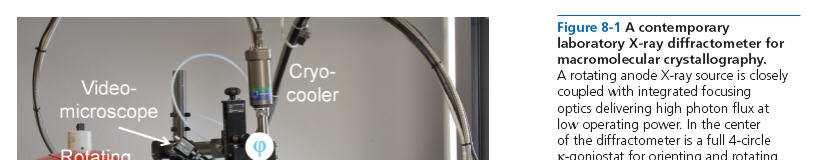
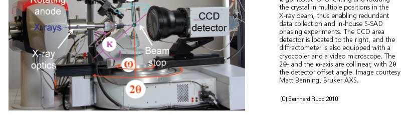
16 X-ray Diffractometer
17 Growing Protein Crystals We all are familiar with crystals from rock collections or small molecules, such as salt or sugar. They are usually associated with properties such as hardness, durability, and prettiness. Unfortunately, only the latter is true for protein crystals.
18 Proteins Proteins consist of long macromolecule chains made up from 20 different amino acids. The chains can be several hundred residues long and fold into a 3-dimensional structure. It is therefore quite understandable that protein molecules have irregular shapes and are not ideally suited to be stacked into a periodic lattice, i.e., a crystal. Protein crystals are thus very fragile, soft (think of a cube of jelly instead of a brick) and sensitive to all kind of environmental variations.
19 Proteins Protein crystals contain on average 50% solvent, mostly in large channels between the stacked molecules on the crystal. The interactions holding the molecules together are usually weak, hydrogen bonds, salt bridges, and hydrophobic interactions, compared to strong covalent or ionic interactions in mineral crystals. This explains the fragility of the crystals, but allows for the possibility of soaking metal solutions (important for phasing) or even large enzyme substrates or inhibitors, into the crystals.
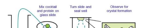
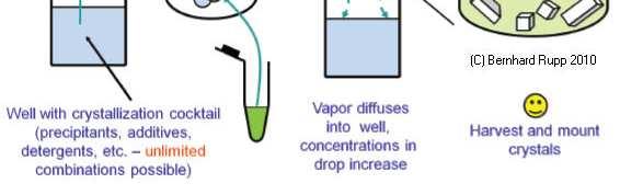
20 The Experimental Set-up In order to obtain a crystal, the protein molecules must assemble into a periodic lattice. One starts with a solution of the protein with a fairly high concentration (2-50 mg/ml) and adds reagents that reduce the solubility close to spontaneous precipitation. By slow further concentration, and under conditions suitable for the formation of a few nucleation sites, small crystals may start to grow. Often very many conditions have to be tried to succeed. Crystal size should to be from a few hundred down to about 20 micron in each direction to be useful for diffraction experiments.
21 The Experimental Set-up
22 The Experimental Set-up The most common setup to grow protein crystals is by the hanging drop technique : A few microliters of protein solution are mixed with an about equal amount of reservoir solution containing the precipitants. A drop of this mixture is put on a glass slide which covers the reservoir. As the protein/precipitant mixture in the drop is less concentrated than the reservoir solution (The protein solution is mixed with the reservior solution about 1:1), water evaporates from the drop into the reservoir. As a result the concentration of both protein and precipitant in the drop slowly increases, and crystals may form. There is a variety of other techniques available such as sitting drops, dialysis buttons, and gel and microbatch techniques
23 Cryocrystallography- Radiation Damage Just as any organic or living material, proteins are sensitive to X- ray radiation damage. The energy range of X-rays used for diffraction is in the 6 to 15 kev range, which is in fact severely ionizing radiation. The ionizing absorption events create radicals, which rapidly destroy any protein crystal, particularly at dose rates experienced at synchrotrons. An efficient way to suppress radiation damage by slowing down the kinetics of the radical reactions is cryogenic cooling. Rapidly quenching or flash-cooling crystals to liquid nitrogen temperatures, either in cold nitrogen gas streams or directly into liquid nitrogen, will strongly reduce radiation damage. To prevent the formation of crystalline ice during flash-cooling of the crystals, cryoprotectants, present in the mother liqor or added to the mother liquor, are necessary.
24 Cryomounting Cryoprotection is effectively accomplished during harvesting, when the crystals are scooped up from the drop in cryo-loops and briefly swept though a cryoprotectant before being dipped into liquid nitrogen. Common cryoprotectants are ethylene glycol (the anti-freeze in automobile radiators), glycerol, higher alcohols, polyethylene glycols (PEGs), or high concentration solutions of sucrose or salts. Once the protein crystals are flash-cooled and stored in pucks in a liquid nitrogen dewar (dry shipper), they can be safely sent to a synchrotron for data collection.
25 Benefits of Cryo-cooling Many factors contribute to improvements in data quality during cryoprotection: Obvious benefits are reduced thermal vibrations, enhanced signal-tonoise ratio, reduced conformational disorder, and in many cases, a higher limiting resolution. The most important effect is the suppression of radiation damage, permitting a complete data set to be collected from one single crystal. This in turn eliminates errors from merging and scaling of data sub-sets from multiple crystals or non-isomorphism between MAD data sets. In addition, crystal mounting is vastly simplified over conventional capillary techniques. These improvements combined lead to enhanced contrast and sharper detail in electron density maps, facilitating model building and reducing the total time required for structure determination.
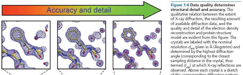
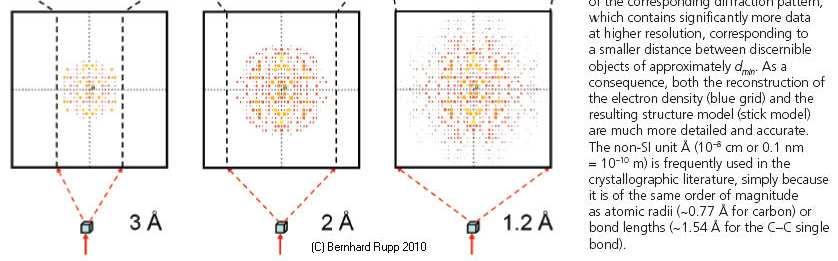
26 What does High Resolution Mean? The figure on the following slide shows what a certain resolution, given in Ångström (Å) means for the user of structural models derived from X- ray data. One always has to remember that the obtainedmodel you see was built into an experimental electron density. The model may look as good at 3 Å as it does at 1.2 Å, but is it a correct and unique description of reality?
27
28 Resolution The pictures of the electron density at different data set resolution of the same region of a molecule leave no question that a model of a phenylalanine-containing ligand (the 6-ring structure) can be correctly placed into the 1.2 Å data. This still can be done with confidence in the 2 Å case, but at 3 Å we already observe a deviation of the centroid of the ring from the correct model. The bottom panel visualizes the relation between diffraction limit, amount of data and nominal resolution. The more and better data, the more acurate and detailed the final structure model will be. Most protein crystals diffract between 1.8 and 3 Å, a few to very high resolution (the term high resolution is used loosely in macromolecular crystallography, we apply it to data of Å, below 'atomic' or 'ultrahigh' resolution are commonly used). The most efficient way to increase resolution (short of trying to grow better crystals) is to cryo-cool the crystals to near liquid nitrogen temperature.
X-ray crystallography
 X-ray crystallography Sources: 1. The Elements of Physical Chemistry by Peter Atkins (including some nice color slides incorporated into these lectures) 2. Physical Chemistry by Tinoco, Sauer, Wang and
Two Lectures in X-ray Crystallography
 Biochemistry 503 Michael Wiener (mwiener@virginia.edu, 3-2731, Snyder 360) Two Lectures in X-ray Crystallography Outline 1. Justification & introductory remarks 2. Experimental setup 3. Protein crystals
X-ray Crystallography. Kalyan Das
 X-ray Crystallography Kalyan Das Electromagnetic Spectrum NMR 10 um - 10 mm 700 to 10 4 nm 400 to 700 nm 10 to 400 nm 10-1 to 10 nm 10-4 to 10-1 nm X-ray radiation was discovered by Roentgen in 1895. X-rays
Protein Crystallography
 Protein Crystallography Part II Tim Grüne Dept. of Structural Chemistry Prof. G. Sheldrick University of Göttingen http://shelx.uni-ac.gwdg.de tg@shelx.uni-ac.gwdg.de Overview The Reciprocal Lattice The
Protein Structure Analysis and Verification. Course S Basics for Biosystems of the Cell exercise work. Maija Nevala, BIO, 67485U 16.1.
 Protein Structure Analysis and Verification Course S-114.2500 Basics for Biosystems of the Cell exercise work Maija Nevala, BIO, 67485U 16.1.2008 1. Preface When faced with an unknown protein, scientists
Why do We Trust X-ray Crystallography?
 Why do We Trust X-ray Crystallography? Andrew D Bond All chemists know that X-ray crystallography is the gold standard characterisation technique: an X-ray crystal structure provides definitive proof of
Determining Protein Structure BIBC 100
 Determining Protein Structure BIBC 100 Determining Protein Structure X-Ray Diffraction Interactions of x-rays with electrons in molecules in a crystal NMR- Nuclear Magnetic Resonance Interactions of magnetic
Lecture- 08 Emission and absorption spectra
 Atomic and Molecular Absorption Spectrometry for Pollution Monitoring Dr. J R Mudakavi Department of Chemical Engineering Indian Institute of Science, Bangalore Lecture- 08 Emission and absorption spectra
Chapter Two Test Chemistry. 1. If an atom contains 11 protons and 12 neutrons, its atomic number is A. 1 C. 12 B. 11 D. 23
 Name Chapter Two Test Chemistry 1. If an atom contains 11 protons and 12 neutrons, its atomic number is A. 1 C. 12 B. 11 D. 23 2. The nucleus is made up of all of the following: A. Electrons C. Protons
Molecular Biology Course 2006 Protein Crystallography Part I
 Molecular Biology Course 2006 Protein Crystallography Part I Tim Grüne University of Göttingen Dept. of Structural Chemistry November 2006 http://shelx.uni-ac.gwdg.de tg@shelx.uni-ac.gwdg.de Overview Overview
CS273: Algorithms for Structure Handout # 13 and Motion in Biology Stanford University Tuesday, 11 May 2003
 CS273: Algorithms for Structure Handout # 13 and Motion in Biology Stanford University Tuesday, 11 May 2003 Lecture #13: 11 May 2004 Topics: Protein Structure Determination Scribe: Minli Zhu We acknowledge
Principles of Physical Biochemistry
 Principles of Physical Biochemistry Kensal E. van Hold e W. Curtis Johnso n P. Shing Ho Preface x i PART 1 MACROMOLECULAR STRUCTURE AND DYNAMICS 1 1 Biological Macromolecules 2 1.1 General Principles
BIOCHEMISTRY Course Outline (Fall, 2011)
") BIOCHEMISTRY 402 - Course Outline (Fall, 2011) Number OVERVIEW OF LECTURE TOPICS: of Lectures INSTRUCTOR 1. Structural Components of Proteins G. Brayer (a) Amino Acids and the Polypeptide Chain Backbone...2
BIOCHEMISTRY 402 - Course Outline (Fall, 2011) Number OVERVIEW OF LECTURE TOPICS: of Lectures INSTRUCTOR 1. Structural Components of Proteins G. Brayer (a) Amino Acids and the Polypeptide Chain Backbone...2
Scattering Lecture. February 24, 2014
 Scattering Lecture February 24, 2014 Structure Determination by Scattering Waves of radiation scattered by different objects interfere to give rise to an observable pattern! The wavelength needs to close
SAXS and SANS facilities and experimental practice. Clement Blanchet
 SAXS and SANS facilities and experimental practice Clement Blanchet SAS experiment Detector X-ray or neutron Beam Sample 2 s Buffer X-rays Roengten, 1895 Electromagnetic wave The electromagnetic spectrum
Overview - Macromolecular Crystallography
 Overview - Macromolecular Crystallography 1. Overexpression and crystallization 2. Crystal characterization and data collection 3. The diffraction experiment 4. Phase problem 1. MIR (Multiple Isomorphous
The Solid State. Phase diagrams Crystals and symmetry Unit cells and packing Types of solid
 The Solid State Phase diagrams Crystals and symmetry Unit cells and packing Types of solid Learning objectives Apply phase diagrams to prediction of phase behaviour Describe distinguishing features of
The basics of structural biology. And Why we use synchrotron sources Sean McSweeney ESRF Structural Biology Group
 The basics of structural biology And Why we use synchrotron sources Sean McSweeney ESRF Structural Biology Group The rise and rise of structural biology. 2 The aim of the game 3 What information does structure
Structure factors again
 Structure factors again Remember 1D, structure factor for order h F h = F h exp[iα h ] = I 01 ρ(x)exp[2πihx]dx Where x is fractional position along unit cell distance (repeating distance, origin arbitrary)
Crystals! Table of Contents. Vocabulary 2. Word Search 6. What is a Crystal? 7. Atoms, Ions, Molecules. and the Unit Cell 13.
 Crystals! Table of Contents Vocabulary 2 Word Search 6 What is a Crystal? 7 Atoms, Ions, Molecules and the Unit Cell 13 Crystal Shapes 15 X-Ray Crystallography 17 Recipes for Making A Booklet for Elementary
CHEMISTRY (CHEM) CHEM 5800 Principles Of Materials Chemistry. Tutorial in selected topics in materials chemistry. S/U grading only.
 CHEM 5800 Principles Of Materials Chemistry. Tutorial in selected topics in materials chemistry. S/U grading only.") Chemistry (CHEM) 1 CHEMISTRY (CHEM) CHEM 5100 Principles of Organic and Inorganic Chemistry Study of coordination compounds with a focus on ligand bonding, electron counting, molecular orbital theory,
Chemistry (CHEM) 1 CHEMISTRY (CHEM) CHEM 5100 Principles of Organic and Inorganic Chemistry Study of coordination compounds with a focus on ligand bonding, electron counting, molecular orbital theory,
Name Biology Chapter 2 Note-taking worksheet
 Name Biology Chapter 2 Note-taking worksheet The Nature of Matter 1. Life depends on Atoms 1. The study of chemistry starts with the basic unit of matter, the. 2. The atom was first used by the Greek philosopher
CHEM 463: Advanced Inorganic Chemistry Modeling Metalloproteins for Structural Analysis
 CHEM 463: Advanced Inorganic Chemistry Modeling Metalloproteins for Structural Analysis Purpose: The purpose of this laboratory is to introduce some of the basic visualization and modeling tools for viewing
Crystals, X-rays and Proteins
 Crystals, X-rays and Proteins Comprehensive Protein Crystallography Dennis Sherwood MA (Hons), MPhil, PhD Jon Cooper BA (Hons), PhD OXFORD UNIVERSITY PRESS Contents List of symbols xiv PART I FUNDAMENTALS
X-Ray Damage to Biological Crystalline Samples
 X-Ray Damage to Biological Crystalline Samples Gerd Rosenbaum Structural Biology Center, ANL and Dept. of Biochemistry, UGA ACA Summer School IIT, 19 July 2007 A U.S. Department of Energy laboratory managed
Chemistry A: States of Matter Packet Name: Hour: Page!1. Chemistry A States of Matter Packet
 Chemistry A: States of Matter Packet Name: Hour: Page!1 Chemistry A States of Matter Packet Chemistry A: States of Matter Packet Name: Hour: Page!2 Worksheet #1: States of Matter In this packet we will
CRYSTALLOGRAPHY AND STORYTELLING WITH DATA. President, Association of Women in Science, Bethesda Chapter STEM Consultant
 CRYSTALLOGRAPHY AND STORYTELLING WITH DATA President, Association of Women in Science, Bethesda Chapter STEM Consultant MY STORY Passion for Science BS Biology Major MS Biotechnology & Project in Bioinformatics
Protein crystallography. Garry Taylor
 Protein crystallography Garry Taylor X-ray Crystallography - the Basics Grow crystals Collect X-ray data Determine phases Calculate ρ-map Interpret map Refine coordinates Do the biology. Nitrogen at -180
Application Note SC-XRD 505 Single Crystal Diffraction
 Application Note SC-XRD 505 Single Crystal Diffraction Introduction Single-crystal X-ray diffraction, commonly referred to as X-ray crystallography, is an analytical technique in which X-ray methods are
UNIT 10: Water. Essential Idea(s): Water is the medium of life. IB Assessment Statements
: Water is the medium of life. IB Assessment Statements") UNIT 10: Water Name: Essential Idea(s): Water is the medium of life. IB Assessment Statements 2.2.U1 2.2.NOS 2.2.U2 2.2.A1 2.2.A2 2.2.U3 2.2.A3 Water molecules are polar and hydrogen bonds form between
UNIT 10: Water Name: Essential Idea(s): Water is the medium of life. IB Assessment Statements 2.2.U1 2.2.NOS 2.2.U2 2.2.A1 2.2.A2 2.2.U3 2.2.A3 Water molecules are polar and hydrogen bonds form between
Crystallography past, present and future
 Crystallography past, present and future Jenny P. Glusker Philadelphia, PA, U. S. A. International Year of Crystallography UNESCO, Paris, France 20 January 2014 QUARTZ CRYSTALS Quartz crystals found growing
PDBe TUTORIAL. PDBePISA (Protein Interfaces, Surfaces and Assemblies)
") PDBe TUTORIAL PDBePISA (Protein Interfaces, Surfaces and Assemblies) http://pdbe.org/pisa/ This tutorial introduces the PDBePISA (PISA for short) service, which is a webbased interactive tool offered by
PDBe TUTORIAL PDBePISA (Protein Interfaces, Surfaces and Assemblies) http://pdbe.org/pisa/ This tutorial introduces the PDBePISA (PISA for short) service, which is a webbased interactive tool offered by
Chapter 6 Chemistry in Biology
 Section 1: Atoms, Elements, and Compounds Section 2: Chemical Reactions Section 3: Water and Solutions Section 4: The Building Blocks of Life Click on a lesson name to select. 6.1 Atoms, Elements, and
Structure Report for J. Reibenspies
 X-ray Diffraction Laboratory Center for Chemical Characterization and Analysis Department of Chemistry Texas A & M University Structure Report for J. Reibenspies Project Name: Sucrose Date: January 29,
- Introduction of x-ray crystallography: what it s used for, how it works, applications in science - Different methods used to generate data - Case
 - Introduction of x-ray crystallography: what it s used for, how it works, applications in science - Different methods used to generate data - Case studies emphasizing the importance of the technique -
Changing and challenging times for service crystallography. Electronic Supplementary Information
 Changing and challenging times for service crystallography Simon J Coles,* a and Philip A Gale* a Electronic Supplementary Information Instrument descriptions and experimental protocols The following firstly
Solids / Crystal Structure
 The first crystal analysis proved that in the typical inorganic salt, NaCl, there is no molecular grouping. The inference that the structure consists of alternate ions of sodium and chlorine was an obvious
Radioactivity. Lecture 6 Detectors and Instrumentation
 Radioactivity Lecture 6 Detectors and Instrumentation The human organs Neither humans nor animals have an organ for detecting radiation from radioactive decay! We can not hear it, smell it, feel it or
Preparing a PDB File
 Figure 1: Schematic view of the ligand-binding domain from the vitamin D receptor (PDB file 1IE9). The crystallographic waters are shown as small spheres and the bound ligand is shown as a CPK model. HO
2 How Substances Dissolve
 CHAPTER 8 SECTION Solutions 2 How Substances Dissolve KEY IDEAS As you read this section, keep these questions in mind: Why is water called the universal solvent? How do substances dissolve? Why Do Substances
X-ray Crystallography
 2009/11/25 [ 1 ] X-ray Crystallography Andrew Torda, wintersemester 2009 / 2010 X-ray numerically most important more than 4/5 structures Goal a set of x, y, z coordinates different properties to NMR History
CHEMISTRY (CHEM) CHEM 1200 Problem Solving In General Chemistry
 CHEM 1200 Problem Solving In General Chemistry") Chemistry (CHEM) 1 CHEMISTRY (CHEM) CHEM 1090 Elementary Chemistry [0-3 credit hours (0-2, 0, 0-1)] For students who major in science, engineering or other fields which require chemistry as a prerequisite
Chemistry (CHEM) 1 CHEMISTRY (CHEM) CHEM 1090 Elementary Chemistry [0-3 credit hours (0-2, 0, 0-1)] For students who major in science, engineering or other fields which require chemistry as a prerequisite
BIOLOGY 101. CHAPTER 3: Water and Life: The Molecule that supports all Live
 BIOLOGY 101 CHAPTER 3: Water and Life: The Molecule that supports all Live The Molecule that Supports all Life CONCEPTS: 3.1 Polar covalent bonds in water molecules result in hydrogen bonding 3.2 Four
Name: Class: Date: ID: A
 Name: Class: _ Date: _ ID: A Ch 2 Review Multiple Choice Identify the choice that best completes the statement or answers the question. 1. Isotopes are atoms of the same element with the same number of
ZAHID IQBAL WARRAICH
 Q1 Chromatography is an important analytical technique in chemistry. There is a number of techniques under the general heading of chromatography. (a) Paper and gas chromatography rely on partition to separate
Saba Al Fayoumi. Tamer Barakat. Dr. Mamoun Ahram + Dr. Diala Abu-Hassan
 1 Saba Al Fayoumi Tamer Barakat Dr. Mamoun Ahram + Dr. Diala Abu-Hassan What is BIOCHEMISTRY??? Biochemistry = understanding life Chemical reactions are what makes an organism (An organism is simply atoms
Protein Structure Determination 9/25/2007
 One-dimensional NMR spectra Ethanol Cellulase (36 a.a.) Branden & Tooze, Fig. 18.16 1D and 2D NMR spectra of inhibitor K (57 a.a.) K. Wuthrich, NMR of Proteins and Nucleic Acids. (Wiley, 1986.) p. 54-55.
X-Ray structure analysis
 X-Ray structure analysis Kay Diederichs kay.diederichs@uni-konstanz.de Analysis of what? Proteins ( /ˈproʊˌtiːnz/ or /ˈproʊti.ɨnz/) are biochemical compounds consisting of one or more polypeptides typically
Structure and Dynamics : An Atomic View of Materials
 Structure and Dynamics : An Atomic View of Materials MARTIN T. DOVE Department ofearth Sciences University of Cambridge OXFORD UNIVERSITY PRESS Contents 1 Introduction 1 1.1 Observations 1 1.1.1 Microscopic
Copyright Mark Brandt, Ph.D A third method, cryogenic electron microscopy has seen increasing use over the past few years.
 Structure Determination and Sequence Analysis The vast majority of the experimentally determined three-dimensional protein structures have been solved by one of two methods: X-ray diffraction and Nuclear
Quality Assurance Plan. March 30, Chemical Crystallography Laboratory
 Quality Assurance Plan Page 1 of 7 Quality Assurance Plan March 30, 2017 Chemical Crystallography Laboratory Author: Douglas R. Powell Quality Assurance Plan Page 2 of 7 Distribution Douglas R. Powell,
Paper No. 01. Paper Title: Food Chemistry. Module-02: Water in Food Systems
 Paper No. 01 Paper Title: Food Chemistry Module-02: Water in Food Systems Water is abundant in all living things and consequently is in almost all foods, unless steps have been taken to remove it. It is
Chemistry in Biology. Section 1. Atoms, Elements, and Compounds
 Section 1 Atoms, Elements, and Compounds Atoms! Chemistry is the study of matter.! Atoms are the building blocks of matter.! Neutrons and protons are located at the center of the atom.! Protons are positively
Receptor Based Drug Design (1)
") Induced Fit Model For more than 100 years, the behaviour of enzymes had been explained by the "lock-and-key" mechanism developed by pioneering German chemist Emil Fischer. Fischer thought that the chemicals
Induced Fit Model For more than 100 years, the behaviour of enzymes had been explained by the "lock-and-key" mechanism developed by pioneering German chemist Emil Fischer. Fischer thought that the chemicals
2 How Substances Dissolve
 CHAPTER 8 SECTION Solutions 2 How Substances Dissolve KEY IDEAS As you read this section, keep these questions in mind: Why is water called the universal solvent? How do substances dissolve? Why Do Substances
Scattering by two Electrons
 Scattering by two Electrons p = -r k in k in p r e 2 q k in /λ θ θ k out /λ S q = r k out p + q = r (k out - k in ) e 1 Phase difference of wave 2 with respect to wave 1: 2π λ (k out - k in ) r= 2π S r
Supporting Information
 Supporting Information Structural Basis of the Antiproliferative Activity of Largazole, a Depsipeptide Inhibitor of the Histone Deacetylases Kathryn E. Cole 1, Daniel P. Dowling 1,2, Matthew A. Boone 3,
X-rays. X-ray Radiography - absorption is a function of Z and density. X-ray crystallography. X-ray spectrometry
 X-rays Wilhelm K. Roentgen (1845-1923) NP in Physics 1901 X-ray Radiography - absorption is a function of Z and density X-ray crystallography X-ray spectrometry X-rays Cu K α E = 8.05 kev λ = 1.541 Å Interaction
Section 2: How Substances Dissolve. Preview Key Ideas Bellringer Water: A Common Solvent The Dissolving Process Surface Area
 : How Substances Dissolve Preview Key Ideas Bellringer Water: A Common Solvent The Dissolving Process Surface Area Key Ideas Why is water called the universal solvent? Why do substances dissolve? Bellringer
Supplementary Figure S1 a, wireframe view of the crystal structure of compound 11. b, view of the pyridinium sites. c, crystal packing of compound
 a b c Supplementary Figure S1 a, wireframe view of the crystal structure of compound 11. b, view of the pyridinium sites. c, crystal packing of compound 11. 1 a b c Supplementary Figure S2 a, wireframe
1 WHAT IS SPECTROSCOPY?
 1 WHAT IS SPECTROSCOPY? 1.1 The Nature Of Electromagnetic Radiation Anyone who has been sunburnt will know that light packs a punch: in scientific terms, it contains considerable amounts of energy. All
Contents. xiii. Preface v
 Contents Preface Chapter 1 Biological Macromolecules 1.1 General PrincipIes 1.1.1 Macrornolecules 1.2 1.1.2 Configuration and Conformation Molecular lnteractions in Macromolecular Structures 1.2.1 Weak
Chapter 1 X-ray Absorption Fine Structure (EXAFS)
") 1 Chapter 1 X-ray Absorption Fine Structure (EXAFS) 1.1 What is EXAFS? X-ray absorption fine structure (EXAFS, XAFS) is an oscillatory modulation in the X-ray absorption coefficient on the high-energy
1 Chapter 1 X-ray Absorption Fine Structure (EXAFS) 1.1 What is EXAFS? X-ray absorption fine structure (EXAFS, XAFS) is an oscillatory modulation in the X-ray absorption coefficient on the high-energy
Chapter Two (Chemistry of Life)
") 1 Chapter Two (Chemistry of Life) SECTION ONE: THE COMPOSITION OF MATTER MATTER Everything in the universe is made of matter. Matter is anything that occupies space and has mass. Mass is the quantity of
1 Chapter Two (Chemistry of Life) SECTION ONE: THE COMPOSITION OF MATTER MATTER Everything in the universe is made of matter. Matter is anything that occupies space and has mass. Mass is the quantity of
The OTHER TWO states of matter
 ` The OTHER TWO states of matter LIQUIDS A decrease in the average kinetic energy of gas particles causes the temperature to decrease. As it cools, the particles tend to move more slowly if they slow down
Name Class Date. How do mixtures differ from elements and compounds? How can mixtures be separated? What are solutions?
 CHAPTER 3 3 Mixtures SECTION Elements, Compounds, and Mixtures BEFORE YOU READ After you read this section, you should be able to answer these questions: How do mixtures differ from elements and compounds?
X-ray practical: Crystallography
 X-ray practical: Crystallography Aim: To familiarise oneself with the operation of Tex-X-Ometer spectrometer and to use it to determine the lattice spacing in NaCl and LiF single crystals. Background:
Physical Science DCI Progression Chart
 DCI Progression Chart PS1: Matter and Its Interactions Grade Bands PS1.A Structure & Properties of Matter Grades K-2 Grades 3-5 Grades 6-8 Grades 9-12 Second Grade * Different kinds of matter exist and
 Helpful resources for all X ray lectures Crystallization http://www.hamptonresearch.com under tech support: crystal growth 101 literature Spacegroup tables http://img.chem.ucl.ac.uk/sgp/mainmenu.htm Crystallography
Name Date Period Molecular Nature of Water
 Name Date Period Molecular Nature of Water Purpose: To determine how water molecules react using molecular models and Lab demos. Materials: I cup of 12 water molecules (red & white), 1 Na (blue), 1 Cl
BIOCHEMISTRY NOTES - UNIT 2-
 BIOCHEMISTRY NOTES - UNIT 2- ATOMS - the basic unit of matter. Contains subatomic particles o (+ charge) o (no charge/neutral) o (- charge) Protons and neutrons have about the same mass. Electrons are
c cm 3 d. a, b, and c c ng d g
 Mr. Stone Honors Biology Practice Test 1. Which one is equal to 1.5 10 3 ml? a. 1.5 10-1 L b. 150 10 0 ml c. 1.5 10 3 cm 3 d. a, b, and c e. b and c only 2. Which one is NOT equal to 2.74 10 2 milligrams?
Introduction to" Protein Structure
 Introduction to" Protein Structure Function, evolution & experimental methods Thomas Blicher, Center for Biological Sequence Analysis Learning Objectives Outline the basic levels of protein structure.
Advanced Pharmaceutical Analysis
 Lecture 2 Advanced Pharmaceutical Analysis IR spectroscopy Dr. Baraa Ramzi Infrared Spectroscopy It is a powerful tool for identifying pure organic and inorganic compounds. Every molecular compound has
Chemistry A: States of Matter Packet Name: Hour: Page 1. Chemistry A States of Matter Packet
 Chemistry A: States of Matter Packet Name: Hour: Page 1 Chemistry A States of Matter Packet Chemistry A: States of Matter Packet Name: Hour: Page 2 Worksheet #1: States of Matter In this packet we will
Basics of protein structure
 Today: 1. Projects a. Requirements: i. Critical review of one paper ii. At least one computational result b. Noon, Dec. 3 rd written report and oral presentation are due; submit via email to bphys101@fas.harvard.edu
Denaturation and renaturation of proteins
 Denaturation and renaturation of proteins Higher levels of protein structure are formed without covalent bonds. Therefore, they are not as stable as peptide covalent bonds which make protein primary structure
Chemistry Review. Structure of an Atom. The six most abundant elements of life. Types of chemical bonds. U n i t 2 - B i o c h e m i s t r y
 Chemistry Review Structure of an Atom are organized into shells or levels around the nucleus. Atoms are most stable when their outer or valence shell is. The six most abundant elements of life Types of
Section Objectives: Section Objectives: Distinguish mixtures and solutions. Define acids and bases and relate their importance to biological systems.
 Section Objectives: Relate the structure of an atom to the identity of elements. Relate the formation of covalent and ionic chemical bonds to the stability of atoms. Section Objectives: Distinguish mixtures
Why study Carbon? Chemistry of Life. Chemistry of Life. Hydrocarbons can grow. Hydrocarbons. Building Blocks. Combinations of C & H
 Chemistry of Life Building Blocks Why study Carbon? All of life is built on carbon Cells ~72% 2 O ~25% carbon compounds carbohydrates lipids proteins nucleic acids ~3% salts Na, Cl, K Chemistry of Life
M2 TP. Low-Energy Electron Diffraction (LEED)
") M2 TP Low-Energy Electron Diffraction (LEED) Guide for report preparation I. Introduction: Elastic scattering or diffraction of electrons is the standard technique in surface science for obtaining structural
M2 TP Low-Energy Electron Diffraction (LEED) Guide for report preparation I. Introduction: Elastic scattering or diffraction of electrons is the standard technique in surface science for obtaining structural
NORTH CENTRAL HIGH SCHOOL NOTE & STUDY GUIDE. Honors Biology I
 NOTE/STUDY GUIDE: Unit 1-2, Biochemistry Honors Biology I, Mr. Doc Miller, M.Ed. North Central High School Name: Period: Seat #: Date: NORTH CENTRAL HIGH SCHOOL NOTE & STUDY GUIDE Honors Biology I Unit
Roger Johnson Structure and Dynamics: X-ray Diffraction Lecture 6
 6.1. Summary In this Lecture we cover the theory of x-ray diffraction, which gives direct information about the atomic structure of crystals. In these experiments, the wavelength of the incident beam must
1. (5) Draw a diagram of an isomeric molecule to demonstrate a structural, geometric, and an enantiomer organization.
 Draw a diagram of an isomeric molecule to demonstrate a structural, geometric, and an enantiomer organization.") Organic Chemistry Assignment Score. Name Sec.. Date. Working by yourself or in a group, answer the following questions about the Organic Chemistry material. This assignment is worth 35 points with the
Organic Chemistry Assignment Score. Name Sec.. Date. Working by yourself or in a group, answer the following questions about the Organic Chemistry material. This assignment is worth 35 points with the
Chem 1075 Chapter 13 Liquids and Solids Lecture Outline
 Chem 1075 Chapter 13 Liquids and Solids Lecture Outline Slide 2-3 Properties of Liquids Unlike gases, liquids respond dramatically to temperature and pressure changes. We can study the liquid state and
A Primer in X-ray Crystallography for Redox Biologists. Mark Wilson Karolinska Institute June 3 rd, 2014
 A Primer in X-ray Crystallography for Redox Biologists Mark Wilson Karolinska Institute June 3 rd, 2014 X-ray Crystallography Basics Optimistic workflow for crystallography Experiment Schematic Fourier
Biological Small Angle X-ray Scattering (SAXS) Dec 2, 2013
 Dec 2, 2013") Biological Small Angle X-ray Scattering (SAXS) Dec 2, 2013 Structural Biology Shape Dynamic Light Scattering Electron Microscopy Small Angle X-ray Scattering Cryo-Electron Microscopy Wide Angle X- ray
Biological Small Angle X-ray Scattering (SAXS) Dec 2, 2013 Structural Biology Shape Dynamic Light Scattering Electron Microscopy Small Angle X-ray Scattering Cryo-Electron Microscopy Wide Angle X- ray
X-ray crystallography has been responsible for results. leading to fundamental and important structural concepts. Each
 Chapter 9 CONCLUSION AND THE SCOPE FOR FUTURE STUDIES X-ray crystallography has been responsible for results leading to fundamental and important structural concepts. Each crystal structure reveals something
What Are Atoms? Chapter 2: Atoms, Molecules & Life
 Chapter 2: Atoms, Molecules & Life What Are Atoms? An atom are the smallest unit of matter. Atoms are composed of Electrons = negatively charged particles. Neutrons = particles with no charge (neutral).
Rule 2. Rule 1. Rule 4. Rule 3. Rule 5. Rule 6. Rule 7. Rule 8
 Rule 1 Follow the directions in your course reader, of your teaching assistant and of your instructor. They are usually much more experienced doing chemistry. Rule 3 When in doubt, ask. This will make
Modern physics ideas are strange! L 36 Modern Physics [2] The Photon Concept. How are x-rays produced? The uncertainty principle
![Modern physics ideas are strange! L 36 Modern Physics [2] The Photon Concept. How are x-rays produced? The uncertainty principle](/thumbs/88/117098787.jpg "Modern physics ideas are strange! L 36 Modern Physics [2] The Photon Concept. How are x-rays produced? The uncertainty principle") L 36 Modern Physics [2] X-rays & gamma rays How lasers work Medical applications of lasers Applications of high power lasers Medical imaging techniques CAT scans MRI s Modern physics ideas are strange!
L 36 Modern Physics [2] X-rays & gamma rays How lasers work Medical applications of lasers Applications of high power lasers Medical imaging techniques CAT scans MRI s Modern physics ideas are strange!
CHEMICAL BONDS. Attraction that holds molecules together Involves valence electrons. Ionic Bonds Covalent Bonds. Involves sharing of.
 CHEMICAL BONDS DEFINITION/DESCRIPTION: Attraction that holds molecules together Involves valence electrons TYPES: Ionic Bonds Covalent Bonds Involves sharing of electrons Electronegativities O = 3.5 N
PSD '17 -- Xray Lecture 5, 6. Patterson Space, Molecular Replacement and Heavy Atom Isomorphous Replacement
 PSD '17 -- Xray Lecture 5, 6 Patterson Space, Molecular Replacement and Heavy Atom Isomorphous Replacement The Phase Problem We can t measure the phases! X-ray detectors (film, photomultiplier tubes, CCDs,
Notes: Matter and Change
 Name Chemistry-PAP Notes: Matter and Change Period: I. What is Chemistry? is the study of composition, structure, and properties of matter and energy associated with the changes it undergoes. is defined
UNIT 2 PHYSICAL & CHEMICAL PROPERTIES
 UNIT 2 PHYSICAL & CHEMICAL PROPERTIES What Is Matter? How matter is made of Elements? What atoms make up? Theory Law and Hypothesis Physical and Chemical Changes Heterogenous and Homogenous Substances
H 2 O WHAT PROPERTIES OF WATER MAKE IT ESSENTIAL TO LIFE OF EARTH? Good solvent High Surface tension Low vapor pressure High boiling point
 Unit 9: Solutions H 2 O WHAT PROPERTIES OF WATER MAKE IT ESSENTIAL TO LIFE OF EARTH? Good solvent High Surface tension Low vapor pressure High boiling point Water is a polar molecule. It experiences hydrogen
What is the Phase Problem? Overview of the Phase Problem. Phases. 201 Phases. Diffraction vector for a Bragg spot. In General for Any Atom (x, y, z)
") Protein Overview of the Phase Problem Crystal Data Phases Structure John Rose ACA Summer School 2006 Reorganized by Andy Howard,, Spring 2008 Remember We can measure reflection intensities We can calculate
Protein Overview of the Phase Problem Crystal Data Phases Structure John Rose ACA Summer School 2006 Reorganized by Andy Howard,, Spring 2008 Remember We can measure reflection intensities We can calculate
Protein separation and characterization
 Address:800 S Wineville Avenue, Ontario, CA 91761,USA Website:www.aladdin-e.com Email USA: tech@aladdin-e.com Email EU: eutech@aladdin-e.com Email Asia Pacific: cntech@aladdin-e.com Protein separation
Solids, liquids and gases
 Solids, liquids and gases Solids, liquids, and gases are held together by intermolecular forces. Intermolecular forces occur between molecules, not within molecules (as in bonding). When a molecule changes
Electronic Supplementary Information for: Gram-scale Synthesis of a Bench-Stable 5,5 -Unsubstituted Terpyrrole
 Electronic Supplementary Information for: Gram-scale Synthesis of a Bench-Stable 5,5 -Unsubstituted Terpyrrole James T. Brewster II, a Hadiqa Zafar, a Matthew McVeigh, a Christopher D. Wight, a Gonzalo