Two Lectures in X-ray Crystallography
|
|
|
- Delilah Warren
- 6 years ago
- Views:
Transcription
1 Biochemistry 503 Michael Wiener , Snyder 360) Two Lectures in X-ray Crystallography Outline 1. Justification & introductory remarks 2. Experimental setup 3. Protein crystals how to obtain them 4. Resolution & Bragg s Law 5. Crystals what are they? 6. The reciprocal lattice & the Ewald sphere 7. X-ray data to electron density maps 8. The fundamental equation of crystallography 9. Fourier transforms (pictures & examples) 10. Depiction of phases via the Argand diagram 11. Crystallographic phases and how to get them 12. Design of phasing experiments 13. Graphical image of phase determination 14. Crystallographic B-factors 15. Crystallographic Refinement 16. Evaluating a structure 17. (Graphical examples/demonstration) Goal of lectures (what you should know/extract/learn) 1. Qualitative - description of how to get from x-rays to a structure (i.e., everything not covered by 2 or 3) 2. Semi-quantitative - reciprocal lattice, phasing, evaluation of a structure 3. Quantitative - Bragg s Law, design a phasing experiment, Argand diagram, B-factors
2 What is X-ray Crystallography? X-ray crystallography is an experimental technique that exploits the fact that X-rays are diffracted by crystals. It is not an imaging technique. X-rays have the proper wavelength (in the Ångström range, ~10-10 m) to be scattered by the electron cloud of an atom of comparable size. Based on the diffraction pattern obtained from X-ray scattering off the periodic assembly of molecules or atoms in the crystal, the electron density can be reconstructed. Additional phase information must be extracted either from the diffraction data or from supplementing diffraction experiments to complete the reconstruction (the phase problem in crystallography). y) A model is then progressively built into the experimental electron density, refined against the data and the result is a quite accurate molecular structure. Why Crystallography? The knowledge of accurate molecular structures is a prerequisite for rational drug design and for structure based functional studies to aid the development of effective therapeutic agents and drugs. Crystallography can reliably provide the answer to many structure related questions, from global folds to atomic details of bonding. In contrast to NMR, which is an indirect spectroscopic method, no size limitation exists for the molecule or complex to be studied. The price for the high accuracy of crystallographic structures is that a good crystal must be found, and that only limited information about the molecule's dynamic behavior is available from one single diffraction experiment. Outline of the experiment In a macromolecular X-ray diffraction experiment a small protein crystal is placed into an intense X-ray beam and the diffracted X-rays are collected with an area detector (it is advantageous to cool the crystals to low temperatures, primarily to prevent radiation damage). The diffraction pattern consists of reflections of different intensity, and a lot of patterns need to be collected to cover all necessary crystal orientations. After some data processing, we end up with a list of indexed reflections and their intensities.


3 The diffracted X-rays are scattered by the crystal at a certain angle. The further backwards the x-rays scatter, the higher we say is the resolution of the data set. The extent to which the crystal diffracts determines how fine a detail we can actually distinguish in our final model of the structure. High resolution is thus desirable. Knowing the wavelength and the diffraction angle of a reflection, its resolution d can be easily calculated : This is just a reformulation of the famous Bragg equation X-ray Diffraction Equipment. The Experimental Setup To perform an X-ray diffraction experiment, we need an x-ray source. In most cases a rotating tti anode generator producing an X-ray beam of a characteristic ti wavelength this used. Intense, tunable X-ray radiation produced by a Synchrotron provides additional advantages. The primary X-ray beam is monochromated by either crystal monochromators or focusing mirrors. After the beam passes through a helium flushed collimator it passes through the crystal mounted on a pin on a goniometer head. The head is mounted to a goniometer which allows to position the crystal in different orientations
 offers many advantages, the most significant of which is the elimination of radiation damage.")
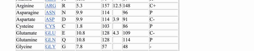
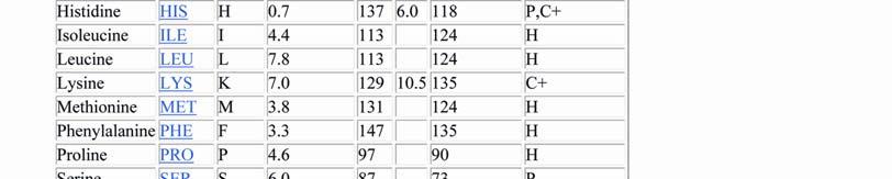
4 in the beam. The diffracted X-rays are recorded using image plates, Multiwire detectors or CCD cameras. Flash cooling protein crystals to cryogenic temperatures (~100 K) offers many advantages, the most significant of which is the elimination of radiation damage. A part of the X-rays passing through the crystal is scattered in different directions into a detector. The detector delivers an image of the diffraction spots. A large number of these images recorded from different crystal orientations are processed (scaled and merged) into a final list of indexed reflection intensities. How to grow protein crystals We all are familiar with crystals from rock collections or small molecules, such as salt or sugar. We usually associate them with properties like hard, durable, and pretty. Unfortunately, only the latter is true for protein crystals. Proteins Proteins consist of long macromolecule chains made up from 20 different amino acids. The chains can be several hundred residues long and fold into a 3-dimensional structure. It is therefore quite understandable that protein molecules have irregular shapes and are not ideally suited to be stacked into a periodic lattice, i.e., a crystal. Protein crystals are thus very fragile, soft (think of a cube of jelly instead of a brick) and sensitive to all kind of environmental variations. Protein crystals contain on average 50% solvent, mostly in large channels between the stacked molecules on the crystal. The interactions holding the molecules together are usually weak, hydrogen bonds, salt bridges, and hydrophobic interactions, compared to strong covalent or ionic interactions in mineral crystals. This
5 explains the fragility of the crystals, but allows for the possibility of soaking metal solutions (important for phasing) or even large enzyme substrates or inhibitors, into the crystals. The Experimental Setup In order to obtain a crystal, the protein molecules must assemble into a periodic lattice. One starts with a solution of the protein with a fairly high hconcentration (2-50 mg/ml) and adds reagents that reduce the solubility close to spontaneous precipitation. By slow further concentration, and under conditions suitable for the formation of a few nucleation sites, small crystals may start to grow. Often very many conditions have to be tried to succeed. This is usually done by initial screening, followed by a systematic optimization of conditions Crystals should to be at least a tenth of a mm in each direction to be useful for in-house diffraction experiments; smaller crystals can be used at the synchrotron. Right : The hanging drop technique. The most common setup to grow protein crystals is by the hanging drop technique : A few microliters of protein solution are mixed with an about equal amount of reservoir solution containing the precipitants. A drop of this mixture is put on a glass slide which covers the reservoir. As the protein/precipitant mixture in the drop is less concentrated than the reservoir solution (remember: we mixed the protein solution with the reservior solution about 1:1), water evaporates from the drop into the reservoir. As a result the concentration of both protein and precipitant i in the drop slowly l increases, and crystals may form. There is a variety of other techniques available such as sitting drops, dialysis buttons, and gel and microbatch techniques. Robots are useful for automatic screening and optimization of crystallization conditions. The main advantage is the small sample size a crystallization robot can handle reproducibly, but it needs some effort to set it up. Helpful techniques to screen proteins The chances for success in crystallizaion experiments depend strongly on the conformational purity of a protein. A single band on a denaturing SDS gel is a good sign, but other methods such as dynamic or static light scattering, circular dichroism, gel filtration, analytical ultracentrifugation, and native-gel electrophoresis can be useful to
 means for the user of structural models derived from X-ray data.")


6 characterize aggregation and dispersity or the folding of your protein sample. A functional assay can also certainly be of utility! What does high resolution mean? The following figure shows what a certain resolution, given in Ångström (Å) means for the user of structural models derived from X-ray data. One always has to remember that the cute model you see was built into an experimental electron density. The model may look as good at 3 Å as it does at 1.5 Å, but is it a correct and unique description of reality? Above: pictures of the electron density at different data set resolution of the same region of a molecule. There is no question that a model of phenylalanine (the 6-ring structure) can be correctly placed into the 1.1 Å data. This still can be done with confidence in the 2 Å case, but at 3 Å we already observe a deviation of the centroid of the ring from the correct model.. The left panels shows the same nominal resolution structure at room temperature (293K) and nitrogen cooled (125K). The difference is a striking example for improvements that can be achieved using cryo-techniques. Most protein crystals diffract between 1.8 and 3 Å, a few to very high resolution (the term high resolution is used loosely in macromolecular crystallography, we apply it to data of 1.2 Å or better resolution). The most efficient way to increase resolution (short of trying to grow better crystals) is to cryo-cool the crystals to near liquid nitrogen temperature.
7 Bragg's Law and Diffraction: How waves reveal the atomic structure of crystals What is Bragg's Law and Why is it Important? Bragg's Law refers to the simple equation: nλ = 2d sinθ (1) derived by the English physicists Sir W.H. Bragg and his son Sir W.L. Bragg in 1913 to explain why the cleavage faces of crystals appear to reflect X-ray beams at certain angles of incidence (theta, θ). The variable d is the distance between atomic layers in a crystal, and the variable lambda λ is the wavelength of the incident X-ray beam, n is an integer. This observation was an example of X-ray wave interference (Roentgenstrahlinterferenzen) commonly known as X-ray diffraction (XRD), and was direct evidence for the periodic atomic structure of crystals postulated for several centuries. The Braggs were awarded the Nobel Prize in physics in 1915 for their work in determining crystal structures beginning with NaCl, ZnS and diamond. Although Bragg's law was used to explain the interference pattern of X-rays scattered by crystals, diffraction has been developed to study the structure of all states of matter with any beam, e.g., ions, electrons, neutrons, and protons, with a wavelength similar to the distance between the atomic or molecular structures of interest. Deriving Bragg's Law Bragg's Law can easily be derived by considering the conditions necessary to make the phases of the beams coincide when the incident angle equals and reflecting angle. The rays of the incident beam are always in phase and parallel up to the point at which the top beam strikes the top layer at atom z (Fig. 1). The second beam continues to the next layer where it is scattered by atom B. The second beam must travel the extra distance AB + BC if the two beams are to continue traveling adjacent and parallel. This extra distance must be an integral (n) multiple of the wavelength (λ) for the phases of the two beams to be the same:
8 Fig. 1 Deriving i Bragg's Law using the reflection geometry and applying trigonometry. The lower beam must travel the extra distance (AB + BC) to continue traveling parallel and adjacent to the top beam. Recognizing d as the hypotenuse of the right triangle Abz, we can use trigonometry to relate d and θ to the distance (AB + BC). The distance AB is opposite θ so, AB = d sinθ (3). Because AB = BC eq. (2) becomes, nλ =2AB(4) Substituting eq. (3) in eq. (4) we have, nλ = 2 d sinθ, (1)


9 and Bragg's Law has been derived. The location of the surface does not change the derivation of Bragg's Law. Experimental Diffraction Patterns The following figure shows experimental x-ray diffraction patterns of a protein crystal (in this case, a membrane protein crystal) obtained with synchrotron radiation.


10 About Crystals, Symmetry and Space Groups How can proteins be assembled in a periodic lattice? A crystal is a periodic arrangement of a motif in a lattice. The motif can be a single atom, a small molecule, a protein or any combination thereof. So here is our protein, RGFP (Red-Green Fluorescent Protein) serving as a structural motif : If we just repeat this motif in three dimensions, we have realized the most simple way to form a crystal. Very often the motif, also referred to as to the 'asymmetric unit', is subjected to a number of symmetry operations yielding differently oriented copies. Let's just use a 2-fold axis for now : If there are no additional symmetry operations, we have already created the contents of the unit cell. The crystal is build from the unit cells arranged into a three-dimensional lattice :


11 So here is our final crystal of RGFP. The question arises, how many ways are there to build crystals by combination of symmetry operations and lattice translations? Infinite? A few 1000? Time to look at this in more detail.
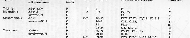
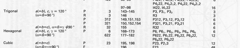
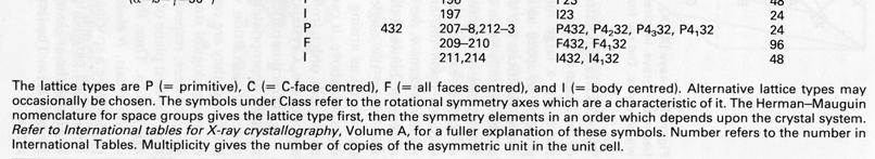
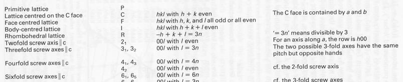
12 Crystal Systems and Bravais Lattices Let us now consider in which way we can translate our cell contents in 3 dimensions to obtain a crystal. The translations which are allowed create 14 Bravais lattices which belong to 7 crystal systems. Each system has a primitive cell, and some allow face or body centering, as well as the rhombohedral h centering in the trigonal system (the rhombohedral lattice can be derived from a cube by pulling along its space diagonal).
13 The Reciprocal Lattice Introduction In the introduction to crystal symmetry I have shown that a crystal consists of a periodic arrangement of fthe unit cell (filled with the motif and dits symmetry generated equivalents), into a lattice. In the same fashion we can define the reciprocal lattice, whose lattice dimensions are reciprocal to the original cell (and correspond to the reflection positions) and whose 'size' (the intensity of the reflection) corresponds to the contents of the unit cell. The following picture will make this clear. Each of the lattice points corresponds to the diffraction from a periodic set of specific crystal lattice planes defined by the index triple hkl. The dimensions of the reciprocal lattice are reciprocally related to the real lattice. In the case of the orthorhombic system I have drawn, the relations are simple: c* = 1/c etc., but in a generic oblique system the relation is more complicated. The length of a reciprocal lattice vector d(hkl)* (from origin to reciprocal lattice point h,k,l) again corresponds to the reciprocal distance d(hkl) of the crystal lattice planes with this index. In our simple case, for 001 this is just the cell dimension c for d(001) or 1/2 c for 002 etc.(d(001)*=1/c, thus d=c).
14 Resolution revisited The vector d(hkl) also determines the location of the diffraction spot in the diffraction image. The diffraction angle at which we observe the reflection is given by Bragg's formula sin(θ)=λ/2d. The higher the index of a reciprocal lattice point, the larger the diffraction angle will be. We have already seen that the larger the diffraction angle, the higher the resolution, i.e. the finer the detail we can observe in the reconstruction of the crystal structure. This can be easily understood now : we need to 'slice' the crystal fine enough (i.e, small d(hkl) = high indices hkl) to have enough information contained in our diffraction pattern to reconstruct details. In the next chapter we will use the reciprocal lattice and the Ewald construction to visualize some important concepts in data collection. The Ewald Construction Intensity of diffraction spots The observed intensity I of the diffraction spots can be thought of as corresponding to the 'size' of the reciprocal lattice point ( I(hkl) is proportional to F(hkl) 2 ). Clearly, either depends on the contents of the unit cell, and we already suspect that the space group symmetry will thus have some implications on the diffraction pattern symmetry. Before we investigate further, it may be useful to understand how the diffraction pattern can be derived from the reciprocal lattice (RL). Let us look at a RL with spots in it :
15 Here we see different magnitudes for the lattice points. The largest spot is at the origin corresponding to F(000), which we know already is the sum of all electrons in the unit cell. The reflection itself is at zero diffraction angle, i.e., in the primary beam path and not observable. Now, where do we expect all the other diffraction spots to appear? Ewald construction A most useful means to understand the occurrence of diffraction spots is the Ewald construction. Let's begin slowly: We draw a sphere of radius 1/λ, in the center of which we imagine the real crystal. The origin of the reciprocal lattice (RL, see above) lies in the transmitted beam, at the edge of the Ewald sphere.


16 We know already that diffraction maxima (reflections, diffraction spots) occur only when the 3 Laue equations, or equivalent, the Bragg equation in vector form, are satisfied. This condition occurs whenever a reciprocal lattice point lies exactly on the Ewald sphere. As you may have assumed already, the chance for this to occur is modest, and we need to rotate the crystal in order to move more reciprocal lattice points through the Ewald sphere. In the following, I have drawn a reciprocal lattice in the origin, and we rotate it along the vertical axis of the drawing. We actually accomplish this by rotating the crystal along the same axis.
17 Just imagine turning the RL through the Ewald sphere : in the beginning, only (101) and (10-1) give rise to a reflection. After you turned the RL a bit (which actually means turning the crystal around the same axis), the reciprocal lattice point 201 will enter the sphere and create a diffraction spot.
18 How do we go from x-ray diffraction data to electron density maps?
19 Outline Goal of the experiment is to determine ρ(x,y,z), the electron density for all x, y, z in the unit cell. What we measure in the experiment, F hkl What we still need, φ hkl (the phase problem) Methods for solving the phase problem Molecular Replacement (MR) Multiple/Single l l Isomorphous replacement (MIR/SIR) Multiple/Single wavelength Anomalous Diffraction (MAD/SAD)
20 Goal of the Experiment 1 ρ( xyz) = Fhkl exp[ 2π i( hx + ky + lz) + iϕ V h k l Need to solve the equation above for all x, y, z in the unit cell What do we know? We record the position (the triple index hkl) and p ( p ) intensity, I hkl, of each reflection (spots on the detector) hkl ]
21 Diffraction Data The measured intensities are proportional to the coefficients of the electron density equation I hkl Fhkl 2
22 From Diffraction Data to Electron Density From the structure factor equation we can see that if we know the contents of the unit cell, we can calculate F hkl We are dealing with the inverse problem. We have information about F hkl but need to know the contents of the crystal.
23 From F hkl to ρ(x,y,z) The structure factor equation is periodic, and is represented a Fourier series. Taking the Fourier Transform (FT) of the equation for F hkl we get the necessary equation which describes the electron density in the crystal. So the FT of the diffraction data gives us a representation of the contents of the crystal. (The FT of the contents of the crystal gives us the diffraction pattern.)
24 Fourier Transforms, examples An atom, and its Fourier Transform: Note the both functions have circular symmetry. The atom is a sharp feature, whereas its transform is a broad smooth function. This illustrates the reciprocal relationship between a function and its Fourier transform.
25 Fourier Transforms, examples A molecule and its FT The molecule consists of seven atoms. Its transform shows some detail, but the overall shape is still that of the atomic transform. We can consider the molecule as the convolution of the point atom structure and the atomic shape. Thus its transform is the product of the point atom transform and the atomic transform.
26 Fourier Transforms, examples An lattice, and its Fourier Transform: The grid points (delta functions) are exaggerated for clarity. Note that the Fourier transform of a grid is a grid with reciprocal directions and spacings. This is the origin of the reciprocal lattice.
27 Fourier Transforms, examples A crystal, and its Fourier Transform: i ll b ild l b l i h l l Finally, we build up a crystal by convoluting the molecule with the grid. The result is a crystal structure. The Fourier transform of the crystal is thus the product of the molecular transform and the reciprocal lattice. This is the diffraction pattern.
28 Data Quality Related to Structure A duck and its Fourier Transform

29 Data Quality Related to Structure If we only have the low resolution terms of the diffraction pattern, we only get a low resolution duck: Crystallographic Interpretation: There is considerable loss of detail. At low resolution,,your atomic model may reflect more what you expect to see than what is actually there.


30 Data Quality Related to Structure If a segment of data is missing, features perpendicular to that segment will be blurred. Crystallographic Interpretation: Helices parallel to the missing data axis will become cylinders. Beta sheets parallel may merge into a flat blob. Beta sheets perpendicular to the missing data may be very weak. You could get into a lot of trouble with anisotropic temperature factors in this case.


31 Animal Magic Here is our old friend; the Fourier Duck, and his Fourier transform: And here is a new friend; the Fourier Cat and his Fourier transform:


32 Animal Magic Now we will mix them up. Let us combine the the magnitudes from the Duck transform with the phases from the Cat transform. (You can see the bi brightness from the duck and the colours from the cat). If we then transform the mixture, we get the following: We can do the same thing the other way round. Using the magnitudes from the Cat transform and the phases from the Duck transform, we get:


33 Animal Magic In each case, the image which contributed the phases is still visible, whereas the image which contributed the magnitudes has gone
34 Crystallographic Cys Interpretation: In X-ray diffraction experiments, we collect only the diffraction magnitudes, and not the phases. Unfortunately the phases contain the bulk of the structural information. That is why crystallography is difficult. This is also why incorrect phases can cause big problems (e.g. in molecular replacement)


35 The Phase Problem F hkl is complex and can be represented with an Argand diagram. F hkl =A+iB We measured F hkl in the experiment but we still need φ hkl.
36 Solving the Phase Problem Molecular Replacement (MR) source of initial phases structure of similar molecule (model) The model is repositioned (replaced) to obtain the best agreement with the x-ray dt data. Phases are calculated from the model (using the structure factor equation). Calculated phases are combined with the experimental data.
37 Solving the Phase Problem Multiple/Single Isomorphous Replacement (MIR/SIR) Source of phases intensity differences between data from native and derivative (heavy atom containing) crystals Positions of heavy atoms identified from isomorphous difference Patterson maps Similar to MAD/SAD
38 Solving the Phase Problem Multiple/Single wavelength Anomalous Diffraction (MAD/SAD) Source of phases anomalous intensity differences Bijvoet differences are between symmetry related reflections at a single wavelength. Dispersive differences are between the same reflection at different wavelengths. Positions of anomalous scatterers identified from Positions of anomalous scatterers identified from anomalous difference Patterson maps.
39 Summary 1 ρ ( xyz ) F hkl exp[ 2 π i ( hx + ky + lz ) + i ϕ V = hkl h k l Need to solve the electron density equation which reveals the contents of the crystal. From the diffraction data we measure the positions and intensities of the reflections. Intensities, I hkl, are proportional to the square of the structure factor magnitudes, F 2 hkl. F hkl is the vector sum of the scattering factors of all the atoms in the crystal (f j ). ]
40 Summary We measure the magnitudes of fthe F hkl s, but still need the phases, φ hkl. Several methods commonly used to obtain phases Molecular Replacement MIR/SIR MAD/SAD
41 Estimation of the signal from an anomalous scatterer or heavy atom: Signal size = relative change (%) in F (2N/N 1/2 T ) (X/Z eff ) N = number of anomalous or heavy-atom scatterers N T = total number of nonhydrogen atoms ~ 7.8*N res or 68*M (kd) Z eff = effective normal scattering at zero angle ~ 6.7 electrons X = Z (# of electrons) for isomorphous replacement = f for anomalous diffraction (Bijvoet differences) What is the corresponding fractional change in intensity (i.e., in the measured diffraction)? What size signal is required? It should be larger than or at What size signal is required? It should be larger than or at least comparable to the experimental noise of the data. What is the experimental noise of the data? What are features of a dataset (or of the information on a dataset in the table of a paper) that we should examine?
42 Excerpt from the summary (logfile) of a data-reduction program (HKL2000, W. Minor & Z. Otwinowski): Summary of reflections intensities and R-factors by shells R linear = SUM ( ABS(I - <I>)) / SUM (I) R square = SUM ( (I - <I>) ** 2) / SUM (I ** 2) Chi**2 = SUM ( (I - <I>) ** 2) / (Error ** 2 * N / (N-1) ) ) In all sums single measurements are excluded Shell Lower Upper Average Average Norm. Linear Square limit Angstrom I error stat. Chi**2 R-fac R-fac All reflections Resolution shells: An approximately equal number of reflections are in each shell. Note that equal shells of reciprocal space do NOT correspond to equal shells of real- space. The linear R-factor (often called R sym note that there are a bunch of R-things used in crystallography and each means something different) is used to estimate the precision with which diffraction data are measured. Because of symmetry, for a given reflection I(hkl) there will be some other I(h k l ) that are equal to it [or that exact same reflection can be measured again]. Thus, each I(hkl) will belong to some set of reflections that are equal, and deviation of each reflection from the average of the set can be used to measure the noise in the data. (This R sym formula should remind you somewhat of the formula for calculating a standard deviation).
43 Graphical depiction of experimental phase determination The following discussion is for a heavy-atom derivative used in isomorphous replacement. A similar il presentation ti can be made for anomalous scattering. The fundamental point is that neither a single heavy-atom derivative nor a singlewavelength anomalous diffraction (SAD) dataset is sufficient to determine the experimental phase.
44
45
46 B-factors X Y Z Q B ATOM 1 N GLY N ATOM 2 CA GLY C ATOM 3 C GLY C ATOM 4 O GLY O ATOM 8 N TRP N ATOM 9 CA TRP C ATOM 10 C TRP C ATOM 11 O TRP O ATOM 12 CB TRP C ATOM 13 CG TRP C ATOM 14 CD1 TRP C ATOM 15 CD2 TRP C ATOM 16 NE1 TRP N ATOM 17 CE2 TRP C ATOM 18 CE3 TRP C ATOM 19 CZ2 TRP C ATOM 20 CZ3 TRP C ATOM 21 CH2 TRP C What is B? If the atoms in a crystal were idealized points, then specification of each atom s positions by x,y,z would be sufficient. However, atoms are not idealized points and do occupy some volume of space. What is the volume of space that an atom occupies? That is related to the degree of average motion it undergoes; for example, you might expect the atoms in amino acids that are in the core of a protein to be very tightly packed and to not move too much, while surface residues may be more mobile. This B-factor is a measure of this envelope of motion. The contents t of this envelope or distribution is the number of electrons in that atom, but the distribution can be very sharp (little or no motion) or broad (more motion). If a particular part of a protein is very mobile and has no long-time-averaged average center-of-mass position, then it will not possess long-range order through the crystal. Therefore, that portion of the protein will not contribute to the diffraction pattern, and will not be determined in the structure. Note: the best that you can
47 achieve in a crystal structure determination is an accurate image of the average structure of the contents of the asu. By using a simple harmonic oscillator model (atoms as balls on springs), B can be used to estimate the average rms displacement <u> 2 B = 8π 2 <u> 2 B has units of Å 2 So, some dynamics information can be obtained from a crystal structure. Crystallographic Refinement Crystallographic refinement can be formulated as a chemically-restrained non-linear optimization problem. The goal is to optimize the simultaneous agreement of an atomic model with observed data and known chemical information. E = E chem + w data E data where: E chem comprises empirical information about chemical interactions between atoms in the model. It is a function of all atomic positions and includes information about both covalent and non-bonded interactions. E data describes the difference between observed and calculated data.
48 w data is a weight chosen to balance the gradients arising from the two terms. Note that refinement is generally NOT an unrestrained fit solely to experimental data. Stereochemical and other outside information is used as well. A well-refined model will have good stereochemistry and good agreement with the observed amplitude data. The crystallographic R-factor is used. R = F ( hkl ) k F ( hkl ) obs l hkl hkl F obs ( hkl) calc [In some cases (SAD or MAD) the φ calc,exp can be so accurate that they are also fit to with φ calc.] Various minimization i i i methods are used to minimize i i an error function such as the one above, which will lead to a reduction in R over the course of the refinement. However, use of R alone can lead to problems. Specifically, determination of the proper number of parameters to use in refinement is nontrivial. For simple functional fitting, one can count the datapoints and count the number of free parameters in the fitting function and easily determine if the procedure is overor under-determined. However, because the atoms are not
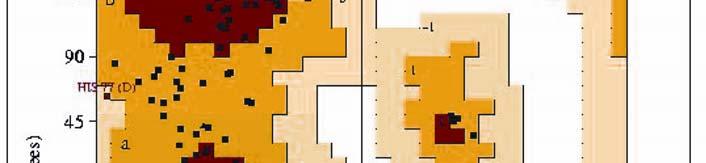
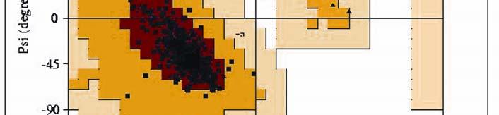
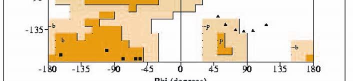
49 clear what parameters are statistically and physically valid to use. (For instance, x,y,z of atoms, but what about group or individual B-factors? Use of ordered water molecules?) More parameters in a fitting function (in this case the function is the macromolecular l model) will fit the data better and will lower R. What to do? The free-rfactor(r factor free ) is always utilized in modern macromolecular crystallographic refinement. R free is an example of the statistical method of cross-validation. The idea is simple. Before any refinement is performed, a fraction of the F obs dataset (typically ~ 5%) is selected randomly and omitted from refinement; this is the test set. That is, the refinement calculation is seeking to minimize some error function that includes minimizing the differences between F calc and F obs. So, of course one expects R to decrease because the minimization is trying to reduce those differences. However, in addition to calculating R for this working set, an R-factor (R free ) is calculated for the test set. This test set has not been used to refine against, so if R free drops then the improvement in the model is statistically justified and has been shown to be so by this cross-validation technique. In a refined structure, R free is typically 2-6% higher than R with R having a value between 15 and 25%. These precise values are dependent upon resolution, data quality, etc. Evaluation of a structure rms deviations of bond-angle and bond-length. These are usually reported, and should typically be degrees or less
 will indicate other issues such as close contacts, etc.")
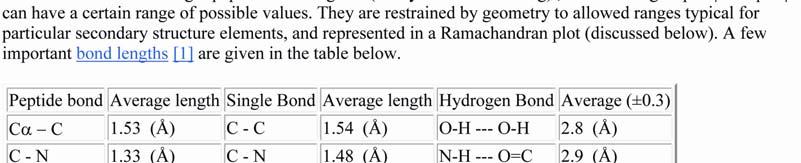
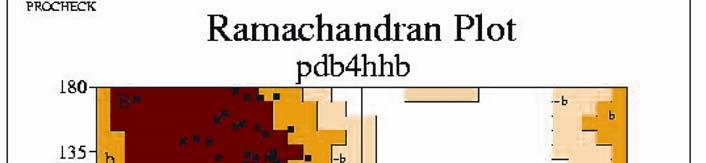
50 (or up to degrees for structures of lower resolution). Bond-length rms should be less than about 0.02Å (up to ~ 0.03Å for lower-resolution). The header of the pdb file (especially of more recent depositions) will indicate other issues such as close contacts, etc. There are also programs that can be run on a pdb file to check for features that may be peculiar. Also, the chemical environment of sidechains should make sense. What do I mean by that?
51
52
53
54
55
X-Ray Crystallography
 X-Ray Crystallography BECAUSE The underlying principle of function is structure. X-ray crystallography is the study of crystal structures through X-ray diffraction techniques. When an X-ray beam bombards
X-ray Crystallography. Kalyan Das
 X-ray Crystallography Kalyan Das Electromagnetic Spectrum NMR 10 um - 10 mm 700 to 10 4 nm 400 to 700 nm 10 to 400 nm 10-1 to 10 nm 10-4 to 10-1 nm X-ray radiation was discovered by Roentgen in 1895. X-rays
Protein crystallography. Garry Taylor
 Protein crystallography Garry Taylor X-ray Crystallography - the Basics Grow crystals Collect X-ray data Determine phases Calculate ρ-map Interpret map Refine coordinates Do the biology. Nitrogen at -180
Scattering by two Electrons
 Scattering by two Electrons p = -r k in k in p r e 2 q k in /λ θ θ k out /λ S q = r k out p + q = r (k out - k in ) e 1 Phase difference of wave 2 with respect to wave 1: 2π λ (k out - k in ) r= 2π S r
Protein Crystallography
 Protein Crystallography Part II Tim Grüne Dept. of Structural Chemistry Prof. G. Sheldrick University of Göttingen http://shelx.uni-ac.gwdg.de tg@shelx.uni-ac.gwdg.de Overview The Reciprocal Lattice The
X-ray Crystallography
 2009/11/25 [ 1 ] X-ray Crystallography Andrew Torda, wintersemester 2009 / 2010 X-ray numerically most important more than 4/5 structures Goal a set of x, y, z coordinates different properties to NMR History
Crystals, X-rays and Proteins
 Crystals, X-rays and Proteins Comprehensive Protein Crystallography Dennis Sherwood MA (Hons), MPhil, PhD Jon Cooper BA (Hons), PhD OXFORD UNIVERSITY PRESS Contents List of symbols xiv PART I FUNDAMENTALS
PSD '17 -- Xray Lecture 5, 6. Patterson Space, Molecular Replacement and Heavy Atom Isomorphous Replacement
 PSD '17 -- Xray Lecture 5, 6 Patterson Space, Molecular Replacement and Heavy Atom Isomorphous Replacement The Phase Problem We can t measure the phases! X-ray detectors (film, photomultiplier tubes, CCDs,
X-ray crystallography
 X-ray crystallography Sources: 1. The Elements of Physical Chemistry by Peter Atkins (including some nice color slides incorporated into these lectures) 2. Physical Chemistry by Tinoco, Sauer, Wang and
Structure factors again
 Structure factors again Remember 1D, structure factor for order h F h = F h exp[iα h ] = I 01 ρ(x)exp[2πihx]dx Where x is fractional position along unit cell distance (repeating distance, origin arbitrary)
 Helpful resources for all X ray lectures Crystallization http://www.hamptonresearch.com under tech support: crystal growth 101 literature Spacegroup tables http://img.chem.ucl.ac.uk/sgp/mainmenu.htm Crystallography
Basic Crystallography Part 1. Theory and Practice of X-ray Crystal Structure Determination
 Basic Crystallography Part 1 Theory and Practice of X-ray Crystal Structure Determination We have a crystal How do we get there? we want a structure! The Unit Cell Concept Ralph Krätzner Unit Cell Description
Phase problem: Determining an initial phase angle α hkl for each recorded reflection. 1 ρ(x,y,z) = F hkl cos 2π (hx+ky+ lz - α hkl ) V h k l
 = F hkl cos 2π (hx+ky+ lz - α hkl ) V h k l") Phase problem: Determining an initial phase angle α hkl for each recorded reflection 1 ρ(x,y,z) = F hkl cos 2π (hx+ky+ lz - α hkl ) V h k l Methods: Heavy atom methods (isomorphous replacement Hg, Pt)
Phase problem: Determining an initial phase angle α hkl for each recorded reflection 1 ρ(x,y,z) = F hkl cos 2π (hx+ky+ lz - α hkl ) V h k l Methods: Heavy atom methods (isomorphous replacement Hg, Pt)
Crystal lattice Real Space. Reflections Reciprocal Space. I. Solving Phases II. Model Building for CHEM 645. Purified Protein. Build model.
 I. Solving Phases II. Model Building for CHEM 645 Purified Protein Solve Phase Build model and refine Crystal lattice Real Space Reflections Reciprocal Space ρ (x, y, z) pronounced rho F hkl 2 I F (h,
Molecular Biology Course 2006 Protein Crystallography Part I
 Molecular Biology Course 2006 Protein Crystallography Part I Tim Grüne University of Göttingen Dept. of Structural Chemistry November 2006 http://shelx.uni-ac.gwdg.de tg@shelx.uni-ac.gwdg.de Overview Overview
6. X-ray Crystallography and Fourier Series
 6. X-ray Crystallography and Fourier Series Most of the information that we have on protein structure comes from x-ray crystallography. The basic steps in finding a protein structure using this method
Resolution: maximum limit of diffraction (asymmetric)
") Resolution: maximum limit of diffraction (asymmetric) crystal Y X-ray source 2θ X direct beam tan 2θ = Y X d = resolution 2d sinθ = λ detector 1 Unit Cell: two vectors in plane of image c* Observe: b*
Resolution: maximum limit of diffraction (asymmetric) crystal Y X-ray source 2θ X direct beam tan 2θ = Y X d = resolution 2d sinθ = λ detector 1 Unit Cell: two vectors in plane of image c* Observe: b*
Probing Atomic Crystals: Bragg Diffraction
 1 Probing Atomic Crystals: Bragg Diffraction OBJECTIVE: To learn how scientists probe the structure of solids, using a scaled-up version of X-ray Diffraction. APPARATUS: Steel ball "crystal", microwave
Chapter 2. X-ray X. Diffraction and Reciprocal Lattice. Scattering from Lattices
 Chapter. X-ray X Diffraction and Reciprocal Lattice Diffraction of waves by crystals Reciprocal Lattice Diffraction of X-rays Powder diffraction Single crystal X-ray diffraction Scattering from Lattices
What is the Phase Problem? Overview of the Phase Problem. Phases. 201 Phases. Diffraction vector for a Bragg spot. In General for Any Atom (x, y, z)
") Protein Overview of the Phase Problem Crystal Data Phases Structure John Rose ACA Summer School 2006 Reorganized by Andy Howard,, Spring 2008 Remember We can measure reflection intensities We can calculate
Protein Overview of the Phase Problem Crystal Data Phases Structure John Rose ACA Summer School 2006 Reorganized by Andy Howard,, Spring 2008 Remember We can measure reflection intensities We can calculate
3.012 Structure An Introduction to X-ray Diffraction
 3.012 Structure An Introduction to X-ray Diffraction This handout summarizes some topics that are important for understanding x-ray diffraction. The following references provide a thorough explanation
Protein Structure Determination 9/25/2007
 One-dimensional NMR spectra Ethanol Cellulase (36 a.a.) Branden & Tooze, Fig. 18.16 1D and 2D NMR spectra of inhibitor K (57 a.a.) K. Wuthrich, NMR of Proteins and Nucleic Acids. (Wiley, 1986.) p. 54-55.
Overview - Macromolecular Crystallography
 Overview - Macromolecular Crystallography 1. Overexpression and crystallization 2. Crystal characterization and data collection 3. The diffraction experiment 4. Phase problem 1. MIR (Multiple Isomorphous
The Reciprocal Lattice
 59-553 The Reciprocal Lattice 61 Because of the reciprocal nature of d spacings and θ from Bragg s Law, the pattern of the diffraction we observe can be related to the crystal lattice by a mathematical
SOLID STATE 18. Reciprocal Space
 SOLID STATE 8 Reciprocal Space Wave vectors and the concept of K-space can simplify the explanation of several properties of the solid state. They will be introduced to provide more information on diffraction
X-ray, Neutron and e-beam scattering
 X-ray, Neutron and e-beam scattering Introduction Why scattering? Diffraction basics Neutrons and x-rays Techniques Direct and reciprocal space Single crystals Powders CaFe 2 As 2 an example What is the
Molecular Biology Course 2006 Protein Crystallography Part II
 Molecular Biology Course 2006 Protein Crystallography Part II Tim Grüne University of Göttingen Dept. of Structural Chemistry December 2006 http://shelx.uni-ac.gwdg.de tg@shelx.uni-ac.gwdg.de Overview
SHELXC/D/E. Andrea Thorn
 SHELXC/D/E Andrea Thorn What is experimental phasing? Experimental phasing is what you do if MR doesn t work. What is experimental phasing? Experimental phasing methods depend on intensity differences.
Electron Density at various resolutions, and fitting a model as accurately as possible.
 Section 9, Electron Density Maps 900 Electron Density at various resolutions, and fitting a model as accurately as possible. ρ xyz = (Vol) -1 h k l m hkl F hkl e iφ hkl e-i2π( hx + ky + lz ) Amplitude
CS273: Algorithms for Structure Handout # 13 and Motion in Biology Stanford University Tuesday, 11 May 2003
 CS273: Algorithms for Structure Handout # 13 and Motion in Biology Stanford University Tuesday, 11 May 2003 Lecture #13: 11 May 2004 Topics: Protein Structure Determination Scribe: Minli Zhu We acknowledge
Macromolecular X-ray Crystallography
 Protein Structural Models for CHEM 641 Fall 07 Brian Bahnson Department of Chemistry & Biochemistry University of Delaware Macromolecular X-ray Crystallography Purified Protein X-ray Diffraction Data collection
(Re-write, January 2011, from notes of S. C. Fain Jr., L. Sorensen, O. E. Vilches, J. Stoltenberg and D. B. Pengra, Version 1, preliminary)
") Electron Diffraction (Re-write, January 011, from notes of S. C. Fain Jr., L. Sorensen, O. E. Vilches, J. Stoltenberg and D. B. Pengra, Version 1, preliminary) References: Any introductory physics text
Electron Diffraction (Re-write, January 011, from notes of S. C. Fain Jr., L. Sorensen, O. E. Vilches, J. Stoltenberg and D. B. Pengra, Version 1, preliminary) References: Any introductory physics text
Handout 7 Reciprocal Space
 Handout 7 Reciprocal Space Useful concepts for the analysis of diffraction data http://homepages.utoledo.edu/clind/ Concepts versus reality Reflection from lattice planes is just a concept that helps us
Application Note SC-XRD 505 Single Crystal Diffraction
 Application Note SC-XRD 505 Single Crystal Diffraction Introduction Single-crystal X-ray diffraction, commonly referred to as X-ray crystallography, is an analytical technique in which X-ray methods are
Patterson Methods
 59-553 Patterson Methods 113 In 1935, Patterson showed that the unknown phase information in the equation for electron density: ρ(xyz) = 1/V h k l F(hkl) exp[iα(hkl)] exp[-2πi(h x + k y + l z)] can be
PSD '18 -- Xray lecture 4. Laue conditions Fourier Transform The reciprocal lattice data collection
 PSD '18 -- Xray lecture 4 Laue conditions Fourier Transform The reciprocal lattice data collection 1 Fourier Transform The Fourier Transform is a conversion of one space into another space with reciprocal
General theory of diffraction
 General theory of diffraction X-rays scatter off the charge density (r), neutrons scatter off the spin density. Coherent scattering (diffraction) creates the Fourier transform of (r) from real to reciprocal
X-Ray structure analysis
 X-Ray structure analysis Kay Diederichs kay.diederichs@uni-konstanz.de Analysis of what? Proteins ( /ˈproʊˌtiːnz/ or /ˈproʊti.ɨnz/) are biochemical compounds consisting of one or more polypeptides typically
Anomalous dispersion
 Selenomethionine MAD Selenomethionine is the amino acid methionine with the Sulfur replaced by a Selenium. Selenium is a heavy atom that also has the propery of "anomalous scatter" at some wavelengths,
Fourier Syntheses, Analyses, and Transforms
 Fourier Syntheses, Analyses, and Transforms http://homepages.utoledo.edu/clind/ The electron density The electron density in a crystal can be described as a periodic function - same contents in each unit
X-ray Data Collection. Bio5325 Spring 2006
 X-ray Data Collection Bio535 Spring 006 Obtaining I hkl and α (Ihkl) from Frame Images Braggs Law -predicts conditions for in-phase scattering by equivalent atoms lying in planes that transect a crystal.
The structure of liquids and glasses. The lattice and unit cell in 1D. The structure of crystalline materials. Describing condensed phase structures
 Describing condensed phase structures Describing the structure of an isolated small molecule is easy to do Just specify the bond distances and angles How do we describe the structure of a condensed phase?
Direct Method. Very few protein diffraction data meet the 2nd condition
 Direct Method Two conditions: -atoms in the structure are equal-weighted -resolution of data are higher than the distance between the atoms in the structure Very few protein diffraction data meet the 2nd
Data processing and reduction
 Data processing and reduction Leopoldo Suescun International School on Fundamental Crystallography 2014 May 1st, 2014 Reciprocal lattice c* b* b * dh' k' l' 1 dh' k' l' * dhkl 1 dhkl a a* 0 d hkl c bc
Determining Protein Structure BIBC 100
 Determining Protein Structure BIBC 100 Determining Protein Structure X-Ray Diffraction Interactions of x-rays with electrons in molecules in a crystal NMR- Nuclear Magnetic Resonance Interactions of magnetic
Roger Johnson Structure and Dynamics: X-ray Diffraction Lecture 6
 6.1. Summary In this Lecture we cover the theory of x-ray diffraction, which gives direct information about the atomic structure of crystals. In these experiments, the wavelength of the incident beam must
Diffraction Geometry
 Diffraction Geometry Diffraction from a crystal - Laue equations Reciprocal lattice Ewald construction Data collection strategy Phil Evans LMB May 2013 MRC Laboratory of Molecular Biology Cambridge UK
Surface Sensitivity & Surface Specificity
 Surface Sensitivity & Surface Specificity The problems of sensitivity and detection limits are common to all forms of spectroscopy. In its simplest form, the question of sensitivity boils down to whether
Determination of the Substructure
 Monday, June 15 th, 2009 Determination of the Substructure EMBO / MAX-INF2 Practical Course http://shelx.uni-ac.gwdg.de Overview Substructure Definition and Motivation Extracting Substructure Data from
Physics I : Oscillations and Waves Prof. S. Bharadwaj Department of Physics and Meteorology Indian Institute of Technology, Kharagpur
 Physics I : Oscillations and Waves Prof. S. Bharadwaj Department of Physics and Meteorology Indian Institute of Technology, Kharagpur Lecture - 21 Diffraction-II Good morning. In the last class, we had
Phys 460 Describing and Classifying Crystal Lattices
 Phys 460 Describing and Classifying Crystal Lattices What is a material? ^ crystalline Regular lattice of atoms Each atom has a positively charged nucleus surrounded by negative electrons Electrons are
Michelson Interferometer
 Michelson Interferometer Objective Determination of the wave length of the light of the helium-neon laser by means of Michelson interferometer subsectionprinciple and Task Light is made to produce interference
Quantum Condensed Matter Physics Lecture 5
 Quantum Condensed Matter Physics Lecture 5 detector sample X-ray source monochromator David Ritchie http://www.sp.phy.cam.ac.uk/drp2/home QCMP Lent/Easter 2019 5.1 Quantum Condensed Matter Physics 1. Classical
Principles of Physical Biochemistry
 Principles of Physical Biochemistry Kensal E. van Hold e W. Curtis Johnso n P. Shing Ho Preface x i PART 1 MACROMOLECULAR STRUCTURE AND DYNAMICS 1 1 Biological Macromolecules 2 1.1 General Principles
4. Other diffraction techniques
 4. Other diffraction techniques 4.1 Reflection High Energy Electron Diffraction (RHEED) Setup: - Grazing-incidence high energy electron beam (3-5 kev: MEED,
Molecular Modeling lecture 2
 Molecular Modeling 2018 -- lecture 2 Topics 1. Secondary structure 3. Sequence similarity and homology 2. Secondary structure prediction 4. Where do protein structures come from? X-ray crystallography
Fundamentals of X-ray diffraction
 Fundamentals of X-ray diffraction Elena Willinger Lecture series: Modern Methods in Heterogeneous Catalysis Research Outline History of X-ray Sources of X-ray radiation Physics of X-ray scattering Fundamentals
X-ray Diffraction. Diffraction. X-ray Generation. X-ray Generation. X-ray Generation. X-ray Spectrum from Tube
 X-ray Diffraction Mineral identification Mode analysis Structure Studies X-ray Generation X-ray tube (sealed) Pure metal target (Cu) Electrons remover inner-shell electrons from target. Other electrons
disordered, ordered and coherent with the substrate, and ordered but incoherent with the substrate.
 5. Nomenclature of overlayer structures Thus far, we have been discussing an ideal surface, which is in effect the structure of the topmost substrate layer. The surface (selvedge) layers of the solid however
X-ray analysis. 1. Basic crystallography 2. Basic diffraction physics 3. Experimental methods
 X-ray analysis 1. Basic crystallography 2. Basic diffraction physics 3. Experimental methods Introduction Noble prizes associated with X-ray diffraction 1901 W. C. Roentgen (Physics) for the discovery
Physical Chemistry I. Crystal Structure
 Physical Chemistry I Crystal Structure Crystal Structure Introduction Crystal Lattice Bravis Lattices Crytal Planes, Miller indices Distances between planes Diffraction patters Bragg s law X-ray radiation
Scattering Lecture. February 24, 2014
 Scattering Lecture February 24, 2014 Structure Determination by Scattering Waves of radiation scattered by different objects interfere to give rise to an observable pattern! The wavelength needs to close
Crystallography past, present and future
 Crystallography past, present and future Jenny P. Glusker Philadelphia, PA, U. S. A. International Year of Crystallography UNESCO, Paris, France 20 January 2014 QUARTZ CRYSTALS Quartz crystals found growing
PX-CBMSO Course (2) of Symmetry
 of Symmetry") PX-CBMSO Course (2) The mathematical description of Symmetry y PX-CBMSO-June 2011 Cele Abad-Zapatero University of Illinois at Chicago Center for Pharmaceutical Biotechnology. Lecture no. 2 This material
PX-CBMSO Course (2) The mathematical description of Symmetry y PX-CBMSO-June 2011 Cele Abad-Zapatero University of Illinois at Chicago Center for Pharmaceutical Biotechnology. Lecture no. 2 This material
Physical Chemistry Analyzing a Crystal Structure and the Diffraction Pattern Virginia B. Pett The College of Wooster
 Physical Chemistry Analyzing a Crystal Structure and the Diffraction Pattern Virginia B. Pett The College of Wooster L. W. Haynes and his Senior Independent Study students conducted the 2 + 2 photo addition
Keble College - Hilary 2012 Section VI: Condensed matter physics Tutorial 2 - Lattices and scattering
 Tomi Johnson Keble College - Hilary 2012 Section VI: Condensed matter physics Tutorial 2 - Lattices and scattering Please leave your work in the Clarendon laboratory s J pigeon hole by 5pm on Monday of
Materials 286C/UCSB: Class VI Structure factors (continued), the phase problem, Patterson techniques and direct methods
, the phase problem, Patterson techniques and direct methods") Materials 286C/UCSB: Class VI Structure factors (continued), the phase problem, Patterson techniques and direct methods Ram Seshadri (seshadri@mrl.ucsb.edu) Structure factors The structure factor for a
Materials 286C/UCSB: Class VI Structure factors (continued), the phase problem, Patterson techniques and direct methods Ram Seshadri (seshadri@mrl.ucsb.edu) Structure factors The structure factor for a
Structure and Dynamics : An Atomic View of Materials
 Structure and Dynamics : An Atomic View of Materials MARTIN T. DOVE Department ofearth Sciences University of Cambridge OXFORD UNIVERSITY PRESS Contents 1 Introduction 1 1.1 Observations 1 1.1.1 Microscopic
Protein Structure Determination. Part 1 -- X-ray Crystallography
 Protein Structure Determination Part 1 -- X-ray Crystallography Topics covering in this 1/2 course Crystal growth Diffraction theory Symmetry Solving phases using heavy atoms Solving phases using a model
Scattering and Diffraction
 Scattering and Diffraction Andreas Kreyssig, Alan Goldman, Rob McQueeney Ames Laboratory Iowa State University All rights reserved, 2018. Atomic scale structure - crystals Crystalline materials... atoms
Proteins. Central Dogma : DNA RNA protein Amino acid polymers - defined composition & order. Perform nearly all cellular functions Drug Targets
 Proteins Central Dogma : DNA RNA protein Amino acid polymers - defined composition & order Perform nearly all cellular functions Drug Targets Fold into discrete shapes. Proteins - cont. Specific shapes
Symmetry Crystallography
 Crystallography Motif: the fundamental part of a symmetric design that, when repeated, creates the whole pattern In 3-D, translation defines operations which move the motif into infinitely repeating patterns
An Introduction to Diffraction and Scattering. School of Chemistry The University of Sydney
 An Introduction to Diffraction and Scattering Brendan J. Kennedy School of Chemistry The University of Sydney 1) Strong forces 2) Weak forces Types of Forces 3) Electromagnetic forces 4) Gravity Types
Chapter 20: Convergent-beam diffraction Selected-area diffraction: Influence of thickness Selected-area vs. convergent-beam diffraction
 1 Chapter 0: Convergent-beam diffraction Selected-area diffraction: Influence of thickness Selected-area diffraction patterns don t generally get much better when the specimen gets thicker. Sometimes a
Protein Crystallography. Mitchell Guss University of Sydney Australia
 Protein Crystallography Mitchell Guss University of Sydney Australia Outline of the talk Recap some basic crystallography and history Highlight the special requirements for protein (macromolecular) structure
Crystal planes. Neutrons: magnetic moment - interacts with magnetic materials or nuclei of non-magnetic materials. (in Å)
") Crystallography: neutron, electron, and X-ray scattering from periodic lattice, scattering of waves by periodic structures, Miller indices, reciprocal space, Ewald construction. Diffraction: Specular,
Crystallography: neutron, electron, and X-ray scattering from periodic lattice, scattering of waves by periodic structures, Miller indices, reciprocal space, Ewald construction. Diffraction: Specular,
Why do We Trust X-ray Crystallography?
 Why do We Trust X-ray Crystallography? Andrew D Bond All chemists know that X-ray crystallography is the gold standard characterisation technique: an X-ray crystal structure provides definitive proof of
Chem 728 Introduction to Solid Surfaces
 Chem 728 Introduction to Solid Surfaces Solids: hard; fracture; not compressible; molecules close to each other Liquids: molecules mobile, but quite close to each other Gases: molecules very mobile; compressible
Solids. properties & structure
 Solids properties & structure Determining Crystal Structure crystalline solids have a very regular geometric arrangement of their particles the arrangement of the particles and distances between them is
Changing and challenging times for service crystallography. Electronic Supplementary Information
 Changing and challenging times for service crystallography Simon J Coles,* a and Philip A Gale* a Electronic Supplementary Information Instrument descriptions and experimental protocols The following firstly
Lattice (Sieć) A collection of nodes, i.e. points with integral coordinates. In crystallography, a lattice is an
 A collection of nodes, i.e. points with integral coordinates. In crystallography, a lattice is an") Prof. dr hab. Mariusz Jaskólski GLOSSARYUSZ TERMINÓW KRYSTALOGRAFICZNYCH (dla osób nie znających jeszcze krystalografii, ale znających język angielski) Symmetry (Symetria) Property of physical and mathematical
Prof. dr hab. Mariusz Jaskólski GLOSSARYUSZ TERMINÓW KRYSTALOGRAFICZNYCH (dla osób nie znających jeszcze krystalografii, ale znających język angielski) Symmetry (Symetria) Property of physical and mathematical
Applications of X-ray and Neutron Scattering in Biological Sciences: Symmetry in direct and reciprocal space 2012
 Department of Drug Design and Pharmacology Applications of X-ray and Neutron Scattering in Biological Sciences: Symmetry in direct and reciprocal space 2012 Michael Gajhede Biostructural Research Copenhagen
Experimental Determination of Crystal Structure
 Experimental Determination of Crystal Structure Branislav K. Nikolić Department of Physics and Astronomy, University of Delaware, U.S.A. PHYS 624: Introduction to Solid State Physics http://www.physics.udel.edu/~bnikolic/teaching/phys624/phys624.html
Chapter 1 X-ray Absorption Fine Structure (EXAFS)
") 1 Chapter 1 X-ray Absorption Fine Structure (EXAFS) 1.1 What is EXAFS? X-ray absorption fine structure (EXAFS, XAFS) is an oscillatory modulation in the X-ray absorption coefficient on the high-energy
1 Chapter 1 X-ray Absorption Fine Structure (EXAFS) 1.1 What is EXAFS? X-ray absorption fine structure (EXAFS, XAFS) is an oscillatory modulation in the X-ray absorption coefficient on the high-energy
Crystal Structure and Electron Diffraction
 Crystal Structure and Electron Diffraction References: Kittel C.: Introduction to Solid State Physics, 8 th ed. Wiley 005 University of Michigan, PHY441-44 (Advanced Physics Laboratory Experiments, Electron
Solid State Physics Lecture 3 Diffraction and the Reciprocal Lattice (Kittel Ch. 2)
") Solid State Physics 460 - Lecture 3 Diffraction and the Reciprocal Lattice (Kittel Ch. 2) Diffraction (Bragg Scattering) from a powder of crystallites - real example of image at right from http://www.uni-wuerzburg.de/mineralogie/crystal/teaching/pow.html
Solid State Physics 460 - Lecture 3 Diffraction and the Reciprocal Lattice (Kittel Ch. 2) Diffraction (Bragg Scattering) from a powder of crystallites - real example of image at right from http://www.uni-wuerzburg.de/mineralogie/crystal/teaching/pow.html
Biochemistry,530:,, Introduc5on,to,Structural,Biology, Autumn,Quarter,2015,
 Biochemistry,530:,, Introduc5on,to,Structural,Biology, Autumn,Quarter,2015, Course,Informa5on, BIOC%530% GraduateAlevel,discussion,of,the,structure,,func5on,,and,chemistry,of,proteins,and, nucleic,acids,,control,of,enzyma5c,reac5ons.,please,see,the,course,syllabus,and,
PROBING CRYSTAL STRUCTURE
 PROBING CRYSTAL STRUCTURE Andrew Baczewski PHY 491, October 10th, 2011 OVERVIEW First - we ll briefly discuss Friday s quiz. Today, we will answer the following questions: How do we experimentally probe
Rajesh Prasad Department of Applied Mechanics Indian Institute of Technology New Delhi
 TEQIP WORKSHOP ON HIGH RESOLUTION X-RAY AND ELECTRON DIFFRACTION, FEB 01, 2016, IIT-K. Introduction to x-ray diffraction Peak Positions and Intensities Rajesh Prasad Department of Applied Mechanics Indian
Lecture- 08 Emission and absorption spectra
 Atomic and Molecular Absorption Spectrometry for Pollution Monitoring Dr. J R Mudakavi Department of Chemical Engineering Indian Institute of Science, Bangalore Lecture- 08 Emission and absorption spectra
Protein Crystallography Part II
 Molecular Biology Course 2007 Protein Crystallography Part II Tim Grüne University of Göttingen Dept. of Structural Chemistry November 2007 http://shelx.uni-ac.gwdg.de tg@shelx.uni-ac.gwdg.de Overview
X- ray crystallography. CS/CME/Biophys/BMI 279 Nov. 12, 2015 Ron Dror
 X- ray crystallography CS/CME/Biophys/BMI 279 Nov. 12, 2015 Ron Dror 1 Outline Overview of x-ray crystallography Crystals Electron density Diffraction patterns The computational problem: determining structure
CCP4 Diamond 2014 SHELXC/D/E. Andrea Thorn
 CCP4 Diamond 2014 SHELXC/D/E Andrea Thorn SHELXC/D/E workflow SHELXC: α calculation, file preparation SHELXD: Marker atom search = substructure search SHELXE: density modification Maps and coordinate files
Handout 13 Interpreting your results. What to make of your atomic coordinates, bond distances and angles
 Handout 13 Interpreting your results What to make of your atomic coordinates, bond distances and angles 1 What to make of the outcome of your refinement There are several ways of judging whether the outcome
Name Biology Chapter 2 Note-taking worksheet
 Name Biology Chapter 2 Note-taking worksheet The Nature of Matter 1. Life depends on Atoms 1. The study of chemistry starts with the basic unit of matter, the. 2. The atom was first used by the Greek philosopher
Experiment 3 1. The Michelson Interferometer and the He- Ne Laser Physics 2150 Experiment No. 3 University of Colorado
 Experiment 3 1 Introduction The Michelson Interferometer and the He- Ne Laser Physics 2150 Experiment No. 3 University of Colorado The Michelson interferometer is one example of an optical interferometer.
How to study minerals?!
 How to study minerals?! ü What tools did scientists have from pre-history to Renaissance? Eyes and measuring devices Calcite Crystal faces! ü One of the most spectacular aspect of minerals ü NOTE: No mention
What use is Reciprocal Space? An Introduction
 What use is Reciprocal Space? An Introduction a* b* x You are here John Bargar 5th Annual SSRL Workshop on Synchrotron X-ray Scattering Techniques in Materials and Environmental Sciences June 1-3, 2010
TEP Examination of the structure of NaCl monocrystals with different orientations
 Examination of the structure of NaCl TEP Related topics Characteristic X-radiation, energy levels, crystal structures, reciprocal lattices, Miller indices, atomic form factor, structure factor, and Bragg
The Solid State. Phase diagrams Crystals and symmetry Unit cells and packing Types of solid
 The Solid State Phase diagrams Crystals and symmetry Unit cells and packing Types of solid Learning objectives Apply phase diagrams to prediction of phase behaviour Describe distinguishing features of