Direct Method. Very few protein diffraction data meet the 2nd condition
|
|
|
- Merryl Mills
- 5 years ago
- Views:
Transcription
1 Direct Method Two conditions: -atoms in the structure are equal-weighted -resolution of data are higher than the distance between the atoms in the structure Very few protein diffraction data meet the 2nd condition Heavy atoms in protein => sub-structure of heavy atoms in a derivative F H = F PH - F P
2 Direct Method 1. Determine the substructure of heavy atoms 2. Determine overall protein structure at 1.2 Å 3. Programs: - Shake and bake (SnB) - SHELXD 4. Phases refined by Sharp
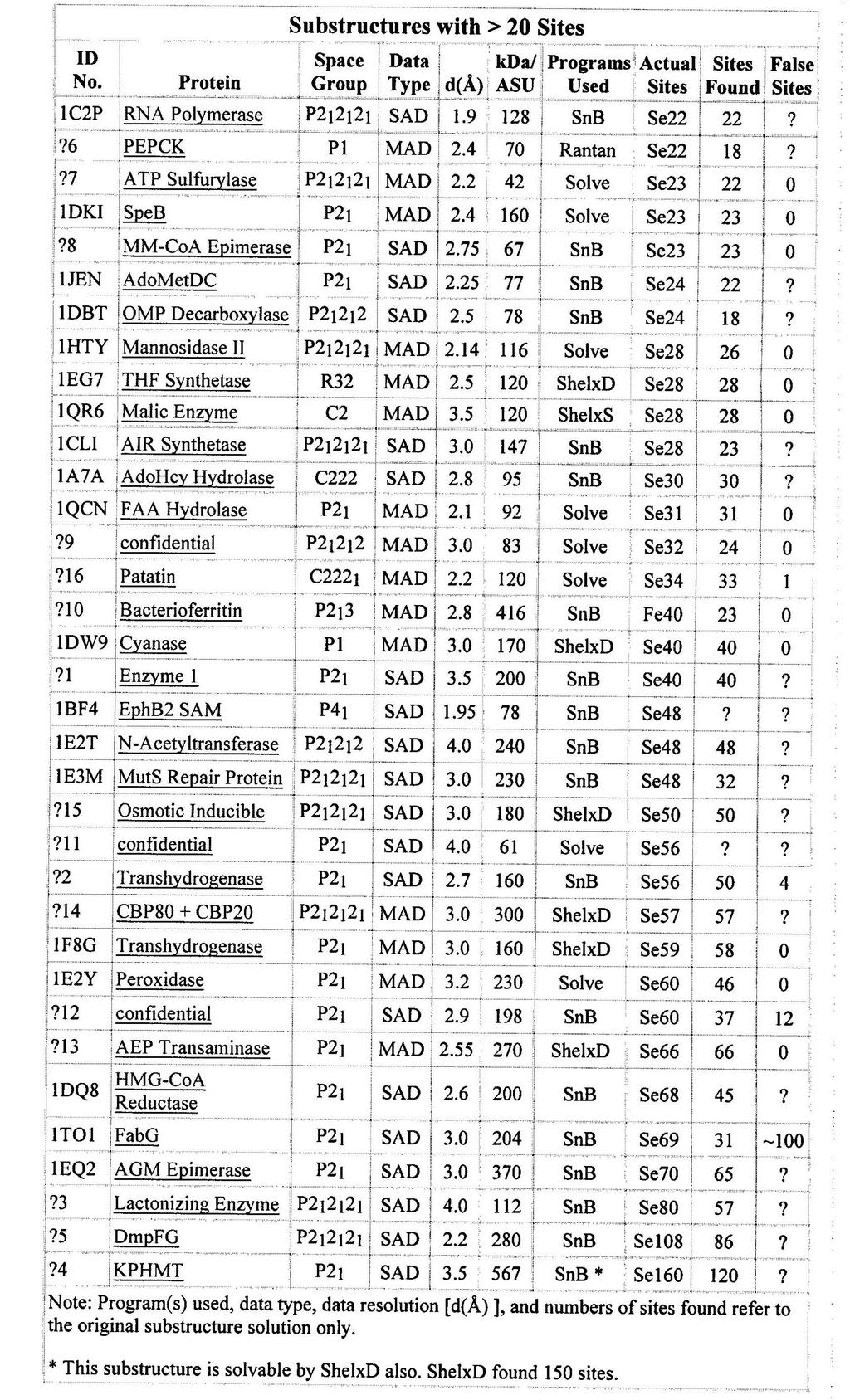
3 Large substructure solved mainly by direct method The number of heavy atoms (anomalous scatters) > 20 Mainly apply to protein crystals contain many Se-Met and halide soaking
4
5 Molecular Replacement 1. A homologous model (structure) available 2. A structure determined by putting the model in proper orientation and precise position in the target unit cell. - rotation search (rotation matrix) - translation search (translation vector) 3. New structure X B X B = [C] X A + d 4. Amore, Molrep, CNS, Phaser 5. Over 50% protein structures solved by MR
6 Solvent Content Mathew coefficient (V M ) V O V M = Z x n x MW V M = 1.7~ 3.5 V O Unit cell volume Z number of au in a unit cell M W molecular weight of protein n number of protein molecules in the au Protein content: V protein = 1.23/V M, 1.23 = density of protein crystals Solvent content: V sol = /V M When V M = 2.5 Å 3 /Da, V sol = 0.51 =>Most common Vm and Vsol -Solvent fractions of are common. -Crystals with high solvent contents general diffract poorly and are fragile. -But high solvent content is a great advantage in phase improvement by density modification.
7 Fourier transforms Electron density ρ (x,y,z) = 1/V F hkl exp (-2πi(hX+kY+lZ)) h k l F hkl = F hkl exp(i 2π(hxj+kyj+lzj) ) Structure factor ρ (x) FT - FT F(h)
8 Density modification We use current phases (model) to calculate a density map. Then we modify that map to make it conform better to some idea about what an electron density map should look like. The new and better map is then back-transformed to the calculate structure factors, which should have more accurate phases than original map. By iterating, we are getting a map that does not change anymore and should be closer to the true map
9 Solvent flattening/flipping Density in protein and solvent regions: Solvent flattening: -replacing all the density values within the solvent region with the average value throughout the solvent region. ρ out (x) = (ρ in (x) - ρ sol ) * µ (x) + ρ sol Solvent flipping: - modified solvent flattening to remove biases from original map

10 NCS averaging NCS copies in AU The NCS copies in AU should have same electron density The difference in a noisy density map between NCS copies caused by random errors. Non-crystallographic symmetry: symmetry relations among identical copies in AU A new map made by averaging the copies of density related by non-crystallographic symmetry should be more accurate, since the noise is averaged out.
11 Model Building Putting blocks of protein structures into electron density Mainly use interactive computer graphic programs: - O, XtalView, Coot Automatic model-building programs: - ARP/wARP, RESOLVE, MAID

12 Map and Model 6.0 Å map

13 Map and Model 1.0 Å map
14 Fourier Method F F(h) = F exp (iα c ) -F is the true structure factor but only F measured -Fc is from an initial model or a molecular replacement search model -F(h) is model-phased structured factor closer to the true F than Fc was -So the map will the map will have the features of the true structure -Used to solve ligand- soaked structures

15 Map from Fourier Method o Model-phased map
indicates atoms should be added -negative density (red) indicates the atoms should be moved elsewhere (or")
16 Difference Map F(h) = ( F - F C ) exp (iα c ) F o - F c map The difference map highlights the difference of true structure and the model -positive density (blue)indicates atoms should be added -negative density (red) indicates the atoms should be moved elsewhere (or removed)
17 Refinement of Model Now we have 1) X-ray diffraction intensities (h, k, l) from data 2) x, y, z positions of atoms in unit cell from models 3) known scattering factors of atoms Let s adjust the model to find a closer agreement between the calculated and observed structure factors - minimize the difference below: Σ ( F cal - F obs ) 2 hkl - correct the errors in the initial atomic model
18 Thermal Motion - atoms undergo motion in crystal - not exactly fixed at x j, y j, z j - B factor B j = 8π 2 u j 2 B j is a measure of motion u j is degree of vibration B j = 80 Å 2, u j = 1.00Å B j = 20 Å 2, u j = 0.5Å atoms F (h,k,l) = Σ f (j) exp [2π * i(hx (j) + ky (j) +lz (j) ]* exp [-B j * (sinθ/λ) 2 ] j=1
19 Resolution and Refinement Resolution Observations/parameters 3.5 Å Å Å Å Å 6.2 -For a protein crystal with a typical packing density, and 4 parameters (x, y, z, and B) per atom (non-h). -At resolutions < 1.0, the ratio of observations to parameters is low and the refinement is poor over-determined.


20 Resolution and Structure 1.0 Å 2.5 Å 3.0 Å 4.0 Å Cambridge course
21 Restraints and Constraints Additional observations are incorporated in the refinement -Stereochemical data from small molecular structures e.g. bond lengths and angles, etc Constraints The stereo data taken as rigid and only dihedral angles varied in models -effectively reduce the # of parameters Restraints The stereo data allowed to vary around a standard value and controlled by an energy term E = E chem + w E xray E xay = Σ( F cal (h) - k F obs (h) ) 2 h -define the difference of models to x-ray data E chem = Σ (M ideal M model ) M bond lengths and angles, torsion angles and van der Waals contacts, etc
22 R Factor Difference between F obs and F calc R factor = Σ F obs - F calc hkl Σ F obs hkl Quality of Model R factor = 0.00 perfect fit 0.20 good fit 0.60 random fit
23 Subjectivity and Overfitting Subjectivity: misinterpret density map Overfitting: lower R-factors without removing errors in the model Protein crystals usually could not diffract to atomic resolution, which provide a room for above two error-inducing phenomena Overfit the diffraction data by introduction of too many adjustable parameters. e.g., too many water molecules are fitted to the diffraction data, which compensates for errors in the model or the data. Certain subtle errors introduced by overfitting can produce a low R factor.
24 Validation and Model Evaluation Ramachandran plot Bond lengths/angles Homolog structure comparison Independent structural solutions
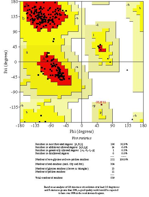
25 Ramachandran Plot
26 R free and Cross-validation R free = hkl T Σ F obs - F calc hkl T Σ F obs Difference between F obs and F calc for the test data hkl T: All the reflections belong to test set, random selection of ~5% of the observed reflections which never used in refinement. Every observation contains information from all the atoms in a structure.
27 Structure Quality What to look for 1) R free near ) Resolution (better than 3.5 Å) 3) Completeness ~ 95% 4) Ramachandran plot 5) RMSD of bond lengths and angles from ideal values (<0.02 Å, <2.0 o )
28 Refine and Rebuild Model - Check the correctness of the model - 2Fo-Fc and Fo-Fc maps (re-building) - database in O, coot, XtalView - Refine the updated model against diffraction data - use R free as monitor - Evaluate the structure - torsion angles by Ramachandran Plot - geometry, bond lengths and angles - Iterate the three steps until satisfaction
Crystal lattice Real Space. Reflections Reciprocal Space. I. Solving Phases II. Model Building for CHEM 645. Purified Protein. Build model.
 I. Solving Phases II. Model Building for CHEM 645 Purified Protein Solve Phase Build model and refine Crystal lattice Real Space Reflections Reciprocal Space ρ (x, y, z) pronounced rho F hkl 2 I F (h,
Phase problem: Determining an initial phase angle α hkl for each recorded reflection. 1 ρ(x,y,z) = F hkl cos 2π (hx+ky+ lz - α hkl ) V h k l
 = F hkl cos 2π (hx+ky+ lz - α hkl ) V h k l") Phase problem: Determining an initial phase angle α hkl for each recorded reflection 1 ρ(x,y,z) = F hkl cos 2π (hx+ky+ lz - α hkl ) V h k l Methods: Heavy atom methods (isomorphous replacement Hg, Pt)
Phase problem: Determining an initial phase angle α hkl for each recorded reflection 1 ρ(x,y,z) = F hkl cos 2π (hx+ky+ lz - α hkl ) V h k l Methods: Heavy atom methods (isomorphous replacement Hg, Pt)
Protein crystallography. Garry Taylor
 Protein crystallography Garry Taylor X-ray Crystallography - the Basics Grow crystals Collect X-ray data Determine phases Calculate ρ-map Interpret map Refine coordinates Do the biology. Nitrogen at -180
X-ray Crystallography. Kalyan Das
 X-ray Crystallography Kalyan Das Electromagnetic Spectrum NMR 10 um - 10 mm 700 to 10 4 nm 400 to 700 nm 10 to 400 nm 10-1 to 10 nm 10-4 to 10-1 nm X-ray radiation was discovered by Roentgen in 1895. X-rays
Structure factors again
 Structure factors again Remember 1D, structure factor for order h F h = F h exp[iα h ] = I 01 ρ(x)exp[2πihx]dx Where x is fractional position along unit cell distance (repeating distance, origin arbitrary)
PSD '17 -- Xray Lecture 5, 6. Patterson Space, Molecular Replacement and Heavy Atom Isomorphous Replacement
 PSD '17 -- Xray Lecture 5, 6 Patterson Space, Molecular Replacement and Heavy Atom Isomorphous Replacement The Phase Problem We can t measure the phases! X-ray detectors (film, photomultiplier tubes, CCDs,
X-ray Crystallography
 2009/11/25 [ 1 ] X-ray Crystallography Andrew Torda, wintersemester 2009 / 2010 X-ray numerically most important more than 4/5 structures Goal a set of x, y, z coordinates different properties to NMR History
Molecular Biology Course 2006 Protein Crystallography Part II
 Molecular Biology Course 2006 Protein Crystallography Part II Tim Grüne University of Göttingen Dept. of Structural Chemistry December 2006 http://shelx.uni-ac.gwdg.de tg@shelx.uni-ac.gwdg.de Overview
Anomalous dispersion
 Selenomethionine MAD Selenomethionine is the amino acid methionine with the Sulfur replaced by a Selenium. Selenium is a heavy atom that also has the propery of "anomalous scatter" at some wavelengths,
Molecular Replacement (Alexei Vagin s lecture)
") Molecular Replacement (Alexei Vagin s lecture) Contents What is Molecular Replacement Functions in Molecular Replacement Weighting scheme Information from data and model Some special techniques of Molecular
Molecular Replacement (Alexei Vagin s lecture) Contents What is Molecular Replacement Functions in Molecular Replacement Weighting scheme Information from data and model Some special techniques of Molecular
Scattering by two Electrons
 Scattering by two Electrons p = -r k in k in p r e 2 q k in /λ θ θ k out /λ S q = r k out p + q = r (k out - k in ) e 1 Phase difference of wave 2 with respect to wave 1: 2π λ (k out - k in ) r= 2π S r
Phaser: Experimental phasing
 Phaser: Experimental phasing Using SAD data in Phaser R J Read, Department of Haematology Cambridge Institute for Medical Research Diffraction with anomalous scatterers SAD: single-wavelength anomalous
From x-ray crystallography to electron microscopy and back -- how best to exploit the continuum of structure-determination methods now available
 From x-ray crystallography to electron microscopy and back -- how best to exploit the continuum of structure-determination methods now available Scripps EM course, November 14, 2007 What aspects of contemporary
Likelihood and SAD phasing in Phaser. R J Read, Department of Haematology Cambridge Institute for Medical Research
 Likelihood and SAD phasing in Phaser R J Read, Department of Haematology Cambridge Institute for Medical Research Concept of likelihood Likelihood with dice 4 6 8 10 Roll a seven. Which die?? p(4)=p(6)=0
Protein Crystallography Part II
 Molecular Biology Course 2007 Protein Crystallography Part II Tim Grüne University of Göttingen Dept. of Structural Chemistry November 2007 http://shelx.uni-ac.gwdg.de tg@shelx.uni-ac.gwdg.de Overview
X-ray Crystallography I. James Fraser Macromolecluar Interactions BP204
 X-ray Crystallography I James Fraser Macromolecluar Interactions BP204 Key take-aways 1. X-ray crystallography results from an ensemble of Billions and Billions of molecules in the crystal 2. Models in
Overview - Macromolecular Crystallography
 Overview - Macromolecular Crystallography 1. Overexpression and crystallization 2. Crystal characterization and data collection 3. The diffraction experiment 4. Phase problem 1. MIR (Multiple Isomorphous
Protein Structure Determination. Part 1 -- X-ray Crystallography
 Protein Structure Determination Part 1 -- X-ray Crystallography Topics covering in this 1/2 course Crystal growth Diffraction theory Symmetry Solving phases using heavy atoms Solving phases using a model
Fourier Syntheses, Analyses, and Transforms
 Fourier Syntheses, Analyses, and Transforms http://homepages.utoledo.edu/clind/ The electron density The electron density in a crystal can be described as a periodic function - same contents in each unit
Determination of the Substructure
 Monday, June 15 th, 2009 Determination of the Substructure EMBO / MAX-INF2 Practical Course http://shelx.uni-ac.gwdg.de Overview Substructure Definition and Motivation Extracting Substructure Data from
Macromolecular Crystallography Part II
 Molecular Biology Course 2009 Macromolecular Crystallography Part II Tim Grüne University of Göttingen Dept. of Structural Chemistry November 2009 http://shelx.uni-ac.gwdg.de tg@shelx.uni-ac.gwdg.de From
 Helpful resources for all X ray lectures Crystallization http://www.hamptonresearch.com under tech support: crystal growth 101 literature Spacegroup tables http://img.chem.ucl.ac.uk/sgp/mainmenu.htm Crystallography
Molecular replacement. New structures from old
 Molecular replacement New structures from old The Phase Problem phase amplitude Phasing by molecular replacement Phases can be calculated from atomic model Rotate and translate related structure Models
Electron Density at various resolutions, and fitting a model as accurately as possible.
 Section 9, Electron Density Maps 900 Electron Density at various resolutions, and fitting a model as accurately as possible. ρ xyz = (Vol) -1 h k l m hkl F hkl e iφ hkl e-i2π( hx + ky + lz ) Amplitude
SUPPLEMENTARY INFORMATION
 Table of Contents Page Supplementary Table 1. Diffraction data collection statistics 2 Supplementary Table 2. Crystallographic refinement statistics 3 Supplementary Fig. 1. casic1mfc packing in the R3
Macromolecular X-ray Crystallography
 Protein Structural Models for CHEM 641 Fall 07 Brian Bahnson Department of Chemistry & Biochemistry University of Delaware Macromolecular X-ray Crystallography Purified Protein X-ray Diffraction Data collection
Protein Crystallography
 Protein Crystallography Part II Tim Grüne Dept. of Structural Chemistry Prof. G. Sheldrick University of Göttingen http://shelx.uni-ac.gwdg.de tg@shelx.uni-ac.gwdg.de Overview The Reciprocal Lattice The
Summary of Experimental Protein Structure Determination. Key Elements
 Programme 8.00-8.20 Summary of last week s lecture and quiz 8.20-9.00 Structure validation 9.00-9.15 Break 9.15-11.00 Exercise: Structure validation tutorial 11.00-11.10 Break 11.10-11.40 Summary & discussion
Biology III: Crystallographic phases
 Haupt/Masterstudiengang Physik Methoden moderner Röntgenphysik II: Streuung und Abbildung SS 2013 Biology III: Crystallographic phases Thomas R. Schneider, EMBL Hamburg 25/6/2013 thomas.schneider@embl-hamburg.de
Crystals, X-rays and Proteins
 Crystals, X-rays and Proteins Comprehensive Protein Crystallography Dennis Sherwood MA (Hons), MPhil, PhD Jon Cooper BA (Hons), PhD OXFORD UNIVERSITY PRESS Contents List of symbols xiv PART I FUNDAMENTALS
CCP4 Diamond 2014 SHELXC/D/E. Andrea Thorn
 CCP4 Diamond 2014 SHELXC/D/E Andrea Thorn SHELXC/D/E workflow SHELXC: α calculation, file preparation SHELXD: Marker atom search = substructure search SHELXE: density modification Maps and coordinate files
SOLID STATE 9. Determination of Crystal Structures
 SOLID STATE 9 Determination of Crystal Structures In the diffraction experiment, we measure intensities as a function of d hkl. Intensities are the sum of the x-rays scattered by all the atoms in a crystal.
Why do We Trust X-ray Crystallography?
 Why do We Trust X-ray Crystallography? Andrew D Bond All chemists know that X-ray crystallography is the gold standard characterisation technique: an X-ray crystal structure provides definitive proof of
Materials 286C/UCSB: Class VI Structure factors (continued), the phase problem, Patterson techniques and direct methods
, the phase problem, Patterson techniques and direct methods") Materials 286C/UCSB: Class VI Structure factors (continued), the phase problem, Patterson techniques and direct methods Ram Seshadri (seshadri@mrl.ucsb.edu) Structure factors The structure factor for a
Materials 286C/UCSB: Class VI Structure factors (continued), the phase problem, Patterson techniques and direct methods Ram Seshadri (seshadri@mrl.ucsb.edu) Structure factors The structure factor for a
SHELXC/D/E. Andrea Thorn
 SHELXC/D/E Andrea Thorn What is experimental phasing? Experimental phasing is what you do if MR doesn t work. What is experimental phasing? Experimental phasing methods depend on intensity differences.
Direct Methods and Many Site Se-Met MAD Problems using BnP. W. Furey
 Direct Methods and Many Site Se-Met MAD Problems using BnP W. Furey Classical Direct Methods Main method for small molecule structure determination Highly automated (almost totally black box ) Solves structures
Ultra-high resolution structures in validation
 Ultra-high resolution structures in validation (and not only...) Mariusz Jaskolski Department of Crystallography,, A. Mickiewicz University Center for Biocrystallographic Research, Polish Academy of Sciences,
What is the Phase Problem? Overview of the Phase Problem. Phases. 201 Phases. Diffraction vector for a Bragg spot. In General for Any Atom (x, y, z)
") Protein Overview of the Phase Problem Crystal Data Phases Structure John Rose ACA Summer School 2006 Reorganized by Andy Howard,, Spring 2008 Remember We can measure reflection intensities We can calculate
Protein Overview of the Phase Problem Crystal Data Phases Structure John Rose ACA Summer School 2006 Reorganized by Andy Howard,, Spring 2008 Remember We can measure reflection intensities We can calculate
Handout 12 Structure refinement. Completing the structure and evaluating how good your data and model agree
 Handout 1 Structure refinement Completing the structure and evaluating how good your data and model agree Why you should refine a structure We have considered how atoms are located by Patterson, direct
Fast, Intuitive Structure Determination IV: Space Group Determination and Structure Solution
 Fast, Intuitive Structure Determination IV: Space Group Determination and Structure Solution November 25, 2013 Welcome I I Dr. Michael Ruf Product Manager Crystallography Bruker AXS Inc. Madison, WI, USA
Supporting Information. Synthesis of Aspartame by Thermolysin : An X-ray Structural Study
 Supporting Information Synthesis of Aspartame by Thermolysin : An X-ray Structural Study Gabriel Birrane, Balaji Bhyravbhatla, and Manuel A. Navia METHODS Crystallization. Thermolysin (TLN) from Calbiochem
Web-based Auto-Rickshaw for validation of the X-ray experiment at the synchrotron beamline
 Web-based Auto-Rickshaw for validation of the X-ray experiment at the synchrotron beamline Auto-Rickshaw http://www.embl-hamburg.de/auto-rickshaw A platform for automated crystal structure determination
Resolution: maximum limit of diffraction (asymmetric)
") Resolution: maximum limit of diffraction (asymmetric) crystal Y X-ray source 2θ X direct beam tan 2θ = Y X d = resolution 2d sinθ = λ detector 1 Unit Cell: two vectors in plane of image c* Observe: b*
Resolution: maximum limit of diffraction (asymmetric) crystal Y X-ray source 2θ X direct beam tan 2θ = Y X d = resolution 2d sinθ = λ detector 1 Unit Cell: two vectors in plane of image c* Observe: b*
BC530 Class notes on X-ray Crystallography
 BC530 Class notes on X-ray Crystallography web material: Ethan A Merritt http://skuld.bmsc.washington.edu/~merritt/bc530/ October 11, 2016 Growing Crystals It should be self-evident that in order to do
SUPPLEMENTARY INFORMATION
 Supplementary Results DNA binding property of the SRA domain was examined by an electrophoresis mobility shift assay (EMSA) using synthesized 12-bp oligonucleotide duplexes containing unmodified, hemi-methylated,
Experimental Phasing with SHELX C/D/E
 WIR SCHAFFEN WISSEN HEUTE FÜR MORGEN Dr. Tim Grüne :: Paul Scherrer Institut :: tim.gruene@psi.ch Experimental Phasing with SHELX C/D/E CCP4 / APS School Chicago 2017 22 nd June 2017 1 - The Phase Problem
Macromolecular Phasing with shelxc/d/e
 Sunday, June 13 th, 2010 CCP4 Workshop APS Chicago, June 2010 http://shelx.uni-ac.gwdg.de Overview Substructure Definition and Motivation Extracting Substructure Data from measured Data Substructure Solution
Image definition evaluation functions for X-ray crystallography: A new perspective on the phase. problem. Hui LI*, Meng HE* and Ze ZHANG
 Image definition evaluation functions for X-ray crystallography: A new perspective on the phase problem Hui LI*, Meng HE* and Ze ZHANG Beijing University of Technology, Beijing 100124, People s Republic
Exploring symmetry related bias in conformational data from the Cambridge Structural Database: A rare phenomenon?
 Exploring symmetry related bias in conformational data from the Cambridge Structural Database: A rare phenomenon? Aim To explore some well known cases where symmetry effects bias the distribution of conformational
Joana Pereira Lamzin Group EMBL Hamburg, Germany. Small molecules How to identify and build them (with ARP/wARP)
") Joana Pereira Lamzin Group EMBL Hamburg, Germany Small molecules How to identify and build them (with ARP/wARP) The task at hand To find ligand density and build it! Fitting a ligand We have: electron
Joana Pereira Lamzin Group EMBL Hamburg, Germany Small molecules How to identify and build them (with ARP/wARP) The task at hand To find ligand density and build it! Fitting a ligand We have: electron
Practical aspects of SAD/MAD. Judit É Debreczeni
 Practical aspects of SAD/MAD Judit É Debreczeni anomalous scattering Hg sinθ/λ CuKα 0 Å - 0.4 Å - 0.6 Å - Å.5Å 0.83Å f 80 53 4 f total (θ, λ) f(θ) + f (λ) + if (λ) f f -5 8-5 8-5 8 f total increasing with
Proteins. Central Dogma : DNA RNA protein Amino acid polymers - defined composition & order. Perform nearly all cellular functions Drug Targets
 Proteins Central Dogma : DNA RNA protein Amino acid polymers - defined composition & order Perform nearly all cellular functions Drug Targets Fold into discrete shapes. Proteins - cont. Specific shapes
APPENDIX E. Crystallographic Data for TBA Eu(DO2A)(DPA) Temperature Dependence
(DPA) Temperature Dependence") APPENDIX E Crystallographic Data for TBA Eu(DO2A)(DPA) Temperature Dependence Temperature Designation CCDC Page 100 K MLC18 761599 E2 200 K MLC17 762705 E17 300 K MLC19 763335 E31 E2 CALIFORNIA INSTITUTE
APPENDIX E Crystallographic Data for TBA Eu(DO2A)(DPA) Temperature Dependence Temperature Designation CCDC Page 100 K MLC18 761599 E2 200 K MLC17 762705 E17 300 K MLC19 763335 E31 E2 CALIFORNIA INSTITUTE
Practical applications of synchrotron radiation in the determination of bio-macromolecule three-dimensional structures. M. Nardini and M.
 Practical applications of synchrotron radiation in the determination of bio-macromolecule three-dimensional structures M. Nardini and M. Bolognesi Department of Biomolecular Sciences and Biotechnology,
SUPPLEMENTARY INFORMATION
 SUPPLEMENTARY INFORMATION doi:10.1038/nature11524 Supplementary discussion Functional analysis of the sugar porter family (SP) signature motifs. As seen in Fig. 5c, single point mutation of the conserved
Experimental phasing in Crank2
 Experimental phasing in Crank2 Pavol Skubak and Navraj Pannu Biophysical Structural Chemistry, Leiden University, The Netherlands http://www.bfsc.leidenuniv.nl/software/crank/ X-ray structure solution
Part 1 X-ray Crystallography
 Part 1 X-ray Crystallography What happens to electron when it is hit by x-rays? 1. The electron starts vibrating with the same frequency as the x-ray beam 2. As a result, secondary beams will be scattered
11/6/2013. Refinement. Fourier Methods. Fourier Methods. Difference Map. Difference Map Find H s. Difference Map No C 1
 Refinement Fourier Methods find heavy atom or some F s phases using direct methods locate new atoms, improve phases continue until all atoms found in more or less correct position starting point of refinement
ACORN - a flexible and efficient ab initio procedure to solve a protein structure when atomic resolution data is available
 ACORN - a flexible and efficient ab initio procedure to solve a protein structure when atomic resolution data is available Yao Jia-xing Department of Chemistry, University of York, Heslington, York, YO10
Garib N Murshudov MRC-LMB, Cambridge
 Garib N Murshudov MRC-LMB, Cambridge Contents Introduction AceDRG: two functions Validation of entries in the DB and derived data Generation of new ligand description Jligand for link description Conclusions
Jack D. Dunitz. X-Ray Analysis and the Structure of Organic Molecules VCHP. (2nd Corrected Reprint) $ Verlag Helvetica Chimica Acta, Basel
 $ Verlag Helvetica Chimica Acta, Basel") Jack D. Dunitz X-Ray Analysis and the Structure of Organic Molecules (2nd Corrected Reprint) $ Verlag Helvetica Chimica Acta, Basel VCHP Weinheim New York Basel Cambridge Tokyo Introduction 17 PART ONE:
Jack D. Dunitz X-Ray Analysis and the Structure of Organic Molecules (2nd Corrected Reprint) $ Verlag Helvetica Chimica Acta, Basel VCHP Weinheim New York Basel Cambridge Tokyo Introduction 17 PART ONE:
CALIFORNIA INSTITUTE OF TECHNOLOGY BECKMAN INSTITUTE X-RAY CRYSTALLOGRAPHY LABORATORY
 APPENDIX F Crystallographic Data for TBA Tb(DO2A)(F-DPA) CALIFORNIA INSTITUTE OF TECHNOLOGY BECKMAN INSTITUTE X-RAY CRYSTALLOGRAPHY LABORATORY Date 11 January 2010 Crystal Structure Analysis of: MLC23
Electronic Supplementary Information (ESI) for Chem. Commun. Unveiling the three- dimensional structure of the green pigment of nitrite- cured meat
 for Chem. Commun. Unveiling the three- dimensional structure of the green pigment of nitrite- cured meat") Electronic Supplementary Information (ESI) for Chem. Commun. Unveiling the three- dimensional structure of the green pigment of nitrite- cured meat Jun Yi* and George B. Richter- Addo* Department of Chemistry
Electronic Supplementary Information (ESI) for Chem. Commun. Unveiling the three- dimensional structure of the green pigment of nitrite- cured meat Jun Yi* and George B. Richter- Addo* Department of Chemistry
Protein Structure Determination 9/25/2007
 One-dimensional NMR spectra Ethanol Cellulase (36 a.a.) Branden & Tooze, Fig. 18.16 1D and 2D NMR spectra of inhibitor K (57 a.a.) K. Wuthrich, NMR of Proteins and Nucleic Acids. (Wiley, 1986.) p. 54-55.
Acta Crystallographica Section F
 Supporting information Acta Crystallographica Section F Volume 70 (2014) Supporting information for article: Chemical conversion of cisplatin and carboplatin with histidine in a model protein crystallised
Supporting Information
 Supporting Information Structural Basis of the Antiproliferative Activity of Largazole, a Depsipeptide Inhibitor of the Histone Deacetylases Kathryn E. Cole 1, Daniel P. Dowling 1,2, Matthew A. Boone 3,
General theory of diffraction
 General theory of diffraction X-rays scatter off the charge density (r), neutrons scatter off the spin density. Coherent scattering (diffraction) creates the Fourier transform of (r) from real to reciprocal
Examples of Protein Modeling. Protein Modeling. Primary Structure. Protein Structure Description. Protein Sequence Sources. Importing Sequences to MOE
 Examples of Protein Modeling Protein Modeling Visualization Examination of an experimental structure to gain insight about a research question Dynamics To examine the dynamics of protein structures To
Structure solution from weak anomalous data
 Structure solution from weak anomalous data Phenix Workshop SBGrid-NE-CAT Computing School Harvard Medical School, Boston June 7, 2014 Gábor Bunkóczi, Airlie McCoy, Randy Read (Cambridge University) Nat
Summary: Crystallography in a nutshell. Lecture no. 4. (Crystallography without tears, part 2)
") Structure Determination Summary: Crystallography in a nutshell. Lecture no. 4. (Crystallography without tears, part 2) Cele Abad-Zapatero University of Illinois at Chicago Center for Pharmaceutical Biotechnology.
Structure Determination Summary: Crystallography in a nutshell. Lecture no. 4. (Crystallography without tears, part 2) Cele Abad-Zapatero University of Illinois at Chicago Center for Pharmaceutical Biotechnology.
HOMOLOGY MODELING. The sequence alignment and template structure are then used to produce a structural model of the target.
 HOMOLOGY MODELING Homology modeling, also known as comparative modeling of protein refers to constructing an atomic-resolution model of the "target" protein from its amino acid sequence and an experimental
7.91 Amy Keating. Solving structures using X-ray crystallography & NMR spectroscopy
 7.91 Amy Keating Solving structures using X-ray crystallography & NMR spectroscopy How are X-ray crystal structures determined? 1. Grow crystals - structure determination by X-ray crystallography relies
Table 1. Crystallographic data collection, phasing and refinement statistics. Native Hg soaked Mn soaked 1 Mn soaked 2
 Table 1. Crystallographic data collection, phasing and refinement statistics Native Hg soaked Mn soaked 1 Mn soaked 2 Data collection Space group P2 1 2 1 2 1 P2 1 2 1 2 1 P2 1 2 1 2 1 P2 1 2 1 2 1 Cell
4. Constraints and Hydrogen Atoms
 4. Constraints and ydrogen Atoms 4.1 Constraints versus restraints In crystal structure refinement, there is an important distinction between a constraint and a restraint. A constraint is an exact mathematical
organic papers Malonamide: an orthorhombic polymorph Comment
 organic papers Acta Crystallographica Section E Structure Reports Online ISSN 1600-5368 Malonamide: an orthorhombic polymorph Gary S. Nichol and William Clegg* School of Natural Sciences (Chemistry), Bedson
Molecular Modeling lecture 2
 Molecular Modeling 2018 -- lecture 2 Topics 1. Secondary structure 3. Sequence similarity and homology 2. Secondary structure prediction 4. Where do protein structures come from? X-ray crystallography
1.b What are current best practices for selecting an initial target ligand atomic model(s) for structure refinement from X-ray diffraction data?!
 for structure refinement from X-ray diffraction data?!") 1.b What are current best practices for selecting an initial target ligand atomic model(s) for structure refinement from X-ray diffraction data?! Visual analysis: Identification of ligand density from
1.b What are current best practices for selecting an initial target ligand atomic model(s) for structure refinement from X-ray diffraction data?! Visual analysis: Identification of ligand density from
The SHELX approach to the experimental phasing of macromolecules. George M. Sheldrick, Göttingen University
 The SHELX approach to the experimental phasing of macromolecules IUCr 2011 Madrid George M. Sheldrick, Göttingen University http://shelx.uni-ac.gwdg.de/shelx/ Experimental phasing of macromolecules Except
SUPPLEMENTARY INFORMATION
 doi:10.1038/nature11054 Supplementary Fig. 1 Sequence alignment of Na v Rh with NaChBac, Na v Ab, and eukaryotic Na v and Ca v homologs. Secondary structural elements of Na v Rh are indicated above the
Charles Ballard (original GáborBunkóczi) CCP4 Workshop 7 December 2011
 CCP4 Workshop 7 December 2011") Experimental phasing Charles Ballard (original GáborBunkóczi) CCP4 Workshop 7 December 2011 Anomalous diffraction F P protein F A anomalous substructure ano F A " -FA" A F F -* F A F P Phasing Substructure
Experimental phasing Charles Ballard (original GáborBunkóczi) CCP4 Workshop 7 December 2011 Anomalous diffraction F P protein F A anomalous substructure ano F A " -FA" A F F -* F A F P Phasing Substructure
Patterson Methods
 59-553 Patterson Methods 113 In 1935, Patterson showed that the unknown phase information in the equation for electron density: ρ(xyz) = 1/V h k l F(hkl) exp[iα(hkl)] exp[-2πi(h x + k y + l z)] can be
Full wwpdb X-ray Structure Validation Report i
 Full wwpdb X-ray Structure Validation Report i Jan 28, 2019 11:10 AM EST PDB ID : 6A5H Title : The structure of [4+2] and [6+4] cyclase in the biosynthetic pathway of unidentified natural product Authors
The Crystal and Molecular Structures of Hydrazine Adducts with Isomeric Pyrazine Dicarboxylic Acids
 The Open Crystallography Journal, 2008, 1, 31-36 31 Open Access The Crystal and Molecular Structures of Hydrazine Adducts with Isomeric Pyrazine Dicarboxylic Acids Wojciech Starosta and Janusz Leciejewicz*
Full wwpdb X-ray Structure Validation Report i
 Full wwpdb X-ray Structure Validation Report i Jan 14, 2019 11:10 AM EST PDB ID : 6GYW Title : Crystal structure of DacA from Staphylococcus aureus Authors : Tosi, T.; Freemont, P.S.; Grundling, A. Deposited
Supporting Information
 Submitted to Cryst. Growth Des. Version 1 of August 22, 2007 Supporting Information Engineering Hydrogen-Bonded Molecular Crystals Built from 1,3,5-Substituted Derivatives of Benzene: 6,6',6''-(1,3,5-Phenylene)tris-1,3,5-triazine-2,4-diamines
Full wwpdb X-ray Structure Validation Report i
 Full wwpdb X-ray Structure Validation Report i Mar 14, 2018 02:00 pm GMT PDB ID : 3RRQ Title : Crystal structure of the extracellular domain of human PD-1 Authors : Lazar-Molnar, E.; Ramagopal, U.A.; Nathenson,
Supplementary figure 1. Comparison of unbound ogm-csf and ogm-csf as captured in the GIF:GM-CSF complex. Alignment of two copies of unbound ovine
 Supplementary figure 1. Comparison of unbound and as captured in the GIF:GM-CSF complex. Alignment of two copies of unbound ovine GM-CSF (slate) with bound GM-CSF in the GIF:GM-CSF complex (GIF: green,
Principles of Physical Biochemistry
 Principles of Physical Biochemistry Kensal E. van Hold e W. Curtis Johnso n P. Shing Ho Preface x i PART 1 MACROMOLECULAR STRUCTURE AND DYNAMICS 1 1 Biological Macromolecules 2 1.1 General Principles
6. X-ray Crystallography and Fourier Series
 6. X-ray Crystallography and Fourier Series Most of the information that we have on protein structure comes from x-ray crystallography. The basic steps in finding a protein structure using this method
Rietveld Structure Refinement of Protein Powder Diffraction Data using GSAS
 Rietveld Structure Refinement of Protein Powder Diffraction Data using GSAS Jon Wright ESRF, Grenoble, France Plan This is a users perspective Cover the protein specific aspects (assuming knowledge of
Supporting Information
 Supporting Information Wiley-VCH 2006 69451 Weinheim, Germany Snapshots of the Reaction Mechanism of Matrix Metalloproteinases Ivano Bertini, *,1,2 Vito Calderone, 1 Marco Fragai, 1,3 Claudio Luchinat,
DISCRETE TUTORIAL. Agustí Emperador. Institute for Research in Biomedicine, Barcelona APPLICATION OF DISCRETE TO FLEXIBLE PROTEIN-PROTEIN DOCKING:
 DISCRETE TUTORIAL Agustí Emperador Institute for Research in Biomedicine, Barcelona APPLICATION OF DISCRETE TO FLEXIBLE PROTEIN-PROTEIN DOCKING: STRUCTURAL REFINEMENT OF DOCKING CONFORMATIONS Emperador
This is an author produced version of Privateer: : software for the conformational validation of carbohydrate structures.
 This is an author produced version of Privateer: : software for the conformational validation of carbohydrate structures. White Rose Research Online URL for this paper: http://eprints.whiterose.ac.uk/95794/
The Phase Problem of X-ray Crystallography
 163 The Phase Problem of X-ray Crystallography H.A. Hauptman Hauptman-Woodward Medical Research Institute, Inc. 73 High Street Buffalo, NY, USA hauptman@hwi.buffalo.edu ABSTRACT. The intensities of a sufficient
Protein structure analysis. Risto Laakso 10th January 2005
 Protein structure analysis Risto Laakso risto.laakso@hut.fi 10th January 2005 1 1 Summary Various methods of protein structure analysis were examined. Two proteins, 1HLB (Sea cucumber hemoglobin) and 1HLM
TLS and all that. Ethan A Merritt. CCP4 Summer School 2011 (Argonne, IL) Abstract
 Abstract") TLS and all that Ethan A Merritt CCP4 Summer School 2011 (Argonne, IL) Abstract We can never know the position of every atom in a crystal structure perfectly. Each atom has an associated positional uncertainty.
TLS and all that Ethan A Merritt CCP4 Summer School 2011 (Argonne, IL) Abstract We can never know the position of every atom in a crystal structure perfectly. Each atom has an associated positional uncertainty.
Crystal structure of DL-Tryptophan at 173K
 Cryst. Res. Technol. 39, No. 3, 274 278 (2004) / DOI 10.1002/crat.200310182 Crystal structure of DL-Tryptophan at 173K Ch. B. Hübschle, M. Messerschmidt, and P. Luger* Institut für Chemie / Kristallographie,
A Primer in X-ray Crystallography for Redox Biologists. Mark Wilson Karolinska Institute June 3 rd, 2014
 A Primer in X-ray Crystallography for Redox Biologists Mark Wilson Karolinska Institute June 3 rd, 2014 X-ray Crystallography Basics Optimistic workflow for crystallography Experiment Schematic Fourier
Full wwpdb X-ray Structure Validation Report i
 Full wwpdb X-ray Structure Validation Report i Mar 13, 2018 04:03 pm GMT PDB ID : 5NMJ Title : Chicken GRIFIN (crystallisation ph: 6.5) Authors : Ruiz, F.M.; Romero, A. Deposited on : 2017-04-06 Resolution