Direct Methods and Many Site Se-Met MAD Problems using BnP. W. Furey
|
|
|
- Leslie Norman
- 5 years ago
- Views:
Transcription
1 Direct Methods and Many Site Se-Met MAD Problems using BnP W. Furey
2 Classical Direct Methods Main method for small molecule structure determination Highly automated (almost totally black box ) Solves structures containing up to a few hundred non-hydrogen atoms in the asymmetric unit.
3 Direct Methods Assumptions and Requirements Non-negativity of electron density Atoms are resolved, i.e. atomic resolution data are available Unit cell, symmetry and contents are known
4 Important Concepts - 1 Normalized Structure Factors E H given by E H = F H / < F H 2 > 1/2 with averaging in resolution shells The phase φ H of E H is the same as for F H < E H 2 > = 1 hence normalized
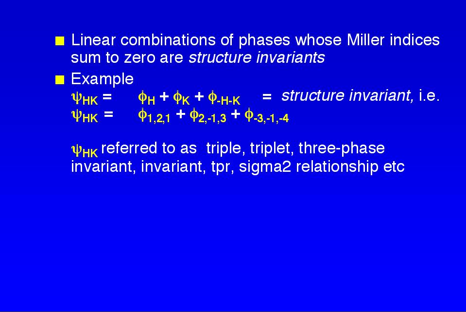
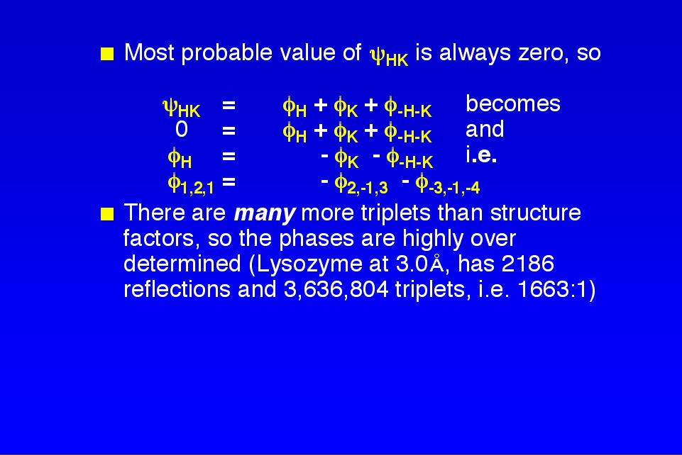
5 Important Concepts - 2 Structure Invariant - structural quantity independent of choice of unit cell origin Probabilistic estimates can be made for the values of structure invariants given the associated E magnitudes and cell contents
6
7 Fundamental formulas involving individual triplets P(ψ HK ) = [2π I 0 (A HK )] -1 exp(a HK cos ψ HK ) where P(ψ HK ) is the probability of the structure invariant having the value ψ HK A HK = 2 E H E K E -H-K / N 1/2 where N is the number of atoms in the cell and the E s are normalized structure factors
8 Note probability P(ψ HK ) increases as A HK increases, and that A HK is proportional to product of E s and inversely proportional to N 1/2 Expected value of cos ψ HK is given by <cos ψ HK > = I 1 (A HK ) / I 0 (A HK )
9 Φ 3 = Ψ HK, K=A HK Cochran Distribution for various K s σ vs K
10

11

12
13 Classical Direct Methods Applications for Proteins Used for phase extension to very high resolution Used with moderate success to locate heavy atom sites in isomorphous derivatives E values used in molecular replacement calculations
14 Current Direct Methods Applications for Proteins Shake n Bake (based on minimum function) used to solve complete protein structures with over 1,000 atoms (rubredoxin, lysozyme, calmodulin etc.), provided data to 1.1Å or better is available Used to locate anomalous scatterer sites from MAD or SAS data
15 General Shake n Bake Concept Use a multi-solution method starting with random phases (or randomly positioned atoms) in each trial. For each trial phase set, use a dual space procedure iterating between real and reciprocal space optimization/constraints.
16 Reciprocal space optimization based on shifting phases to reduce the minimum function R(ψ) Real space optimization and constraints based on computing new phases only from the largest peaks in map based on previous cycle phases Each trial phase set ranked by value of R(ψ)
17 SnB inner loop for trial structure Generate random trial structure Stop after N iterations Compute phases from structure Select structure from largest peaks Shift phases to reduce R(ψ) Compute map from new phases
18 Choice of data for Se determination Use F H + - F H - (anomalous) difference at single λ Use F H λi - F H l λj (dispersive) difference between two λ s Use F A values (derived from data at all λ s) Use F HLE values based on max anomalous and max dispersive differences

19
20
21 MAD Phasing For data collected at λ1, λ2 etc, choose a wavelength λn as native data, and reduce that data set by averaging Bijvoet pairs. For other derivative wavelengths λd, reduce both by averaging Bijvoet pairs to form isomorphous data sets, and without averaging to form anomalous data sets.
22 MAD Phasing For isomorphous and derivative anomalous data sets, scale derivative to native and use scattering factors of f 0 = 0, f = f (λd) - f (λn), f = f (λd) For native anomalous data use original native Bijvoet pairs and scattering factors of f 0 = 0, f = 0, f = f (λn)
23 Phase Refinement Minimizing W h P φ P FPHobs h FPHcalc φ P h φ P FPHcalc ( ) ( φ P ) where ( ) h 2 = h FPobs 2 + h FHcalc 2 h + 2 FPobs h FHcalc h cos ( φ P φ H )
24 h Phase Refinement Options ( ) W P FPHobs FPHcalc ( φ ) h φ P h P h φ P Classical - φ P = centroid, W h =1/E 2,1/ <E 2 > or unity, Pφ P =1, use reflections with FOM > Maximum Likelihood - φ P stepped over allowed phases, Pφ P = corresponding probability, W h =1/E 2, 1/ <E 2 > or unity, use reflections with FOM > 0.2 φ P, Pφ P can also come from external source, i.e solvent flattened or NC-symmetry averaged maps. 2
25
26

27
28 Projection of peaks down NC twofold
29 MAD λ1, λ2, λ3 data (Scalepack files) CMBISO iso and ano scaled files all native (λ3) data CMBANO PHASIT phase file final map FSFOUR EXTRMP submap file MAPAVG averag mask MISSNG extension file BNDRY BLDCEL MAPINV

30
31 MAD Phasing/Averaging Statistics Wavelength type dmin (Å) No. refl Rano Riso dmin (Å) (phasing) Rc Phasing Power <FOM>!1, edge ano , !2, peak ano , !3, remote ano , !1-!3 iso , !2-!3 iso , Mean FOM (combined) = for 48,632 reflections (2.6Å) Correlation coefficient between monomer density prior to NCS averaging = Correlation coefficient between monomer density after NCS averaging/phase combination = 0.906
32
33

34 Peak anomalous (λ2)( difference Patterson

35
36
37
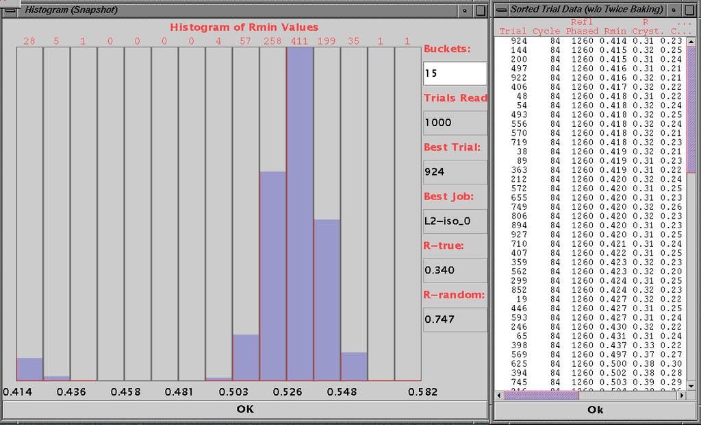
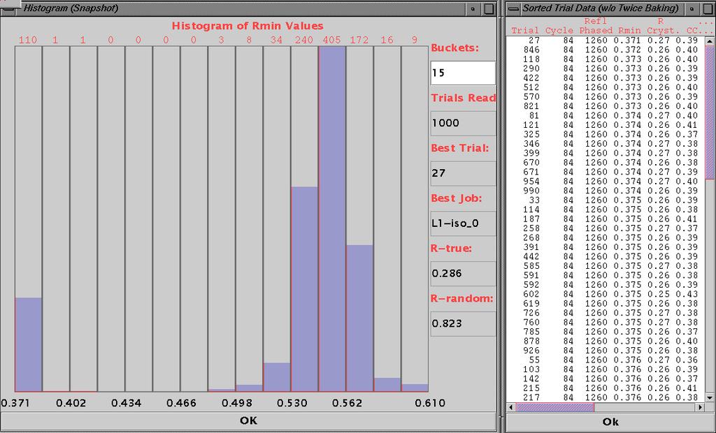
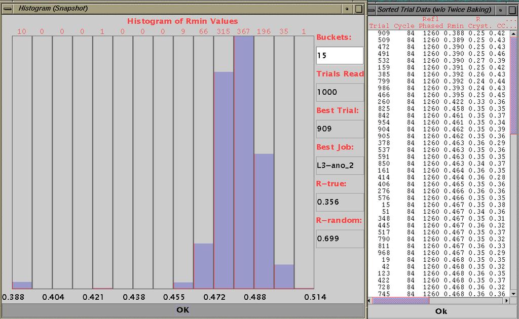
38
39
40 With SnB it s possible to automatically locate the anomalous scatterer substructure with data from any one of the dispersive combinations or anomalous pair sets As expected, sets with the maximum dispersive or anomalous signal typically yield a greater frequency of success
41 Automated Applications of BnP: Methodology W. Furey, 1 L. Pasupulati, 1 S. Potter 2, H. Xu 2, R. Miller 3 & C. Weeks 2 1 University of Pittsburgh School of Medicine and VA Medical Center 2 Hauptman-Woodward Medical Research Institute 3 Center for Computational Research, SUNY at Buffalo
42 Goal: Provide user-friendly software for automatic determination of protein crystal structures SnB Strengths 1. Powerful, state-of-the-art direct methods for automatically locating heavy atom sites 2. Friendly graphical user interface. SnB Weaknesses 1. Stops after finding sites, i.e no protein phasing 2. No software interface PHASES Strengths 1. Proven protein phasing (MAD MIRAS, etc), solvent flattenin NCS averaging, external program interfacing 2. Interactive graphics PHASES Weaknesses 1. Doesn t automatically find heavy atom sites 2. Script based, i.e. no GUI
43 Adopted Strategy Combine the SnB program with the PHASES package, putting everything under GUI control Establish default parameters and procedures allowing a aspects of the structure determination to be fully automated Also provide a manual mode allowing experienced users more control, and to facilitate development Provide graphical feedback when possible Facilitate coupling with popular external software
44 Main Developments Required for Automated Structure Determination Automatic substructure solution detection Automatic substructure validation Automatic hand determination (including space group changes, when needed)


45 Automatic Substructure Solution Original Method Based on histogram (Manual, time consuming, requires user interaction) Detection Current Method Based on R min and R cryst statistics (Automatic, fast, no user interaction)
46 Automatic Substructure Validation Original Method Left up to user to decide which peaks correspond to true sites (Manual) Current Method (auto mode) Based on occupancy refinement against Bijvoet differences (Automatic, fast, requires no coordinate refinement, hand insensitive) Current Method (manual mode) As in auto but can also compare peaks from different solutions (Manual)
47 Automatic Substructure Validation
 Current Method (MAD, SIRAS")

48 Automatic Hand Determination Original Method Visual inspection of map projections (Manual, requires user interaction) Current Method (MAD, SIRAS or MIRAS) Based on variance differences in protein and solvent regions (Automatic, fast since requires no refinement, also requires no user interaction)
49 Automatic Hand Determination Current Method (SAS data only) Comparative analysis of R, FOM and CC after solvent flattening/phase combination. (Automatic, fast, requires no refinement) Current Method (SIR, MIR data only) Both hands tried, map examination needed. (Requires user interaction)
50 No man (or program) is an island Importing data files Scalepack files D*Trek files MTZ files $ Free format files Exporting data files Exporting control files O RESOLVE 2.08 Arp/wARP Job submission from GUI Free format files CNS files MTZ files $ O files CHAIN files PDB files RESOLVE $ 2.08 Arp/wARP $ $ RESOLVE, Arp/wARP and/or CCP4 must be obtained from their respective authors/distributors for these options to work
51 Results for 1jc4 a=43.6 b=78.6, c=89.4 Å, β= 91.95, P2 1 4 molecules (592 residues) in asu 2.1Å data, 3λ MAD data Substructure: Found 24 of 24 Se Phasing: mean PP- 2.95; mean FOM Time to map: ~41 min on G4 (1.5 GHz) Powerbook ~13 min on G5 (2.7 GHz) Desktop Auto Tracability: Resolve- 87% main chain, 68% side chain Arp/wARP- 82% main chain, 73% side chain
52 SeMet ASU Size & Data Resolution PDB No. No. PDB No. No. Code Sites Residues NCS d(å) Code Sites Residues NCS d(å 1QC CLI BX A7A CB L8A T5H E3M JXH HI GSO GKP TPS DQ DBT E2Y JEN M JC EQ

53 Phasing Flexibility (Manual Mode)
54 Conclusion BnP is a user friendly, efficient, package for the automated determination of protein structures from x-ray diffraction data BnP downloads for Linux, Apple G4, G5, & Intel, and SGI s available (academic & non-profit institutions) at
SHELXC/D/E. Andrea Thorn
 SHELXC/D/E Andrea Thorn What is experimental phasing? Experimental phasing is what you do if MR doesn t work. What is experimental phasing? Experimental phasing methods depend on intensity differences.
Web-based Auto-Rickshaw for validation of the X-ray experiment at the synchrotron beamline
 Web-based Auto-Rickshaw for validation of the X-ray experiment at the synchrotron beamline Auto-Rickshaw http://www.embl-hamburg.de/auto-rickshaw A platform for automated crystal structure determination
Likelihood and SAD phasing in Phaser. R J Read, Department of Haematology Cambridge Institute for Medical Research
 Likelihood and SAD phasing in Phaser R J Read, Department of Haematology Cambridge Institute for Medical Research Concept of likelihood Likelihood with dice 4 6 8 10 Roll a seven. Which die?? p(4)=p(6)=0
Phaser: Experimental phasing
 Phaser: Experimental phasing Using SAD data in Phaser R J Read, Department of Haematology Cambridge Institute for Medical Research Diffraction with anomalous scatterers SAD: single-wavelength anomalous
The Phase Problem of X-ray Crystallography
 163 The Phase Problem of X-ray Crystallography H.A. Hauptman Hauptman-Woodward Medical Research Institute, Inc. 73 High Street Buffalo, NY, USA hauptman@hwi.buffalo.edu ABSTRACT. The intensities of a sufficient
Experimental Phasing with SHELX C/D/E
 WIR SCHAFFEN WISSEN HEUTE FÜR MORGEN Dr. Tim Grüne :: Paul Scherrer Institut :: tim.gruene@psi.ch Experimental Phasing with SHELX C/D/E CCP4 / APS School Chicago 2017 22 nd June 2017 1 - The Phase Problem
Fast, Intuitive Structure Determination IV: Space Group Determination and Structure Solution
 Fast, Intuitive Structure Determination IV: Space Group Determination and Structure Solution November 25, 2013 Welcome I I Dr. Michael Ruf Product Manager Crystallography Bruker AXS Inc. Madison, WI, USA
Determination of the Substructure
 Monday, June 15 th, 2009 Determination of the Substructure EMBO / MAX-INF2 Practical Course http://shelx.uni-ac.gwdg.de Overview Substructure Definition and Motivation Extracting Substructure Data from
Shake-and-Bake: Applications and Advances. Russ Miller & Charles M. Weeks
 Shake-and-Bake: Applications and Advances Russ Miller & Charles M. Weeks Hauptman-Woodward Med. Res. Inst. Principal Contributors: C.-S. Chang G.T. DeTitta S.M. Gallo H.A. Hauptman H.G. Khalak D.A. Langs
Direct Method. Very few protein diffraction data meet the 2nd condition
 Direct Method Two conditions: -atoms in the structure are equal-weighted -resolution of data are higher than the distance between the atoms in the structure Very few protein diffraction data meet the 2nd
Macromolecular Phasing with shelxc/d/e
 Sunday, June 13 th, 2010 CCP4 Workshop APS Chicago, June 2010 http://shelx.uni-ac.gwdg.de Overview Substructure Definition and Motivation Extracting Substructure Data from measured Data Substructure Solution
CCP4 Diamond 2014 SHELXC/D/E. Andrea Thorn
 CCP4 Diamond 2014 SHELXC/D/E Andrea Thorn SHELXC/D/E workflow SHELXC: α calculation, file preparation SHELXD: Marker atom search = substructure search SHELXE: density modification Maps and coordinate files
Protein Crystallography
 Protein Crystallography Part II Tim Grüne Dept. of Structural Chemistry Prof. G. Sheldrick University of Göttingen http://shelx.uni-ac.gwdg.de tg@shelx.uni-ac.gwdg.de Overview The Reciprocal Lattice The
Experimental phasing, Pattersons and SHELX Andrea Thorn
 Experimental phasing, Pattersons and SHELX Andrea Thorn What is experimental phasing? Experimental phasing is what you do if MR doesn t work. What is experimental phasing? Experimental phasing methods
Experimental phasing in Crank2
 Experimental phasing in Crank2 Pavol Skubak and Navraj Pannu Biophysical Structural Chemistry, Leiden University, The Netherlands http://www.bfsc.leidenuniv.nl/software/crank/ X-ray structure solution
(716) Partial funding from NIH and NSF. Computing from TMC, PSC, Intel, and NIH. Principal Contributors
 Partial funding from NIH and NSF. Computing from TMC, PSC, Intel, and NIH. Principal Contributors") SnB: A STATUS REPORT Russ Miller 1;2 and Charles M. Weeks 2 1 Dept. of Comp. Sci., SUNY-Bualo 2 The Medical Foundation of Bualo fmiller,weeksg@mfb.bualo.edu (716) 856-9600 Partial funding from NIH and
SnB: A STATUS REPORT Russ Miller 1;2 and Charles M. Weeks 2 1 Dept. of Comp. Sci., SUNY-Bualo 2 The Medical Foundation of Bualo fmiller,weeksg@mfb.bualo.edu (716) 856-9600 Partial funding from NIH and
Patterson Methods
 59-553 Patterson Methods 113 In 1935, Patterson showed that the unknown phase information in the equation for electron density: ρ(xyz) = 1/V h k l F(hkl) exp[iα(hkl)] exp[-2πi(h x + k y + l z)] can be
research papers 1. Introduction Thomas C. Terwilliger a * and Joel Berendzen b
 Acta Crystallographica Section D Biological Crystallography ISSN 0907-4449 Discrimination of solvent from protein regions in native Fouriers as a means of evaluating heavy-atom solutions in the MIR and
X-ray Crystallography. Kalyan Das
 X-ray Crystallography Kalyan Das Electromagnetic Spectrum NMR 10 um - 10 mm 700 to 10 4 nm 400 to 700 nm 10 to 400 nm 10-1 to 10 nm 10-4 to 10-1 nm X-ray radiation was discovered by Roentgen in 1895. X-rays
ACORN in CCP4 and its applications
 Acta Crystallographica Section D Biological Crystallography ISSN 0907-4449 ACORN in CCP4 and its applications Jia-xing Yao York Structural Biology Laboratory, Department of Chemistry, University of York,
ACORN - a flexible and efficient ab initio procedure to solve a protein structure when atomic resolution data is available
 ACORN - a flexible and efficient ab initio procedure to solve a protein structure when atomic resolution data is available Yao Jia-xing Department of Chemistry, University of York, Heslington, York, YO10
Crystal lattice Real Space. Reflections Reciprocal Space. I. Solving Phases II. Model Building for CHEM 645. Purified Protein. Build model.
 I. Solving Phases II. Model Building for CHEM 645 Purified Protein Solve Phase Build model and refine Crystal lattice Real Space Reflections Reciprocal Space ρ (x, y, z) pronounced rho F hkl 2 I F (h,
Protein crystallography. Garry Taylor
 Protein crystallography Garry Taylor X-ray Crystallography - the Basics Grow crystals Collect X-ray data Determine phases Calculate ρ-map Interpret map Refine coordinates Do the biology. Nitrogen at -180
Experimental phasing in Crank2
 Experimental phasing in Crank2 Pavol Skubak and Navraj Pannu Biophysical Structural Chemistry, Leiden University, The Netherlands http://www.bfsc.leidenuniv.nl/software/crank/ Crank2 for experimental phasing
Direct-method SAD phasing with partial-structure iteration: towards automation
 Acta Crystallographica Section D Biological Crystallography ISSN 0907-4449 Editors: E. N. Baker and Z. Dauter Direct-method SAD phasing with partial-structure iteration: towards automation J. W. Wang,
 Helpful resources for all X ray lectures Crystallization http://www.hamptonresearch.com under tech support: crystal growth 101 literature Spacegroup tables http://img.chem.ucl.ac.uk/sgp/mainmenu.htm Crystallography
The SHELX approach to the experimental phasing of macromolecules. George M. Sheldrick, Göttingen University
 The SHELX approach to the experimental phasing of macromolecules IUCr 2011 Madrid George M. Sheldrick, Göttingen University http://shelx.uni-ac.gwdg.de/shelx/ Experimental phasing of macromolecules Except
What is the Phase Problem? Overview of the Phase Problem. Phases. 201 Phases. Diffraction vector for a Bragg spot. In General for Any Atom (x, y, z)
") Protein Overview of the Phase Problem Crystal Data Phases Structure John Rose ACA Summer School 2006 Reorganized by Andy Howard,, Spring 2008 Remember We can measure reflection intensities We can calculate
Protein Overview of the Phase Problem Crystal Data Phases Structure John Rose ACA Summer School 2006 Reorganized by Andy Howard,, Spring 2008 Remember We can measure reflection intensities We can calculate
Scattering by two Electrons
 Scattering by two Electrons p = -r k in k in p r e 2 q k in /λ θ θ k out /λ S q = r k out p + q = r (k out - k in ) e 1 Phase difference of wave 2 with respect to wave 1: 2π λ (k out - k in ) r= 2π S r
Structure solution from weak anomalous data
 Structure solution from weak anomalous data Phenix Workshop SBGrid-NE-CAT Computing School Harvard Medical School, Boston June 7, 2014 Gábor Bunkóczi, Airlie McCoy, Randy Read (Cambridge University) Nat
SOLVE and RESOLVE: automated structure solution, density modification and model building
 Journal of Synchrotron Radiation ISSN 0909-0495 SOLVE and RESOLVE: automated structure solution, density modification and model building Thomas Terwilliger Copyright International Union of Crystallography
Biology III: Crystallographic phases
 Haupt/Masterstudiengang Physik Methoden moderner Röntgenphysik II: Streuung und Abbildung SS 2013 Biology III: Crystallographic phases Thomas R. Schneider, EMBL Hamburg 25/6/2013 thomas.schneider@embl-hamburg.de
Determining Protein Structure BIBC 100
 Determining Protein Structure BIBC 100 Determining Protein Structure X-Ray Diffraction Interactions of x-rays with electrons in molecules in a crystal NMR- Nuclear Magnetic Resonance Interactions of magnetic
Molecular Replacement (Alexei Vagin s lecture)
") Molecular Replacement (Alexei Vagin s lecture) Contents What is Molecular Replacement Functions in Molecular Replacement Weighting scheme Information from data and model Some special techniques of Molecular
Molecular Replacement (Alexei Vagin s lecture) Contents What is Molecular Replacement Functions in Molecular Replacement Weighting scheme Information from data and model Some special techniques of Molecular
PAN-modular Structure of Parasite Sarcocystis muris Microneme Protein SML-2 at 1.95 Å Resolution and the Complex with 1-Thio-β-D-Galactose
 Supplementary Material to the paper: PAN-modular Structure of Parasite Sarcocystis muris Microneme Protein SML-2 at 1.95 Å Resolution and the Complex with 1-Thio-β-D-Galactose Jürgen J. Müller, a Manfred
Overview - Macromolecular Crystallography
 Overview - Macromolecular Crystallography 1. Overexpression and crystallization 2. Crystal characterization and data collection 3. The diffraction experiment 4. Phase problem 1. MIR (Multiple Isomorphous
PSD '17 -- Xray Lecture 5, 6. Patterson Space, Molecular Replacement and Heavy Atom Isomorphous Replacement
 PSD '17 -- Xray Lecture 5, 6 Patterson Space, Molecular Replacement and Heavy Atom Isomorphous Replacement The Phase Problem We can t measure the phases! X-ray detectors (film, photomultiplier tubes, CCDs,
BCM Protein crystallography - II Isomorphous Replacement Anomalous Scattering and Molecular Replacement Model Building and Refinement
 BCM 6200 - Protein crystallography - II Isomorphous Replacement Anomalous Scattering and Molecular Replacement Model Building and Refinement Changing practice in de novo structure determination Hendrickson
Charles Ballard (original GáborBunkóczi) CCP4 Workshop 7 December 2011
 CCP4 Workshop 7 December 2011") Experimental phasing Charles Ballard (original GáborBunkóczi) CCP4 Workshop 7 December 2011 Anomalous diffraction F P protein F A anomalous substructure ano F A " -FA" A F F -* F A F P Phasing Substructure
Experimental phasing Charles Ballard (original GáborBunkóczi) CCP4 Workshop 7 December 2011 Anomalous diffraction F P protein F A anomalous substructure ano F A " -FA" A F F -* F A F P Phasing Substructure
Rietveld Structure Refinement of Protein Powder Diffraction Data using GSAS
 Rietveld Structure Refinement of Protein Powder Diffraction Data using GSAS Jon Wright ESRF, Grenoble, France Plan This is a users perspective Cover the protein specific aspects (assuming knowledge of
Tutorial on how to solve a Se-substructure using
 1 Introduction Tutorial on how to solve a Se-substructure using SHELXD Thomas R. Schneider Dept. of Structural Chemistry University of Göttingen trs@shelx.uni-ac.gwdg.de July 4, 2002 The Solution of the
Practical aspects of SAD/MAD. Judit É Debreczeni
 Practical aspects of SAD/MAD Judit É Debreczeni anomalous scattering Hg sinθ/λ CuKα 0 Å - 0.4 Å - 0.6 Å - Å.5Å 0.83Å f 80 53 4 f total (θ, λ) f(θ) + f (λ) + if (λ) f f -5 8-5 8-5 8 f total increasing with
X-ray Crystallography
 2009/11/25 [ 1 ] X-ray Crystallography Andrew Torda, wintersemester 2009 / 2010 X-ray numerically most important more than 4/5 structures Goal a set of x, y, z coordinates different properties to NMR History
Preparing a PDB File
 Figure 1: Schematic view of the ligand-binding domain from the vitamin D receptor (PDB file 1IE9). The crystallographic waters are shown as small spheres and the bound ligand is shown as a CPK model. HO
Anomalous dispersion
 Selenomethionine MAD Selenomethionine is the amino acid methionine with the Sulfur replaced by a Selenium. Selenium is a heavy atom that also has the propery of "anomalous scatter" at some wavelengths,
Supplementary materials. Crystal structure of the carboxyltransferase domain. of acetyl coenzyme A carboxylase. Department of Biological Sciences
 Supplementary materials Crystal structure of the carboxyltransferase domain of acetyl coenzyme A carboxylase Hailong Zhang, Zhiru Yang, 1 Yang Shen, 1 Liang Tong Department of Biological Sciences Columbia
Pipelining Ligands in PHENIX: elbow and REEL
 Pipelining Ligands in PHENIX: elbow and REEL Nigel W. Moriarty Lawrence Berkeley National Laboratory Physical Biosciences Division Ligands in Crystallography Drug design Biological function studies Generate
General theory of diffraction
 General theory of diffraction X-rays scatter off the charge density (r), neutrons scatter off the spin density. Coherent scattering (diffraction) creates the Fourier transform of (r) from real to reciprocal
Crystals, X-rays and Proteins
 Crystals, X-rays and Proteins Comprehensive Protein Crystallography Dennis Sherwood MA (Hons), MPhil, PhD Jon Cooper BA (Hons), PhD OXFORD UNIVERSITY PRESS Contents List of symbols xiv PART I FUNDAMENTALS
wwpdb X-ray Structure Validation Summary Report
 wwpdb X-ray Structure Validation Summary Report io Jan 31, 2016 06:45 PM GMT PDB ID : 1CBS Title : CRYSTAL STRUCTURE OF CELLULAR RETINOIC-ACID-BINDING PROTEINS I AND II IN COMPLEX WITH ALL-TRANS-RETINOIC
Garib N Murshudov MRC-LMB, Cambridge
 Garib N Murshudov MRC-LMB, Cambridge Contents Introduction AceDRG: two functions Validation of entries in the DB and derived data Generation of new ligand description Jligand for link description Conclusions
Space Group & Structure Solution
 Space Group & Structure Solution Determine the Space Group Space group determination can always be performed by hand by examining the intensity data. A program that can facilitate this step is the command-prompt
A GUI FOR EVOLVE ZAMS
 A GUI FOR EVOLVE ZAMS D. R. Schlegel Computer Science Department Here the early work on a new user interface for the Evolve ZAMS stellar evolution code is presented. The initial goal of this project is
research papers HKL-3000: the integration of data reduction and structure solution from diffraction images to an initial model in minutes
 Acta Crystallographica Section D Biological Crystallography ISSN 0907-4449 HKL-3000: the integration of data reduction and structure solution from diffraction images to an initial model in minutes Wladek
SUPPLEMENTARY INFORMATION
 doi:10.1038/nature11054 Supplementary Fig. 1 Sequence alignment of Na v Rh with NaChBac, Na v Ab, and eukaryotic Na v and Ca v homologs. Secondary structural elements of Na v Rh are indicated above the
Electronic Supplementary Information (ESI) for Chem. Commun. Unveiling the three- dimensional structure of the green pigment of nitrite- cured meat
 for Chem. Commun. Unveiling the three- dimensional structure of the green pigment of nitrite- cured meat") Electronic Supplementary Information (ESI) for Chem. Commun. Unveiling the three- dimensional structure of the green pigment of nitrite- cured meat Jun Yi* and George B. Richter- Addo* Department of Chemistry
Electronic Supplementary Information (ESI) for Chem. Commun. Unveiling the three- dimensional structure of the green pigment of nitrite- cured meat Jun Yi* and George B. Richter- Addo* Department of Chemistry
Molecular replacement. New structures from old
 Molecular replacement New structures from old The Phase Problem phase amplitude Phasing by molecular replacement Phases can be calculated from atomic model Rotate and translate related structure Models
Protein Structure and Visualisation. Introduction to PDB and PyMOL
 Protein Structure and Visualisation Introduction to PDB and PyMOL 1 Feedback Persons http://www.bio-evaluering.dk/ 2 Program 8.00-8.15 Quiz results 8.15-8.50 Introduction to PDB & PyMOL 8.50-9.00 Break
Image definition evaluation functions for X-ray crystallography: A new perspective on the phase. problem. Hui LI*, Meng HE* and Ze ZHANG
 Image definition evaluation functions for X-ray crystallography: A new perspective on the phase problem Hui LI*, Meng HE* and Ze ZHANG Beijing University of Technology, Beijing 100124, People s Republic
Modelling against small angle scattering data. Al Kikhney EMBL Hamburg, Germany
 Modelling against small angle scattering data Al Kikhney EMBL Hamburg, Germany Validation of atomic models CRYSOL Rigid body modelling SASREF BUNCH CORAL Oligomeric mixtures OLIGOMER Flexible systems EOM
Resolution and data formats. Andrea Thorn
 Resolution and data formats Andrea Thorn RESOLUTION Motivation Courtesy of M. Sawaya Map resolution http://www.bmsc.washington.edu/people/verlinde/experiment.html Data quality indicators Resolution accounts
Recent developments in Crank. Leiden University, The Netherlands
 Recent developments in Crank Navraj js. Pannu Leiden University, The Netherlands Current developers Pavol Skubak Ruben Zubac Irakli Sikharulidze Jan Pieter Abrahams RAG de Graaff Willem-Jan Waterreus Substructure
Molecular Biology Course 2006 Protein Crystallography Part II
 Molecular Biology Course 2006 Protein Crystallography Part II Tim Grüne University of Göttingen Dept. of Structural Chemistry December 2006 http://shelx.uni-ac.gwdg.de tg@shelx.uni-ac.gwdg.de Overview
PDBe TUTORIAL. PDBePISA (Protein Interfaces, Surfaces and Assemblies)
") PDBe TUTORIAL PDBePISA (Protein Interfaces, Surfaces and Assemblies) http://pdbe.org/pisa/ This tutorial introduces the PDBePISA (PISA for short) service, which is a webbased interactive tool offered by
PDBe TUTORIAL PDBePISA (Protein Interfaces, Surfaces and Assemblies) http://pdbe.org/pisa/ This tutorial introduces the PDBePISA (PISA for short) service, which is a webbased interactive tool offered by
Tutorial. Getting started. Sample to Insight. March 31, 2016
 Getting started March 31, 2016 Sample to Insight CLC bio, a QIAGEN Company Silkeborgvej 2 Prismet 8000 Aarhus C Denmark Telephone: +45 70 22 32 44 www.clcbio.com support-clcbio@qiagen.com Getting started
electronic reprint Optimizing DREAR and SnB parameters for determining Se-atom substructures
 Acta Crystallographica Section D Biological Crystallography ISSN 0907-4449 Optimizing DREAR and SnB parameters for determining Se-atom substructures P. Lynne Howell, Robert H. Blessing, G. David Smith
Full wwpdb X-ray Structure Validation Report i
 Full wwpdb X-ray Structure Validation Report i Mar 13, 2018 04:03 pm GMT PDB ID : 5NMJ Title : Chicken GRIFIN (crystallisation ph: 6.5) Authors : Ruiz, F.M.; Romero, A. Deposited on : 2017-04-06 Resolution
Ab initio crystal structure analysis based on powder diffraction data using PDXL
 Ab initio crystal structure analysis based on powder diffraction data using PDXL Akito Sasaki*, Akihiro Himeda*, Hisashi Konaka* and Norihiro Muroyama* 1. Introduction Physical and chemical properties
Scattering Lecture. February 24, 2014
 Scattering Lecture February 24, 2014 Structure Determination by Scattering Waves of radiation scattered by different objects interfere to give rise to an observable pattern! The wavelength needs to close
Phase problem: Determining an initial phase angle α hkl for each recorded reflection. 1 ρ(x,y,z) = F hkl cos 2π (hx+ky+ lz - α hkl ) V h k l
 = F hkl cos 2π (hx+ky+ lz - α hkl ) V h k l") Phase problem: Determining an initial phase angle α hkl for each recorded reflection 1 ρ(x,y,z) = F hkl cos 2π (hx+ky+ lz - α hkl ) V h k l Methods: Heavy atom methods (isomorphous replacement Hg, Pt)
Phase problem: Determining an initial phase angle α hkl for each recorded reflection 1 ρ(x,y,z) = F hkl cos 2π (hx+ky+ lz - α hkl ) V h k l Methods: Heavy atom methods (isomorphous replacement Hg, Pt)
research papers Detecting outliers in non-redundant diffraction data 1. Introduction Randy J. Read
 Acta Crystallographica Section D Biological Crystallography ISSN 0907-4449 Detecting outliers in non-redundant diffraction data Randy J. Read Department of Haematology, University of Cambridge, Cambridge
Full wwpdb X-ray Structure Validation Report i
 Full wwpdb X-ray Structure Validation Report i Jan 28, 2019 11:10 AM EST PDB ID : 6A5H Title : The structure of [4+2] and [6+4] cyclase in the biosynthetic pathway of unidentified natural product Authors
Principles of Protein X-Ray Crystallography
 Principles of Protein X-Ray Crystallography Jan Drenth Principles of Protein X-Ray Crystallography Third Edition With Major Contribution from Jeroen Mesters University of Lübeck, Germany Jan Drenth Laboratory
Prediction and refinement of NMR structures from sparse experimental data
 Prediction and refinement of NMR structures from sparse experimental data Jeff Skolnick Director Center for the Study of Systems Biology School of Biology Georgia Institute of Technology Overview of talk
The Development of a Quality Control and Analysis Application for the ThermoFluor High Throughput Screening Assay
 The Development of a Quality Control and Analysis Application for the ThermoFluor High Throughput Screening Assay Robert B. Nachbar 1 Delphine Collin 2 Jonathan Robinson 1 Thomas J. Mildorf 3 Eugen Buehler
Full wwpdb X-ray Structure Validation Report i
 Full wwpdb X-ray Structure Validation Report i Jan 14, 2019 11:10 AM EST PDB ID : 6GYW Title : Crystal structure of DacA from Staphylococcus aureus Authors : Tosi, T.; Freemont, P.S.; Grundling, A. Deposited
Physical Chemistry Analyzing a Crystal Structure and the Diffraction Pattern Virginia B. Pett The College of Wooster
 Physical Chemistry Analyzing a Crystal Structure and the Diffraction Pattern Virginia B. Pett The College of Wooster L. W. Haynes and his Senior Independent Study students conducted the 2 + 2 photo addition
Working with protein structures. Benjamin Jack
 Working with protein structures Benjamin Jack Structure of Triosephosphate Isomerase PDB ID: 1HTI loop beta sheet alpha helix Different perspectives of the same structure Structure of Truncated Hemoglobin
4. Constraints and Hydrogen Atoms
 4. Constraints and ydrogen Atoms 4.1 Constraints versus restraints In crystal structure refinement, there is an important distinction between a constraint and a restraint. A constraint is an exact mathematical
Molecular Biology Course 2006 Protein Crystallography Part I
 Molecular Biology Course 2006 Protein Crystallography Part I Tim Grüne University of Göttingen Dept. of Structural Chemistry November 2006 http://shelx.uni-ac.gwdg.de tg@shelx.uni-ac.gwdg.de Overview Overview
Full wwpdb X-ray Structure Validation Report i
 Full wwpdb X-ray Structure Validation Report i Mar 14, 2018 02:00 pm GMT PDB ID : 3RRQ Title : Crystal structure of the extracellular domain of human PD-1 Authors : Lazar-Molnar, E.; Ramagopal, U.A.; Nathenson,
research papers Reduction of density-modification bias by b correction 1. Introduction Pavol Skubák* and Navraj S. Pannu
 Acta Crystallographica Section D Biological Crystallography ISSN 0907-4449 Reduction of density-modification bias by b correction Pavol Skubák* and Navraj S. Pannu Biophysical Structural Chemistry, Leiden
IgE binds asymmetrically to its B cell receptor CD23
 Supplementary Information IgE binds asymmetrically to its B cell receptor CD23 Balvinder Dhaliwal 1*, Marie O. Y. Pang 2, Anthony H. Keeble 2,3, Louisa K. James 2,4, Hannah J. Gould 2, James M. McDonnell
Protein Structure Determination 9/25/2007
 One-dimensional NMR spectra Ethanol Cellulase (36 a.a.) Branden & Tooze, Fig. 18.16 1D and 2D NMR spectra of inhibitor K (57 a.a.) K. Wuthrich, NMR of Proteins and Nucleic Acids. (Wiley, 1986.) p. 54-55.
Protein Crystallography. Mitchell Guss University of Sydney Australia
 Protein Crystallography Mitchell Guss University of Sydney Australia Outline of the talk Recap some basic crystallography and history Highlight the special requirements for protein (macromolecular) structure
Changing and challenging times for service crystallography. Electronic Supplementary Information
 Changing and challenging times for service crystallography Simon J Coles,* a and Philip A Gale* a Electronic Supplementary Information Instrument descriptions and experimental protocols The following firstly
This is an author produced version of Privateer: : software for the conformational validation of carbohydrate structures.
 This is an author produced version of Privateer: : software for the conformational validation of carbohydrate structures. White Rose Research Online URL for this paper: http://eprints.whiterose.ac.uk/95794/
Full wwpdb X-ray Structure Validation Report i
 Full wwpdb X-ray Structure Validation Report i Mar 10, 2018 01:44 am GMT PDB ID : 1MWP Title : N-TERMINAL DOMAIN OF THE AMYLOID PRECURSOR PROTEIN Authors : Rossjohn, J.; Cappai, R.; Feil, S.C.; Henry,
Full wwpdb X-ray Structure Validation Report i
 Full wwpdb X-ray Structure Validation Report i Mar 8, 2018 06:13 pm GMT PDB ID : 5G5C Title : Structure of the Pyrococcus furiosus Esterase Pf2001 with space group C2221 Authors : Varejao, N.; Reverter,
ECS8020 ORGANIC ELEMENTAL ANALYZER CHNS-O Analyzer
 ECS8020 ORGANIC ELEMENTAL ANALYZER CHNS-O Analyzer ECS 8020 CHNS-O Analyzer Organic Elemental Analysis ECS 8020 is a C-H-N-S-O Elemental Analyzer Model based on the Dumas combustion method. ECS 8020 is
TLS and all that. Ethan A Merritt. CCP4 Summer School 2011 (Argonne, IL) Abstract
 Abstract") TLS and all that Ethan A Merritt CCP4 Summer School 2011 (Argonne, IL) Abstract We can never know the position of every atom in a crystal structure perfectly. Each atom has an associated positional uncertainty.
TLS and all that Ethan A Merritt CCP4 Summer School 2011 (Argonne, IL) Abstract We can never know the position of every atom in a crystal structure perfectly. Each atom has an associated positional uncertainty.
New Features in Agilent's CrysAlis Pro X-ray Diffractometer Software
 New Features in Agilent's CrysAlis Pro X-ray Diffractometer Software Mathias Meyer Agilent Technologies Poland Oliver Presly Agilent Technologies UK 1 30+ Years of Innovation Sapphire2- First Back illuminated
Institute of Physics, Prague 6, Cukrovarnická street
 Jana2006 Institute of Physics, Prague 6, Cukrovarnická street Jana2006 Program for structure analysis of crystals periodic in three or more dimensions from diffraction data Václav Petříček, Michal Dušek
11/6/2013. Refinement. Fourier Methods. Fourier Methods. Difference Map. Difference Map Find H s. Difference Map No C 1
 Refinement Fourier Methods find heavy atom or some F s phases using direct methods locate new atoms, improve phases continue until all atoms found in more or less correct position starting point of refinement
Web Knowledge Base on Low Energy Nuclear Physics
 Stellenbosch University Web Knowledge Base on Low Energy Nuclear Physics http://nrv.jinr.ru/nrv/ http://nrv.sun.ac.za/ Everybody time to time does the following: Search for available experimental data
organic papers 2-[(Dimethylamino)(phenyl)methyl]benzoic acid
![organic papers 2-[(Dimethylamino)(phenyl)methyl]benzoic acid](/thumbs/82/85896414.jpg "organic papers 2-[(Dimethylamino)(phenyl)methyl]benzoic acid") organic papers Acta Crystallographica Section E Structure Reports Online ISSN 1600-5368 2-[(Dimethylamino)(phenyl)methyl]benzoic acid Yvette L. Dann, Andrew R. Cowley and Harry L. Anderson* University
organic papers Acta Crystallographica Section E Structure Reports Online ISSN 1600-5368 2-[(Dimethylamino)(phenyl)methyl]benzoic acid Yvette L. Dann, Andrew R. Cowley and Harry L. Anderson* University
X-ray Data Collection. Bio5325 Spring 2006
 X-ray Data Collection Bio535 Spring 006 Obtaining I hkl and α (Ihkl) from Frame Images Braggs Law -predicts conditions for in-phase scattering by equivalent atoms lying in planes that transect a crystal.
Applications of X-ray and Neutron Scattering in Biological Sciences: Symmetry in direct and reciprocal space 2012
 Department of Drug Design and Pharmacology Applications of X-ray and Neutron Scattering in Biological Sciences: Symmetry in direct and reciprocal space 2012 Michael Gajhede Biostructural Research Copenhagen
Small-Angle Scattering Atomic Structure Based Modeling
 Small-Angle Scattering Atomic Structure Based Modeling Alejandro Panjkovich EMBL Hamburg 07.12.2017 A. Panjkovich (EMBL) BioSAS atomic modeling 07.12.2017 1 / 49 From the forest to the particle accelerator