Biology III: Crystallographic phases
|
|
|
- Veronica Underwood
- 5 years ago
- Views:
Transcription
1 Haupt/Masterstudiengang Physik Methoden moderner Röntgenphysik II: Streuung und Abbildung SS 2013 Biology III: Crystallographic phases Thomas R. Schneider, EMBL Hamburg 25/6/2013


 j =1 For chiral")

2 Recap! Real and reciprocal space! The crystallographic phase problem! Symmetry in real and reciprocal space N F hkl = f j e 2πi(hx j +ky j +lz j ) j =1 For chiral objects forming crystals, only 65 space groups (lattice + point group) are possible. That is all!
3 Today! Phasing methods! Experiments Thomas R. Schneider Meth. moderner Röntgenphysik II 20/6/2013
4 Phase Determination in Practise! Ab Initio! INPUT: F hkl only! Needs data to a resolution where atoms are separated, i.e. 1.1 Å! Molecular Replacement! INPUT: F hkl / homologues structure model / position and orientation! Needs homologues model! Model Bias! Multiple Isomorphous Replacement (MIR)! INPUT: F hkl for native, F hkl for derivative 1, F hkl for derivative 2! Derivatization! Non-isomorphism! Anomalous Diffraction (MAD, SAD)! INPUT: F hkl from one crystal
5 Molecular Replacement! Assuming that the structure of the molecule of interest and the structure of a molecule with similar amino-acid sequence (the search model) are similar, an approximate model of the content of the crystal can be made by placing the search model into the crystallographic unit cell via a 6D-search.! Consistent packing is a strong criterion for correctness.! From this model, phases can be calculated, combined with the measured structure factors amplitudes and used to calculate an (F obs, phi calc )electron density map. N f j j=1 F hkl,calc = e 2πi(hx j+ky j +lz j )
6 Model bias I Adapted%from%xtal.ohsu.edu/teaching/lewis_clark/Models.pptx%! The first electron density map after molecular replacement is calculated with phases derived from the potentially/likely incorrect search model. Meas obs F hkl real%structure% FT F calc hkl,ϕ calc hkl posi-oned%search%model%
 synthesis can be misleading. 'phase bias'.")
7 Model bias II h<p:// If the search model is very different from the target model, the image resulting from the (F obs, phi calc ) synthesis can be misleading. 'phase bias'. Meas obs F hkl real%structure% FT F calc hkl,ϕ calc hkl In reality, distinguishing between I and II is difficult posi-oned%search%model%


8 Model Bias

9 Isomorphous Replacement Taylor%(2010)%Acta%Cryst%D66:325%! By introducing extraatoms into a crystal, its diffraction properties become altered.! Determining phases experimentally is better than inventing phases based on insecure assumptions.
10 A protein and an extra scatterer N F hkl = f j e 2πi(hx j +ky j +lz j ) j=1 F PH F H F P Φ PH
11 Isomorphous replacement! Addition of heavy atoms leads to changes in diffracted intensities. E.g. a single Hgatom (80 e - ) in 1000 CNOatoms (6-8 e - ) changes the intensities on average by 25%.! The isomorphous difference: F H = F PH F P can be used as an estimate of the heavy atom structure factor amplitude Taylor%(2010)%Acta%Cryst%D66:325%
12 Single Isomorphous Replacement! After finding the heavy atoms, structure factors for the heavy atoms can be calculated. N F H,hkl,calc = f j e 2πi(hx j+ky j +lz j ) j=1! However, for a given set of heavy atom positions, two phases are possible for F P and F PH
13 Tafel Harker construction
14 Isomorphous Replacement Taylor%(2010)%Acta%Cryst%D66:325%! From the differences observed between the measurements on the native and the derivate crystal, the positions of the extra atoms can be found.! By clever combination of the knowledge about the positions of the extra atoms and the differences in the measurements, phase information can be derived.! Unfortunately, usually two phase angles are consistent with the data. 'Phase ambiguity'
15 Multiple Isomorphous Replacement! When two 'derivatives' are used, the phase ambiguity can be resolved and a unimodel phase distribution can be constructed.! An electron density map can be calculated. Taylor%(2010)%Acta%Cryst%D66:325%
16 Non-isomorphism!!
17 Anomalous Scattering
18 Normal Scattering (centrosymmetry) Friedel's law F hkl = F hkl Anomalous Scattering Friedel's law is broken F hkl F hkl F hkl F hkl can be measured experimentally
19 Physics of Anomalous Scattering! Normal scattering! elastic! direction-dependent! Anomalous scattering! resonant / inelastic! direction independent Pictures courtesy of Zbyszek Dauter
20 Normal diffraction into hkl and h-k-l F hkl F hkl = F hkl ϕ hkl = ϕ hkl F hkl
21 Anomalous diffraction into hkl and h-k-l f = f 0 + f '+if " F hkl F hkl F + F
22 Anomalous Diffraction into hkl and h-k-l Pictures courtesy of Zbyszek Dauter
23 A protein and an anomalous scatterer A A non-elastic interaction between the X-rays and the electrons can be incorporated into the structure factor by modifying the scattering factor for the anomalous scatterer 'A': f = f 0 + f '+if " ΔF F + F T F A F - F P
24 Anomalous Dispersion f " f ' E f"(e) can be measured by X-ray absorption spectroscopy: f "(E) = mc 2he 2 Eµ(E) f'(e) can be calculated from f"(e) by a Kramers- Kronig transformation f '(E) = 2 π 0 E' f "(E') de' (E 2 E' 2 )
25 A MAD Experiment f'' f' E 'high E remote' 'peak' 'inflection point' F A F P for one pair of refl: F + F - F + F - F + F - from the same crystal
26 The MAD Observational Equations pk F pk F + ip F + ip F Hendrickson WA et al. (1985) Meth.Enz. 115:41-55 F T F P F A λ 2 a F ( obs λ) = ( f ' + ± 2 = F T 2 f " 2 ) / f o2 2 + a(λ) of b ( λ) = 2 f '/ f A + b(λ) F T F A cosα o c ( λ) = 2 f "/ f ± c(λ) F T F A sinα pk + F obs = F T + a(pk) FA + b(pk) FT F A cosα + c(pk) F T F A sinα pk F obs = F T + a(pk) FA + b(pk) FT F A cosα c(pk) F T F A sinα ip + F obs = F T + a(ip) FA + b(ip) FT F A cosα + c(ip) F T F A sinα ip F obs 2 = F T 2 + a(ip) FA 2 + b(ip) FT F A cosα c(ip) F T F A sinα
27 The meaning of FH/A-values! In principle, F H/A -values represent the substructure of anomalous / heavy scatterers only. This reduces the initial step of phasing to finding a few atoms in a large unit cell.! Ab initio methods can be used for finding the anomalous / heavy scatterers.! ΔF-values can be considered as lower limit estimates of F A - values.

28 Solving the substructure against F A hkl F A σ FA #atoms SHELXD positions occupancies f.o.m's
29 Knowing the substructure allows to calculate φ A and φ T F A φ A φ T Real life is not that simple - ML as implemented e.g. in SHARP is the way to go. N Se F A,hkl = f j e 2πi(hx j +ky j +lz j ) j=1 F A,hkl,e iϕ A,hkl


30 ... in proteins C Selenium O N S H f" f" f' f' E

- map 0.")
 σ A")
-map 0.")
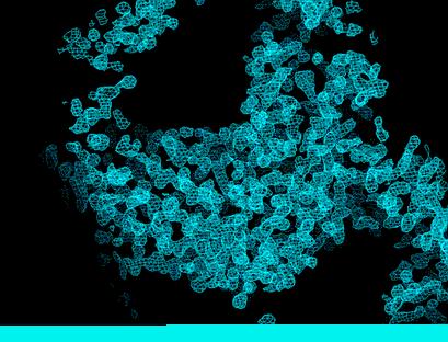
31 Aldose Reductase - experimental vs. refined experimental (F obs,φ MAD )- map 0.9 Å, contoured at 1σ refined (remote) σ A weighted (2F obs -1F calc,φ calc )-map 0.9 Å, contoured at 1σ


32 Aldose Reductase - Level of Detail! Multiple conformations! O > N > C

")
33 Choosing the right hand! The correct solution for the substructure and its enantiomorph have the same agreement with the experimental data.! But they give rise to different phase sets for the crystal structure 'Contrast' (SHELXE) inverted original ( x, y, z) ( x, y, z)! local r.m.s. density Terwilliger & Berendsen (1999) Acta Cryst. D55:1872! size of the PNG-file: vs Bytes


34 Back to the experiment! Anomalous diffraction occurs when when the energy / wavelength used is close to an absorption edge where transitions of electrons can be excited.

35 Accessible Edges Ca-K: 3.1 Å = 4.0 kev Se-K: 0.98 Å = kev Se-L: 8.64 Å = 1.44 kev 12.4 kev / 20 kev = 0.62 Å 12.4 kev / 5 kev = 2.48 Å EMBO Course, ESRF Grenoble Thomas R. Schneider 6/6/2011

36 Accessible Edges SSRL: Å / kev
 Uranyl' compounds' Chemicals'")


37 Derivatization Laboratory, (11 m 2 ) Uranyl' compounds' Chemicals' Fume'hood' with' analy6cal' Scale' Heavy'atom' library.'44' elemens'in'152' compounds' gas'cabinet' xenon' chamber' minispin'ph' meter'etc.'
38 SAD Phasing Only one wavelength! A good option when:! the absorption edge is not reachable, e.g. for sulphur, the K edge is at 2.4 kev, 5.04 A ( when crystals can not stand 3 successive data collections! when the beamline / home source is not tunable! when the beamline is not sufficiently stable! Best explanation: Dauter (2002) Acta Cryst D58:1958 EMBO Course, ESRF Grenoble Thomas R. Schneider 6/6/2011
39 SAD! How can we derive phase information if we have only one wavelength data? 1. extract substructure structure factors 2. solve substructure 3. derive phase angles EMBO Course, ESRF Grenoble Thomas R. Schneider 6/6/2011
40 A protein and an anomalous scatterer A A non-elastic interaction between the X-rays and the electrons can be incorporated into the structure factor by modifying the scattering factor for the anomalous scatterer: f = f 0 + f '+if " ΔF F + F T F A F - F P
41 Relation between F A and ΔF ΔF ± = 2 F A ΔF ± = 0! The magnitude of ΔF depends on the relative orientation between FP and F A Picture courtesy of Zbyszek Dauter EMBO Course, ESRF Grenoble Thomas R. Schneider 6/6/2011
42 Sinusoidal dependence of ΔF and F A ΔF ± = F + F = 2F" A sin(ϕ T ϕ A ) Hendrickson (1979) Acta Cryst. A35:245 For large anomalous differences: ΔF ± 2F A " = 2F A ( f " / f 0 ) Rossmann (1961) Acta Cryst.14:383 Picture courtesy of Zbyszek Dauter EMBO Course, ESRF Grenoble Thomas R. Schneider 6/6/2011
43 SAD Phasing Substructure Solution! Anomalous differences can be considered as lower limit estimates to the F A values for the anomalously scattering substructure.! If we only use the large anomalous differences, these are actually proportional to the F A values for the anomalously scattering substructure.! As ab initio methods rely only on the 10-20% strongest reflections anyway, these methods can solve substructures based on anomalous differences.! When we know the anomalous substructure, we can calculate the φ A and then orient the ΔF pair of vectors. EMBO Course, ESRF Grenoble Thomas R. Schneider 6/6/2011
44 SAD Phasing Phase Ambiguity! If the anomalous sites are known (ΔF±, F A, F A, F A, ϕ A ), there are two possible solutions for the phase ϕ T of the crystal structure.! There are various tricks to resolve the phase ambiguity! resolved anomalous phasing! density modification! direct methods! NCS Picture courtesy of Zbyszek Dauter EMBO Course, ESRF Grenoble Thomas R. Schneider 6/6/2011
45 Anomalous Phasing! By using X-rays with a wavelength/energy close to an absorption edge of atoms inside the crystal, Friedel's law F + =F - is broken.! 'The crystal becomes a derivative of itself'! From the knowledge of the positions of the anomalous scatterers and the differences between the F + and the F - reflections, phase information can be derived.! This phase information is bimodal.! The bimodality can be resolved! experimentally by collecting data at different energies on the same crystal! computationally by using various tricks, including density modification (e.g. solvent flattening).
 Acta Cryst D65:220 ElectronMdensity%map%aNer%density%modifica-on.")
46 Sulphur SAD phasing of a 66.3 kda protein! 1120 images of 1.0 for space group C2 (redundancy = 23) at a wavelength of 1.9 Å at BESSY (Berlin).! Expected Bijvoet ratio with f S =0.82 at 1.9 Å was 1.8%.! 21 S-sites were found with SHELXD.! Phasing with SHARP / Density modification with SOLOMON Lakomek et al. (2009) Acta Cryst D65:220 ElectronMdensity%map%aNer%density%modifica-on.%One%NM acetylglucosamine%moiety%of%the%glycan%a<ached%to% Asn115%(for%example)%is%already%clearly%visible%in%the% experimental%map%at%a%level%of%1.5%å%before%the%inclusion% of%any%model%phases%and%manual%interven-on.% EMBO Course, ESRF Grenoble Thomas R. Schneider 6/6/2011

47 Data Quality Pictures from Wikipedia High precision, low accuracy High accuracy, low precision Of course we would like to achieve high accuracy and high precision. We need accurate measures of accuracy and precision to make sensible decisions during difficult structure determinations EMBO Course, ESRF Grenoble Thomas R. Schneider 7/6/2011

48 Anomalous signal to-noise-ratio! Plot ΔF / σ(δf) as a function of resolution! It is better to use scaled but unmerged data to do this (in order to not depend on the accuracy of the error estimates from data processing...) Plot is for unpublished MAD data (Schneider et al.) on Rabex-5 * Ub complex EMBO Course, ESRF Grenoble Thomas R. Schneider 7/6/2011
49 CORR(ΔF,ΔF) ΔF (pk) ΔF (pk) 83.7 ΔF (hrm) ΔF (hrm) ΔF (hrm) ideal case good real case EMBO Course, ESRF Grenoble Thomas R. Schneider 7/6/2011
 ΔF (hrm) acceptable real case good real case unacceptable real case EMBO Course,")
50 CORR(ΔF,ΔF) ΔF ΔF (pk) (pk) ΔF (pk) ΔF (hrm) ΔF (hrm) acceptable real case good real case unacceptable real case EMBO Course, ESRF Grenoble Thomas R. Schneider 7/6/2011
51 CORR(ΔF,ΔF) Apical Domain: P x3Se/224 res = 1/74 SC = 42% Martin Walsh et al. (1999) Acta Cryst. D55: hrm/pk hrm/ip pk/ip RRF: P x3Se/185 res = 1/63 SC = 65% Maria Selmer et al. (1999) Science 286: 2349 PFH: P2 1 4x4Se/350 res SC = 40% not published...
52 Statistically independent data? Cordell et al. EMBO J. (2001). 20:2454
SHELXC/D/E. Andrea Thorn
 SHELXC/D/E Andrea Thorn What is experimental phasing? Experimental phasing is what you do if MR doesn t work. What is experimental phasing? Experimental phasing methods depend on intensity differences.
Experimental phasing, Pattersons and SHELX Andrea Thorn
 Experimental phasing, Pattersons and SHELX Andrea Thorn What is experimental phasing? Experimental phasing is what you do if MR doesn t work. What is experimental phasing? Experimental phasing methods
Scattering by two Electrons
 Scattering by two Electrons p = -r k in k in p r e 2 q k in /λ θ θ k out /λ S q = r k out p + q = r (k out - k in ) e 1 Phase difference of wave 2 with respect to wave 1: 2π λ (k out - k in ) r= 2π S r
Determination of the Substructure
 Monday, June 15 th, 2009 Determination of the Substructure EMBO / MAX-INF2 Practical Course http://shelx.uni-ac.gwdg.de Overview Substructure Definition and Motivation Extracting Substructure Data from
Protein Crystallography
 Protein Crystallography Part II Tim Grüne Dept. of Structural Chemistry Prof. G. Sheldrick University of Göttingen http://shelx.uni-ac.gwdg.de tg@shelx.uni-ac.gwdg.de Overview The Reciprocal Lattice The
Anomalous dispersion
 Selenomethionine MAD Selenomethionine is the amino acid methionine with the Sulfur replaced by a Selenium. Selenium is a heavy atom that also has the propery of "anomalous scatter" at some wavelengths,
Phase problem: Determining an initial phase angle α hkl for each recorded reflection. 1 ρ(x,y,z) = F hkl cos 2π (hx+ky+ lz - α hkl ) V h k l
 = F hkl cos 2π (hx+ky+ lz - α hkl ) V h k l") Phase problem: Determining an initial phase angle α hkl for each recorded reflection 1 ρ(x,y,z) = F hkl cos 2π (hx+ky+ lz - α hkl ) V h k l Methods: Heavy atom methods (isomorphous replacement Hg, Pt)
Phase problem: Determining an initial phase angle α hkl for each recorded reflection 1 ρ(x,y,z) = F hkl cos 2π (hx+ky+ lz - α hkl ) V h k l Methods: Heavy atom methods (isomorphous replacement Hg, Pt)
Protein crystallography. Garry Taylor
 Protein crystallography Garry Taylor X-ray Crystallography - the Basics Grow crystals Collect X-ray data Determine phases Calculate ρ-map Interpret map Refine coordinates Do the biology. Nitrogen at -180
X-ray Crystallography. Kalyan Das
 X-ray Crystallography Kalyan Das Electromagnetic Spectrum NMR 10 um - 10 mm 700 to 10 4 nm 400 to 700 nm 10 to 400 nm 10-1 to 10 nm 10-4 to 10-1 nm X-ray radiation was discovered by Roentgen in 1895. X-rays
BCM Protein crystallography - II Isomorphous Replacement Anomalous Scattering and Molecular Replacement Model Building and Refinement
 BCM 6200 - Protein crystallography - II Isomorphous Replacement Anomalous Scattering and Molecular Replacement Model Building and Refinement Changing practice in de novo structure determination Hendrickson
CCP4 Diamond 2014 SHELXC/D/E. Andrea Thorn
 CCP4 Diamond 2014 SHELXC/D/E Andrea Thorn SHELXC/D/E workflow SHELXC: α calculation, file preparation SHELXD: Marker atom search = substructure search SHELXE: density modification Maps and coordinate files
Direct Method. Very few protein diffraction data meet the 2nd condition
 Direct Method Two conditions: -atoms in the structure are equal-weighted -resolution of data are higher than the distance between the atoms in the structure Very few protein diffraction data meet the 2nd
Experimental phasing in Crank2
 Experimental phasing in Crank2 Pavol Skubak and Navraj Pannu Biophysical Structural Chemistry, Leiden University, The Netherlands http://www.bfsc.leidenuniv.nl/software/crank/ X-ray structure solution
Crystal lattice Real Space. Reflections Reciprocal Space. I. Solving Phases II. Model Building for CHEM 645. Purified Protein. Build model.
 I. Solving Phases II. Model Building for CHEM 645 Purified Protein Solve Phase Build model and refine Crystal lattice Real Space Reflections Reciprocal Space ρ (x, y, z) pronounced rho F hkl 2 I F (h,
Web-based Auto-Rickshaw for validation of the X-ray experiment at the synchrotron beamline
 Web-based Auto-Rickshaw for validation of the X-ray experiment at the synchrotron beamline Auto-Rickshaw http://www.embl-hamburg.de/auto-rickshaw A platform for automated crystal structure determination
Methoden moderner Röntgenphysik: Streuung und Abbildung
 Methoden moderner Röntgenphysik: Streuung und Abbildung Lecture 8 Location Vorlesung zum Haupt- oder Masterstudiengang Physik, SoSe 018 G. Grübel, A. Philippi-Kobs, O. Seeck, L. Frenzel, F. Lehmkühler,
Materials 286C/UCSB: Class VI Structure factors (continued), the phase problem, Patterson techniques and direct methods
, the phase problem, Patterson techniques and direct methods") Materials 286C/UCSB: Class VI Structure factors (continued), the phase problem, Patterson techniques and direct methods Ram Seshadri (seshadri@mrl.ucsb.edu) Structure factors The structure factor for a
Materials 286C/UCSB: Class VI Structure factors (continued), the phase problem, Patterson techniques and direct methods Ram Seshadri (seshadri@mrl.ucsb.edu) Structure factors The structure factor for a
Likelihood and SAD phasing in Phaser. R J Read, Department of Haematology Cambridge Institute for Medical Research
 Likelihood and SAD phasing in Phaser R J Read, Department of Haematology Cambridge Institute for Medical Research Concept of likelihood Likelihood with dice 4 6 8 10 Roll a seven. Which die?? p(4)=p(6)=0
Overview - Macromolecular Crystallography
 Overview - Macromolecular Crystallography 1. Overexpression and crystallization 2. Crystal characterization and data collection 3. The diffraction experiment 4. Phase problem 1. MIR (Multiple Isomorphous
Phaser: Experimental phasing
 Phaser: Experimental phasing Using SAD data in Phaser R J Read, Department of Haematology Cambridge Institute for Medical Research Diffraction with anomalous scatterers SAD: single-wavelength anomalous
Macromolecular Phasing with shelxc/d/e
 Sunday, June 13 th, 2010 CCP4 Workshop APS Chicago, June 2010 http://shelx.uni-ac.gwdg.de Overview Substructure Definition and Motivation Extracting Substructure Data from measured Data Substructure Solution
Practical aspects of SAD/MAD. Judit É Debreczeni
 Practical aspects of SAD/MAD Judit É Debreczeni anomalous scattering Hg sinθ/λ CuKα 0 Å - 0.4 Å - 0.6 Å - Å.5Å 0.83Å f 80 53 4 f total (θ, λ) f(θ) + f (λ) + if (λ) f f -5 8-5 8-5 8 f total increasing with
What is the Phase Problem? Overview of the Phase Problem. Phases. 201 Phases. Diffraction vector for a Bragg spot. In General for Any Atom (x, y, z)
") Protein Overview of the Phase Problem Crystal Data Phases Structure John Rose ACA Summer School 2006 Reorganized by Andy Howard,, Spring 2008 Remember We can measure reflection intensities We can calculate
Protein Overview of the Phase Problem Crystal Data Phases Structure John Rose ACA Summer School 2006 Reorganized by Andy Howard,, Spring 2008 Remember We can measure reflection intensities We can calculate
X-ray Crystallography
 2009/11/25 [ 1 ] X-ray Crystallography Andrew Torda, wintersemester 2009 / 2010 X-ray numerically most important more than 4/5 structures Goal a set of x, y, z coordinates different properties to NMR History
research papers HKL-3000: the integration of data reduction and structure solution from diffraction images to an initial model in minutes
 Acta Crystallographica Section D Biological Crystallography ISSN 0907-4449 HKL-3000: the integration of data reduction and structure solution from diffraction images to an initial model in minutes Wladek
Patterson Methods
 59-553 Patterson Methods 113 In 1935, Patterson showed that the unknown phase information in the equation for electron density: ρ(xyz) = 1/V h k l F(hkl) exp[iα(hkl)] exp[-2πi(h x + k y + l z)] can be
Molecular Biology Course 2006 Protein Crystallography Part I
 Molecular Biology Course 2006 Protein Crystallography Part I Tim Grüne University of Göttingen Dept. of Structural Chemistry November 2006 http://shelx.uni-ac.gwdg.de tg@shelx.uni-ac.gwdg.de Overview Overview
ANODE: ANOmalous and heavy atom DEnsity
 Bruker User s Meeting 2012 ANODE: ANOmalous and heavy atom DEnsity Andrea Thorn September 26 th, 2012 Typical SHELXC/D/E workflow The anomalous or heavy atom signal is used to find the substructure of
ANODE: ANOmalous and heavy atom DEnsity
 Bruker Webinar ANODE: ANOmalous and heavy atom DEnsity Andrea Thorn 03 December, 2013 Michael Ruf Welcome Dr. Michael Ruf Product Manager, SC-XRD Bruker AXS Inc. Madison, WI, USA Andrea Thorn Crystallographic
Professor G N Ramachandran's Contributions to X-ray Crystallography
 Professor G N Ramachandran's Contributions to X-ray Crystallography K Venkatesan This article presents briefly the original contributions of Professor G N Ramachandran to the methods of the structure determination
Methoden moderner Röntgenphysik II: Streuung und Abbildung
 . Methoden moderner Röntgenphysik II: Streuung und Abbildung Lecture 5 Vorlesung zum Haupt/Masterstudiengang Physik SS 2014 G. Grübel, M. Martins, E. Weckert Today: 1 st exercises!!!!!!!!!!!!!!!!!!!!!!!!!!!!!!!!
Methoden moderner Röntgenphysik II: Streuung und Abbildung
 Methoden moderner Röntgenphysik II: Streuung und Abbildung Lecture 4 Location Vorlesung zum Haupt- oder Masterstudiengang Physik, SoSe 2015 G. Grübel, M. Martins, E. Weckert Lecture hall AP, Physics, Jungiusstraße
Experimental phasing in Crank2
 Experimental phasing in Crank2 Pavol Skubak and Navraj Pannu Biophysical Structural Chemistry, Leiden University, The Netherlands http://www.bfsc.leidenuniv.nl/software/crank/ Crank2 for experimental phasing
Experimental Phasing with SHELX C/D/E
 WIR SCHAFFEN WISSEN HEUTE FÜR MORGEN Dr. Tim Grüne :: Paul Scherrer Institut :: tim.gruene@psi.ch Experimental Phasing with SHELX C/D/E CCP4 / APS School Chicago 2017 22 nd June 2017 1 - The Phase Problem
PAN-modular Structure of Parasite Sarcocystis muris Microneme Protein SML-2 at 1.95 Å Resolution and the Complex with 1-Thio-β-D-Galactose
 Supplementary Material to the paper: PAN-modular Structure of Parasite Sarcocystis muris Microneme Protein SML-2 at 1.95 Å Resolution and the Complex with 1-Thio-β-D-Galactose Jürgen J. Müller, a Manfred
S-SAD and Fe-SAD Phasing using X8 PROTEUM
 S-SAD and Fe-SAD Phasing using X8 PROTEUM Kristina Djinovic Carugo Dept. for Structural and Computational Biology Max F. Perutz Labs Univ. Vienna, Austria Outline Fe-SAD on chlorite dismutase from Candidatus
Direct Methods and Many Site Se-Met MAD Problems using BnP. W. Furey
 Direct Methods and Many Site Se-Met MAD Problems using BnP W. Furey Classical Direct Methods Main method for small molecule structure determination Highly automated (almost totally black box ) Solves structures
SOLVE and RESOLVE: automated structure solution, density modification and model building
 Journal of Synchrotron Radiation ISSN 0909-0495 SOLVE and RESOLVE: automated structure solution, density modification and model building Thomas Terwilliger Copyright International Union of Crystallography
PSD '17 -- Xray Lecture 5, 6. Patterson Space, Molecular Replacement and Heavy Atom Isomorphous Replacement
 PSD '17 -- Xray Lecture 5, 6 Patterson Space, Molecular Replacement and Heavy Atom Isomorphous Replacement The Phase Problem We can t measure the phases! X-ray detectors (film, photomultiplier tubes, CCDs,
The SHELX approach to the experimental phasing of macromolecules. George M. Sheldrick, Göttingen University
 The SHELX approach to the experimental phasing of macromolecules IUCr 2011 Madrid George M. Sheldrick, Göttingen University http://shelx.uni-ac.gwdg.de/shelx/ Experimental phasing of macromolecules Except
Practical applications of synchrotron radiation in the determination of bio-macromolecule three-dimensional structures. M. Nardini and M.
 Practical applications of synchrotron radiation in the determination of bio-macromolecule three-dimensional structures M. Nardini and M. Bolognesi Department of Biomolecular Sciences and Biotechnology,
ACORN - a flexible and efficient ab initio procedure to solve a protein structure when atomic resolution data is available
 ACORN - a flexible and efficient ab initio procedure to solve a protein structure when atomic resolution data is available Yao Jia-xing Department of Chemistry, University of York, Heslington, York, YO10
Crystals, X-rays and Proteins
 Crystals, X-rays and Proteins Comprehensive Protein Crystallography Dennis Sherwood MA (Hons), MPhil, PhD Jon Cooper BA (Hons), PhD OXFORD UNIVERSITY PRESS Contents List of symbols xiv PART I FUNDAMENTALS
Tutorial on how to solve a Se-substructure using
 1 Introduction Tutorial on how to solve a Se-substructure using SHELXD Thomas R. Schneider Dept. of Structural Chemistry University of Göttingen trs@shelx.uni-ac.gwdg.de July 4, 2002 The Solution of the
General theory of diffraction
 General theory of diffraction X-rays scatter off the charge density (r), neutrons scatter off the spin density. Coherent scattering (diffraction) creates the Fourier transform of (r) from real to reciprocal
research papers Reduction of density-modification bias by b correction 1. Introduction Pavol Skubák* and Navraj S. Pannu
 Acta Crystallographica Section D Biological Crystallography ISSN 0907-4449 Reduction of density-modification bias by b correction Pavol Skubák* and Navraj S. Pannu Biophysical Structural Chemistry, Leiden
Structure factors again
 Structure factors again Remember 1D, structure factor for order h F h = F h exp[iα h ] = I 01 ρ(x)exp[2πihx]dx Where x is fractional position along unit cell distance (repeating distance, origin arbitrary)
Macromolecular X-ray Crystallography
 Protein Structural Models for CHEM 641 Fall 07 Brian Bahnson Department of Chemistry & Biochemistry University of Delaware Macromolecular X-ray Crystallography Purified Protein X-ray Diffraction Data collection
Electron Density at various resolutions, and fitting a model as accurately as possible.
 Section 9, Electron Density Maps 900 Electron Density at various resolutions, and fitting a model as accurately as possible. ρ xyz = (Vol) -1 h k l m hkl F hkl e iφ hkl e-i2π( hx + ky + lz ) Amplitude
Summary: Crystallography in a nutshell. Lecture no. 4. (Crystallography without tears, part 2)
") Structure Determination Summary: Crystallography in a nutshell. Lecture no. 4. (Crystallography without tears, part 2) Cele Abad-Zapatero University of Illinois at Chicago Center for Pharmaceutical Biotechnology.
Structure Determination Summary: Crystallography in a nutshell. Lecture no. 4. (Crystallography without tears, part 2) Cele Abad-Zapatero University of Illinois at Chicago Center for Pharmaceutical Biotechnology.
The Crystallographic Process
 Experimental Phasing Macromolecular Crystallography School Madrid, May 2017 Paul Adams Lawrence Berkeley Laboratory and Department of Bioengineering UC Berkeley The Crystallographic Process Crystallization
SOLID STATE 9. Determination of Crystal Structures
 SOLID STATE 9 Determination of Crystal Structures In the diffraction experiment, we measure intensities as a function of d hkl. Intensities are the sum of the x-rays scattered by all the atoms in a crystal.
Non-merohedral Twinning in Protein Crystallography
 Bruker Users Meeting 2010 Karlsruhe, 22 nd September 2010 Non-merohedral Twinning in Protein Crystallography Regine Herbst-Irmer rherbst@shelx.uni-ac.gwdg.de http://shelx.uni-ac.gwdg.de/~rherbst/twin.html
Proteins. Central Dogma : DNA RNA protein Amino acid polymers - defined composition & order. Perform nearly all cellular functions Drug Targets
 Proteins Central Dogma : DNA RNA protein Amino acid polymers - defined composition & order Perform nearly all cellular functions Drug Targets Fold into discrete shapes. Proteins - cont. Specific shapes
Protein Crystallography. Mitchell Guss University of Sydney Australia
 Protein Crystallography Mitchell Guss University of Sydney Australia Outline of the talk Recap some basic crystallography and history Highlight the special requirements for protein (macromolecular) structure
Structure solution from weak anomalous data
 Structure solution from weak anomalous data Phenix Workshop SBGrid-NE-CAT Computing School Harvard Medical School, Boston June 7, 2014 Gábor Bunkóczi, Airlie McCoy, Randy Read (Cambridge University) Nat
Molecular replacement. New structures from old
 Molecular replacement New structures from old The Phase Problem phase amplitude Phasing by molecular replacement Phases can be calculated from atomic model Rotate and translate related structure Models
Fan, Hai-fu Institute of Physics, Chinese Academy of Sciences, Beijing , China
 Direct Methods in Crystallography Fan, Hai-fu Institute of Physics, Chinese Academy of Sciences, Beijing 100080, China An important branch of crystallography is the X-ray diffraction analysis of crystal
research papers 1. Introduction Thomas C. Terwilliger a * and Joel Berendzen b
 Acta Crystallographica Section D Biological Crystallography ISSN 0907-4449 Discrimination of solvent from protein regions in native Fouriers as a means of evaluating heavy-atom solutions in the MIR and
X-ray Diffraction. Diffraction. X-ray Generation. X-ray Generation. X-ray Generation. X-ray Spectrum from Tube
 X-ray Diffraction Mineral identification Mode analysis Structure Studies X-ray Generation X-ray tube (sealed) Pure metal target (Cu) Electrons remover inner-shell electrons from target. Other electrons
Protein Structure Determination 9/25/2007
 One-dimensional NMR spectra Ethanol Cellulase (36 a.a.) Branden & Tooze, Fig. 18.16 1D and 2D NMR spectra of inhibitor K (57 a.a.) K. Wuthrich, NMR of Proteins and Nucleic Acids. (Wiley, 1986.) p. 54-55.
Why do We Trust X-ray Crystallography?
 Why do We Trust X-ray Crystallography? Andrew D Bond All chemists know that X-ray crystallography is the gold standard characterisation technique: an X-ray crystal structure provides definitive proof of
Calculating strategies for data collection
 Calculating strategies for data collection Alexander Popov A.Popov 1 Data quality Completeness, Resolution, Statistics Detector Size Size N.of N.of pixels pixels T T readout readout Noise Noise Sensitivity
Resolution: maximum limit of diffraction (asymmetric)
") Resolution: maximum limit of diffraction (asymmetric) crystal Y X-ray source 2θ X direct beam tan 2θ = Y X d = resolution 2d sinθ = λ detector 1 Unit Cell: two vectors in plane of image c* Observe: b*
Resolution: maximum limit of diffraction (asymmetric) crystal Y X-ray source 2θ X direct beam tan 2θ = Y X d = resolution 2d sinθ = λ detector 1 Unit Cell: two vectors in plane of image c* Observe: b*
Protein Crystallography Part II
 Molecular Biology Course 2007 Protein Crystallography Part II Tim Grüne University of Göttingen Dept. of Structural Chemistry November 2007 http://shelx.uni-ac.gwdg.de tg@shelx.uni-ac.gwdg.de Overview
Protein Structure Determination. Part 1 -- X-ray Crystallography
 Protein Structure Determination Part 1 -- X-ray Crystallography Topics covering in this 1/2 course Crystal growth Diffraction theory Symmetry Solving phases using heavy atoms Solving phases using a model
Molecular Biology Course 2006 Protein Crystallography Part II
 Molecular Biology Course 2006 Protein Crystallography Part II Tim Grüne University of Göttingen Dept. of Structural Chemistry December 2006 http://shelx.uni-ac.gwdg.de tg@shelx.uni-ac.gwdg.de Overview
Schematic representation of relation between disorder and scattering
 Crystal lattice Reciprocal lattice FT Schematic representation of relation between disorder and scattering ρ = Δρ + Occupational disorder Diffuse scattering Bragg scattering ρ = Δρ + Positional
Roger Johnson Structure and Dynamics: X-ray Diffraction Lecture 6
 6.1. Summary In this Lecture we cover the theory of x-ray diffraction, which gives direct information about the atomic structure of crystals. In these experiments, the wavelength of the incident beam must
Fourier Syntheses, Analyses, and Transforms
 Fourier Syntheses, Analyses, and Transforms http://homepages.utoledo.edu/clind/ The electron density The electron density in a crystal can be described as a periodic function - same contents in each unit
Resonant Diffraction on Powders
 Resonant Diffraction on Powders - Site Selective Spectroscopy - Chemical Selective Diffraction H. Palancher, S. Bos, Ch. Pichon, J. Lynch, B. Rebours, E. Lorenzo, J.-F. Berar, J.-L. Hodeau Lab. Cristallographie-CNRS
Twinning. Andrea Thorn
 Twinning Andrea Thorn OVERVIEW Introduction: Definitions, origins of twinning Merohedral twins: Recognition, statistical analysis: H plot, Yeates Padilla plot Example Refinement and R values Reticular
Image definition evaluation functions for X-ray crystallography: A new perspective on the phase. problem. Hui LI*, Meng HE* and Ze ZHANG
 Image definition evaluation functions for X-ray crystallography: A new perspective on the phase problem Hui LI*, Meng HE* and Ze ZHANG Beijing University of Technology, Beijing 100124, People s Republic
Scattering Lecture. February 24, 2014
 Scattering Lecture February 24, 2014 Structure Determination by Scattering Waves of radiation scattered by different objects interfere to give rise to an observable pattern! The wavelength needs to close
Cover Page. The handle holds various files of this Leiden University dissertation.
 Cover Page The handle http://hdl.handle.net/1887/22715 holds various files of this Leiden University dissertation. Author: Waterreus, Willem-Jan Title: Software developments in automated structure solution
Data quality indicators. Kay Diederichs
 Data quality indicators Kay Diederichs Crystallography has been highly successful Now 105839 Could it be any better? 2 Confusion what do these mean? CC1/2 Rmerge Rsym Mn(I/sd) I/σ Rmeas CCanom Rpim Rcum
Scattering Techniques and Geometries How to choose a beamline. Christopher J. Tassone
 Scattering Techniques and Geometries How to choose a beamline Christopher J. Tassone Why Care About Geometries? How do you decide which beamline you want to use? Questions you should be asking Do I want
CS273: Algorithms for Structure Handout # 13 and Motion in Biology Stanford University Tuesday, 11 May 2003
 CS273: Algorithms for Structure Handout # 13 and Motion in Biology Stanford University Tuesday, 11 May 2003 Lecture #13: 11 May 2004 Topics: Protein Structure Determination Scribe: Minli Zhu We acknowledge
11/6/2013. Refinement. Fourier Methods. Fourier Methods. Difference Map. Difference Map Find H s. Difference Map No C 1
 Refinement Fourier Methods find heavy atom or some F s phases using direct methods locate new atoms, improve phases continue until all atoms found in more or less correct position starting point of refinement
Macromolecular Crystallography Part II
 Molecular Biology Course 2009 Macromolecular Crystallography Part II Tim Grüne University of Göttingen Dept. of Structural Chemistry November 2009 http://shelx.uni-ac.gwdg.de tg@shelx.uni-ac.gwdg.de From
Determining Protein Structure BIBC 100
 Determining Protein Structure BIBC 100 Determining Protein Structure X-Ray Diffraction Interactions of x-rays with electrons in molecules in a crystal NMR- Nuclear Magnetic Resonance Interactions of magnetic
Methoden moderner Röntgenphysik I. Coherence based techniques II. Christian Gutt DESY, Hamburg
 Methoden moderner Röntgenphysik I Coherence based techniques II Christian Gutt DESY Hamburg christian.gutt@desy.de 8. January 009 Outline 18.1. 008 Introduction to Coherence 8.01. 009 Structure determination
Anisotropy in macromolecular crystal structures. Andrea Thorn July 19 th, 2012
 Anisotropy in macromolecular crystal structures Andrea Thorn July 19 th, 2012 Motivation Courtesy of M. Sawaya Motivation Crystal structures are inherently anisotropic. X-ray diffraction reflects this
Linking data and model quality in macromolecular crystallography. Kay Diederichs
 Linking data and model quality in macromolecular crystallography Kay Diederichs Crystallography is highly successful Can we do better? Error in experimental data Error = random + systematic Multiplicity
The application of multivariate statistical techniques improves single-wavelength anomalous diffraction phasing
 Acta Crystallographica Section D Biological Crystallography ISSN 97-4449 Editors: E. N. Baker and Z. Dauter The application of multivariate statistical techniques improves single-wavelength anomalous diffraction
research papers Ab initio structure determination using dispersive differences from multiple-wavelength synchrotronradiation powder diffraction data
 Acta Crystallographica Section A Foundations of Crystallography ISSN 008-7673 Ab initio structure determination using dispersive differences from multiple-wavelength synchrotronradiation powder diffraction
SI Text S1 Solution Scattering Data Collection and Analysis. SI references
 SI Text S1 Solution Scattering Data Collection and Analysis. The X-ray photon energy was set to 8 kev. The PILATUS hybrid pixel array detector (RIGAKU) was positioned at a distance of 606 mm from the sample.
SUPPLEMENTARY INFORMATION
 In the format provided by the authors and unedited. DOI: 10.1038/NMAT4890 Electron-crystallography for determining the handedness of a chiral zeolite nano-crystal Yanhang Ma 1,2, Peter Oleynikov 1,2,*
Summary of Experimental Protein Structure Determination. Key Elements
 Programme 8.00-8.20 Summary of last week s lecture and quiz 8.20-9.00 Structure validation 9.00-9.15 Break 9.15-11.00 Exercise: Structure validation tutorial 11.00-11.10 Break 11.10-11.40 Summary & discussion
ID14-EH3. Adam Round
 Bio-SAXS @ ID14-EH3 Adam Round Contents What can be obtained from Bio-SAXS Measurable parameters Modelling strategies How to collect data at Bio-SAXS Procedure Data collection tests Data Verification and
X- ray crystallography. CS/CME/Biophys/BMI 279 Nov. 12, 2015 Ron Dror
 X- ray crystallography CS/CME/Biophys/BMI 279 Nov. 12, 2015 Ron Dror 1 Outline Overview of x-ray crystallography Crystals Electron density Diffraction patterns The computational problem: determining structure
Recent developments in Crank. Leiden University, The Netherlands
 Recent developments in Crank Navraj js. Pannu Leiden University, The Netherlands Current developers Pavol Skubak Ruben Zubac Irakli Sikharulidze Jan Pieter Abrahams RAG de Graaff Willem-Jan Waterreus Substructure
X-ray Data Collection. Bio5325 Spring 2006
 X-ray Data Collection Bio535 Spring 006 Obtaining I hkl and α (Ihkl) from Frame Images Braggs Law -predicts conditions for in-phase scattering by equivalent atoms lying in planes that transect a crystal.
research papers Single-wavelength anomalous diffraction phasing revisited 1. Introduction Luke M. Rice, a Thomas N. Earnest b and Axel T.
 Acta Crystallographica Section D Biological Crystallography ISSN 0907-4449 Single-wavelength anomalous diffraction phasing revisited Luke M. Rice, a Thomas N. Earnest b and Axel T. Brunger c * a Department
Electronic Supplementary Information (ESI) for Chem. Commun. Unveiling the three- dimensional structure of the green pigment of nitrite- cured meat
 for Chem. Commun. Unveiling the three- dimensional structure of the green pigment of nitrite- cured meat") Electronic Supplementary Information (ESI) for Chem. Commun. Unveiling the three- dimensional structure of the green pigment of nitrite- cured meat Jun Yi* and George B. Richter- Addo* Department of Chemistry
Electronic Supplementary Information (ESI) for Chem. Commun. Unveiling the three- dimensional structure of the green pigment of nitrite- cured meat Jun Yi* and George B. Richter- Addo* Department of Chemistry
BC530 Class notes on X-ray Crystallography
 BC530 Class notes on X-ray Crystallography web material: Ethan A Merritt http://skuld.bmsc.washington.edu/~merritt/bc530/ October 11, 2016 Growing Crystals It should be self-evident that in order to do
An Introduction to Diffraction and Scattering. School of Chemistry The University of Sydney
 An Introduction to Diffraction and Scattering Brendan J. Kennedy School of Chemistry The University of Sydney 1) Strong forces 2) Weak forces Types of Forces 3) Electromagnetic forces 4) Gravity Types
Part 1 X-ray Crystallography
 Part 1 X-ray Crystallography What happens to electron when it is hit by x-rays? 1. The electron starts vibrating with the same frequency as the x-ray beam 2. As a result, secondary beams will be scattered
From x-ray crystallography to electron microscopy and back -- how best to exploit the continuum of structure-determination methods now available
 From x-ray crystallography to electron microscopy and back -- how best to exploit the continuum of structure-determination methods now available Scripps EM course, November 14, 2007 What aspects of contemporary