Summary: Crystallography in a nutshell. Lecture no. 4. (Crystallography without tears, part 2)
|
|
|
- Cathleen Booker
- 5 years ago
- Views:
Transcription
 Cele Abad-Zapatero University")

1 Structure Determination Summary: Crystallography in a nutshell. Lecture no. 4. (Crystallography without tears, part 2) Cele Abad-Zapatero University of Illinois at Chicago Center for Pharmaceutical Biotechnology. Lecture no. 4: Summary This material copyrighted by Cele Abad- Zapatero. PX_CBMSO_2011
2 Crystallography: Description of symmetry Theory of Diffraction Structure Determination Refinement
3 Structure determination Overview Crystallize Characterize Crystals (Characterize the lattice) Collect the Data (gather together: h,k,l,,, Fo, σ) For all the unique reflections in the crystal) Determine phases for those reflections Calculate electron density map (Fourier equation or elec. Density) Interpret the map in terms of your polypeptide chain(s) and build a model Refine the model to optimize the fit to your Experimental values Validate (Confirm and interpret)
4 Metaphor of a Diffraction Experiment as a Symphonic Concert comb of wind to change color sound waves Audience l, r, s; S Wind Orchestra stage crystal: h, k, l; F o, Detector surface
5 Protein (macromolecular) crystals when exposed to X-ray they produce a diffraction pattern with thousands of spots. ρ FT FT-1 real space:x,y,z reciprocal space: Fhkl, phase real space real numbers complex numbers real numbers Taylor (2003) Acta Cryst. D59, 1881
 Wave with amplitude F and")

6 Review of Waves, Vectors, and Complex Numbers (The large and the small details: low and high resolution) Wave with amplitude F and phase angle α, and the frequency of oscillation ω. F α α Vector F in complex plane with modulus F and phase angle α. F B α A Stout and Jensen (1989)
7 The collapsing of the incoming wave on the detector On a crystallographic experiment. h,k,l X-rays or Sound waves DETECTOR h,k,l, Fo, σ It is as if all the time (or frecuency) information of the incoming wave would be lost when the wave hits the detector. The detector seems to absorb the wave and there is only a record how Intense the wave was: Amplitude: Fo(hkl). These are the Fo s for all the reflections of the data set. These are the data from a xtallographic experiment.
8 The structure factor equation (from atoms to Fhkl) Depends on λ and for some λ anomalous effects. Structure factor: contribution of all atoms to a reflection Fhkl Contribution of all instruments to sound S at seat labeled by l, r, s
9 Example for a 5 Atom Structure For one diffraction spot as an example:- The phases (all the α angles: α1-α5) are lost in the experiment! Total structure Factor. Total phase Five atoms contribution to Fh is shown here but many more atoms Five atoms contribution to Fh is shown here but many more atoms contributions can be vectorially added for a more complicated structure like a protein.
10 Protein (macromolecular) crystals when exposed to X-ray they produce a diffraction pattern with thousands of spots. ρ FT FT-1 real space:x,y,z reciprocal space: Fhkl, phase real space real numbers complex numbers real numbers Taylor (2003) Acta Cryst. D59, 1881
11 How to go backwards? From measurements on a diffraction pattern to the molecules/atoms in the unit cell? Easy if we have good phases for each of the reflections (Fhkl) of the data set.
12 The electron density equation Electron density equation: electron density at xyz x,y,z in unit cell from all reflections (all h,k,l). Summation of all the people p in Depends the audience on λ and for some at the λ anomalous effects. concert Structure to factor: produce contribution density of all of atoms instruments/unit to reflection Fhkl volume. Contribution of all instruments to sound S at seat labeled by l, r, s
13 Protein structure determination (Conceptual notion 1) Can be compared to reconstructing a complex spatial pattern using simpler component parts. Th t h ld b dd d t th The parts should be added together using the same scale and origin.
14 Protein structure determination (Conceptual notion 2) The parts in your toolkit are of different sizes, varying from: Large (low resolution): major features Small (high resolution): finer detail. The parts should be added together using the same scale and origin.
15 Protein structure determination (Conceptual notion 3) The parts are not physical parts but waves of different sizes, frequencies and phases y = A. sin (ωt + φ) A: Amplitud of the wave ω: frequency of waving (oscillation) φ : individual phase.
16 Protein structure determination (Conceptual notion 4) It is like composing a symphony (or major piece of music) using the sounds (waves) of each instrument properly in phase with the rest: violin, violas, cellos, flutes.. or better: Reconstructing the position & movement of the instruments on a concert, based on which each and every person in the concert-hall did hear at the concert.

 can be expressed as a sum of other")

17 A mathematical insight Given a complex function, how can you approximate its value using well-behaved analytical functions? J.B.J. J Fourier ( ) 1830) French mathematician, showed that any periodic function ψ(x) can be expressed as a sum of other simpler functions as: low medium high resolution terms ψ(x)= ao/2+ a1cos α + a2 cos2α + a3 cos3α + b1sin α + b2 sin2α + b3 sin3α + α= 2π(x/c);
18 If the phases are known, everything is simple and straight forward. ΣΣΣ.e ρ(x,y,z) y ) = Fhkl 2πι(hx + ky + lz- α) A simple summation of terms: hkl h,k,l: all the reflections in the data set with the corresponding phase for that reflection.
19 Review of Waves, Vectors, and Complex Numbers (The large and the small details: low and high resolution) Wave with amplitude F and phase angle α, and the frequency of oscillation ω. F α α Vector F in complex plane with modulus F and phase angle α. F B α A Stout and Jensen (1989)
20 A Simple Example of a Fourier Series slow oscillating medium oscillating faster oscillating Any periodic structure can be described by a periodic function: a series of sine and cosine terms. This applies to the simple one-dimensional dimensional, periodic step function shown here, as well as the complicated three-dimensional electron density of molecules in a crystal. Experiment with the Fourier applet at t /f /
21 Breaking a square wave into componen s = a+b+c+d (a) sin (ωt) (c) 1/5.sin (5ωt (d) 1/7.sin (7ωt) a b c d s (b) 1/3.sin (3ωt)
22 The electron density equation Electron density equation: electron density at xyz x,y,z in unit cell from all reflections (all h,k,l). Summation of all the people p in Depends the audience on λ and for some at the λ anomalous effects. concert Structure to factor: produce contribution density of all of atoms instruments/unit to reflection Fhkl volume. Contribution of all instruments to sound S at seat labeled by l, r, s Periodic function: Crystal repeats the unit Cell Fourier components of El. Density: Low, medium, high resolution terms.

23 Relative Importance of Intensities and Phases Fourier transform of duck amplitudes with cat phases yields a cat! Explore the Fourier site at
24 Making a square wave from Wave functions s = a+b+c+d (a) sin (ωt) (c) 1/5.sin (5ωt) (d) 1/7.sin (7ωt) a b c d s (b) 1/3.sin (3ωt) a: low resolution (overall size/shape) c: fine detail: nose, fingers b: medium size features (arms, legs) d: eyebrows, finger nails
25 In our Orchestra-Concert metaphor Determining the structure will be like g locating all the instruments (ie. atoms) on the orchestra (i.e. asymmetric unit of our hypothetical crystal) and their motions.
26 MAKING-UP A CRYSTAL: The motif; i.e. The asymmetric unit
27 A 2-D view of our hypothetical crystal

28 Metaphor of a Diffraction Experiment as a Symphonic Concert comb of wind to change color sound waves Audience l, r, s; S Wind Orchestra stage crystal: h, k, l; F o, Detector surface
29 A critical concept: RESOLUTION OF THE DATA h,k,l Radius of the sphere that contains all the data (max res) Data shell
30 An inspiring metaphor (parallel) Diffraction Experiment X-rays Crystal Crystal rotation Detector Diffraction spot at h,k,l Measure I (Intensity of spot) I (h,k,l) (modulus) Symphonic concert wind behind stage stage with musical instruments repeated by symmetry stage rotation audience person at position l, r, s in the audience Measure Intensity. of sound (S) S(l,r,s) (modulus)
31 Structure determination Overview Crystallize Characterize Crystals (Characterize the lattice) Collect the Data (gather together: h,k,l,,, Fo, σ) For all the unique reflections in the crystal) Determine phases for those reflections Calculate electron density map (Fourier equation or elec. Density) Interpret the map in terms of your polypeptide chain(s) and build a model Refine the model to optimize the fit to your Experimental values Validate (Confirm)
32 Refinement: Fo vs. Fc s Structure factor: contribution of all atoms to reflection Fhkl Contribution of all instruments to sound S at seat labeled by l, r, s The structure factors calculated from all the atoms in your structure (Fc(hkl)) are compared with the ones measured from your diffraction experiment Fo(hkl) to see how they agree: Electron density equation: electron density at x,y,z in unit cell from all reflections (all h,k,l). Σ(hkl) Fo -Σ(hkl) Fc R = Summation of all the people Σ(hkl) Fo in the audience at the concert to produce density of instruments/unit volume. Often given as percent
33 Refinement Permits modeled atoms to move in order to minimize the F o -F c differences. All structures are refined to some degree before publication. The success of refinement depends upon the resolution of the data: a low resolution structure (e.g., 4 Å) will not refine well, whereas a high resolution structure (e.g., better than 3 Å) has many more observed data points (F o s), and should converge very nicely. The higher h the resolution, the more confidence in the correctness of the structure. t This is due to the degree of overdeterminacy of the calculation: the ratio of the number of observations to the number of variables. In refinement, the changes in atomic positions must obey the restraints of idealized bond lengths and angles (see Restraints). Residual: The R-factor is the overall indication of how well the theoretical data calculated from the model (F c ) agree with the observed data (F o ): R = Σ F o -F c x 100 Σ F o The R -factor is usually presented as a percent (as above). The values to expect are as follows: Model Status R-factor Random, totally incorrect structure ~59% Unrefined structure just solved by MIR or MR 40 55% Excellently refined and rebuilt high resolution structure <20% Perfect structure and perfect data 0% As a coincidental but general rule, the R-factor should be approximately 10 times the resolution limit. (i.e., a 1.9 Å structure should have an R-factor of ~19% or better).
34 Structure determination Find position/struc.stage Overview ( crystal) Overview (stage instrum) Crystallize Assemble musicians at concert Hall. No audience, no detector Characterize crystals (Characterize the lattice) Collect the Data (Gather together: h,k,l, Fo, σ) for all the unique reflections in the crystal) Determine phases for those reflections need to find phase of Fo vector Calculate electron density map including the phase of each ref. Interpret the map in terms of polypeptide chain(s) and build a model Refine the model to optimize the fit to your experimental values Validate (Confirm) Characterize the auditorium seat capacity & spacing betw. seats End of concert ushers collect l, r, s, S for all attendees concert Determ. Phases of sound waves to your neighbor s (vectors) Add up all the terms together including the phase for each seat Interpret the result in terms of orchestra composition and loc. adjust the position, details to fit the Intensity you hear Make sure everything seems OK
35 Solving a structure means: Phasing Techniques (crystal) Using isomorphous differences. Soak heavy atoms (Hg, Pt, etc.) and compare the intensities of the reflections to calculate the phases using the position of the heavy atoms. Molecular replacement (faster, simpler if similar structure exists). Critical for SBDD. Refinement
36 Solving a structure means;- Phasing Techniques (music stage) Using isomorphous concerts. That is to say. Ask the musicians to repeat the concert including some additional HEAVY instruments: t i.e. Tuba; gather the data etc. calculate where the Tuba was and synchonize all the waves. Molecular replacement (faster, simpler if similar structure exists):i.e. you might know the orchestra (or similar) from other concert. Refinement
37 Molecular Replacement harnesses a known 3D structure t Molecular replacement is the placement of a known protein structure t into a different ie new crystal form: you already know which object is present in the crystal. VERY COMMON in SBDD. Molecular replacement uses a homology 3D model ie where the amino acid sequence identity with a known 3D structure is >40% and places it into the new crystal unit cell. This is the starting point for further final refinement (next step). Championed by Michael G. Rossmann in the 60 s. Pioneer the method even though the programs (computers) of the time were not the best. Completely routine nowdays. Takes minutes to solve a structure.
38 Method of Isomorphous replacement Max Perutz John Kendrew To show the applicability of this method for the determination of the first two protein structures: hemoglobin and myoglobin was the crowning achievement of these two scientists: i.e. birth of protein crystallography HOW DID THEY DO IT?
39 Addition of one Hg Same unit cell - isomorphous Intensity changed, position of spot unchanged Two precession photos of spots, slightly displaced for clarity, of the same protein one with Hg bound the other without.
40 16 0 1
41 Ways to estimate the phase If we can change each reflection in a known way, and measure the amplitude F with and without the change, then we can work out tthe phase. We can change the structure factor for each reflection in two ways: add a few (heavy) atoms (isomorphous replacement) Imagina α H aryfhαh F P α P F PH α PH Real For each reflexion or seat in the concert add tuba change the scattering of some atoms by changing the wavelength (anomalous scattering) F(h) = Σ j f j exp(2πih.r j ) Replace bridge in violin For a golden bridge


42 Pig2Zninsulin insulin Pb derivative Diffraction pattern Pb sites. Pig 2Zn insulin Diffraction pattern Figure courtesy of E J Dodson
43 The Harker Construction for SIR Follow these steps to graphically solve the phase problem: 1) Draw a circle of radius FP centered at the origin. 2) Draw -FH from the origin (both FH and αh are known). 3) Draw a circle of radius FPH at the end of the -FH vector. the phase solution is at an intersection of the circles! αh + α αh - α Major problem with SIR: Which intersection? There is a twofold ambiguity in the phase solution. The ambiguity is a function of FP - FH worst when they are perpendicular, and best (single solution) when they are collinear.
44 Phase Probability for SIR Phase probability for one reflection in a SIR experiment. Fbest is the centroid of the distribution. The map calculated with Fbest e iα best (or m FP e iα best, where m is the figure of merit, cosδα ) has the least error. In this example, m = 0.29 implies a 73 error in the phase angle.
45 SIR Electron Density Map: Un-interpretable (a) A 2.6 Å SIR electron-density map with the final α-carbon trace of the structure superimposed. ρx = (1/V)Σm FP exp(iαbest)exp(-2πih x). (b) A close-up of the map with all atoms of the the final structure t superimposed. Note that the map is NOT-interpretable. Taylor (2003) Acta Cryst. D59, 1881
46 Multiple Isomorphous Replacement (MIR) Because of the phase ambiguity, SIR is usually not good enough to solve the phase problem. The best approach? Use multiple heavy atom derivatives! Their combination should yield an unambiguous phase solution. The Harker construction for two derivatives: Two or more derivatives should give a unique solution, with only one intersection of all circles.
47 MIR (cont) The phase probability for each reflection of the multiple derivatives can be plotted linearly: The optimum phase is calculated for every hkl, and then all the structure factors and phases are combined in a Fourier summation to calculate an electron density map. Practical problems with MIR: 1) Harsh chemistry: heavy atom reagents often damage protein crystals. 2) Heavy atom reagents often enhance radiation sensitivity. 3) Derivatives are not always isomorphous. 4) Substitution is often incomplete, giving a weak FH signal. 5) The FH signal is low, ~10% of FP signal, and may not be detectable. 6) The electron density near the site of a heavy atom in an MIR map is usually un-interpretable. MIR jargon: lack of closure errors, most probable vs. best phases, figure of merit, phasing power, etc.
48 Altering the scattering properties of certain atoms in the protein This use of multiple wavelengths (at least 2 or more) is called Mult. Annomalous Diff. Selenomethionine protein production is now a reasonably straightforward technique of protein production. One in 57 amino acids, on average, is methionine. Intensity changes with wavelength are small but, being all on one crystal, viable to measure. Very small errors of non-isomorphism.
49 MAD with Seleno-methionine Avoids the search for isomorphous heavy atom derivatives, which is/was a trial and error process. Thus higher throughput of protein crystal structure determination is achievable; even at a genome numbers scale>>> ie the field of structural genomics has arrived!


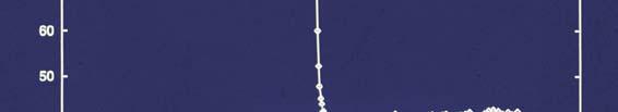
50 Seleno-methionine hydroxymethylbilane synthase fluorescence scan y y y y measured at SRS 9.5
51 Seleno-methionine hydroxymethylbilane synthase has 5 Se-met residues. The protein MW is 34 kda; now known to be near the average MW of proteins based on genome sequencing results. Haedener et al Acta Cryst D55, Determination of the structure of selenomethionine-labelled labelled hydroxymethylbilane synthase in its active form by multi-wavelength anomalous dispersion
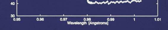
52 MAD and modified electron density maps bf before after Figure courtesy of E J Dodson


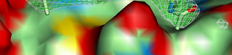
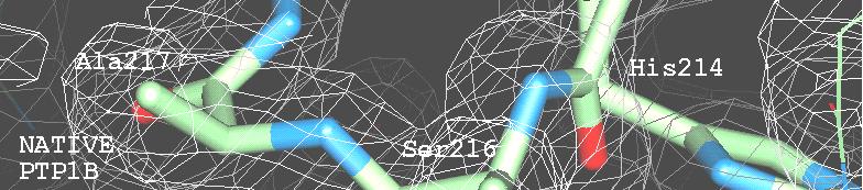


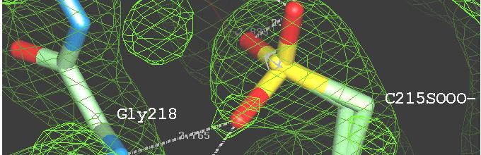
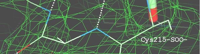
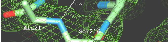
53 The end result is an electron density map that can be interpreted
54 Molecular Replacement harnesses a known 3D structure t Molecular replacement is the placement of a known protein structure into a different ie new crystal form: you already know which object is present in the crystal. VERY COMMON in SBDD. Molecular replacement uses a homology 3D model ie where the amino acid sequence identity i with a known 3D structure is >40% and places it into the new crystal unit cell. This is the starting point for further final refinement (next step).


55 Six parameter Search (Rotation and Translation of the known object) VERY IMPORTANT IN SBDD SINCE YOU SEEK STRUCTURES OF THE If we can SAME TARGET WITH MANY DIFFERENT LIGANDS. find the unknown structure known rotation and MGDKPIWEQIGSSFIQHYYQLFDNDRTQLGAIY PSPLLVGREFVRQYYTLLNKAPEYLHRFYGRNSSY structure IDASCLTWEGQQFQGKAAIVEKLSSLPFQKIQH VHGGVDASGKPQEAVYGQNDIHHKVLSLNFSECHT translation SITAQDHQPTPDSCIISMVVGQLKADEDPIMGF KIRHVDAHATLSDGVVVQVMGLLSNSGQPERKFMQ HQMFLLKNINDAWVCTNDMFRLALHNFG TFVLAPEGSVPNKFYVHNDMFRYEDE that puts the model in the correct position in the crystal cell, THEN we can calculate approx. phases. origin o H K L F φ etc... Figure courtesy of Prof Eleanor Dodson origin o H K L F φ etc...

56 CRYSTALLOGRAPHIC unknown structure MGDKPIWEQIGSSFIQHYYQLFDNDRTQLGAIYID ASCLTWEGQQFQGKAAIVEKLSSLPFQKIQHSITA QDHQPTPDSCIISMVVGQLKADEDPIMGFHQMFLL KNINDAWVCTNDMFRLALHNFG SYMMETRY known structure PSPLLVGREFVRQYYTLLNKAPEYLHRFYGRNSSYV HGGVDASGKPQEAVYGQNDIHHKVLSLNFSECHTKI RHVDAHATLSDGVVVQVMGLLSNSGQPERKFMQTFV LAPEGSVPNKFYVHNDMFRYEDE ROTATION TRANSLATION ROTATION TRANSLATION origin o origin o Figure courtesy of Prof Eleanor Dodson When symmetry is present, we only have to find one rotation and translation operator; the other one is given by the symmetry.
57 This is a summary of the key Protein Crystallography Concepts in a nutshell. Electron density interpretation Refinement Analysis of a Structural Paper. Questions?
58 The collapsing of the incoming wave on the detector On a crystallographic experiment. X-rays or Sound waves DETECTOR h,k,l, Fo, σ It is as if all the time (or frecuency) information of the incoming wave would be lost when the wave hits the detector. The detector seems to absorb the wave and there is only a record how Intense the wave was: Amplitude: Fo(hkl). These are the Fo s for all the reflections of the data set. These are the data from a xtallographic experiment.
59 Refinement: Fo vs. Fc s Structure factor: contribution of all atoms to reflection Fhkl Contribution of all instruments to sound S at seat labeled by l, r, s The structure factors calculated from all the atoms in your structure (Fc(hkl)) are compared with the ones measured from your diffraction experiment Fo(hkl) to see how they agree: Electron density equation: electron density at x,y,z in unit cell from all reflections (all h,k,l). Σ(hkl) Fo -Σ(hkl) Fc R = Summation of all the people Σ(hkl) Fo in the audience at the concert to produce density of instruments/unit volume.
Protein Crystallography
 Protein Crystallography Part II Tim Grüne Dept. of Structural Chemistry Prof. G. Sheldrick University of Göttingen http://shelx.uni-ac.gwdg.de tg@shelx.uni-ac.gwdg.de Overview The Reciprocal Lattice The
Scattering by two Electrons
 Scattering by two Electrons p = -r k in k in p r e 2 q k in /λ θ θ k out /λ S q = r k out p + q = r (k out - k in ) e 1 Phase difference of wave 2 with respect to wave 1: 2π λ (k out - k in ) r= 2π S r
Protein crystallography. Garry Taylor
 Protein crystallography Garry Taylor X-ray Crystallography - the Basics Grow crystals Collect X-ray data Determine phases Calculate ρ-map Interpret map Refine coordinates Do the biology. Nitrogen at -180
Crystal lattice Real Space. Reflections Reciprocal Space. I. Solving Phases II. Model Building for CHEM 645. Purified Protein. Build model.
 I. Solving Phases II. Model Building for CHEM 645 Purified Protein Solve Phase Build model and refine Crystal lattice Real Space Reflections Reciprocal Space ρ (x, y, z) pronounced rho F hkl 2 I F (h,
PSD '17 -- Xray Lecture 5, 6. Patterson Space, Molecular Replacement and Heavy Atom Isomorphous Replacement
 PSD '17 -- Xray Lecture 5, 6 Patterson Space, Molecular Replacement and Heavy Atom Isomorphous Replacement The Phase Problem We can t measure the phases! X-ray detectors (film, photomultiplier tubes, CCDs,
SHELXC/D/E. Andrea Thorn
 SHELXC/D/E Andrea Thorn What is experimental phasing? Experimental phasing is what you do if MR doesn t work. What is experimental phasing? Experimental phasing methods depend on intensity differences.
X-ray Crystallography
 2009/11/25 [ 1 ] X-ray Crystallography Andrew Torda, wintersemester 2009 / 2010 X-ray numerically most important more than 4/5 structures Goal a set of x, y, z coordinates different properties to NMR History
Anomalous dispersion
 Selenomethionine MAD Selenomethionine is the amino acid methionine with the Sulfur replaced by a Selenium. Selenium is a heavy atom that also has the propery of "anomalous scatter" at some wavelengths,
Patterson Methods
 59-553 Patterson Methods 113 In 1935, Patterson showed that the unknown phase information in the equation for electron density: ρ(xyz) = 1/V h k l F(hkl) exp[iα(hkl)] exp[-2πi(h x + k y + l z)] can be
Macromolecular X-ray Crystallography
 Protein Structural Models for CHEM 641 Fall 07 Brian Bahnson Department of Chemistry & Biochemistry University of Delaware Macromolecular X-ray Crystallography Purified Protein X-ray Diffraction Data collection
X-ray Crystallography. Kalyan Das
 X-ray Crystallography Kalyan Das Electromagnetic Spectrum NMR 10 um - 10 mm 700 to 10 4 nm 400 to 700 nm 10 to 400 nm 10-1 to 10 nm 10-4 to 10-1 nm X-ray radiation was discovered by Roentgen in 1895. X-rays
Phase problem: Determining an initial phase angle α hkl for each recorded reflection. 1 ρ(x,y,z) = F hkl cos 2π (hx+ky+ lz - α hkl ) V h k l
 = F hkl cos 2π (hx+ky+ lz - α hkl ) V h k l") Phase problem: Determining an initial phase angle α hkl for each recorded reflection 1 ρ(x,y,z) = F hkl cos 2π (hx+ky+ lz - α hkl ) V h k l Methods: Heavy atom methods (isomorphous replacement Hg, Pt)
Phase problem: Determining an initial phase angle α hkl for each recorded reflection 1 ρ(x,y,z) = F hkl cos 2π (hx+ky+ lz - α hkl ) V h k l Methods: Heavy atom methods (isomorphous replacement Hg, Pt)
Molecular Biology Course 2006 Protein Crystallography Part I
 Molecular Biology Course 2006 Protein Crystallography Part I Tim Grüne University of Göttingen Dept. of Structural Chemistry November 2006 http://shelx.uni-ac.gwdg.de tg@shelx.uni-ac.gwdg.de Overview Overview
What is the Phase Problem? Overview of the Phase Problem. Phases. 201 Phases. Diffraction vector for a Bragg spot. In General for Any Atom (x, y, z)
") Protein Overview of the Phase Problem Crystal Data Phases Structure John Rose ACA Summer School 2006 Reorganized by Andy Howard,, Spring 2008 Remember We can measure reflection intensities We can calculate
Protein Overview of the Phase Problem Crystal Data Phases Structure John Rose ACA Summer School 2006 Reorganized by Andy Howard,, Spring 2008 Remember We can measure reflection intensities We can calculate
Direct Method. Very few protein diffraction data meet the 2nd condition
 Direct Method Two conditions: -atoms in the structure are equal-weighted -resolution of data are higher than the distance between the atoms in the structure Very few protein diffraction data meet the 2nd
Fourier Syntheses, Analyses, and Transforms
 Fourier Syntheses, Analyses, and Transforms http://homepages.utoledo.edu/clind/ The electron density The electron density in a crystal can be described as a periodic function - same contents in each unit
PX-CBMSO Course (2) of Symmetry
 of Symmetry") PX-CBMSO Course (2) The mathematical description of Symmetry y PX-CBMSO-June 2011 Cele Abad-Zapatero University of Illinois at Chicago Center for Pharmaceutical Biotechnology. Lecture no. 2 This material
PX-CBMSO Course (2) The mathematical description of Symmetry y PX-CBMSO-June 2011 Cele Abad-Zapatero University of Illinois at Chicago Center for Pharmaceutical Biotechnology. Lecture no. 2 This material
Experimental phasing, Pattersons and SHELX Andrea Thorn
 Experimental phasing, Pattersons and SHELX Andrea Thorn What is experimental phasing? Experimental phasing is what you do if MR doesn t work. What is experimental phasing? Experimental phasing methods
Overview - Macromolecular Crystallography
 Overview - Macromolecular Crystallography 1. Overexpression and crystallization 2. Crystal characterization and data collection 3. The diffraction experiment 4. Phase problem 1. MIR (Multiple Isomorphous
Resolution: maximum limit of diffraction (asymmetric)
") Resolution: maximum limit of diffraction (asymmetric) crystal Y X-ray source 2θ X direct beam tan 2θ = Y X d = resolution 2d sinθ = λ detector 1 Unit Cell: two vectors in plane of image c* Observe: b*
Resolution: maximum limit of diffraction (asymmetric) crystal Y X-ray source 2θ X direct beam tan 2θ = Y X d = resolution 2d sinθ = λ detector 1 Unit Cell: two vectors in plane of image c* Observe: b*
Protein Structure Determination 9/25/2007
 One-dimensional NMR spectra Ethanol Cellulase (36 a.a.) Branden & Tooze, Fig. 18.16 1D and 2D NMR spectra of inhibitor K (57 a.a.) K. Wuthrich, NMR of Proteins and Nucleic Acids. (Wiley, 1986.) p. 54-55.
Scattering Lecture. February 24, 2014
 Scattering Lecture February 24, 2014 Structure Determination by Scattering Waves of radiation scattered by different objects interfere to give rise to an observable pattern! The wavelength needs to close
Roger Johnson Structure and Dynamics: X-ray Diffraction Lecture 6
 6.1. Summary In this Lecture we cover the theory of x-ray diffraction, which gives direct information about the atomic structure of crystals. In these experiments, the wavelength of the incident beam must
Determining Protein Structure BIBC 100
 Determining Protein Structure BIBC 100 Determining Protein Structure X-Ray Diffraction Interactions of x-rays with electrons in molecules in a crystal NMR- Nuclear Magnetic Resonance Interactions of magnetic
CCP4 Diamond 2014 SHELXC/D/E. Andrea Thorn
 CCP4 Diamond 2014 SHELXC/D/E Andrea Thorn SHELXC/D/E workflow SHELXC: α calculation, file preparation SHELXD: Marker atom search = substructure search SHELXE: density modification Maps and coordinate files
PSD '18 -- Xray lecture 4. Laue conditions Fourier Transform The reciprocal lattice data collection
 PSD '18 -- Xray lecture 4 Laue conditions Fourier Transform The reciprocal lattice data collection 1 Fourier Transform The Fourier Transform is a conversion of one space into another space with reciprocal
Determination of the Substructure
 Monday, June 15 th, 2009 Determination of the Substructure EMBO / MAX-INF2 Practical Course http://shelx.uni-ac.gwdg.de Overview Substructure Definition and Motivation Extracting Substructure Data from
6. X-ray Crystallography and Fourier Series
 6. X-ray Crystallography and Fourier Series Most of the information that we have on protein structure comes from x-ray crystallography. The basic steps in finding a protein structure using this method
Structure factors again
 Structure factors again Remember 1D, structure factor for order h F h = F h exp[iα h ] = I 01 ρ(x)exp[2πihx]dx Where x is fractional position along unit cell distance (repeating distance, origin arbitrary)
Protein Structure Determination. Part 1 -- X-ray Crystallography
 Protein Structure Determination Part 1 -- X-ray Crystallography Topics covering in this 1/2 course Crystal growth Diffraction theory Symmetry Solving phases using heavy atoms Solving phases using a model
BC530 Class notes on X-ray Crystallography
 BC530 Class notes on X-ray Crystallography web material: Ethan A Merritt http://skuld.bmsc.washington.edu/~merritt/bc530/ October 11, 2016 Growing Crystals It should be self-evident that in order to do
Crystals, X-rays and Proteins
 Crystals, X-rays and Proteins Comprehensive Protein Crystallography Dennis Sherwood MA (Hons), MPhil, PhD Jon Cooper BA (Hons), PhD OXFORD UNIVERSITY PRESS Contents List of symbols xiv PART I FUNDAMENTALS
Basic Crystallography Part 1. Theory and Practice of X-ray Crystal Structure Determination
 Basic Crystallography Part 1 Theory and Practice of X-ray Crystal Structure Determination We have a crystal How do we get there? we want a structure! The Unit Cell Concept Ralph Krätzner Unit Cell Description
Image definition evaluation functions for X-ray crystallography: A new perspective on the phase. problem. Hui LI*, Meng HE* and Ze ZHANG
 Image definition evaluation functions for X-ray crystallography: A new perspective on the phase problem Hui LI*, Meng HE* and Ze ZHANG Beijing University of Technology, Beijing 100124, People s Republic
Handout 7 Reciprocal Space
 Handout 7 Reciprocal Space Useful concepts for the analysis of diffraction data http://homepages.utoledo.edu/clind/ Concepts versus reality Reflection from lattice planes is just a concept that helps us
Protein Crystallography. Mitchell Guss University of Sydney Australia
 Protein Crystallography Mitchell Guss University of Sydney Australia Outline of the talk Recap some basic crystallography and history Highlight the special requirements for protein (macromolecular) structure
Electron Density at various resolutions, and fitting a model as accurately as possible.
 Section 9, Electron Density Maps 900 Electron Density at various resolutions, and fitting a model as accurately as possible. ρ xyz = (Vol) -1 h k l m hkl F hkl e iφ hkl e-i2π( hx + ky + lz ) Amplitude
Materials 286C/UCSB: Class VI Structure factors (continued), the phase problem, Patterson techniques and direct methods
, the phase problem, Patterson techniques and direct methods") Materials 286C/UCSB: Class VI Structure factors (continued), the phase problem, Patterson techniques and direct methods Ram Seshadri (seshadri@mrl.ucsb.edu) Structure factors The structure factor for a
Materials 286C/UCSB: Class VI Structure factors (continued), the phase problem, Patterson techniques and direct methods Ram Seshadri (seshadri@mrl.ucsb.edu) Structure factors The structure factor for a
GBS765 Electron microscopy
 GBS765 Electron microscopy Lecture 1 Waves and Fourier transforms 10/14/14 9:05 AM Some fundamental concepts: Periodicity! If there is some a, for a function f(x), such that f(x) = f(x + na) then function
Proteins. Central Dogma : DNA RNA protein Amino acid polymers - defined composition & order. Perform nearly all cellular functions Drug Targets
 Proteins Central Dogma : DNA RNA protein Amino acid polymers - defined composition & order Perform nearly all cellular functions Drug Targets Fold into discrete shapes. Proteins - cont. Specific shapes
Diffraction Geometry
 Diffraction Geometry Diffraction from a crystal - Laue equations Reciprocal lattice Ewald construction Data collection strategy Phil Evans LMB May 2013 MRC Laboratory of Molecular Biology Cambridge UK
General theory of diffraction
 General theory of diffraction X-rays scatter off the charge density (r), neutrons scatter off the spin density. Coherent scattering (diffraction) creates the Fourier transform of (r) from real to reciprocal
Biological Small Angle X-ray Scattering (SAXS) Dec 2, 2013
 Dec 2, 2013") Biological Small Angle X-ray Scattering (SAXS) Dec 2, 2013 Structural Biology Shape Dynamic Light Scattering Electron Microscopy Small Angle X-ray Scattering Cryo-Electron Microscopy Wide Angle X- ray
Biological Small Angle X-ray Scattering (SAXS) Dec 2, 2013 Structural Biology Shape Dynamic Light Scattering Electron Microscopy Small Angle X-ray Scattering Cryo-Electron Microscopy Wide Angle X- ray
Practical applications of synchrotron radiation in the determination of bio-macromolecule three-dimensional structures. M. Nardini and M.
 Practical applications of synchrotron radiation in the determination of bio-macromolecule three-dimensional structures M. Nardini and M. Bolognesi Department of Biomolecular Sciences and Biotechnology,
CS273: Algorithms for Structure Handout # 13 and Motion in Biology Stanford University Tuesday, 11 May 2003
 CS273: Algorithms for Structure Handout # 13 and Motion in Biology Stanford University Tuesday, 11 May 2003 Lecture #13: 11 May 2004 Topics: Protein Structure Determination Scribe: Minli Zhu We acknowledge
SUPPLEMENTARY INFORMATION
 Table of Contents Page Supplementary Table 1. Diffraction data collection statistics 2 Supplementary Table 2. Crystallographic refinement statistics 3 Supplementary Fig. 1. casic1mfc packing in the R3
Experimental phasing in Crank2
 Experimental phasing in Crank2 Pavol Skubak and Navraj Pannu Biophysical Structural Chemistry, Leiden University, The Netherlands http://www.bfsc.leidenuniv.nl/software/crank/ X-ray structure solution
Biology III: Crystallographic phases
 Haupt/Masterstudiengang Physik Methoden moderner Röntgenphysik II: Streuung und Abbildung SS 2013 Biology III: Crystallographic phases Thomas R. Schneider, EMBL Hamburg 25/6/2013 thomas.schneider@embl-hamburg.de
 Helpful resources for all X ray lectures Crystallization http://www.hamptonresearch.com under tech support: crystal growth 101 literature Spacegroup tables http://img.chem.ucl.ac.uk/sgp/mainmenu.htm Crystallography
Fan, Hai-fu Institute of Physics, Chinese Academy of Sciences, Beijing , China
 Direct Methods in Crystallography Fan, Hai-fu Institute of Physics, Chinese Academy of Sciences, Beijing 100080, China An important branch of crystallography is the X-ray diffraction analysis of crystal
Data Collection. Overview. Methods. Counter Methods. Crystal Quality with -Scans
 Data Collection Overview with a unit cell, possible space group and computer reference frame (orientation matrix); the location of diffracted x-rays can be calculated (h k l) and intercepted by something
Two Lectures in X-ray Crystallography
 Biochemistry 503 Michael Wiener (mwiener@virginia.edu, 3-2731, Snyder 360) Two Lectures in X-ray Crystallography Outline 1. Justification & introductory remarks 2. Experimental setup 3. Protein crystals
The ideal fiber pattern exhibits 4-quadrant symmetry. In the ideal pattern the fiber axis is called the meridian, the perpendicular direction is
 Fiber diffraction is a method used to determine the structural information of a molecule by using scattering data from X-rays. Rosalind Franklin used this technique in discovering structural information
SOLID STATE 18. Reciprocal Space
 SOLID STATE 8 Reciprocal Space Wave vectors and the concept of K-space can simplify the explanation of several properties of the solid state. They will be introduced to provide more information on diffraction
3.012 Structure An Introduction to X-ray Diffraction
 3.012 Structure An Introduction to X-ray Diffraction This handout summarizes some topics that are important for understanding x-ray diffraction. The following references provide a thorough explanation
Why do We Trust X-ray Crystallography?
 Why do We Trust X-ray Crystallography? Andrew D Bond All chemists know that X-ray crystallography is the gold standard characterisation technique: an X-ray crystal structure provides definitive proof of
Materials Science and Engineering 102 Structure and Bonding. Prof. Stephen L. Sass. Midterm Examination Duration: 1 hour 20 minutes
 October 9, 008 MSE 0: Structure and Bonding Midterm Exam SOLUTIONS SID: Signature: Materials Science and Engineering 0 Structure and Bonding Prof. Stephen L. Sass Midterm Examination Duration: hour 0 minutes
SOLID STATE 9. Determination of Crystal Structures
 SOLID STATE 9 Determination of Crystal Structures In the diffraction experiment, we measure intensities as a function of d hkl. Intensities are the sum of the x-rays scattered by all the atoms in a crystal.
Maximum Likelihood. Maximum Likelihood in X-ray Crystallography. Kevin Cowtan Kevin Cowtan,
 Maximum Likelihood Maximum Likelihood in X-ray Crystallography Kevin Cowtan cowtan@ysbl.york.ac.uk Maximum Likelihood Inspired by Airlie McCoy's lectures. http://www-structmed.cimr.cam.ac.uk/phaser/publications.html
Chapter 2. X-ray X. Diffraction and Reciprocal Lattice. Scattering from Lattices
 Chapter. X-ray X Diffraction and Reciprocal Lattice Diffraction of waves by crystals Reciprocal Lattice Diffraction of X-rays Powder diffraction Single crystal X-ray diffraction Scattering from Lattices
X-ray analysis. 1. Basic crystallography 2. Basic diffraction physics 3. Experimental methods
 X-ray analysis 1. Basic crystallography 2. Basic diffraction physics 3. Experimental methods Introduction Noble prizes associated with X-ray diffraction 1901 W. C. Roentgen (Physics) for the discovery
Fast, Intuitive Structure Determination IV: Space Group Determination and Structure Solution
 Fast, Intuitive Structure Determination IV: Space Group Determination and Structure Solution November 25, 2013 Welcome I I Dr. Michael Ruf Product Manager Crystallography Bruker AXS Inc. Madison, WI, USA
Principles of Protein X-Ray Crystallography
 Principles of Protein X-Ray Crystallography Jan Drenth Principles of Protein X-Ray Crystallography Third Edition With Major Contribution from Jeroen Mesters University of Lübeck, Germany Jan Drenth Laboratory
Twinning. Andrea Thorn
 Twinning Andrea Thorn OVERVIEW Introduction: Definitions, origins of twinning Merohedral twins: Recognition, statistical analysis: H plot, Yeates Padilla plot Example Refinement and R values Reticular
CALIFORNIA INSTITUTE OF TECHNOLOGY BECKMAN INSTITUTE X-RAY CRYSTALLOGRAPHY LABORATORY
 APPENDIX F Crystallographic Data for TBA Tb(DO2A)(F-DPA) CALIFORNIA INSTITUTE OF TECHNOLOGY BECKMAN INSTITUTE X-RAY CRYSTALLOGRAPHY LABORATORY Date 11 January 2010 Crystal Structure Analysis of: MLC23
Crystal Structure SOLID STATE PHYSICS. Lecture 5. A.H. Harker. thelecture thenextlecture. Physics and Astronomy UCL
 Crystal Structure thelecture thenextlecture SOLID STATE PHYSICS Lecture 5 A.H. Harker Physics and Astronomy UCL Structure & Diffraction Crystal Diffraction (continued) 2.4 Experimental Methods Notes: examples
Keble College - Hilary 2012 Section VI: Condensed matter physics Tutorial 2 - Lattices and scattering
 Tomi Johnson Keble College - Hilary 2012 Section VI: Condensed matter physics Tutorial 2 - Lattices and scattering Please leave your work in the Clarendon laboratory s J pigeon hole by 5pm on Monday of
There and back again A short trip to Fourier Space. Janet Vonck 23 April 2014
 There and back again A short trip to Fourier Space Janet Vonck 23 April 2014 Where can I find a Fourier Transform? Fourier Transforms are ubiquitous in structural biology: X-ray diffraction Spectroscopy
3.012 PS Issued: Fall 2003 Graded problems due:
 3.012 PS 4 3.012 Issued: 10.07.03 Fall 2003 Graded problems due: 10.15.03 Graded problems: 1. Planes and directions. Consider a 2-dimensional lattice defined by translations T 1 and T 2. a. Is the direction
Synthetic, Structural, and Mechanistic Aspects of an Amine Activation Process Mediated at a Zwitterionic Pd(II) Center
 Center") Synthetic, Structural, and Mechanistic Aspects of an Amine Activation Process Mediated at a Zwitterionic Pd(II) Center Supporting Information Connie C. Lu and Jonas C. Peters* Division of Chemistry and
Synthetic, Structural, and Mechanistic Aspects of an Amine Activation Process Mediated at a Zwitterionic Pd(II) Center Supporting Information Connie C. Lu and Jonas C. Peters* Division of Chemistry and
Schematic representation of relation between disorder and scattering
 Crystal lattice Reciprocal lattice FT Schematic representation of relation between disorder and scattering ρ = Δρ + Occupational disorder Diffuse scattering Bragg scattering ρ = Δρ + Positional
Structure Factors. How to get more than unit cell sizes from your diffraction data.
 Structure Factors How to get more than unit cell sizes from your diffraction data http://homepages.utoledo.edu/clind/ Yet again expanding convenient concepts First concept introduced: Reflection from lattice
Experimental Phasing with SHELX C/D/E
 WIR SCHAFFEN WISSEN HEUTE FÜR MORGEN Dr. Tim Grüne :: Paul Scherrer Institut :: tim.gruene@psi.ch Experimental Phasing with SHELX C/D/E CCP4 / APS School Chicago 2017 22 nd June 2017 1 - The Phase Problem
Cele Abad-Zapatero University of Illinois at Chicago Center for Pharmaceutical Biotechnology. Lecture no. 7 This material copyrighted by
 PX_CBMSO Course (7) Key crystallographic concepts: X-ray sources, instrumentation (adapted from K.Volz lectures on the course Structure of Biopolymers at UIC ) PX-CBMSO_June_2011 Cele Abad-Zapatero University
Handout 13 Interpreting your results. What to make of your atomic coordinates, bond distances and angles
 Handout 13 Interpreting your results What to make of your atomic coordinates, bond distances and angles 1 What to make of the outcome of your refinement There are several ways of judging whether the outcome
Crystallography past, present and future
 Crystallography past, present and future Jenny P. Glusker Philadelphia, PA, U. S. A. International Year of Crystallography UNESCO, Paris, France 20 January 2014 QUARTZ CRYSTALS Quartz crystals found growing
The Phase Problem of X-ray Crystallography
 163 The Phase Problem of X-ray Crystallography H.A. Hauptman Hauptman-Woodward Medical Research Institute, Inc. 73 High Street Buffalo, NY, USA hauptman@hwi.buffalo.edu ABSTRACT. The intensities of a sufficient
Space Group & Structure Solution
 Space Group & Structure Solution Determine the Space Group Space group determination can always be performed by hand by examining the intensity data. A program that can facilitate this step is the command-prompt
Handout 12 Structure refinement. Completing the structure and evaluating how good your data and model agree
 Handout 1 Structure refinement Completing the structure and evaluating how good your data and model agree Why you should refine a structure We have considered how atoms are located by Patterson, direct
Physical Chemistry Analyzing a Crystal Structure and the Diffraction Pattern Virginia B. Pett The College of Wooster
 Physical Chemistry Analyzing a Crystal Structure and the Diffraction Pattern Virginia B. Pett The College of Wooster L. W. Haynes and his Senior Independent Study students conducted the 2 + 2 photo addition
Quantum Condensed Matter Physics Lecture 5
 Quantum Condensed Matter Physics Lecture 5 detector sample X-ray source monochromator David Ritchie http://www.sp.phy.cam.ac.uk/drp2/home QCMP Lent/Easter 2019 5.1 Quantum Condensed Matter Physics 1. Classical
Electronic Supplementary Information (ESI) for Chem. Commun. Unveiling the three- dimensional structure of the green pigment of nitrite- cured meat
 for Chem. Commun. Unveiling the three- dimensional structure of the green pigment of nitrite- cured meat") Electronic Supplementary Information (ESI) for Chem. Commun. Unveiling the three- dimensional structure of the green pigment of nitrite- cured meat Jun Yi* and George B. Richter- Addo* Department of Chemistry
Electronic Supplementary Information (ESI) for Chem. Commun. Unveiling the three- dimensional structure of the green pigment of nitrite- cured meat Jun Yi* and George B. Richter- Addo* Department of Chemistry
Summary of Experimental Protein Structure Determination. Key Elements
 Programme 8.00-8.20 Summary of last week s lecture and quiz 8.20-9.00 Structure validation 9.00-9.15 Break 9.15-11.00 Exercise: Structure validation tutorial 11.00-11.10 Break 11.10-11.40 Summary & discussion
Molecular Biology Course 2006 Protein Crystallography Part II
 Molecular Biology Course 2006 Protein Crystallography Part II Tim Grüne University of Göttingen Dept. of Structural Chemistry December 2006 http://shelx.uni-ac.gwdg.de tg@shelx.uni-ac.gwdg.de Overview
Protein Crystallography Part II
 Molecular Biology Course 2007 Protein Crystallography Part II Tim Grüne University of Göttingen Dept. of Structural Chemistry November 2007 http://shelx.uni-ac.gwdg.de tg@shelx.uni-ac.gwdg.de Overview
Part 1 X-ray Crystallography
 Part 1 X-ray Crystallography What happens to electron when it is hit by x-rays? 1. The electron starts vibrating with the same frequency as the x-ray beam 2. As a result, secondary beams will be scattered
Phys 460 Describing and Classifying Crystal Lattices
 Phys 460 Describing and Classifying Crystal Lattices What is a material? ^ crystalline Regular lattice of atoms Each atom has a positively charged nucleus surrounded by negative electrons Electrons are
Electron microscopy in molecular cell biology II
 Electron microscopy in molecular cell biology II Cryo-EM and image processing Werner Kühlbrandt Max Planck Institute of Biophysics Sample preparation for cryo-em Preparation laboratory Specimen preparation
APPENDIX E. Crystallographic Data for TBA Eu(DO2A)(DPA) Temperature Dependence
(DPA) Temperature Dependence") APPENDIX E Crystallographic Data for TBA Eu(DO2A)(DPA) Temperature Dependence Temperature Designation CCDC Page 100 K MLC18 761599 E2 200 K MLC17 762705 E17 300 K MLC19 763335 E31 E2 CALIFORNIA INSTITUTE
APPENDIX E Crystallographic Data for TBA Eu(DO2A)(DPA) Temperature Dependence Temperature Designation CCDC Page 100 K MLC18 761599 E2 200 K MLC17 762705 E17 300 K MLC19 763335 E31 E2 CALIFORNIA INSTITUTE
Understanding Single-Crystal X-Ray Crystallography Exercises and Solutions
 Understanding Single-Crystal X-Ray Crystallography Exercises and Solutions Dennis W. Bennett Department of Chemistry and Biochemistry University of Wisconsin-Milwaukee Chapter Crystal Lattices. The copper
Lecture Notes for Math 251: ODE and PDE. Lecture 16: 3.8 Forced Vibrations Without Damping
 Lecture Notes for Math 25: ODE and PDE. Lecture 6:.8 Forced Vibrations Without Damping Shawn D. Ryan Spring 202 Forced Vibrations Last Time: We studied non-forced vibrations with and without damping. We
The Reciprocal Lattice
 59-553 The Reciprocal Lattice 61 Because of the reciprocal nature of d spacings and θ from Bragg s Law, the pattern of the diffraction we observe can be related to the crystal lattice by a mathematical
Diffraction. X-ray diffraction
 Diffraction Definition (from Cambridge Advanced Learner s Dictionary ): - diffraction noun [U] SPECIALIZED (a pattern caused by) a change in the direction of light, water or sound waves - diffract verb
Copyright WILEY-VCH Verlag GmbH, D Weinheim, 2000 Angew. Chem Supporting Information For Binding Cesium Ion with Nucleoside Pentamers.
 Copyright WILEY-VCH Verlag GmbH, D-69451 Weinheim, 2000 Angew. Chem. 2000 Supporting Information For Binding Cesium Ion with Nucleoside Pentamers. Templated Self-Assembly of an Isoguanosine Decamer.**
Anisotropy in macromolecular crystal structures. Andrea Thorn July 19 th, 2012
 Anisotropy in macromolecular crystal structures Andrea Thorn July 19 th, 2012 Motivation Courtesy of M. Sawaya Motivation Crystal structures are inherently anisotropic. X-ray diffraction reflects this
Theory of X-ray diffraction
 Theory of X-ray diffraction A users perspective Disclaimer: I am not a physicist but there will be equations! Phil Evans Diamond December 2016 MRC Laboratory of Molecular Biology Cambridge UK Acknowledgements:
Supplementary Material (ESI) for CrystEngComm This journal is The Royal Society of Chemistry 2010
 for CrystEngComm This journal is The Royal Society of Chemistry 2010") Electronic Supplementary Information (ESI) for: A bifunctionalized porous material containing discrete assemblies of copper-porphyrins and calixarenes metallated by ion diffusion Rita De Zorzi, Nicol Guidolin,
Electronic Supplementary Information (ESI) for: A bifunctionalized porous material containing discrete assemblies of copper-porphyrins and calixarenes metallated by ion diffusion Rita De Zorzi, Nicol Guidolin,
X-Ray structure analysis
 X-Ray structure analysis Kay Diederichs kay.diederichs@uni-konstanz.de Analysis of what? Proteins ( /ˈproʊˌtiːnz/ or /ˈproʊti.ɨnz/) are biochemical compounds consisting of one or more polypeptides typically
5.62 Physical Chemistry II Spring 2008
 MIT OpenCourseWare http://ocw.mit.edu 5.62 Physical Chemistry II Spring 2008 For information about citing these materials or our Terms of Use, visit: http://ocw.mit.edu/terms. 5.62 Spring 2008 Lecture