Data quality indicators. Kay Diederichs
|
|
|
- Ralf Ross
- 6 years ago
- Views:
Transcription
1 Data quality indicators Kay Diederichs

2 Crystallography has been highly successful Now Could it be any better? 2
3 Confusion what do these mean? CC1/2 Rmerge Rsym Mn(I/sd) I/σ Rmeas CCanom Rpim Rcum 3
4 Topics Signal versus noise Random versus systematic error Accuracy versus precision Unmerged versus merged data R-values versus correlation coefficients Choice of high-resolution cutoff 4
5 signal vs noise easy hard threshold of solvability impossible James Holton slide5
6 noise : what is noise? what kinds of errors exist? noise = random error + systematic error random error results from quantum effects systematic error results from everything else: technical or other macroscopic aspects of the experiment 6
7 Random error (noise) Statistical events: photon emission from xtal photon absorption in detector electron hopping in semiconductors (amplifier etc) 7
8 non-obvious Systematic errors (noise) beam flicker (instability) in flux or direction shutter jitter vibration due to cryo stream split reflections, secondary lattice(s) absorption from crystal and loop radiation damage detector calibration and inhomogeneity; overload shadows on detector deadtime in shutterless mode imperfect assumptions about the experiment and its geometric parameters in the processing software... 8
9 non-obvious Adding noise = = = = 2 σ1 + σ2 σtotal 2 James Holton slide 14
10 This law is only valid if errors are independent! 15
11 non-obvious How do random and systematic error depend on the signal? random error obeys Poisson statistics error = square root of signal Systematic error is proportional to signal error = x * signal (e.g. x= ) (which is why James Holton calls it fractional error ; there are exceptions) 16
12 non-obvious Consequences need to add both types of errors at high resolution, random error dominates at low resolution, systematic error dominates but: radiation damage influences both the low and the high resolution (the factor x is low at low resolution, and high at high resolution) 17
13 non-obvious How to measure quality? B. Rupp, Biomolecular Crystallography Accuracy Precision how close to the true value? how close are measurements? 18
14 non-obvious What is the true value? if only random error exists, accuracy = precision (on average) if unknown systematic error exists, true value cannot be found from the data themselves a good model can provide an approximation to the truth model calculations do provide the truth consequence: precision can easily be calculated, but not accuracy accuracy and precision differ by the unknown systematic error All data quality indicators estimate precision (only), but YOU want to know accuracy! 19
15 Numerical example Repeatedly determine π= as 2.718, 2.716, : high precision, low accuracy. Precision= relative deviation from average value= ( )/( ) = 0.049% Accuracy= relative deviation from true value= ( ) / = 13.5% Repeatedly determine π= as 3.1, 3.2, 3.0 : low precision, high accuracy Precision= relative deviation from average value= ( )/( ) = 2.6% Accuracy= relative deviation from true value: = 1.3% 20
16 Calculating the precision of unmerged data Precision indicators for the unmerged (individual) observations: <Ii/σi > (σi from error propagation) n I i ( hkl ) I ( hkl ) R merge= hkl i=1 n I i (hkl ) hkl i=1 R meas = hkl n n I ( hkl ) I ( hkl ) n 1 i= 1 i n I i (hkl ) Rmeas ~ 0.8 / <Ii/σi > hkl i= 1 21
17 Averaging ( merging ) of observations Intensities: I = Ii/σi2 / 1/σi2 Sigmas: σ2 = 1 / 1/σi2 (see Wikipedia: weighted mean ) 22
18 non-obvious Merging of observations may improve accuracy and precision Averaging ( merging ) requires multiplicity ( redundancy ) (Only) if errors are unrelated, averaging with multiplicity n decreases the error of the averaged data by sqrt(n) Random errors are unrelated by definition: averaging always decreases the random error of merged data Averaging may decrease the systematic error in the merged data. This requires sampling of its possible values - true multiplicity If errors are related, precision improves, but not accuracy 23
19 Calculating the precision of merged data using the sqrt(n) law: <I/σ(I)> n 1 / n 1 I i ( hkl ) I ( hkl ) R pim = hkl i=1 n I i ( hkl ) Rpim ~ 0.8 / <I/σ > hkl i= 1 by comparing averages of two randomly selected half-datasets X,Y: H,K,L 1,2,3 1,2,4 1,2,5... Ii in order of Assignment to measurement half-dataset X, X, Y, X, Y, Y YXYX XYYX Average I of X Y (calculate the R-factor (D&K1997) or correlation coefficient (K&D 2012) on X, Y ) 24

20 I/σ with σ2 = 1 / 1/σi2 25
21 Shall I use an indicator for precision of unmerged data, or of merged data? It is essential to understand the difference between the two types, but you don't find this in the papers / textbooks! Indicators for precision of unmerged data help to e.g. * decide between spacegroups * calculate amount of radiation damage (see XDS tutorial) Indicators for precision of merged data assess suitability * for downstream calculations (MR, phasing, refinement) 26
22 Crystallographic statistics - which indicators are being used? Data R-values: Rpim < Rmerge=Rsym < Rmeas n R pim = 1 /n 1 d Iatia( hkl ) I ( hkl ) hkl edi= 1 merg n I i ( hkl ) hkl i= 1 n I i ( hkl ) a It(ahkl ) ed d g r n e unm I i ( hkl ) Rmerge = hkl i=1 Rmeas = hkl dd g r ne e unm I i ( hkl ) hkl i=1 Model R-values: Rwork/Rfree n n I ( hkl ) I ( hkl ) n 1 i=1 i ata hkl i= 1 (hkl ) F obs ( hkl ) F ta calc a R work / free= hkl rged d me F obs (hkl ) hkl I/σ (for unmerged or merged data!) CC1/2 and CCanom for the merged data 27
23 Decisions and compromises Which high-resolution cutoff for refinement? Higher resolution means better accuracy and maps But: high resolution yields high Rwork/Rfree! Which datasets/frames to include into scaling? Reject negative observations or unique reflections? The reason why it is difficult to answer R-value questions is that no proper mathematical theory exists that uses absolute differences; concerning the use of R-values, Crystallography is disconnected from mainstream Statistics 28
24 Improper crystallographic reasoning typical example: data to 2.0 Å resolution using all data: R work=19%, Rfree=24% (overall) cut at 2.2 Å resolution: R work=17%, Rfree=23% cutting at 2.2 Å is better because it gives lower R-values 31
25 Proper crystallographic reasoning 1. Better data allow to obtain a better model 2. A better model has a lower Rfree, and a lower Rfree-Rwork gap 3. Comparison of model R-values is only meaningful when using the same data 4. Taken together, this leads to the paired refinement technique : compare models in terms of their R-values against the same data. 32
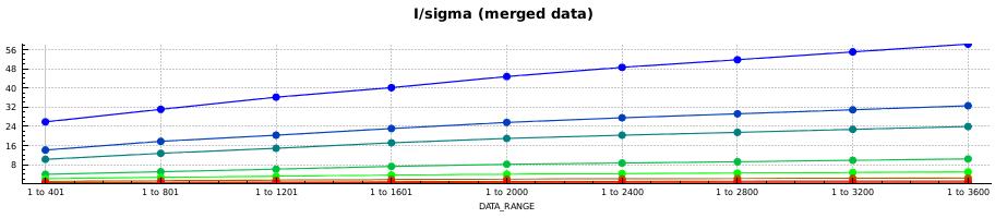
26 Example: Cysteine DiOxygenase (CDO; PDB 3ELN) re-refined against 15-fold weaker data Rmerge Rpim Rfree Rwork I/sigma 33

27 Is there information beyond the conservative hi-res cutoff? Paired refinement technique : refine at (e.g.) 2.0Å and at 1.9Å using the same starting model and refinement parameters since it is meaningless to compare Rvalues at different resolutions, calculate the overall R-values of the 1.9Å model at 2.0Å (main.number_of_macro_cycles=1 strategy=none fix_rotamers=false ordered_solvent=false) ΔR=R1.9(2.0)-R2.0(2.0) 34
28 Measuring the precision of merged data with a correlation coefficient Correlation coefficient has clear meaning and well-known statistical properties Significance of its value can be assessed by Student's ttest (e.g. CC>0.3 is significant at p=0.01 for n>100; CC>0.08 is significant at p=0.01 for n>1000) Apply this idea to crystallographic intensity data: use random half-datasets CC1/2 (called CC_Imean by SCALA/aimless, now CC1/2 ) From CC1/2, we can analytically estimate CC of the merged dataset against the true (usually unmeasurable) intensities using 2 CC 1/ 2 CC*= 1+CC 1/2 (Karplus and Diederichs (2012) Science 336, 1030) 35
29 Data CCs CC1/2 CC* Δ I/sigma 36
30 Model CCs We can define CCwork, CCfree as CCs calculated on Fcalc2 of the working and free set, against the experimental data CCwork and CCfree can be directly compared with CC* CC* Dashes: CCwork, CCfree against weak exp tl data Dots: CC work, CC free against strong 3ELN data 37

31 Four new concepts for improving crystallographic procedures Image courtesy of 38 P.A. Karplus
32 Summary To predict suitability of data for downstream calculations (phasing, MR, refinement), we should use indicators of merged data precision Rmerge should no longer be considered as useful for deciding e.g. on a high-resolution cutoff, or on which datasets to merge, or how large total rotation I/σ has two drawbacks: programs do not agree on σ, and its value can only rise with multiplicity CC1/2 is well understood, reproducible, and directly links to model quality indicators 39
33 References P.A. Karplus and K. Diederichs (2012) Linking Crystallographic Data with Model Quality. Science 336, see also: P.R. Evans (2012) Resolving Some Old Problems in Protein Crystallography. Science 336, K. Diederichs and P.A. Karplus (2013) Better models by discarding data? Acta Cryst. D69, P. R. Evans and G. N. Murshudov (2013) How good are my data and what is the resolution? Acta Cryst. D69, Z. Luo, K. Rajashankar and Z. Dauter (2014) Weak data do not make a free lunch, only a cheap meal. Acta Cryst. D70, J. Wang and R. A. Wing (2014) Diamonds in the rough: a strong case for the inclusion of weak-intensity X-ray diffraction data. Acta Cryst. D70, Diederichs K, "Crystallographic data and model quality" in Nucleic Acids Crystallography. (Ed. E Ennifar), Methods in Molecular Biology (in press). 40
34 Thank you! PDF available pls send to 41
Linking data and model quality in macromolecular crystallography. Kay Diederichs
 Linking data and model quality in macromolecular crystallography Kay Diederichs Crystallography is highly successful Can we do better? Error in experimental data Error = random + systematic Multiplicity
Data quality noise, errors, mistakes
 Data quality noise, errors, mistakes Kay Diederichs Protein Crystallography / Molecular Bioinformatics University of Konstanz, Germany Crystallography has been extremely successful Protein Data Bank on
Resolution and data formats. Andrea Thorn
 Resolution and data formats Andrea Thorn RESOLUTION Motivation Courtesy of M. Sawaya Map resolution http://www.bmsc.washington.edu/people/verlinde/experiment.html Data quality indicators Resolution accounts
X-ray Crystallography I. James Fraser Macromolecluar Interactions BP204
 X-ray Crystallography I James Fraser Macromolecluar Interactions BP204 Key take-aways 1. X-ray crystallography results from an ensemble of Billions and Billions of molecules in the crystal 2. Models in
Twinning. Andrea Thorn
 Twinning Andrea Thorn OVERVIEW Introduction: Definitions, origins of twinning Merohedral twins: Recognition, statistical analysis: H plot, Yeates Padilla plot Example Refinement and R values Reticular
SHELXC/D/E. Andrea Thorn
 SHELXC/D/E Andrea Thorn What is experimental phasing? Experimental phasing is what you do if MR doesn t work. What is experimental phasing? Experimental phasing methods depend on intensity differences.
CCP4 Diamond 2014 SHELXC/D/E. Andrea Thorn
 CCP4 Diamond 2014 SHELXC/D/E Andrea Thorn SHELXC/D/E workflow SHELXC: α calculation, file preparation SHELXD: Marker atom search = substructure search SHELXE: density modification Maps and coordinate files
Diffraction Geometry
 Diffraction Geometry Diffraction from a crystal - Laue equations Reciprocal lattice Ewald construction Data collection strategy Phil Evans LMB May 2013 MRC Laboratory of Molecular Biology Cambridge UK
Data Reduction. Space groups, scaling and data quality. MRC Laboratory of Molecular Biology Cambridge UK. Phil Evans Diamond December 2017
 Data Reduction Space groups, scaling and data quality Phil Evans Diamond December 2017 MRC Laboratory of Molecular Biology Cambridge UK F 2 Scaling and Merging Experiment lots of effects ( errors ) I Our
Experimental phasing, Pattersons and SHELX Andrea Thorn
 Experimental phasing, Pattersons and SHELX Andrea Thorn What is experimental phasing? Experimental phasing is what you do if MR doesn t work. What is experimental phasing? Experimental phasing methods
RNA protects a nucleoprotein complex against radiation damage
 Supporting information Volume 72 (2016) Supporting information for article: RNA protects a nucleoprotein complex against radiation damage Charles S. Bury, John E. McGeehan, Alfred A. Antson, Ian Carmichael,
Quantifying instrument errors in macromolecular X-ray data sets
 Acta Crystallographica Section D Biological Crystallography ISSN 0907-4449 Editors: E. N. Baker and Z. Dauter Quantifying instrument errors in macromolecular X-ray data sets Kay Diederichs Acta Cryst.
Twinning in PDB and REFMAC. Garib N Murshudov Chemistry Department, University of York, UK
 Twinning in PDB and REFMAC Garib N Murshudov Chemistry Department, University of York, UK Contents Crystal peculiarities Problem of twinning Occurrences of twinning in PDB Recognition of potential operators
X-ray Data Collection. Bio5325 Spring 2006
 X-ray Data Collection Bio535 Spring 006 Obtaining I hkl and α (Ihkl) from Frame Images Braggs Law -predicts conditions for in-phase scattering by equivalent atoms lying in planes that transect a crystal.
Electron Crystallography for Structure Solution Latest Results from the PSI Electron Diffraction Group
 WIR SCHAFFEN WISSEN HEUTE FÜR MORGEN Dr. Tim Grüne :: Paul Scherrer Institut :: tim.gruene@psi.ch Electron Crystallography for Structure Solution Latest Results from the PSI Electron Diffraction Group
X-ray Crystallography. Kalyan Das
 X-ray Crystallography Kalyan Das Electromagnetic Spectrum NMR 10 um - 10 mm 700 to 10 4 nm 400 to 700 nm 10 to 400 nm 10-1 to 10 nm 10-4 to 10-1 nm X-ray radiation was discovered by Roentgen in 1895. X-rays
8:10 pm Keynote Lecture: Michael G. Rossmann, Purdue University Current challenges in structural biology
 Sunday, October 11 3:00 pm Check-in 6:00 pm Reception (Lobby) 7:00 pm Dinner 8:00 pm Introductory Notes: Tamir Gonen 8:10 pm Keynote Lecture: Michael G. Rossmann, Purdue University Current challenges in
X-ray Crystallography
 2009/11/25 [ 1 ] X-ray Crystallography Andrew Torda, wintersemester 2009 / 2010 X-ray numerically most important more than 4/5 structures Goal a set of x, y, z coordinates different properties to NMR History
Changing and challenging times for service crystallography. Electronic Supplementary Information
 Changing and challenging times for service crystallography Simon J Coles,* a and Philip A Gale* a Electronic Supplementary Information Instrument descriptions and experimental protocols The following firstly
X-Ray structure analysis
 X-Ray structure analysis Kay Diederichs kay.diederichs@uni-konstanz.de Analysis of what? Proteins ( /ˈproʊˌtiːnz/ or /ˈproʊti.ɨnz/) are biochemical compounds consisting of one or more polypeptides typically
X-Ray Damage to Biological Crystalline Samples
 X-Ray Damage to Biological Crystalline Samples Gerd Rosenbaum Structural Biology Center, ANL and Dept. of Biochemistry, UGA ACA Summer School IIT, 19 July 2007 A U.S. Department of Energy laboratory managed
PSD '18 -- Xray lecture 4. Laue conditions Fourier Transform The reciprocal lattice data collection
 PSD '18 -- Xray lecture 4 Laue conditions Fourier Transform The reciprocal lattice data collection 1 Fourier Transform The Fourier Transform is a conversion of one space into another space with reciprocal
Experimental Phasing of SFX Data. Thomas Barends MPI for Medical Research, Heidelberg. Conclusions
 Experimental Phasing of SFX Data Thomas Barends MP for Medical Researc Heidelberg Conclusions X-ray Free-Electron Lasers are pushing back the boundaries of possibility in biological crystallography: -Data
Calculating strategies for data collection
 Calculating strategies for data collection Alexander Popov A.Popov 1 Data quality Completeness, Resolution, Statistics Detector Size Size N.of N.of pixels pixels T T readout readout Noise Noise Sensitivity
Protein crystallography. Garry Taylor
 Protein crystallography Garry Taylor X-ray Crystallography - the Basics Grow crystals Collect X-ray data Determine phases Calculate ρ-map Interpret map Refine coordinates Do the biology. Nitrogen at -180
Ensemble refinement of protein crystal structures in PHENIX. Tom Burnley Piet Gros
 Ensemble refinement of protein crystal structures in PHENIX Tom Burnley Piet Gros Incomplete modelling of disorder contributes to R factor gap Only ~5% of residues in the PDB are modelled with more than
Practical aspects of SAD/MAD. Judit É Debreczeni
 Practical aspects of SAD/MAD Judit É Debreczeni anomalous scattering Hg sinθ/λ CuKα 0 Å - 0.4 Å - 0.6 Å - Å.5Å 0.83Å f 80 53 4 f total (θ, λ) f(θ) + f (λ) + if (λ) f f -5 8-5 8-5 8 f total increasing with
The Reciprocal Lattice
 59-553 The Reciprocal Lattice 61 Because of the reciprocal nature of d spacings and θ from Bragg s Law, the pattern of the diffraction we observe can be related to the crystal lattice by a mathematical
 Helpful resources for all X ray lectures Crystallization http://www.hamptonresearch.com under tech support: crystal growth 101 literature Spacegroup tables http://img.chem.ucl.ac.uk/sgp/mainmenu.htm Crystallography
Summary of Experimental Protein Structure Determination. Key Elements
 Programme 8.00-8.20 Summary of last week s lecture and quiz 8.20-9.00 Structure validation 9.00-9.15 Break 9.15-11.00 Exercise: Structure validation tutorial 11.00-11.10 Break 11.10-11.40 Summary & discussion
Anisotropy in macromolecular crystal structures. Andrea Thorn July 19 th, 2012
 Anisotropy in macromolecular crystal structures Andrea Thorn July 19 th, 2012 Motivation Courtesy of M. Sawaya Motivation Crystal structures are inherently anisotropic. X-ray diffraction reflects this
Protein Crystallography
 Protein Crystallography Part II Tim Grüne Dept. of Structural Chemistry Prof. G. Sheldrick University of Göttingen http://shelx.uni-ac.gwdg.de tg@shelx.uni-ac.gwdg.de Overview The Reciprocal Lattice The
Basic Crystallography Part 1. Theory and Practice of X-ray Crystal Structure Determination
 Basic Crystallography Part 1 Theory and Practice of X-ray Crystal Structure Determination We have a crystal How do we get there? we want a structure! The Unit Cell Concept Ralph Krätzner Unit Cell Description
High-resolution crystal structure of ERAP1 with bound phosphinic transition-state analogue inhibitor
 High-resolution crystal structure of ERAP1 with bound phosphinic transition-state analogue inhibitor Petros Giastas 1, Margarete Neu 2, Paul Rowland 2, and Efstratios Stratikos 1 1 National Center for
Why do We Trust X-ray Crystallography?
 Why do We Trust X-ray Crystallography? Andrew D Bond All chemists know that X-ray crystallography is the gold standard characterisation technique: an X-ray crystal structure provides definitive proof of
Biological Small Angle X-ray Scattering (SAXS) Dec 2, 2013
 Dec 2, 2013") Biological Small Angle X-ray Scattering (SAXS) Dec 2, 2013 Structural Biology Shape Dynamic Light Scattering Electron Microscopy Small Angle X-ray Scattering Cryo-Electron Microscopy Wide Angle X- ray
Biological Small Angle X-ray Scattering (SAXS) Dec 2, 2013 Structural Biology Shape Dynamic Light Scattering Electron Microscopy Small Angle X-ray Scattering Cryo-Electron Microscopy Wide Angle X- ray
Scattering Lecture. February 24, 2014
 Scattering Lecture February 24, 2014 Structure Determination by Scattering Waves of radiation scattered by different objects interfere to give rise to an observable pattern! The wavelength needs to close
Acta Crystallographica Section F
 Supporting information Acta Crystallographica Section F Volume 70 (2014) Supporting information for article: Chemical conversion of cisplatin and carboplatin with histidine in a model protein crystallised
GC376 (compound 28). Compound 23 (GC373) (0.50 g, 1.24 mmol), sodium bisulfite (0.119 g,
. Compound 23 (GC373) (0.50 g, 1.24 mmol), sodium bisulfite (0.119 g,") Supplemental Material Synthesis of GC376 GC376 (compound 28). Compound 23 (GC373) (0.50 g, 1.24 mmol), sodium bisulfite (0.119 g, 1.12 mmol), ethyl acetate (2 ml), ethanol (1 ml) and water (0.40 ml) were
Supplemental Material Synthesis of GC376 GC376 (compound 28). Compound 23 (GC373) (0.50 g, 1.24 mmol), sodium bisulfite (0.119 g, 1.12 mmol), ethyl acetate (2 ml), ethanol (1 ml) and water (0.40 ml) were
IgE binds asymmetrically to its B cell receptor CD23
 Supplementary Information IgE binds asymmetrically to its B cell receptor CD23 Balvinder Dhaliwal 1*, Marie O. Y. Pang 2, Anthony H. Keeble 2,3, Louisa K. James 2,4, Hannah J. Gould 2, James M. McDonnell
SOLID STATE 9. Determination of Crystal Structures
 SOLID STATE 9 Determination of Crystal Structures In the diffraction experiment, we measure intensities as a function of d hkl. Intensities are the sum of the x-rays scattered by all the atoms in a crystal.
Protein Structure Determination. Part 1 -- X-ray Crystallography
 Protein Structure Determination Part 1 -- X-ray Crystallography Topics covering in this 1/2 course Crystal growth Diffraction theory Symmetry Solving phases using heavy atoms Solving phases using a model
Resolution: maximum limit of diffraction (asymmetric)
") Resolution: maximum limit of diffraction (asymmetric) crystal Y X-ray source 2θ X direct beam tan 2θ = Y X d = resolution 2d sinθ = λ detector 1 Unit Cell: two vectors in plane of image c* Observe: b*
Resolution: maximum limit of diffraction (asymmetric) crystal Y X-ray source 2θ X direct beam tan 2θ = Y X d = resolution 2d sinθ = λ detector 1 Unit Cell: two vectors in plane of image c* Observe: b*
Characterization of low energy ionization signals from Compton scattering in a CCD Dark Matter detector
 Characterization of low energy ionization signals from Compton scattering in a CCD Dark Matter detector Karthik Ramanathan University of Chicago arxiv:1706.06053 (Accepted PRD) TeVPA 2017/08/07 1 Motivation
Biology III: Crystallographic phases
 Haupt/Masterstudiengang Physik Methoden moderner Röntgenphysik II: Streuung und Abbildung SS 2013 Biology III: Crystallographic phases Thomas R. Schneider, EMBL Hamburg 25/6/2013 thomas.schneider@embl-hamburg.de
SUPPLEMENTARY INFORMATION
 Supplementary Table 1: Amplitudes of three current levels. Level 0 (pa) Level 1 (pa) Level 2 (pa) TrkA- TrkH WT 200 K 0.01 ± 0.01 9.5 ± 0.01 18.7 ± 0.03 200 Na * 0.001 ± 0.01 3.9 ± 0.01 12.5 ± 0.03 200
Electronic Supplementary Information (ESI) for Chem. Commun. Unveiling the three- dimensional structure of the green pigment of nitrite- cured meat
 for Chem. Commun. Unveiling the three- dimensional structure of the green pigment of nitrite- cured meat") Electronic Supplementary Information (ESI) for Chem. Commun. Unveiling the three- dimensional structure of the green pigment of nitrite- cured meat Jun Yi* and George B. Richter- Addo* Department of Chemistry
Electronic Supplementary Information (ESI) for Chem. Commun. Unveiling the three- dimensional structure of the green pigment of nitrite- cured meat Jun Yi* and George B. Richter- Addo* Department of Chemistry
Table S1. Overview of used PDZK1 constructs and their binding affinities to peptides. Related to figure 1.
 Table S1. Overview of used PDZK1 constructs and their binding affinities to peptides. Related to figure 1. PDZK1 constru cts Amino acids MW [kda] KD [μm] PEPT2-CT- FITC KD [μm] NHE3-CT- FITC KD [μm] PDZK1-CT-
Non-merohedral Twinning in Protein Crystallography
 Bruker Users Meeting 2010 Karlsruhe, 22 nd September 2010 Non-merohedral Twinning in Protein Crystallography Regine Herbst-Irmer rherbst@shelx.uni-ac.gwdg.de http://shelx.uni-ac.gwdg.de/~rherbst/twin.html
Detection of X-Rays. Solid state detectors Proportional counters Microcalorimeters Detector characteristics
 Detection of X-Rays Solid state detectors Proportional counters Microcalorimeters Detector characteristics Solid State X-ray Detectors X-ray interacts in material to produce photoelectrons which are collected
NIH Public Access Author Manuscript J Phys Conf Ser. Author manuscript; available in PMC 2015 June 01.
 NIH Public Access Author Manuscript Published in final edited form as: J Phys Conf Ser. 2014 June ; 493(1): 012029. doi:10.1088/1742-6596/493/1/012029. Radiation Damage of Myoglobin Crystals in Weak Stationary
Web-based Auto-Rickshaw for validation of the X-ray experiment at the synchrotron beamline
 Web-based Auto-Rickshaw for validation of the X-ray experiment at the synchrotron beamline Auto-Rickshaw http://www.embl-hamburg.de/auto-rickshaw A platform for automated crystal structure determination
Analytical Chemistry II
 Analytical Chemistry II L4: Signal processing (selected slides) Computers in analytical chemistry Data acquisition Printing final results Data processing Data storage Graphical display https://www.creativecontrast.com/formal-revolution-of-computer.html
Pseudo translation and Twinning
 Pseudo translation and Twinning Crystal peculiarities Pseudo translation Twin Order-disorder Pseudo Translation Pseudo translation b Real space a Reciprocal space Distance between spots: 1/a, 1/b Crystallographic
research papers Detecting outliers in non-redundant diffraction data 1. Introduction Randy J. Read
 Acta Crystallographica Section D Biological Crystallography ISSN 0907-4449 Detecting outliers in non-redundant diffraction data Randy J. Read Department of Haematology, University of Cambridge, Cambridge
C. Watson, E. Churchwell, R. Indebetouw, M. Meade, B. Babler, B. Whitney
 Reliability and Completeness for the GLIMPSE Survey C. Watson, E. Churchwell, R. Indebetouw, M. Meade, B. Babler, B. Whitney Abstract This document examines the GLIMPSE observing strategy and criteria
SI Text S1 Solution Scattering Data Collection and Analysis. SI references
 SI Text S1 Solution Scattering Data Collection and Analysis. The X-ray photon energy was set to 8 kev. The PILATUS hybrid pixel array detector (RIGAKU) was positioned at a distance of 606 mm from the sample.
Crystal planes. Neutrons: magnetic moment - interacts with magnetic materials or nuclei of non-magnetic materials. (in Å)
") Crystallography: neutron, electron, and X-ray scattering from periodic lattice, scattering of waves by periodic structures, Miller indices, reciprocal space, Ewald construction. Diffraction: Specular,
Crystallography: neutron, electron, and X-ray scattering from periodic lattice, scattering of waves by periodic structures, Miller indices, reciprocal space, Ewald construction. Diffraction: Specular,
Semiconductor Optoelectronics Prof. M. R. Shenoy Department of Physics Indian Institute of Technology, Delhi
 Semiconductor Optoelectronics Prof. M. R. Shenoy Department of Physics Indian Institute of Technology, Delhi Lecture - 20 Amplification by Stimulated Emission (Refer Slide Time: 00:37) So, we start today
Synthetic, Structural, and Mechanistic Aspects of an Amine Activation Process Mediated at a Zwitterionic Pd(II) Center
 Center") Synthetic, Structural, and Mechanistic Aspects of an Amine Activation Process Mediated at a Zwitterionic Pd(II) Center Supporting Information Connie C. Lu and Jonas C. Peters* Division of Chemistry and
Synthetic, Structural, and Mechanistic Aspects of an Amine Activation Process Mediated at a Zwitterionic Pd(II) Center Supporting Information Connie C. Lu and Jonas C. Peters* Division of Chemistry and
Error Reporting Recommendations: A Report of the Standards and Criteria Committee
 Error Reporting Recommendations: A Report of the Standards and Criteria Committee Adopted by the IXS Standards and Criteria Committee July 26, 2000 1. Introduction The development of the field of x-ray
General theory of diffraction
 General theory of diffraction X-rays scatter off the charge density (r), neutrons scatter off the spin density. Coherent scattering (diffraction) creates the Fourier transform of (r) from real to reciprocal
print first name print last name print student id grade
 print first name print last name print student id grade Experiment 2 X-ray fluorescence X-ray fluorescence (XRF) and X-ray diffraction (XRD) may be used to determine the constituent elements and the crystalline
Detectors in Nuclear Physics: Monte Carlo Methods. Dr. Andrea Mairani. Lectures I-II
 Detectors in Nuclear Physics: Monte Carlo Methods Dr. Andrea Mairani Lectures I-II INTRODUCTION Sampling from a probability distribution Sampling from a probability distribution X λ Sampling from a probability
Direct Method. Very few protein diffraction data meet the 2nd condition
 Direct Method Two conditions: -atoms in the structure are equal-weighted -resolution of data are higher than the distance between the atoms in the structure Very few protein diffraction data meet the 2nd
type GroEL-GroES complex. Crystals were grown in buffer D (100 mm HEPES, ph 7.5,
 Supplementary Material Supplementary Materials and Methods Structure Determination of SR1-GroES-ADP AlF x SR1-GroES-ADP AlF x was purified as described in Materials and Methods for the wild type GroEL-GroES
SOLID STATE 18. Reciprocal Space
 SOLID STATE 8 Reciprocal Space Wave vectors and the concept of K-space can simplify the explanation of several properties of the solid state. They will be introduced to provide more information on diffraction
I. INTRODUCTION AND HISTORICAL PERSPECTIVE
 I. INTRODUCTION AND HISTORICAL PERSPECTIVE A. Failures of Classical Physics At the end of the 19th century, physics was described via two main approaches. Matter was described by Newton s laws while radiation
Data processing and reduction
 Data processing and reduction Leopoldo Suescun International School on Fundamental Crystallography 2014 May 1st, 2014 Reciprocal lattice c* b* b * dh' k' l' 1 dh' k' l' * dhkl 1 dhkl a a* 0 d hkl c bc
Data Collection. Overview. Methods. Counter Methods. Crystal Quality with -Scans
 Data Collection Overview with a unit cell, possible space group and computer reference frame (orientation matrix); the location of diffracted x-rays can be calculated (h k l) and intercepted by something
Quantitative determination of the effect of the harmonic component in. monochromatised synchrotron X-ray beam experiments
 Frascati Physics Series Vol. XXX (1997), pp. 000-000 Conference Title - Conference Town, Oct 3rd, 1996 Quantitative determination of the effect of the harmonic component in monochromatised synchrotron
Proteins. Central Dogma : DNA RNA protein Amino acid polymers - defined composition & order. Perform nearly all cellular functions Drug Targets
 Proteins Central Dogma : DNA RNA protein Amino acid polymers - defined composition & order Perform nearly all cellular functions Drug Targets Fold into discrete shapes. Proteins - cont. Specific shapes
Experimental Phasing with SHELX C/D/E
 WIR SCHAFFEN WISSEN HEUTE FÜR MORGEN Dr. Tim Grüne :: Paul Scherrer Institut :: tim.gruene@psi.ch Experimental Phasing with SHELX C/D/E CCP4 / APS School Chicago 2017 22 nd June 2017 1 - The Phase Problem
ID14-EH3. Adam Round
 Bio-SAXS @ ID14-EH3 Adam Round Contents What can be obtained from Bio-SAXS Measurable parameters Modelling strategies How to collect data at Bio-SAXS Procedure Data collection tests Data Verification and
AP5301/ Name the major parts of an optical microscope and state their functions.
 Review Problems on Optical Microscopy AP5301/8301-2015 1. Name the major parts of an optical microscope and state their functions. 2. Compare the focal lengths of two glass converging lenses, one with
X-ray Diffraction. Diffraction. X-ray Generation. X-ray Generation. X-ray Generation. X-ray Spectrum from Tube
 X-ray Diffraction Mineral identification Mode analysis Structure Studies X-ray Generation X-ray tube (sealed) Pure metal target (Cu) Electrons remover inner-shell electrons from target. Other electrons
Macromolecular X-ray Crystallography
 Protein Structural Models for CHEM 641 Fall 07 Brian Bahnson Department of Chemistry & Biochemistry University of Delaware Macromolecular X-ray Crystallography Purified Protein X-ray Diffraction Data collection
Semiconductor Optoelectronics Prof. M. R. Shenoy Department of physics Indian Institute of Technology, Delhi
 Semiconductor Optoelectronics Prof. M. R. Shenoy Department of physics Indian Institute of Technology, Delhi Lecture - 18 Optical Joint Density of States So, today we will discuss the concept of optical
Scattering by two Electrons
 Scattering by two Electrons p = -r k in k in p r e 2 q k in /λ θ θ k out /λ S q = r k out p + q = r (k out - k in ) e 1 Phase difference of wave 2 with respect to wave 1: 2π λ (k out - k in ) r= 2π S r
F. Elohim Becerra Chavez
 F. Elohim Becerra Chavez Email:fbecerra@unm.edu Office: P&A 19 Phone: 505 277-2673 Lectures: Tuesday and Thursday, 9:30-10:45 P&A Room 184. Textbook: Laser Electronics (3rd Edition) by Joseph T. Verdeyen.
From x-ray crystallography to electron microscopy and back -- how best to exploit the continuum of structure-determination methods now available
 From x-ray crystallography to electron microscopy and back -- how best to exploit the continuum of structure-determination methods now available Scripps EM course, November 14, 2007 What aspects of contemporary
Crystals, X-rays and Proteins
 Crystals, X-rays and Proteins Comprehensive Protein Crystallography Dennis Sherwood MA (Hons), MPhil, PhD Jon Cooper BA (Hons), PhD OXFORD UNIVERSITY PRESS Contents List of symbols xiv PART I FUNDAMENTALS
Determination of the Substructure
 Monday, June 15 th, 2009 Determination of the Substructure EMBO / MAX-INF2 Practical Course http://shelx.uni-ac.gwdg.de Overview Substructure Definition and Motivation Extracting Substructure Data from
X-rays. X-ray Radiography - absorption is a function of Z and density. X-ray crystallography. X-ray spectrometry
 X-rays Wilhelm K. Roentgen (1845-1923) NP in Physics 1901 X-ray Radiography - absorption is a function of Z and density X-ray crystallography X-ray spectrometry X-rays Cu K α E = 8.05 kev λ = 1.541 Å Interaction
Structure solution from weak anomalous data
 Structure solution from weak anomalous data Phenix Workshop SBGrid-NE-CAT Computing School Harvard Medical School, Boston June 7, 2014 Gábor Bunkóczi, Airlie McCoy, Randy Read (Cambridge University) Nat
Recognition of the amber UAG stop codon by release factor RF1
 Manuscript EMBO-2010-73984 Recognition of the amber UAG stop codon by release factor RF1 Andrei Korostelev, Jianyu Zhu, Haruichi Asahara and Harry F. Noller Corresponding author: Harry F. Noller, Univ.
Setting The motor that rotates the sample about an axis normal to the diffraction plane is called (or ).
.") X-Ray Diffraction X-ray diffraction geometry A simple X-ray diffraction (XRD) experiment might be set up as shown below. We need a parallel X-ray source, which is usually an X-ray tube in a fixed position
X-Ray Diffraction X-ray diffraction geometry A simple X-ray diffraction (XRD) experiment might be set up as shown below. We need a parallel X-ray source, which is usually an X-ray tube in a fixed position
Schematic representation of relation between disorder and scattering
 Crystal lattice Reciprocal lattice FT Schematic representation of relation between disorder and scattering ρ = Δρ + Occupational disorder Diffuse scattering Bragg scattering ρ = Δρ + Positional
Sigma Bond Metathesis with Pentamethylcyclopentadienyl Ligands in Sterically. Thomas J. Mueller, Joseph W. Ziller, and William J.
 Sigma Bond Metathesis with Pentamethylcyclopentadienyl Ligands in Sterically Crowded (C 5 Me 5 ) 3 M Complexes Thomas J. Mueller, Joseph W. Ziller, and William J. Evans * Department of Chemistry, University
Copyright Mark Brandt, Ph.D A third method, cryogenic electron microscopy has seen increasing use over the past few years.
 Structure Determination and Sequence Analysis The vast majority of the experimentally determined three-dimensional protein structures have been solved by one of two methods: X-ray diffraction and Nuclear
Swanning about in Reciprocal Space. Kenneth, what is the wavevector?
 Swanning about in Reciprocal Space or, Kenneth, what is the wavevector? Stanford Synchrotron Radiation Laboratory Principles The relationship between the reciprocal lattice vector and the wave vector is
Fast, Intuitive Structure Determination IV: Space Group Determination and Structure Solution
 Fast, Intuitive Structure Determination IV: Space Group Determination and Structure Solution November 25, 2013 Welcome I I Dr. Michael Ruf Product Manager Crystallography Bruker AXS Inc. Madison, WI, USA
X-Ray Emission and Absorption
 X-Ray Emission and Absorption Author: Mike Nill Alex Bryant February 6, 20 Abstract X-rays were produced by two bench-top diffractometers using a copper target. Various nickel filters were placed in front
Small-Angle X-ray Scattering (SAXS) SPring-8/JASRI Naoto Yagi
 SPring-8/JASRI Naoto Yagi") Small-Angle X-ray Scattering (SAXS) SPring-8/JASRI Naoto Yagi 1 Wikipedia Small-angle X-ray scattering (SAXS) is a small-angle scattering (SAS) technique where the elastic scattering of X-rays (wavelength
Small-Angle X-ray Scattering (SAXS) SPring-8/JASRI Naoto Yagi 1 Wikipedia Small-angle X-ray scattering (SAXS) is a small-angle scattering (SAS) technique where the elastic scattering of X-rays (wavelength
Semiconductor Physics and Devices
 Introduction to Quantum Mechanics In order to understand the current-voltage characteristics, we need some knowledge of electron behavior in semiconductor when the electron is subjected to various potential
Supporting Information
 Wiley-VCH 2007 69451 Weinheim, Germany On the polymorphism of aspirin Andrew D. Bond, Roland Boese and Gautam R. Desiraju S1. Comparison of the form I and PZ structures S2. Transforming the unit cells
Structure of Materials Prof. Anandh Subramaniam Department of Material Science and Engineering Indian Institute of Technology, Kanpur
 Structure of Materials Prof. Anandh Subramaniam Department of Material Science and Engineering Indian Institute of Technology, Kanpur Lecture - 5 Geometry of Crystals: Symmetry, Lattices The next question
A Primer in X-ray Crystallography for Redox Biologists. Mark Wilson Karolinska Institute June 3 rd, 2014
 A Primer in X-ray Crystallography for Redox Biologists Mark Wilson Karolinska Institute June 3 rd, 2014 X-ray Crystallography Basics Optimistic workflow for crystallography Experiment Schematic Fourier
arxiv:quant-ph/ v2 7 Nov 2001
 Quantum key distribution using non-classical photon number correlations in macroscopic light pulses A.C. Funk and M.G. Raymer Oregon Center for Optics and Department of Physics, University of Oregon, Eugene,