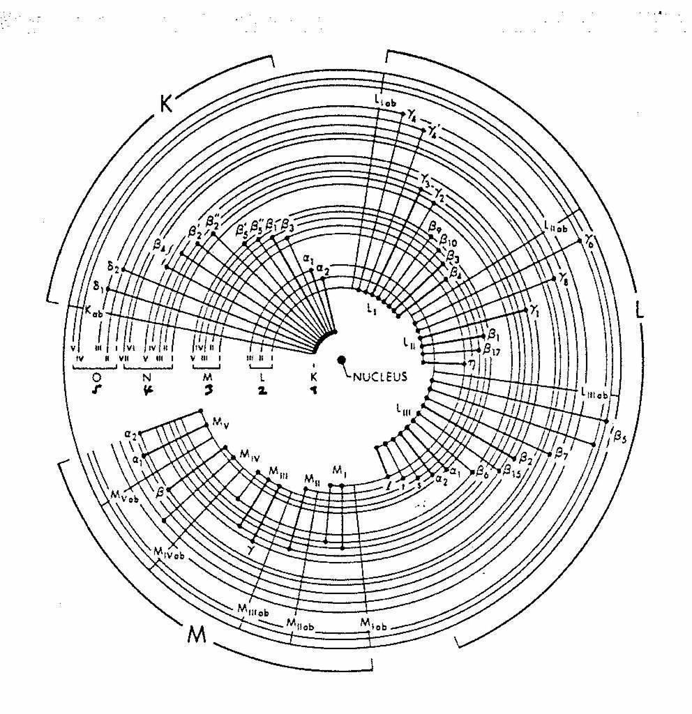
X-rays. X-ray Radiography - absorption is a function of Z and density. X-ray crystallography. X-ray spectrometry
|
|
|
- Tyrone Fowler
- 6 years ago
- Views:
Transcription
 NP in Physics 1901")

1 X-rays Wilhelm K. Roentgen ( ) NP in Physics 1901 X-ray Radiography - absorption is a function of Z and density X-ray crystallography X-ray spectrometry


2 X-rays Cu K α E = 8.05 kev λ = Å
3 Interaction of Electrons with Matter Emission of electromagnetic radiation: Characteristic radiation, discrete energies Bremsstrahlung, continuous energy distribution Luminiscence (UV or visible region) Electron emission: Backscattered electrons (BSE) Auger electrons Secondary electron emission (SE) Effects in the Target: Electron Absorption (ABS) Heat
4 X-ray Tubes
5 X-ray Tubes Tungsten wire at C (about 35 ma heating current) High Voltage kv High max. power kw Typical operating values Cu: 40 kv, 35 ma Mo: 45 kv, 35 ma
6 Spectrum of the X-ray tube Characteristic radiation Bremstrahlung (white radiation) λ min E max = E 0 = e V 0 E = (h c)/λ λ min (A) = hc = ev V ( kv ) 0
7 Characteristic X-ray radiation Primary (incident) electron X-ray (fluorescence) photon K α
8 Selection Rules n = 1, 2, 3. (principal quantum number), corresponds to K, L, M... shells l = 0, 1,..., n-1 (orbital quantum number) j = l±s ; s = 1/2 (spin-orbit coupling) m j = j, j-1, j-2,..., -j Transition only, when Δn 1, Δl = 1, Δj = 0 or 1
9 Selection Rules M = 2J + 1
10 Allowed Transitions
11 Mosley s Law (for multiple electron atoms) 1/λ = c (Z-σ) 2 (1/n 12-1/n 22 ) ΰ(K α ) = ¾R (Z 1) Z = atomic number σ = shielding constant n = quantum number Decreasing wavelength λ with increasing Z
12 Characteristic Wavelengths Element K α2 K α1 K β K abs. edge Cu Mo Ag W
13 X-ray Absorption At the absorption edge, the incident X-ray quantum is energetic enough to knock an electron out of the orbital μ = absorption coefficient Absorption edge
14 Monochromatisation of X-rays Filters (Ni filter for Cu K α ) Crystal Monochromators

15 Sample Holders Capillary Transmission Reflection
16 Detection of X-rays Detectors convert energies of individual photons to electric current convert current into voltage pulses that are counted Film (in the linear range, Guinier, Debye-Scherrer, precession cameras) Gas Proportional Counter Si(Li) solid state detector (powder diffractometers) Scintillation counter (photocathode, dynodes, 4-circle diffractometer, Stoe powder diffractometer) Position Sensitive Detectors (1D or 2D, Stoe powder diffractometer) Image Plate Detectors (2D detection, Stoe IPDS) CCD Detectors (Bruker SMART system)

17 Image plate detectors Metal plate, 18 cm diameter, coated with Eu 2+ doped BaFBr X-rays ionize Eu 2+ to Eu 3+ and the electrons are trapped in color centers Read out process with red laser leads to emission of blue light, when electrons return to ground state The blue light is amplified by a photomultiplier and recorded as a pixel image
18 Detector properties quantum-counting efficiency linearity energy proportionality resolution
19 Resolution
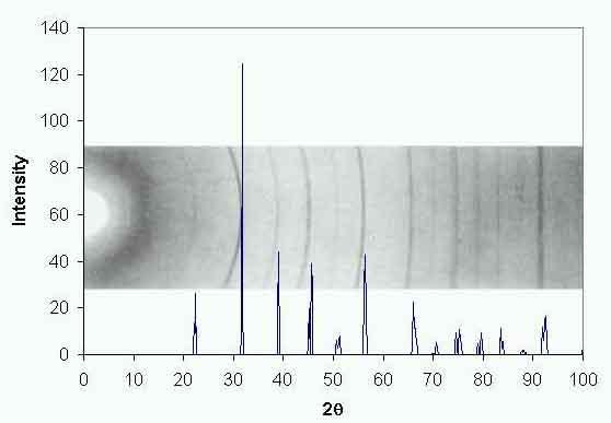
20 X-ray Powder Diffraction William Bragg (1912) n.λ = 2 d sin Θ
21
22 Single crystals polycrystalline
23 Different Geometries of Powder Diffractometers Debye-Scherrer Bragg-Brentano Guinier
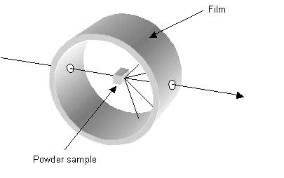
24 Debye-Scherrer
25 Debye-Scherrer
26 Bragg-Brentano
27 Information Extracted from Diffraction Experiments Determination of known phases Crystallinity Determination of lattice constants Structure solution
28 Crystalline and Amorphous Phases

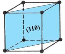
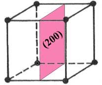
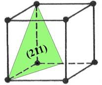
29 X-ray powder diffraction pattern of Fe (110) (200) (211)
30 Quantity More complicated, volume fraction Quality Line position is given by interplanar distance d and wavelenght λ d = λ / 2 sin Θ
![Anode Wavelength [nm] Beta Kα 1 [100] Kα 2 [50] Kβ 1 filter Cr 0.228970 0.229361 0.208487 V Fe 0.193604 0.193998 0.](/docs-images/77/74875048/images/31-1.jpg "175661 Mn Co 0.178897 0.179285 0.162079 Fe Cu 0.154056 0.154439 0.139222 Ni Mo 0.070930 0.071359 0.")

31 Anode Wavelength [nm] Beta Kα 1 [100] Kα 2 [50] Kβ 1 filter Cr V Fe Mn Co Fe Cu Ni Mo Zr d = λ / 2 sin Θ... longer λ... better multiplet separation... shorter λ... more lines Selecting radiation Bcc crystal, Cu radiation a = 1.5 nm --> 2Θ = 11.8 a = 1.2 nm --> 2Θ = 14.8 a = 0.9 nm --> 2Θ = 19.7 a = 0.6 nm --> 2Θ = 29.8 a = 0.3 nm --> 2Θ = 61.8
32 LINE HEIGHT - integral intensity - quantitative analysis -texture LINIE POSITION - qualitative (phase) analysis - lattice macrodistortions LINIE WIDTH - size of diffracting domains - lattice microdistortions GAUSS I(x) = A exp (-x 2 /a 2 ) LORENTZ I(x) = A exp [1+(x 2 /a 2 )] -1 Mod. LORENTZ I(x) = A exp [1+(x 2 /a 2 )] -2 PEARSON VII I(x) = A exp [1+(x 2 /a 2 )] -n Pseudo VOIGT I(x) = A [cl(x) + (1-c)G(x)]
 (220)")
33 (111)??? (200) (220) (311) (222) (400)
")
34 (111) (222)
35 Phase analysis ZrO 2 + Y 2 O 3
36 Results of phase analysis
37 Databases ICSD (Karlsruhe, inorganics, single crystal data) CSD (Cambridge, organics, organometallics, sc data) NRCC CRYSTMET (metals) PDB (proteins, Brookhaven) NIST (NBS) JCPDS = ICDD (PDF files, patterns)
38 Which of these is not involved in the diffraction of X-rays through a crystal? a b c d Electron transitions Crystallographic planes Nuclear interactions Constructive interference
39 What is the largest wavelength of radiation that will be diffracted by a lattice plane of the interplanar spacing d? a 0.5d b c d d 2d No limit
Chemistry Instrumental Analysis Lecture 19 Chapter 12. Chem 4631
 Chemistry 4631 Instrumental Analysis Lecture 19 Chapter 12 There are three major techniques used for elemental analysis: Optical spectrometry Mass spectrometry X-ray spectrometry X-ray Techniques include:
Diffractometer. Geometry Optics Detectors
 Diffractometer Geometry Optics Detectors Diffractometers Debye Scherrer Camera V.K. Pecharsky and P.Y. Zavalij Fundamentals of Powder Diffraction and Structural Characterization of Materials. Diffractometers
Setting The motor that rotates the sample about an axis normal to the diffraction plane is called (or ).
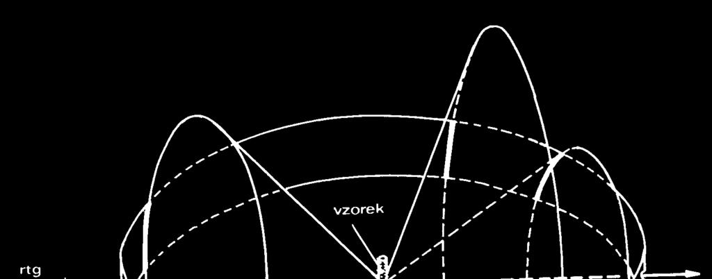
.") X-Ray Diffraction X-ray diffraction geometry A simple X-ray diffraction (XRD) experiment might be set up as shown below. We need a parallel X-ray source, which is usually an X-ray tube in a fixed position
X-Ray Diffraction X-ray diffraction geometry A simple X-ray diffraction (XRD) experiment might be set up as shown below. We need a parallel X-ray source, which is usually an X-ray tube in a fixed position
Röntgenpraktikum. M. Oehzelt. (based on the diploma thesis of T. Haber [1])
![Röntgenpraktikum. M. Oehzelt. (based on the diploma thesis of T. Haber [1])](/thumbs/91/106501237.jpg "Röntgenpraktikum. M. Oehzelt. (based on the diploma thesis of T. Haber [1])") Röntgenpraktikum M. Oehzelt (based on the diploma thesis of T. Haber [1]) October 21, 2004 Contents 1 Fundamentals 2 1.1 X-Ray Radiation......................... 2 1.1.1 Bremsstrahlung......................
Röntgenpraktikum M. Oehzelt (based on the diploma thesis of T. Haber [1]) October 21, 2004 Contents 1 Fundamentals 2 1.1 X-Ray Radiation......................... 2 1.1.1 Bremsstrahlung......................
X-ray Absorption and Emission Prepared By Jose Hodak for BSAC program 2008
 X-ray Absorption and Emission Prepared By Jose Hodak for BSAC program 2008 1- A bit of History: Wilhelm Conrad Röntgen discovered 1895 the X-rays. 1901 he was honored by the Noble prize for physics. In
X-Ray Emission and Absorption
 X-Ray Emission and Absorption Author: Mike Nill Alex Bryant February 6, 20 Abstract X-rays were produced by two bench-top diffractometers using a copper target. Various nickel filters were placed in front
Diffraction: spreading of waves around obstacles (EM waves, matter, or sound) Interference: the interaction of waves
 Interference: the interaction of waves") Diffraction & Interference Diffraction: spreading of waves around obstacles (EM waves, matter, or sound) Interference: the interaction of waves Diffraction in Nature What is Interference? The resultant
Diffraction & Interference Diffraction: spreading of waves around obstacles (EM waves, matter, or sound) Interference: the interaction of waves Diffraction in Nature What is Interference? The resultant
Chemistry 311: Instrumentation Analysis Topic 2: Atomic Spectroscopy. Chemistry 311: Instrumentation Analysis Topic 2: Atomic Spectroscopy
 Topic 2b: X-ray Fluorescence Spectrometry Text: Chapter 12 Rouessac (1 week) 4.0 X-ray Fluorescence Download, read and understand EPA method 6010C ICP-OES Winter 2009 Page 1 Atomic X-ray Spectrometry Fundamental
Atomic Physics. Chapter 6 X ray. Jinniu Hu 24/12/ /20/13
 Atomic Physics Chapter 6 X ray 11/20/13 24/12/2018 Jinniu Hu 1!1 6.1 The discovery of X ray X-rays were discovered in 1895 by the German physicist Wilhelm Roentgen. He found that a beam of high-speed electrons
LAB 01 X-RAY EMISSION & ABSORPTION
 LAB 0 X-RAY EMISSION & ABSORPTION REPORT BY: TEAM MEMBER NAME: Ashley Tsai LAB SECTION No. 05 GROUP 2 EXPERIMENT DATE: Feb., 204 SUBMISSION DATE: Feb. 8, 204 Page of 3 ABSTRACT The goal of this experiment
MS482 Materials Characterization ( 재료분석 ) Lecture Note 4: XRF
 Lecture Note 4: XRF") 2016 Fall Semester MS482 Materials Characterization ( 재료분석 ) Lecture Note 4: XRF Byungha Shin Dept. of MSE, KAIST 1 Course Information Syllabus 1. Overview of various characterization techniques (1 lecture)
2016 Fall Semester MS482 Materials Characterization ( 재료분석 ) Lecture Note 4: XRF Byungha Shin Dept. of MSE, KAIST 1 Course Information Syllabus 1. Overview of various characterization techniques (1 lecture)
X-ray Spectroscopy. c David-Alexander Robinson & Pádraig Ó Conbhuí. 14th March 2011
 X-ray Spectroscopy David-Alexander Robinson; Pádraig Ó Conbhuí; 08332461 14th March 2011 Contents 1 Abstract 2 2 Introduction & Theory 2 2.1 The X-ray Spectrum............................ 2 2.2 X-Ray Absorption
X-ray Absorption Spectroscopy
 X-ray Absorption Spectroscopy Nikki Truss November 26, 2012 Abstract In these experiments, some aspects of x-ray absorption spectroscopy were investigated. The x-ray spectrum of molybdenum was recorded
X Rays & Crystals. Characterizing Mineral Chemistry & Structure. J.D. Price
 X Rays & Crystals Characterizing Mineral Chemistry & Structure J.D. Price Light - electromagnetic spectrum Wave behavior vs. particle behavior If atoms are on the 10-10 m scale, we need to use sufficiently
MT Electron microscopy Scanning electron microscopy and electron probe microanalysis
 MT-0.6026 Electron microscopy Scanning electron microscopy and electron probe microanalysis Eero Haimi Research Manager Outline 1. Introduction Basics of scanning electron microscopy (SEM) and electron
X-ray diffraction geometry
 X-ray diffraction geometry Setting controls sample orientation in the diffraction plane. most important for single-crystal diffraction For any poly- (or nano-) crystalline specimen, we usually set: 1 X-ray
Data Acquisition. What choices need to be made?
 1 Specimen type and preparation Radiation source Wavelength Instrument geometry Detector type Instrument setup Scan parameters 2 Specimen type and preparation Slide mount Front loading cavity Back loading
CHEM*3440. X-Ray Energies. Bremsstrahlung Radiation. X-ray Line Spectra. Chemical Instrumentation. X-Ray Spectroscopy. Topic 13
 X-Ray Energies very short wavelength radiation 0.1Å to 10 nm (100 Å) CHEM*3440 Chemical Instrumentation Topic 13 X-Ray Spectroscopy Visible - Ultraviolet (UV) - Vacuum UV (VUV) - Extreme UV (XUV) - Soft
An Introduction to Diffraction and Scattering. School of Chemistry The University of Sydney
 An Introduction to Diffraction and Scattering Brendan J. Kennedy School of Chemistry The University of Sydney 1) Strong forces 2) Weak forces Types of Forces 3) Electromagnetic forces 4) Gravity Types
Analytical Methods for Materials
 Analytical Methods for Materials Lesson 6 Production & Properties of X-rays Suggested Reading Chapter 1 in Waseda et al. Section 2.1 in Leng Other Reference B.D. Cullity and S.R. Stock, Elements of X-ray
Signal to noise Source of noise Signal to noise enhancement
 Chap. 5 (Signals and Noise), Chap. 6 (Spectroscopy introduction) Signal to noise Source of noise Signal to noise enhancement Signal has the information of the analyte Noise is the extraneous information
EDS User School. Principles of Electron Beam Microanalysis
 EDS User School Principles of Electron Beam Microanalysis Outline 1.) Beam-specimen interactions 2.) EDS spectra: Origin of Bremsstrahlung and characteristic peaks 3.) Moseley s law 4.) Characteristic
Electron probe microanalysis - Electron microprobe analysis EPMA (EMPA) What s EPMA all about? What can you learn?
 What s EPMA all about? What can you learn?") Electron probe microanalysis - Electron microprobe analysis EPMA (EMPA) What s EPMA all about? What can you learn? EPMA - what is it? Precise and accurate quantitative chemical analyses of micron-size
Electron probe microanalysis - Electron microprobe analysis EPMA (EMPA) What s EPMA all about? What can you learn? EPMA - what is it? Precise and accurate quantitative chemical analyses of micron-size
Chapter Six: X-Rays. 6.1 Discovery of X-rays
 Chapter Six: X-Rays 6.1 Discovery of X-rays In late 1895, a German physicist, W. C. Roentgen was working with a cathode ray tube in his laboratory. He was working with tubes similar to our fluorescent
X-RAY SPECTRA. Theory:
 12 Oct 18 X-ray.1 X-RAY SPECTRA In this experiment, a number of measurements involving x-rays will be made. The spectrum of x-rays emitted from a molybdenum target will be measured, and the experimental
Lecture 16 Light transmission and optical detectors
 Lecture 6 Light transmission and optical detectors Charged particle traversing through a material can generate signal in form of light via electromagnetic interactions with orbital electrons of the atoms
XRF books: Analytical Chemistry, Kellner/Mermet/Otto/etc. 3 rd year XRF Spectroscopy Dr. Alan Ryder (R222, Physical Chemistry) 2 lectures:
 2 lectures:") 1 3 rd year XRF Spectroscopy Dr. Alan Ryder (R222, Physical Chemistry) 2 lectures: XRF spectroscopy 1 exam question. Notes on: www.nuigalway.ie/nanoscale/3rdspectroscopy.html XRF books: Analytical Chemistry,
1 3 rd year XRF Spectroscopy Dr. Alan Ryder (R222, Physical Chemistry) 2 lectures: XRF spectroscopy 1 exam question. Notes on: www.nuigalway.ie/nanoscale/3rdspectroscopy.html XRF books: Analytical Chemistry,
Swanning about in Reciprocal Space. Kenneth, what is the wavevector?
 Swanning about in Reciprocal Space or, Kenneth, what is the wavevector? Stanford Synchrotron Radiation Laboratory Principles The relationship between the reciprocal lattice vector and the wave vector is
Laboratory Manual 1.0.6
 Laboratory Manual 1.0.6 Background What is X-ray Diffraction? X-rays scatter off of electrons, in a process of absorption and re-admission. Diffraction is the accumulative result of the x-ray scattering
Characteristic X-rays of molybdenum
 Characteristic X-rays of molybdenum TEP Related Topics X-ray tubes, bremsstrahlung, characteristic X-radiation, energy levels, crystal structures, lattice constant, absorption of X-rays, absorption edges,
Rajesh Prasad Department of Applied Mechanics Indian Institute of Technology New Delhi
 TEQIP WORKSHOP ON HIGH RESOLUTION X-RAY AND ELECTRON DIFFRACTION, FEB 01, 2016, IIT-K. Introduction to x-ray diffraction Peak Positions and Intensities Rajesh Prasad Department of Applied Mechanics Indian
de Broglie Waves h p de Broglie argued Light exhibits both wave and particle properties
 de Broglie argued de Broglie Waves Light exhibits both wave and particle properties Wave interference, diffraction Particle photoelectric effect, Compton effect Then matter (particles) should exhibit both
Photon Instrumentation. First Mexican Particle Accelerator School Guanajuato Oct 6, 2011
 Photon Instrumentation First Mexican Particle Accelerator School Guanajuato Oct 6, 2011 Outline The Electromagnetic Spectrum Photon Detection Interaction of Photons with Matter Photoelectric Effect Compton
print first name print last name print student id grade
 print first name print last name print student id grade Experiment 2 X-ray fluorescence X-ray fluorescence (XRF) and X-ray diffraction (XRD) may be used to determine the constituent elements and the crystalline
Lesson 2 Diffractometers & Phase Identification
 Lesson 2 Diffractometers & Phase Identification Nicola Döbelin RMS Foundation, Bettlach, Switzerland February 11 14, 2013, Riga, Latvia Repetition: Generation of X-rays Kα 1 Target (Cu, Mo, Fe, Co,...)
MT Electron microscopy Scanning electron microscopy and electron probe microanalysis
 MT-0.6026 Electron microscopy Scanning electron microscopy and electron probe microanalysis Eero Haimi Research Manager Outline 1. Introduction Basics of scanning electron microscopy (SEM) and electron
Generation of X-Rays in the SEM specimen
 Generation of X-Rays in the SEM specimen The electron beam generates X-ray photons in the beam-specimen interaction volume beneath the specimen surface. Some X-ray photons emerging from the specimen have
An Introduction to Surface Physics for Engineers and Scientists Jorge A. López Gallardo and Miguel Castro Colín
 An Introduction to Surface Physics for Engineers and Scientists Jorge A. López Gallardo and Miguel Castro Colín Chapter Two: Basic Processes This chapter will review several physical processes that involve
Lecture 23 X-Ray & UV Techniques
 Lecture 23 X-Ray & UV Techniques Schroder: Chapter 11.3 1/50 Announcements Homework 6/6: Will be online on later today. Due Wednesday June 6th at 10:00am. I will return it at the final exam (14 th June).
FUNDAMENTAL PARAMETER METHOD FOR THE LOW ENERGY REGION INCLUDING CASCADE EFFECT AND PHOTOELECTRON EXCITATION
 Copyright (c)jcpds-international Centre for Diffraction Data 2002, Advances in X-ray Analysis, Volume 45. 511 FUNDAMENTAL PARAMETER METHOD FOR THE LOW ENERGY REGION INCLUDING CASCADE EFFECT AND PHOTOELECTRON
X-ray Spectroscopy. Danny Bennett and Maeve Madigan. October 12, 2015
 X-ray Spectroscopy Danny Bennett and Maeve Madigan October 12, 2015 Abstract Various X-ray spectra were obtained, and their properties were investigated. The characteristic peaks were identified for a
Transmission Electron Microscopy
 L. Reimer H. Kohl Transmission Electron Microscopy Physics of Image Formation Fifth Edition el Springer Contents 1 Introduction... 1 1.1 Transmission Electron Microscopy... 1 1.1.1 Conventional Transmission
Chapter 4 Scintillation Detectors
 Med Phys 4RA3, 4RB3/6R03 Radioisotopes and Radiation Methodology 4-1 4.1. Basic principle of the scintillator Chapter 4 Scintillation Detectors Scintillator Light sensor Ionizing radiation Light (visible,
Electron and electromagnetic radiation
 Electron and electromagnetic radiation Generation and interactions with matter Stimuli Interaction with sample Response Stimuli Waves and energy The energy is propotional to 1/λ and 1/λ 2 λ λ 1 Electromagnetic
Particles and Waves Particles Waves
 Particles and Waves Particles Discrete and occupy space Exist in only one location at a time Position and velocity can be determined with infinite accuracy Interact by collisions, scattering. Waves Extended,
2. X-ray Sources 2.1 Electron Impact X-ray Sources - Types of X-ray Source - Bremsstrahlung Emission - Characteristic Emission
 . X-ray Sources.1 Electron Impact X-ray Sources - Types of X-ray Source - Bremsstrahlung Emission - Characteristic Emission. Synchrotron Radiation Sources - Introduction - Characteristics of Bending Magnet
Electron Microprobe Analysis and Scanning Electron Microscopy
 Electron Microprobe Analysis and Scanning Electron Microscopy Electron microprobe analysis (EMPA) Analytical technique in which a beam of electrons is focused on a sample surface, producing X-rays from
Absorption of X-rays
 Absorption of X-rays TEP Related topics Bremsstrahlung, characteristic X-radiation, Bragg scattering, law of absorption, mass absorption coefficient, absorption edges, half-value thickness, photoelectric
THE IMPORTANCE OF THE SPECIMEN DISPLACEMENT CORRECTION IN RIETVELD PATTERN FITTING WITH SYMMETRIC REFLECTION-OPTICS DIFFRACTION DATA
 Copyright(c)JCPDS-International Centre for Diffraction Data 2001,Advances in X-ray Analysis,Vol.44 96 THE IMPORTANCE OF THE SPECIMEN DISPLACEMENT CORRECTION IN RIETVELD PATTERN FITTING WITH SYMMETRIC REFLECTION-OPTICS
V 11: Electron Diffraction
 Martin-Luther-University Halle-Wittenberg Institute of Physics Advanced Practical Lab Course V 11: Electron Diffraction An electron beam conditioned by an electron optical system is diffracted by a polycrystalline,
Bragg reflection :determining the lattice constants of monocrystals
 Bragg reflection :determining the lattice constants of monocrystals Objectives: 1-Investagating Bragg reflection at Nacl monocrystal -determinig the lattice constant a 0 of NaCl. Theory: Bragg's law of
Nuclear Physics and Astrophysics
 Nuclear Physics and Astrophysics PHY-30 Dr. E. Rizvi Lecture 4 - Detectors Binding Energy Nuclear mass MN less than sum of nucleon masses Shows nucleus is a bound (lower energy) state for this configuration
X-ray spectroscopy: Experimental studies of Moseley s law (K-line x-ray fluorescence) and x-ray material s composition determination
 and x-ray material s composition determination") Uppsala University Department of Physics and Astronomy Laboratory exercise X-ray spectroscopy: Experimental studies of Moseley s law (K-line x-ray fluorescence) and x-ray material s composition determination
Uppsala University Department of Physics and Astronomy Laboratory exercise X-ray spectroscopy: Experimental studies of Moseley s law (K-line x-ray fluorescence) and x-ray material s composition determination
AP5301/ Name the major parts of an optical microscope and state their functions.
 Review Problems on Optical Microscopy AP5301/8301-2015 1. Name the major parts of an optical microscope and state their functions. 2. Compare the focal lengths of two glass converging lenses, one with
CHARACTERIZATION of NANOMATERIALS KHP
 CHARACTERIZATION of NANOMATERIALS Overview of the most common nanocharacterization techniques MAIN CHARACTERIZATION TECHNIQUES: 1.Transmission Electron Microscope (TEM) 2. Scanning Electron Microscope
Chemical Engineering 412
 Chemical Engineering 412 Introductory Nuclear Engineering Lecture 26 Radiation Detection & Measurement II Spiritual Thought 2 I would not hold the position in the Church I hold today had I not followed
Electron Microscopy I
 Characterization of Catalysts and Surfaces Characterization Techniques in Heterogeneous Catalysis Electron Microscopy I Introduction Properties of electrons Electron-matter interactions and their applications
FXA UNIT G485 Module X-Rays. Candidates should be able to : I = I 0 e -μx
 1 Candidates should be able to : HISTORY Describe the nature of X-rays. Describe in simple terms how X-rays are produced. X-rays were discovered by Wilhelm Röntgen in 1865, when he found that a fluorescent
Surface Sensitivity & Surface Specificity
 Surface Sensitivity & Surface Specificity The problems of sensitivity and detection limits are common to all forms of spectroscopy. In its simplest form, the question of sensitivity boils down to whether
Understanding X-rays: The electromagnetic spectrum
 Understanding X-rays: The electromagnetic spectrum 1 ULa 13.61 kev 0.09 nm BeKa 0.11 kev 11.27 nm E = hn = h c l where, E : energy, h : Planck's constant, n : frequency c : speed of light in vacuum, l
IMPROVING THE ACCURACY OF RIETVELD-DERIVED LATTICE PARAMETERS BY AN ORDER OF MAGNITUDE
 Copyright (c)jcpds-international Centre for Diffraction Data 2002, Advances in X-ray Analysis, Volume 45. 158 IMPROVING THE ACCURACY OF RIETVELD-DERIVED LATTICE PARAMETERS BY AN ORDER OF MAGNITUDE B. H.
Radioactivity and Ionizing Radiation
 Radioactivity and Ionizing Radiation QuarkNet summer workshop June 24-28, 2013 1 Recent History Most natural phenomena can be explained by a small number of simple rules. You can determine what these rules
Chapter 2. X-ray X. Diffraction and Reciprocal Lattice. Scattering from Lattices
 Chapter. X-ray X Diffraction and Reciprocal Lattice Diffraction of waves by crystals Reciprocal Lattice Diffraction of X-rays Powder diffraction Single crystal X-ray diffraction Scattering from Lattices
CHEM-E5225 :Electron Microscopy X-Ray Spectrometry
 CHEM-E5225 :Electron Microscopy X-Ray Spectrometry 2016.11 Yanling Ge Outline X-ray Spectrometry X-ray Spectra and Images Qualitative and Quantitative X-ray Analysis and Imaging Discussion of homework
hν' Φ e - Gamma spectroscopy - Prelab questions 1. What characteristics distinguish x-rays from gamma rays? Is either more intrinsically dangerous?
 Gamma spectroscopy - Prelab questions 1. What characteristics distinguish x-rays from gamma rays? Is either more intrinsically dangerous? 2. Briefly discuss dead time in a detector. What factors are important
General theory of diffraction
 General theory of diffraction X-rays scatter off the charge density (r), neutrons scatter off the spin density. Coherent scattering (diffraction) creates the Fourier transform of (r) from real to reciprocal
Understanding X-rays: The electromagnetic spectrum
 Understanding X-rays: The electromagnetic spectrum 1 ULa 13.61 kev 0.09 nm BeKa 0.11 kev 11.27 nm E = hn = h c l where, E : energy, h : Planck's constant, n : frequency c : speed of light in vacuum, l
Fundamentals of Nanoscale Film Analysis
 Fundamentals of Nanoscale Film Analysis Terry L. Alford Arizona State University Tempe, AZ, USA Leonard C. Feldman Vanderbilt University Nashville, TN, USA James W. Mayer Arizona State University Tempe,
Chap. 3. Elementary Quantum Physics
 Chap. 3. Elementary Quantum Physics 3.1 Photons - Light: e.m "waves" - interference, diffraction, refraction, reflection with y E y Velocity = c Direction of Propagation z B z Fig. 3.1: The classical view
EEE4106Z Radiation Interactions & Detection
 EEE4106Z Radiation Interactions & Detection 2. Radiation Detection Dr. Steve Peterson 5.14 RW James Department of Physics University of Cape Town steve.peterson@uct.ac.za May 06, 2015 EEE4106Z :: Radiation
PHYS 3446 Lecture #12
 PHYS 3446 Lecture #12 Wednesday, Oct. 18, 2006 Dr. 1. Particle Detection Ionization Detectors MWPC Scintillation Counters Time of Flight 1 Announcements Next LPCC Workshop Preparation work Each group to
6. Analytical Electron Microscopy
 Physical Principles of Electron Microscopy 6. Analytical Electron Microscopy Ray Egerton University of Alberta and National Institute of Nanotechnology Edmonton, Canada www.tem-eels.ca regerton@ualberta.ca
Neutrino Helicity Measurement
 PHYS 851 Introductory Nuclear Physics Instructor: Chary Rangacharyulu University of Saskatchewan Neutrino Helicity Measurement Stefan A. Gärtner stefan.gaertner@gmx.de December 9 th, 2005 2 1 Introduction
GLOSSARY OF BASIC RADIATION PROTECTION TERMINOLOGY
 GLOSSARY OF BASIC RADIATION PROTECTION TERMINOLOGY ABSORBED DOSE: The amount of energy absorbed, as a result of radiation passing through a material, per unit mass of material. Measured in rads (1 rad
Fundamentals of X-ray diffraction
 Fundamentals of X-ray diffraction Elena Willinger Lecture series: Modern Methods in Heterogeneous Catalysis Research Outline History of X-ray Sources of X-ray radiation Physics of X-ray scattering Fundamentals
Lab Manual: Determination of Planck s constant with x-rays
 Lab Manual: Determination of Planck s constant with x-rays 1. Purpose: To obtain a better understanding on the production of X-rays, the bremsstrahlung radiation and the characteristic radiation of a Molybdenum
SCINTILLATION DETECTORS AND PM TUBES
 SCINTILLATION DETECTORS AND PM TUBES General Characteristics Introduction Luminescence Light emission without heat generation Scintillation Luminescence by radiation Scintillation detector Radiation detector
Chemical Analysis in TEM: XEDS, EELS and EFTEM. HRTEM PhD course Lecture 5
 Chemical Analysis in TEM: XEDS, EELS and EFTEM HRTEM PhD course Lecture 5 1 Part IV Subject Chapter Prio x-ray spectrometry 32 1 Spectra and mapping 33 2 Qualitative XEDS 34 1 Quantitative XEDS 35.1-35.4
Detection of X-Rays. Solid state detectors Proportional counters Microcalorimeters Detector characteristics
 Detection of X-Rays Solid state detectors Proportional counters Microcalorimeters Detector characteristics Solid State X-ray Detectors X-ray interacts in material to produce photoelectrons which are collected
1 of 5 14/10/ :21
 X-ray absorption s, characteristic X-ray lines... 4.2.1 Home About Table of Contents Advanced Search Copyright Feedback Privacy You are here: Chapter: 4 Atomic and nuclear physics Section: 4.2 Absorption
Scintillation Detector
 Scintillation Detector Introduction The detection of ionizing radiation by the scintillation light produced in certain materials is one of the oldest techniques on record. In Geiger and Marsden s famous
Interaction X-rays - Matter
 Interaction X-rays - Matter Pair production hν > M ev Photoelectric absorption hν MATTER hν Transmission X-rays hν' < hν Scattering hν Decay processes hν f Compton Thomson Fluorescence Auger electrons
3.012 Fund of Mat Sci: Structure Lecture 18
 3.012 Fund of Mat Sci: Structure Lecture 18 X-RAYS AT WORK An X-ray diffraction image for the protein myoglobin. Source: Wikipedia. Model of helical domains in myoglobin. Image courtesy of Magnus Manske
4. Inelastic Scattering
 1 4. Inelastic Scattering Some inelastic scattering processes A vast range of inelastic scattering processes can occur during illumination of a specimen with a highenergy electron beam. In principle, many
Radionuclide Imaging MII Detection of Nuclear Emission
 Radionuclide Imaging MII 3073 Detection of Nuclear Emission Nuclear radiation detectors Detectors that are commonly used in nuclear medicine: 1. Gas-filled detectors 2. Scintillation detectors 3. Semiconductor
ECE Semiconductor Device and Material Characterization
 ECE 4813 Semiconductor Device and Material Characterization Dr. Alan Doolittle School of Electrical and Computer Engineering Georgia Institute of Technology As with all of these lecture slides, I am indebted
X-RAY SCATTERING AND MOSELEY S LAW. OBJECTIVE: To investigate Moseley s law using X-ray absorption and to observe X- ray scattering.
 X-RAY SCATTERING AND MOSELEY S LAW OBJECTIVE: To investigate Moseley s law using X-ray absorption and to observe X- ray scattering. READING: Krane, Section 8.5. BACKGROUND: In 1913, Henry Moseley measured
X-ray Diffraction. Diffraction. X-ray Generation. X-ray Generation. X-ray Generation. X-ray Spectrum from Tube
 X-ray Diffraction Mineral identification Mode analysis Structure Studies X-ray Generation X-ray tube (sealed) Pure metal target (Cu) Electrons remover inner-shell electrons from target. Other electrons
Neutron facilities and generation. Rob McQueeney, Ames Laboratory and Iowa State University
 Neutron facilities and generation Rob McQueeney, Ames Laboratory and Iowa State University September 12, 2018 19-Sep-18 Physics 502 2 Neutrons compared to other probes of matter Bulk probe Interacts with
Synchrotron Methods in Nanomaterials Research
 Synchrotron Methods in Nanomaterials Research Marcel MiGLiERiNi Slovak University of Technology in Bratislava and Centre for Nanomaterials Research, Olomouc marcel.miglierini@stuba.sk www.nuc.elf.stuba.sk/bruno
Lecture 0. NC State University
 Chemistry 736 Lecture 0 Overview NC State University Overview of Spectroscopy Electronic states and energies Transitions between states Absorption and emission Electronic spectroscopy Instrumentation Concepts
X-ray diffraction is a non-invasive method for determining many types of
 Chapter X-ray Diffraction.1 Introduction X-ray diffraction is a non-invasive method for determining many types of structural features in both crystalline and amorphous materials. In the case of single
EE 5344 Introduction to MEMS CHAPTER 5 Radiation Sensors
 EE 5344 Introduction to MEMS CHAPTER 5 Radiation Sensors 5. Radiation Microsensors Radiation µ-sensors convert incident radiant signals into standard electrical out put signals. Radiant Signals Classification
Supporting Information
 Supporting Information Three-dimensional frameworks of cubic (NH 4 ) 5 Ga 4 SbS 10, (NH 4 ) 4 Ga 4 SbS 9 (OH) H 2 O, and (NH 4 ) 3 Ga 4 SbS 9 (OH 2 ) 2H 2 O. Joshua L. Mertz, Nan Ding, and Mercouri G.
Radiation Detectors. How do we detect ionizing radiation? What are these effects? Types of Ionizing Radiation Detectors
 Radiation Detectors 1 How do we detect ionizing radiation? Indirectly, by its effects as it traverses matter? What are these effects? Ionization and excitation of the atoms and molecules Heat 2 Types of
Rad T 290 Worksheet 2
 Class: Date: Rad T 290 Worksheet 2 1. Projectile electrons travel from a. anode to cathode. c. target to patient. b. cathode to anode. d. inner shell to outer shell. 2. At the target, the projectile electrons
Making the Invisible Visible: Probing Antiferromagnetic Order in Novel Materials
 Making the Invisible Visible: Probing Antiferromagnetic Order in Novel Materials Elke Arenholz Lawrence Berkeley National Laboratory Antiferromagnetic contrast in X-ray absorption Ni in NiO Neel Temperature
Scattering Techniques and Geometries How to choose a beamline. Christopher J. Tassone
 Scattering Techniques and Geometries How to choose a beamline Christopher J. Tassone Why Care About Geometries? How do you decide which beamline you want to use? Questions you should be asking Do I want
Calibration of the IXPE Instrument
 Calibration of the IXPE Instrument Fabio Muleri (INAF-IAPS) On behalf of the IXPE Italian Team 13th IACHEC Meeting 2018 Avigliano Umbro (Italy), 9-12 April 2018 IXPE MISSION IXPE will (re-)open the polarimetric
Photoelectron spectroscopy Instrumentation. Nanomaterials characterization 2
 Photoelectron spectroscopy Instrumentation Nanomaterials characterization 2 RNDr. Věra V Vodičkov ková,, PhD. Photoelectron Spectroscopy general scheme Impact of X-ray emitted from source to the sample