Protein Structure Determination. Why Bother With Structure? Protein Sequences Far Outnumber Structures
|
|
|
- Albert Garrett
- 6 years ago
- Views:
Transcription


1 Protein Structure Determination How are these structures determined? Why Bother With Structure? The amino acid sequence of a protein contains interesting information. A protein sequence can be compared to other protein sequences to establish its evolutionary relationship to other proteins and protein families. However, for the purposes of understanding protein function, the 3D structure of the protein is far more useful than the sequence. Protein Sequences Far Outnumber Structures Only a small number of protein structures have been experimentally determined. PDB ~64,623 protein structures Genebank ~61,132,599 sequences Of the 64,623 structures, only 15,702 are dissimilar in sequence (<30% ID). 1


2 Growth of GenBank Now over 100M sequences and 100B base pairs from 223K species Exponential Increase Growth of Structural Data Currently 64,623 structures deposited Growth of Structural Data Currently 64,623 structures deposited Exponential? 2

3 Structural Proteomics Use experimentally determined structures to model the structures of similar proteins Threading Avoids Ab initio structure determination Homology Modeling Fold recognition Need representative protein structures for the total repertoire of protein folds Provide 3D portraits for all proteins in an organism Goal: Use structure to infer function. More sensitive than primary sequence comparisons Redundancy in PDB (20 April 10) Sequence identity 90% % % % Number of nonredundant chains Unique folds in PDB (SCOP) 3
4 Unique topologies in PDB (CATH) New Topologies and Folds Becoming Rare Structural Genomics Initiated in 1999 by NIH Phase I included 9 large centers for high throughput structure determination Phase I ran from ~ Goal The long-range goal of the Protein Structure Initiative (PSI) is to make the three-dimensional atomic-level structures of most proteins easily obtainable from knowledge of their corresponding DNA sequences. Structural Genomics Benefits Structural descriptions will help researchers illuminate structure-function relationships and thus formulate better hypotheses and design better experiments. The PSI collection of structures will serve as the starting point for structure-based drug development by permitting faster identification of lead compounds and their optimization. The design of better therapeutics will result from comparisons of the structures of proteins that are from pathogenic and host organisms and from normal and diseased human tissues. The PSI collection of structures will assist biomedical investigators in research studies of key biophysical and biochemical problems, such as protein folding, evolution, structure prediction, and the organization of protein families and folds. Technical developments, the availability of reagents and materials, and experimental outcome data in protein production and crystallization will directly benefit all structural biologists and provide valuable assistance to a broad range of biomedical researchers. 4

 In PSI-2, the multi-institutional consortium is rapidly determining the structures of large numbers of strategically selected proteins using x-ray")
5 Structural Genomics Centers in US PSI-1 Winners The Joint Center for Structural Genomics (JCSG) During PSI-2, the JCSG has contributed to the overall goal of maximizing structural coverage of protein families with no structural representation and has continued to developm and disseminate innovative new technologies for structural biology. The JCSG consortium theme is the central machinery of life proteins that are conserved in all kingdoms of life. The Midwest Center for Structural Genomics (MCSG) In PSI-2, the multi-institutional consortium is rapidly determining the structures of large numbers of strategically selected proteins using x-ray crystallography both to provide structural coverage of major protein superfamilies and to elucidate the entire protein folding space. The New York Structural Genomics Research Consortium (NYSGRC) During PSI-2, the consortium s individual project focuses on new targets, principally protein phosphatases and mul tidomain eukaryotic proteins. The Northeast Structural Genomics Consortium (NEGS) In PSI-2, the consortium is solving both prokaryotic and eukaryotic structural representatives from the major domain families constituting the eukaryotic proteome. Structural Genomics Centers in US PSI-1 Losers Center for Eukaryotic Structural Genomics (CESG) The CESG was founded as a collaborative effort to develop the technologies needed for economical high-throughput structure determination of biologically important eukaryotic proteins and to extend the knowledge of fold-function space. This project also aims to further the research of biologically important proteins in Arabidopsis. The protein structures are being determined via X-ray crystallography or NMR spectroscopy. The Berkeley Structural Genomics Center (BSGC)The BSGC is pursuing an integrated structural genomics program designed to obtain a near-complete structural complement of two minimal genomes, Mycoplasma genitalium and Mycoplasma pneumoniae, two related human and animal pathogens. Both NMR spectroscopy and X-ray crystallography are being used for structural determination. The Southeast Collaboratory for Structural Genomics (SECSG) The objective of the SECSG is to develop and test t experimental and computational ti strategies t for high h throughput h t structure determination of proteins by X-ray crystallography and NMR methods and to apply these strategies to scan the entire genome of an organism at a rapid pace. The eukaryotic organisms, Caenorhabditis elegans, Homo sapiens and an ancestrally-related prokaryotic microorganism having a small genome, Pyrococcus furiosus, have been selected as representative genomes. Structural Genomics of Pathogenic Protozoa Consortium (SGPP) The SGPP consortium aims to determine and analyze the structures of a large number of proteins from major global pathogenic protozoa including Leishmania major, Trypanosoma brucei, Trypanosoma cruzi and Plasmodium falciparum. These organisms are responsible for the diseases: leishmaniasis, sleeping sickness, Chagas' disease and malaria. X-ray crystallography is being used for structural determination. The TB Structural Genomics Consortium (TB) The goal of the TB consortium is to determine the structures of over 400 proteins from M. tuberculosis, and to analyze these structures in the context of functional information that currently exists and that is generated by the project. These structures will include about 40 novel folds and 200 new families of protein structures. The protein structures are being determined using X-ray crystallography. 5
6 Current PSI Centers Large-Scale Centers Joint Center for Structural Genomics Midwest Center for Structural Genomics New York SGX Research Center for Structural Genomics Northeast Structural Genomics Consortium Specialized Centers Accelerated Technologies Center for Gene to 3D Structure Center for Eukaryotic Structural Genomics Center for High-Throughput Structural Biology Center for Structures of Membrane Proteins Integrated Center for Structure and Function Innovation New York Consortium on Membrane Protein Structure Homology Modeling Centers Joint Center for Molecular Modeling New Methods for High-Resolution Comparative Modeling Resource Centers PSI-Materials Repository PSI Knowledgebase 60,000 plasmid clones 2008 Structural Genomics Progress Status Total Number of Targets (%) Relative to "Cloned" Targets (%) Relative to "Expressed" Targets (%) Relative to "Purified" Targets (%) Relative to "Crystallized" Targets Cloned Expressed Soluble Purified Crystallized Diffraction-quality Crystals Diffraction NMR Assigned HSQC Crystal Structure NMR Structure In PDB Work Stopped Test Target Other ~40% of structures are from SG in Europe and Asia 2010 Structural Genomics Progress Status Total Number of Targets (%) Relative to "Cloned" Targets (%) Relative to "Expressed" Targets (%) Relative to "Purified" Targets (%) Relative to "Crystallized" Targets Cloned Expressed Soluble Purified Crystallized Diffraction-quality Crystals Diffraction NMR Assigned HSQC Crystal Structure NMR Structure In PDB Work Stopped Test Target Other ~36% of structures are from SG in Europe and Asia 6

7 Project Attrition Unique Folds? Protein Structure Databases Where does protein structural information reside? PDB: Jon MMDB: FSSP: SCOP: Ingo CATH: 7


8 PDB Contents 20 April 2010 Exp.Method Proteins Nucleic Acids X-RAY NMR Protein/NA Complexes Other Total ELECTRON MICROSCOPY HYBRID Other Total X-ray Crystallography 8
 Visible light has a wavelength of ~ 500 nm (5000 Å) Electron beam: c ~ 0.")

9 Optical Microscope Image refocused Lens Scattered rays object Light Atomic Resolution We want to resolve inter-atomic distances (~1.5 Å, 0.15 nm) Visible light has a wavelength of ~ 500 nm (5000 Å) Electron beam: c ~ Å (if e - is moving at c) Electron velocity is less in electron microscopes Typical resolution is ~10 Å, but can be improved X-ray generators produce photons of = Å Use = Å X-ray Crystallography Molecules Refocusing is accomplished with a computer, a crystallographer and a lot of mathematics computer o o o o o o o o o o o o o 3D electron density map m a t h e m O a crystallographer t i c s X-ray detector Crystal size < 0.5 mm Must use X-rays to get atomic resolution (1.5 Å = C-C bond) X-rays 9

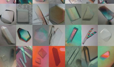
10 X-Ray Crystallography 1. Make crystals of your protein mm in size Proteins must be in an ordered, repeating pattern. 2. X-ray beam is aimed at crystal and data is collected. 3. Structure is determined from the diffraction data. X-Ray Crystallography 1. Make crystals of your protein mm in size Proteins must be in an ordered, repeating pattern. 2. X-ray beam is aimed at crystal and data is collected. 3. Structure is determined from the diffraction data. Protein Crystals Schmid, M. Trends in Microbiolgy, 10:s27-s31. 10

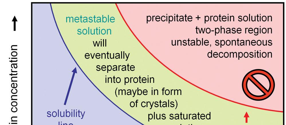
11 Protein Crystals Protein Crystals Protein Crystals 11

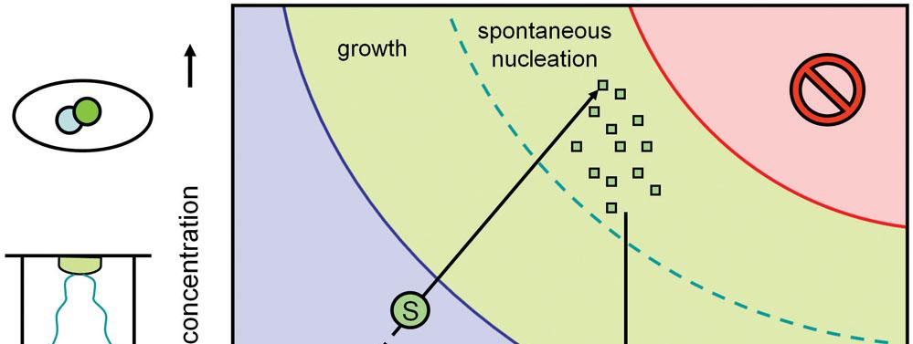
12 Protein Crystals Protein Crystals Protein Crystals 12


13 Protein Crystals Protein Crystals X-Ray Crystallography 1. Make crystals of your protein mm in size Proteins must be in an ordered, repeating pattern. 2. X-ray beam is aimed at crystal and data is collected. 3. Structure is determined from the diffraction data. 13


14 X-Ray Diffraction Experiment Generator Optics Goniometer Detector Optional: Cryo for protein samples X-ray Crystallography Equipment Cryocooling X-ray Generator Detector Mirrors Crystal X-ray Crystallography Equipment 14
15 X-Ray Crystallography 1. Make crystals of your protein mm in size Proteins must be in an ordered, repeating pattern. 2. X-ray beam is aimed at crystal and data is collected. 3. Structure is determined from the diffraction data. Protein Diffraction Image Diffraction Spots Why Spots? X-ray scattering from individual proteins is diffuse Spots arise from a phenomenon called diffraction that is based on the crystal lattice Location of reflections indicates how an object crystallized 230 possibilities Intensity of reflections contains information about the structure of the object in the crystal 15
 and not a diffuse pattern")

")

16 Bragg s Law Why do we get spots (reflections) and not a diffuse pattern of scattered x-rays? 2d hkl sin n d sin = x/d 2d sin = 2x Difference in path (2x) must equal integral number of wavelengths (n ) Constructive Interference d = resolution Condition for reflection Resolution 16
 has a phase and intensity")

 Phase Problem Electron")


17 Phase Problem Every diffraction spot (reflection) has a phase and intensity -The intensities are recorded by the detector -The phases are lost -Must have both to reconstruct the image (structure) Phase Problem Electron Density Phase Problem Electron Density Intensities Phases 17
 -Compare independent datasets collected from native and labeled protein -Heavy atom substructure provides initial phases Anomalous diffraction -Crystal must contain atoms with")
 Structure (model) Crystallography Pros/Cons Advantages -can be fast down to")
18 Solutions to the Phase Problem Molecular replacement -Use known structure of close homologue -Rotational and translational search for solution Heavy atom labeling -Label the protein with electron dense atoms (Hg) -Compare independent datasets collected from native and labeled protein -Heavy atom substructure provides initial phases Anomalous diffraction -Crystal must contain atoms with absorption edges between 0.5 and 2.5 Å -Compare independent datasets collected at pre-edge and post-edge x-ray energies Model Building Electron density (data) Structure (model) Crystallography Pros/Cons Advantages -can be fast down to a few months -large structures possible (ribosome) -very low resolution (down to 0.5 Å) -observables typically > refinement parameters Disadvantages -requires crystal formation -non-physiological conditions -crystal contacts can limit protein motion 18
19 Nuclear Magnetic Resonance Nuclear Magnetic Resonance Magnetically align unpaired proton spins (H 0 ) Probe with radio frequency (RF) Observe resonance RF RF Resonance H0 HSQC: Short ACP construct 19
20 NMR Overview Isotopic labeling ( 15 N, 13 C) Multiple experiments (pulse sequences) Positional refinement typically not possible. Dihedral angles used. As many as 20 structures produced NMR Experimental Observables Backbone conformation from chemical shifts (Chemical Shift Index- CSI) Distance constraints from NOEs Hydrogen bond constraints Backbone and side chain dihedral angle constraints from scalar couplings Orientation constraints from residual dipolar couplings NMR Pros/Cons Advantages -no crystal formation needed -more physiological conditions Disadvantages -results in a set of models that are compatible with data -size limitation to residues (extended recently) -must label protein with 15 N and 13 C -observables typically < refinement parameters 20

 COMPND MOL_ID: 1; COMPND 2 MOLECULE: CHOLESTEROL OXIDASE; COMPND 3")
21 Precision NMR vs. X-ray C N RMSD of the ensemble Mean coordinate error A PDB File Header contains information about protein and structure date of the entry, references, crystallographic data, contents and positions of secondary structure elements HEADER OXIDOREDUCTASE 03-OCT-02 1MXT TITLE ATOMIC RESOLUTION STRUCTURE OF CHOLESTEROL OXIDASE TITLE 2 (STREPTOMYCES SP. SA-COO) COMPND MOL_ID: 1; COMPND 2 MOLECULE: CHOLESTEROL OXIDASE; COMPND 3 CHAIN: A; COMPND 4 SYNONYM: CHOD; COMPND 5 EC: ; COMPND 6 ENGINEERED: YES; COMPND 7 OTHER_DETAILS: FAD COFACTOR NON-COVALENTLY BOUND TO THE COMPND 8 ENZYME A PDB File Header contains information about protein and structure date of the entry, references, crystallographic data, contents and positions of secondary structure elements SOURCE MOL_ID: 1; SOURCE 2 ORGANISM_SCIENTIFIC: STREPTOMYCES SP.; SOURCE 3 ORGANISM_COMMON: BACTERIA; SOURCE 4 GENE: CHOA; SOURCE 5 EXPRESSION_SYSTEM: ESCHERICHIA COLI; SOURCE 6 EXPRESSION_SYSTEM_COMMON: BACTERIA; SOURCE 7 EXPRESSION_SYSTEM_STRAIN: BL21(DE3)PLYSS; SOURCE 8 EXPRESSION_SYSTEM_VECTOR_TYPE: PLASMID; SOURCE 9 EXPRESSION_SYSTEM_PLASMID: PCO202 21


22 A PDB File Header contains information about protein and structure date of the entry, references, crystallographic data, contents and positions of secondary structure elements AUTHOR A.VRIELINK,P.I.LARIO REVDAT 1 25-FEB-03 1MXT 0 JRNL AUTH P.I.LARIO,N.SAMPSON,A.VRIELINK JRNL TITL SUB-ATOMIC RESOLUTION CRYSTAL STRUCTURE OF JRNL TITL 2 CHOLESTEROL OXIDASE: WHAT ATOMIC RESOLUTION JRNL TITL 3 CRYSTALLOGRAPHY REVEALS ABOUT ENZYME MECHANISM AND JRNL TITL 4 THE ROLE OF FAD COFACTOR IN REDOX ACTIVITY JRNL REF J.MOL.BIOL. V JRNL REFN ASTM JMOBAK UK ISSN A PDB File Header contains information about protein and structure date of the entry, references, crystallographic data, contents and positions of secondary structure elements REMARK 3 DATA USED IN REFINEMENT. REMARK 3 RESOLUTION RANGE HIGH (ANGSTROMS) : 0.95 REMARK 3 RESOLUTION RANGE LOW (ANGSTROMS) : REMARK 3 DATA CUTOFF (SIGMA(F)) : REMARK 3 COMPLETENESS FOR RANGE (%) : 94.1 REMARK 3 CROSS-VALIDATION METHOD : FREE R REMARK 3 FREE R VALUE TEST SET SELECTION : RANDOM REMARK 3 REMARK 3 FIT TO DATA USED IN REFINEMENT (NO CUTOFF). REMARK 3 R VALUE (WORKING + TEST SET, NO CUTOFF) : REMARK 3 R VALUE (WORKING SET, NO CUTOFF) : REMARK 3 FREE R VALUE (NO CUTOFF) : REMARK 3 FREE R VALUE TEST SET SIZE (%, NO CUTOFF) : REMARK 3 FREE R VALUE TEST SET COUNT (NO CUTOFF) : REMARK 3 TOTAL NUMBER OF REFLECTIONS (NO CUTOFF) : Resolution: Low > 3 Å Mid 2-3 Å High Å Very High < 1.5 Å R factor (residual): Low resolution ~ 27% Mid resolution ~ 22 % High resolution ~ 29 % Very High res ~ 15% Resolution 22
23 A PDB File Header contains information about protein and structure date of the entry, references, crystallographic data, contents and positions of secondary structure elements HELIX ALA A 289 THR A HELIX THR A 402 GLN A HELIX ASN A 406 GLY A HELIX ASP A 474 ILE A HELIX PRO A 486 VAL A SHEET 1 A 6 HIS A 248 GLN A SHEET 2 A 6 TYR A 261 LYS A O GLU A 266 N GLN A 249 SHEET 3 A 6 LEU A 274 LEU A O LEU A 275 N GLN A 267 SHEET 4 A 6 TYR A 10 ILE A 16 1 N VAL A 14 O PHE A 286 SHEET 5 A 6 THR A 36 GLU A 40 1 O LEU A 37 N VAL A 15 SHEET 6 A 6 VAL A 242 THR A O THR A 243 N MET A 38 A PDB File Body of PDB file contains information about the atoms in the structure ATOM 76 N PRO A N ATOM 77 CA PRO A C ATOM 78 C PRO A C ATOM 79 O PRO A O ATOM 80 CB PRO A C ATOM 81 CG PRO A C ATOM 82 CD PRO A C ATOM 90 N ALA A N ATOM 91 CA ALA A C ATOM 92 C ALA A C ATOM 93 O ALA A O ATOM 94 CB ALA A C Atom number Residue name Residue number Atom name A PDB File Body of PDB file contains information about the atoms in the structure ATOM 76 N PRO A N ATOM 77 CA PRO A C ATOM 78 C PRO A C ATOM 79 O PRO A O ATOM 80 CB PRO A C ATOM 81 CG PRO A C ATOM 82 CD PRO A C ATOM 90 N ALA A N ATOM 91 CA ALA A C ATOM 92 C ALA A C ATOM 93 O ALA A O ATOM 94 CB ALA A C Coordinates in Å (X,Y,Z) Mean coordinate error: Low > 3 Å.4 Å Mid 2-3 Å.3 Å High Å.2 Å Very High < 1.5 Å.1 Å 23
24 A PDB File Body of PDB file contains information about the atoms in the structure ATOM 76 N PRO A N ATOM 77 CA PRO A C ATOM 78 C PRO A C ATOM 79 O PRO A O ATOM 80 CB PRO A C ATOM 81 CG PRO A C ATOM 82 CD PRO A C ATOM 90 N ALA A N ATOM 91 CA ALA A C ATOM 92 C ALA A C ATOM 93 O ALA A O ATOM 94 CB ALA A C < > Occupancy of 0.5 Fractional occupancy A PDB File Body of PDB file contains information about the atoms in the structure ATOM 76 N PRO A N ATOM 77 CA PRO A C ATOM 78 C PRO A C ATOM 79 O PRO A O ATOM 80 CB PRO A C ATOM 81 CG PRO A C ATOM 82 CD PRO A C ATOM 90 N ALA A N ATOM 91 CA ALA A C ATOM 92 C ALA A C ATOM 93 O ALA A O ATOM 94 CB ALA A C B-factor Å 2 24
Protein Structure Determination. How are these structures determined?
 Protein Structure Determination How are these structures determined? Why Bother With Structure? The amino acid sequence of a protein contains interesting information. A protein sequence can be compared
Protein Structure Determination. Why Bother With Structure? Protein Sequences Far Outnumber Structures. Growth of Structural Data
 Protein Structure Determination Why Bother With Structure? The amino acid sequence of a protein contains interesting information. A protein sequence can be compared to other protein sequences to establish
Protein Structure: Data Bases and Classification Ingo Ruczinski
 Protein Structure: Data Bases and Classification Ingo Ruczinski Department of Biostatistics, Johns Hopkins University Reference Bourne and Weissig Structural Bioinformatics Wiley, 2003 More References
Bioinformatics. Macromolecular structure
 Bioinformatics Macromolecular structure Contents Determination of protein structure Structure databases Secondary structure elements (SSE) Tertiary structure Structure analysis Structure alignment Domain
X-ray crystallography NMR Cryoelectron microscopy
 Molecular Graphics with PyMOL Overview of: Protein Data Bank Coordinates Jean-Yves Sgro PyMOL interface Hands-on! Experimental Methods 3 Main: X-ray crystallography NMR Cryoelectron microscopy X-ray source
HIV protease inhibitor. Certain level of function can be found without structure. But a structure is a key to understand the detailed mechanism.
 Proteins are linear polypeptide chains (one or more) Building blocks: 20 types of amino acids. Range from a few 10s-1000s They fold into varying three-dimensional shapes structure medicine Certain level
Molecular Graphics with PyMOL
 Molecular Graphics with PyMOL Jean)YvesSgro Instructors Molecular Graphics & Scientific Communication Ann Palmenberg Jean-Yves Sgro Marchel Hill Holly Basta H. Adam Steinberg 1 Lab Book : Section 1 Computer
Proteins. Central Dogma : DNA RNA protein Amino acid polymers - defined composition & order. Perform nearly all cellular functions Drug Targets
 Proteins Central Dogma : DNA RNA protein Amino acid polymers - defined composition & order Perform nearly all cellular functions Drug Targets Fold into discrete shapes. Proteins - cont. Specific shapes
Molecular Modeling lecture 2
 Molecular Modeling 2018 -- lecture 2 Topics 1. Secondary structure 3. Sequence similarity and homology 2. Secondary structure prediction 4. Where do protein structures come from? X-ray crystallography
Working with protein structures. Benjamin Jack
 Working with protein structures Benjamin Jack Structure of Triosephosphate Isomerase PDB ID: 1HTI loop beta sheet alpha helix Different perspectives of the same structure Structure of Truncated Hemoglobin
Determining Protein Structure BIBC 100
 Determining Protein Structure BIBC 100 Determining Protein Structure X-Ray Diffraction Interactions of x-rays with electrons in molecules in a crystal NMR- Nuclear Magnetic Resonance Interactions of magnetic
Copyright Mark Brandt, Ph.D A third method, cryogenic electron microscopy has seen increasing use over the past few years.
 Structure Determination and Sequence Analysis The vast majority of the experimentally determined three-dimensional protein structures have been solved by one of two methods: X-ray diffraction and Nuclear
Macromolecular X-ray Crystallography
 Protein Structural Models for CHEM 641 Fall 07 Brian Bahnson Department of Chemistry & Biochemistry University of Delaware Macromolecular X-ray Crystallography Purified Protein X-ray Diffraction Data collection
Protein Structures: Experiments and Modeling. Patrice Koehl
 Protein Structures: Experiments and Modeling Patrice Koehl Structural Bioinformatics: Proteins Proteins: Sources of Structure Information Proteins: Homology Modeling Proteins: Ab initio prediction Proteins:
Protein Structure Analysis and Verification. Course S Basics for Biosystems of the Cell exercise work. Maija Nevala, BIO, 67485U 16.1.
 Protein Structure Analysis and Verification Course S-114.2500 Basics for Biosystems of the Cell exercise work Maija Nevala, BIO, 67485U 16.1.2008 1. Preface When faced with an unknown protein, scientists
X-Ray structure analysis
 X-Ray structure analysis Kay Diederichs kay.diederichs@uni-konstanz.de Analysis of what? Proteins ( /ˈproʊˌtiːnz/ or /ˈproʊti.ɨnz/) are biochemical compounds consisting of one or more polypeptides typically
1. Protein Data Bank (PDB) 1. Protein Data Bank (PDB)
 1. Protein Data Bank (PDB)") Protein structure databases; visualization; and classifications 1. Introduction to Protein Data Bank (PDB) 2. Free graphic software for 3D structure visualization 3. Hierarchical classification of protein
Protein structure databases; visualization; and classifications 1. Introduction to Protein Data Bank (PDB) 2. Free graphic software for 3D structure visualization 3. Hierarchical classification of protein
Chapter 2 Structures. 2.1 Introduction Storing Protein Structures The PDB File Format
 Chapter 2 Structures 2.1 Introduction The three-dimensional (3D) structure of a protein contains a lot of information on its function, and can be used for devising ways of modifying it (propose mutants,
Computational structural biology and bioinformatics
 Computational structural biology and bioinformatics What is it all about? Why take it? What are we going to be doing? Organizational notes. Grades etc. Books. CS6104. Spring CS6104. 04. Spring Alexey 04.
Details of Protein Structure
 Details of Protein Structure Function, evolution & experimental methods Thomas Blicher, Center for Biological Sequence Analysis Anne Mølgaard, Kemisk Institut, Københavns Universitet Learning Objectives
Drug targets, Protein Structures and Crystallography
 Drug targets, Protein Structures and Crystallography NS5B viral RNA polymerase (RNA dep) Hepa88s C drug Sofosbuvir (Sovaldi) FDA 2013 Epclusa - combina8on with Velpatasvir approved in in 2016) Prodrug
Introduction to Comparative Protein Modeling. Chapter 4 Part I
 Introduction to Comparative Protein Modeling Chapter 4 Part I 1 Information on Proteins Each modeling study depends on the quality of the known experimental data. Basis of the model Search in the literature
Supplementary Figure 3 a. Structural comparison between the two determined structures for the IL 23:MA12 complex. The overall RMSD between the two
 Supplementary Figure 1. Biopanningg and clone enrichment of Alphabody binders against human IL 23. Positive clones in i phage ELISA with optical density (OD) 3 times higher than background are shown for
Procheck output. Bond angles (Procheck) Structure verification and validation Bond lengths (Procheck) Introduction to Bioinformatics.
 Structure verification and validation Bond lengths (Procheck) Introduction to Bioinformatics.") Structure verification and validation Bond lengths (Procheck) Introduction to Bioinformatics Iosif Vaisman Email: ivaisman@gmu.edu ----------------------------------------------------------------- Bond
Structure verification and validation Bond lengths (Procheck) Introduction to Bioinformatics Iosif Vaisman Email: ivaisman@gmu.edu ----------------------------------------------------------------- Bond
Table 1. Crystallographic data collection, phasing and refinement statistics. Native Hg soaked Mn soaked 1 Mn soaked 2
 Table 1. Crystallographic data collection, phasing and refinement statistics Native Hg soaked Mn soaked 1 Mn soaked 2 Data collection Space group P2 1 2 1 2 1 P2 1 2 1 2 1 P2 1 2 1 2 1 P2 1 2 1 2 1 Cell
Basics of protein structure
 Today: 1. Projects a. Requirements: i. Critical review of one paper ii. At least one computational result b. Noon, Dec. 3 rd written report and oral presentation are due; submit via email to bphys101@fas.harvard.edu
Packing of Secondary Structures
 7.88 Lecture Notes - 4 7.24/7.88J/5.48J The Protein Folding and Human Disease Professor Gossard Retrieving, Viewing Protein Structures from the Protein Data Base Helix helix packing Packing of Secondary
Properties of amino acids in proteins
 Properties of amino acids in proteins one of the primary roles of DNA (but not the only one!) is to code for proteins A typical bacterium builds thousands types of proteins, all from ~20 amino acids repeated
SUPPLEMENTARY INFORMATION
 SUPPLEMENTARY INFORMATION doi:10.1038/nature11524 Supplementary discussion Functional analysis of the sugar porter family (SP) signature motifs. As seen in Fig. 5c, single point mutation of the conserved
Data File Formats. There are dozens of file formats for chemical data.
 1 Introduction There are dozens of file formats for chemical data. We will do an overview of a few that are often used in structural bioinformatics. 2 1 PDB File Format (1) The PDB file format specification
HSQC spectra for three proteins
 HSQC spectra for three proteins SH3 domain from Abp1p Kinase domain from EphB2 apo Calmodulin What do the spectra tell you about the three proteins? HSQC spectra for three proteins Small protein Big protein
Scattering Lecture. February 24, 2014
 Scattering Lecture February 24, 2014 Structure Determination by Scattering Waves of radiation scattered by different objects interfere to give rise to an observable pattern! The wavelength needs to close
Secondary Structure. Bioch/BIMS 503 Lecture 2. Structure and Function of Proteins. Further Reading. Φ, Ψ angles alone determine protein structure
 Bioch/BIMS 503 Lecture 2 Structure and Function of Proteins August 28, 2008 Robert Nakamoto rkn3c@virginia.edu 2-0279 Secondary Structure Φ Ψ angles determine protein structure Φ Ψ angles are restricted
Central Dogma. modifications genome transcriptome proteome
 entral Dogma DA ma protein post-translational modifications genome transcriptome proteome 83 ierarchy of Protein Structure 20 Amino Acids There are 20 n possible sequences for a protein of n residues!
Protein Structure and Visualisation. Introduction to PDB and PyMOL
 Protein Structure and Visualisation Introduction to PDB and PyMOL 1 Feedback Persons http://www.bio-evaluering.dk/ 2 Program 8.00-8.15 Quiz results 8.15-8.50 Introduction to PDB & PyMOL 8.50-9.00 Break
Introduction to" Protein Structure
 Introduction to" Protein Structure Function, evolution & experimental methods Thomas Blicher, Center for Biological Sequence Analysis Learning Objectives Outline the basic levels of protein structure.
CAP 5510 Lecture 3 Protein Structures
 CAP 5510 Lecture 3 Protein Structures Su-Shing Chen Bioinformatics CISE 8/19/2005 Su-Shing Chen, CISE 1 Protein Conformation 8/19/2005 Su-Shing Chen, CISE 2 Protein Conformational Structures Hydrophobicity
Sequential resonance assignments in (small) proteins: homonuclear method 2º structure determination
 proteins: homonuclear method 2º structure determination") Lecture 9 M230 Feigon Sequential resonance assignments in (small) proteins: homonuclear method 2º structure determination Reading resources v Roberts NMR of Macromolecules, Chap 4 by Christina Redfield
Lecture 9 M230 Feigon Sequential resonance assignments in (small) proteins: homonuclear method 2º structure determination Reading resources v Roberts NMR of Macromolecules, Chap 4 by Christina Redfield
Heteropolymer. Mostly in regular secondary structure
 Heteropolymer - + + - Mostly in regular secondary structure 1 2 3 4 C >N trace how you go around the helix C >N C2 >N6 C1 >N5 What s the pattern? Ci>Ni+? 5 6 move around not quite 120 "#$%&'!()*(+2!3/'!4#5'!1/,#64!#6!,6!
Experimental Techniques in Protein Structure Determination
 Experimental Techniques in Protein Structure Determination Homayoun Valafar Department of Computer Science and Engineering, USC Two Main Experimental Methods X-Ray crystallography Nuclear Magnetic Resonance
X-ray Crystallography. Kalyan Das
 X-ray Crystallography Kalyan Das Electromagnetic Spectrum NMR 10 um - 10 mm 700 to 10 4 nm 400 to 700 nm 10 to 400 nm 10-1 to 10 nm 10-4 to 10-1 nm X-ray radiation was discovered by Roentgen in 1895. X-rays
Protein Structure Marianne Øksnes Dalheim, PhD candidate Biopolymers, TBT4135, Autumn 2013
 Protein Structure Marianne Øksnes Dalheim, PhD candidate Biopolymers, TBT4135, Autumn 2013 The presentation is based on the presentation by Professor Alexander Dikiy, which is given in the course compedium:
Protein Data Bank Contents Guide: Atomic Coordinate Entry Format Description. Version Document Published by the wwpdb
 Protein Data Bank Contents Guide: Atomic Coordinate Entry Format Description Version 3.30 Document Published by the wwpdb This format complies with the PDB Exchange Dictionary (PDBx) http://mmcif.pdb.org/dictionaries/mmcif_pdbx.dic/index/index.html.
Lecture 1. Introduction to X-ray Crystallography. Tuesday, February 1, 2011
 Lecture 1 Introduction to X-ray Crystallography Tuesday, February 1, 2011 Protein Crystallography Crystal Structure Determination in Principle: From Crystal to Structure Dr. Susan Yates Contact Information
Programme Last week s quiz results + Summary Fold recognition Break Exercise: Modelling remote homologues
 Programme 8.00-8.20 Last week s quiz results + Summary 8.20-9.00 Fold recognition 9.00-9.15 Break 9.15-11.20 Exercise: Modelling remote homologues 11.20-11.40 Summary & discussion 11.40-12.00 Quiz 1 Feedback
Protein Structure Determination using NMR Spectroscopy. Cesar Trinidad
 Protein Structure Determination using NMR Spectroscopy Cesar Trinidad Introduction Protein NMR Involves the analysis and calculation of data collected from multiple NMR techniques Utilizes Nuclear Magnetic
Ranjit P. Bahadur Assistant Professor Department of Biotechnology Indian Institute of Technology Kharagpur, India. 1 st November, 2013
 Hydration of protein-rna recognition sites Ranjit P. Bahadur Assistant Professor Department of Biotechnology Indian Institute of Technology Kharagpur, India 1 st November, 2013 Central Dogma of life DNA
CS273: Algorithms for Structure Handout # 13 and Motion in Biology Stanford University Tuesday, 11 May 2003
 CS273: Algorithms for Structure Handout # 13 and Motion in Biology Stanford University Tuesday, 11 May 2003 Lecture #13: 11 May 2004 Topics: Protein Structure Determination Scribe: Minli Zhu We acknowledge
CS612 - Algorithms in Bioinformatics
 Fall 2017 Databases and Protein Structure Representation October 2, 2017 Molecular Biology as Information Science > 12, 000 genomes sequenced, mostly bacterial (2013) > 5x10 6 unique sequences available
Viewing and Analyzing Proteins, Ligands and their Complexes 2
 2 Viewing and Analyzing Proteins, Ligands and their Complexes 2 Overview Viewing the accessible surface Analyzing the properties of proteins containing thousands of atoms is best accomplished by representing
DATE A DAtabase of TIM Barrel Enzymes
 DATE A DAtabase of TIM Barrel Enzymes 2 2.1 Introduction.. 2.2 Objective and salient features of the database 2.2.1 Choice of the dataset.. 2.3 Statistical information on the database.. 2.4 Features....
Orientational degeneracy in the presence of one alignment tensor.
 Orientational degeneracy in the presence of one alignment tensor. Rotation about the x, y and z axes can be performed in the aligned mode of the program to examine the four degenerate orientations of two
Protein Structure Prediction and Display
 Protein Structure Prediction and Display Goal Take primary structure (sequence) and, using rules derived from known structures, predict the secondary structure that is most likely to be adopted by each
Nitrogenase MoFe protein from Clostridium pasteurianum at 1.08 Å resolution: comparison with the Azotobacter vinelandii MoFe protein
 Acta Cryst. (2015). D71, 274-282, doi:10.1107/s1399004714025243 Supporting information Volume 71 (2015) Supporting information for article: Nitrogenase MoFe protein from Clostridium pasteurianum at 1.08
Syllabus of BIOINF 528 (2017 Fall, Bioinformatics Program)
") Syllabus of BIOINF 528 (2017 Fall, Bioinformatics Program) Course Name: Structural Bioinformatics Course Description: Instructor: This course introduces fundamental concepts and methods for structural
Syllabus of BIOINF 528 (2017 Fall, Bioinformatics Program) Course Name: Structural Bioinformatics Course Description: Instructor: This course introduces fundamental concepts and methods for structural
Supplementary figure 1. Comparison of unbound ogm-csf and ogm-csf as captured in the GIF:GM-CSF complex. Alignment of two copies of unbound ovine
 Supplementary figure 1. Comparison of unbound and as captured in the GIF:GM-CSF complex. Alignment of two copies of unbound ovine GM-CSF (slate) with bound GM-CSF in the GIF:GM-CSF complex (GIF: green,
April, The energy functions include:
 REDUX A collection of Python scripts for torsion angle Monte Carlo protein molecular simulations and analysis The program is based on unified residue peptide model and is designed for more efficient exploration
Course Notes: Topics in Computational. Structural Biology.
 Course Notes: Topics in Computational Structural Biology. Bruce R. Donald June, 2010 Copyright c 2012 Contents 11 Computational Protein Design 1 11.1 Introduction.........................................
Phase problem: Determining an initial phase angle α hkl for each recorded reflection. 1 ρ(x,y,z) = F hkl cos 2π (hx+ky+ lz - α hkl ) V h k l
 = F hkl cos 2π (hx+ky+ lz - α hkl ) V h k l") Phase problem: Determining an initial phase angle α hkl for each recorded reflection 1 ρ(x,y,z) = F hkl cos 2π (hx+ky+ lz - α hkl ) V h k l Methods: Heavy atom methods (isomorphous replacement Hg, Pt)
Phase problem: Determining an initial phase angle α hkl for each recorded reflection 1 ρ(x,y,z) = F hkl cos 2π (hx+ky+ lz - α hkl ) V h k l Methods: Heavy atom methods (isomorphous replacement Hg, Pt)
Peptides And Proteins
 Kevin Burgess, May 3, 2017 1 Peptides And Proteins from chapter(s) in the recommended text A. Introduction B. omenclature And Conventions by amide bonds. on the left, right. 2 -terminal C-terminal triglycine
Sensitive NMR Approach for Determining the Binding Mode of Tightly Binding Ligand Molecules to Protein Targets
 Supporting information Sensitive NMR Approach for Determining the Binding Mode of Tightly Binding Ligand Molecules to Protein Targets Wan-Na Chen, Christoph Nitsche, Kala Bharath Pilla, Bim Graham, Thomas
Protein Structures. 11/19/2002 Lecture 24 1
 Protein Structures 11/19/2002 Lecture 24 1 All 3 figures are cartoons of an amino acid residue. 11/19/2002 Lecture 24 2 Peptide bonds in chains of residues 11/19/2002 Lecture 24 3 Angles φ and ψ in the
F. Piazza Center for Molecular Biophysics and University of Orléans, France. Selected topic in Physical Biology. Lecture 1
 Zhou Pei-Yuan Centre for Applied Mathematics, Tsinghua University November 2013 F. Piazza Center for Molecular Biophysics and University of Orléans, France Selected topic in Physical Biology Lecture 1
ALL LECTURES IN SB Introduction
 1. Introduction 2. Molecular Architecture I 3. Molecular Architecture II 4. Molecular Simulation I 5. Molecular Simulation II 6. Bioinformatics I 7. Bioinformatics II 8. Prediction I 9. Prediction II ALL
THE UNIVERSITY OF MANITOBA. PAPER NO: 409 LOCATION: Fr. Kennedy Gold Gym PAGE NO: 1 of 6 DEPARTMENT & COURSE NO: CHEM 4630 TIME: 3 HOURS
 PAPER NO: 409 LOCATION: Fr. Kennedy Gold Gym PAGE NO: 1 of 6 DEPARTMENT & COURSE NO: CHEM 4630 TIME: 3 HOURS EXAMINATION: Biochemistry of Proteins EXAMINER: J. O'Neil Section 1: You must answer all of
Protein Structure Prediction II Lecturer: Serafim Batzoglou Scribe: Samy Hamdouche
 Protein Structure Prediction II Lecturer: Serafim Batzoglou Scribe: Samy Hamdouche The molecular structure of a protein can be broken down hierarchically. The primary structure of a protein is simply its
NMR study of complexes between low molecular mass inhibitors and the West Nile virus NS2B-NS3 protease
 University of Wollongong Research Online Faculty of Science - Papers (Archive) Faculty of Science, Medicine and Health 2009 NMR study of complexes between low molecular mass inhibitors and the West Nile
What makes a good graphene-binding peptide? Adsorption of amino acids and peptides at aqueous graphene interfaces: Electronic Supplementary
 Electronic Supplementary Material (ESI) for Journal of Materials Chemistry B. This journal is The Royal Society of Chemistry 21 What makes a good graphene-binding peptide? Adsorption of amino acids and
PROTEIN SECONDARY STRUCTURE PREDICTION: AN APPLICATION OF CHOU-FASMAN ALGORITHM IN A HYPOTHETICAL PROTEIN OF SARS VIRUS
 Int. J. LifeSc. Bt & Pharm. Res. 2012 Kaladhar, 2012 Research Paper ISSN 2250-3137 www.ijlbpr.com Vol.1, Issue. 1, January 2012 2012 IJLBPR. All Rights Reserved PROTEIN SECONDARY STRUCTURE PREDICTION:
Structure and evolution of the spliceosomal peptidyl-prolyl cistrans isomerase Cwc27
 Acta Cryst. (2014). D70, doi:10.1107/s1399004714021695 Supporting information Volume 70 (2014) Supporting information for article: Structure and evolution of the spliceosomal peptidyl-prolyl cistrans isomerase
Giri Narasimhan. CAP 5510: Introduction to Bioinformatics. ECS 254; Phone: x3748
 CAP 5510: Introduction to Bioinformatics Giri Narasimhan ECS 254; Phone: x3748 giri@cis.fiu.edu www.cis.fiu.edu/~giri/teach/bioinfs07.html 2/15/07 CAP5510 1 EM Algorithm Goal: Find θ, Z that maximize Pr
Using Higher Calculus to Study Biologically Important Molecules Julie C. Mitchell
 Using Higher Calculus to Study Biologically Important Molecules Julie C. Mitchell Mathematics and Biochemistry University of Wisconsin - Madison 0 There Are Many Kinds Of Proteins The word protein comes
A Primer in X-ray Crystallography for Redox Biologists. Mark Wilson Karolinska Institute June 3 rd, 2014
 A Primer in X-ray Crystallography for Redox Biologists Mark Wilson Karolinska Institute June 3 rd, 2014 X-ray Crystallography Basics Optimistic workflow for crystallography Experiment Schematic Fourier
Figure 1. Molecules geometries of 5021 and Each neutral group in CHARMM topology was grouped in dash circle.
 Project I Chemistry 8021, Spring 2005/2/23 This document was turned in by a student as a homework paper. 1. Methods First, the cartesian coordinates of 5021 and 8021 molecules (Fig. 1) are generated, in
7.012 Problem Set 1. i) What are two main differences between prokaryotic cells and eukaryotic cells?
 What are two main differences between prokaryotic cells and eukaryotic cells?") ame 7.01 Problem Set 1 Section Question 1 a) What are the four major types of biological molecules discussed in lecture? Give one important function of each type of biological molecule in the cell? b)
ame 7.01 Problem Set 1 Section Question 1 a) What are the four major types of biological molecules discussed in lecture? Give one important function of each type of biological molecule in the cell? b)
Full wwpdb X-ray Structure Validation Report i
 Full wwpdb X-ray Structure Validation Report i Mar 8, 2018 06:13 pm GMT PDB ID : 5G5C Title : Structure of the Pyrococcus furiosus Esterase Pf2001 with space group C2221 Authors : Varejao, N.; Reverter,
Genome Annotation. Bioinformatics and Computational Biology. Genome sequencing Assembly. Gene prediction. Protein targeting.
 Genome Annotation Bioinformatics and Computational Biology Genome Annotation Frank Oliver Glöckner 1 Genome Analysis Roadmap Genome sequencing Assembly Gene prediction Protein targeting trna prediction
COMP 598 Advanced Computational Biology Methods & Research. Introduction. Jérôme Waldispühl School of Computer Science McGill University
 COMP 598 Advanced Computational Biology Methods & Research Introduction Jérôme Waldispühl School of Computer Science McGill University General informations (1) Office hours: by appointment Office: TR3018
Protein Structure Prediction
 Page 1 Protein Structure Prediction Russ B. Altman BMI 214 CS 274 Protein Folding is different from structure prediction --Folding is concerned with the process of taking the 3D shape, usually based on
Principles of Physical Biochemistry
 Principles of Physical Biochemistry Kensal E. van Hold e W. Curtis Johnso n P. Shing Ho Preface x i PART 1 MACROMOLECULAR STRUCTURE AND DYNAMICS 1 1 Biological Macromolecules 2 1.1 General Principles
Lecture 15: Realities of Genome Assembly Protein Sequencing
 Lecture 15: Realities of Genome Assembly Protein Sequencing Study Chapter 8.10-8.15 1 Euler s Theorems A graph is balanced if for every vertex the number of incoming edges equals to the number of outgoing
Full wwpdb X-ray Structure Validation Report i
 Full wwpdb X-ray Structure Validation Report i Mar 14, 2018 02:00 pm GMT PDB ID : 3RRQ Title : Crystal structure of the extracellular domain of human PD-1 Authors : Lazar-Molnar, E.; Ramagopal, U.A.; Nathenson,
BIOINF 4120 Bioinformatics 2 - Structures and Systems -
 BIOINF 4120 Bioinformatics 2 - Structures and Systems - Oliver Kohlbacher Summer 2014 5. Structure Elucidation Overview Protein structure elucidation X-ray diffraction (XRD) Crystallization Physics of
Biochemistry Quiz Review 1I. 1. Of the 20 standard amino acids, only is not optically active. The reason is that its side chain.
 Biochemistry Quiz Review 1I A general note: Short answer questions are just that, short. Writing a paragraph filled with every term you can remember from class won t improve your answer just answer clearly,
 Helpful resources for all X ray lectures Crystallization http://www.hamptonresearch.com under tech support: crystal growth 101 literature Spacegroup tables http://img.chem.ucl.ac.uk/sgp/mainmenu.htm Crystallography
Solid-state NMR and proteins : basic concepts (a pictorial introduction) Barth van Rossum,
 Barth van Rossum,") Solid-state NMR and proteins : basic concepts (a pictorial introduction) Barth van Rossum, 16.02.2009 Solid-state and solution NMR spectroscopy have many things in common Several concepts have been/will
Solid-state NMR and proteins : basic concepts (a pictorial introduction) Barth van Rossum, 16.02.2009 Solid-state and solution NMR spectroscopy have many things in common Several concepts have been/will
SUPPLEMENTARY INFORMATION
 Supplementary Results DNA binding property of the SRA domain was examined by an electrophoresis mobility shift assay (EMSA) using synthesized 12-bp oligonucleotide duplexes containing unmodified, hemi-methylated,
Computational Molecular Modeling
 Computational Molecular Modeling Lecture 1: Structure Models, Properties Chandrajit Bajaj Today s Outline Intro to atoms, bonds, structure, biomolecules, Geometry of Proteins, Nucleic Acids, Ribosomes,
CMPS 6630: Introduction to Computational Biology and Bioinformatics. Structure Comparison
 CMPS 6630: Introduction to Computational Biology and Bioinformatics Structure Comparison Protein Structure Comparison Motivation Understand sequence and structure variability Understand Domain architecture
BCH 4053 Exam I Review Spring 2017
 BCH 4053 SI - Spring 2017 Reed BCH 4053 Exam I Review Spring 2017 Chapter 1 1. Calculate G for the reaction A + A P + Q. Assume the following equilibrium concentrations: [A] = 20mM, [Q] = [P] = 40fM. Assume
Protein structure alignments
 Protein structure alignments Proteins that fold in the same way, i.e. have the same fold are often homologs. Structure evolves slower than sequence Sequence is less conserved than structure If BLAST gives
NMR, X-ray Diffraction, Protein Structure, and RasMol
 NMR, X-ray Diffraction, Protein Structure, and RasMol Introduction So far we have been mostly concerned with the proteins themselves. The techniques (NMR or X-ray diffraction) used to determine a structure
Biological Macromolecules
 Introduction for Chem 493 Chemistry of Biological Macromolecules Dr. L. Luyt January 2008 Dr. L. Luyt Chem 493-2008 1 Biological macromolecules are the molecules of life allow for organization serve a
SCOP. all-β class. all-α class, 3 different folds. T4 endonuclease V. 4-helical cytokines. Globin-like
 SCOP all-β class 4-helical cytokines T4 endonuclease V all-α class, 3 different folds Globin-like TIM-barrel fold α/β class Profilin-like fold α+β class http://scop.mrc-lmb.cam.ac.uk/scop CATH Class, Architecture,
- Introduction of x-ray crystallography: what it s used for, how it works, applications in science - Different methods used to generate data - Case
 - Introduction of x-ray crystallography: what it s used for, how it works, applications in science - Different methods used to generate data - Case studies emphasizing the importance of the technique -
Supporting Information
 Supporting Information Micelle-Triggered b-hairpin to a-helix Transition in a 14-Residue Peptide from a Choline-Binding Repeat of the Pneumococcal Autolysin LytA HØctor Zamora-Carreras, [a] Beatriz Maestro,
An integrated software environment for protein structure refinement
 Graduate Theses and Dissertations Graduate College 2008 An integrated software environment for protein structure refinement Rahul Ravindrudu Iowa State University Follow this and additional works at: http://lib.dr.iastate.edu/etd
Structural Alignment of Proteins
 Goal Align protein structures Structural Alignment of Proteins 1 2 3 4 5 6 7 8 9 10 11 12 13 14 PHE ASP ILE CYS ARG LEU PRO GLY SER ALA GLU ALA VAL CYS PHE ASN VAL CYS ARG THR PRO --- --- --- GLU ALA ILE
Sequence Based Bioinformatics
 Structural and Functional Analysis of Inosine Monophosphate Dehydrogenase using Sequence-Based Bioinformatics Barry Sexton 1,2 and Troy Wymore 3 1 Bioengineering and Bioinformatics Summer Institute, Department
BIOCHEMISTRY Course Outline (Fall, 2011)
") BIOCHEMISTRY 402 - Course Outline (Fall, 2011) Number OVERVIEW OF LECTURE TOPICS: of Lectures INSTRUCTOR 1. Structural Components of Proteins G. Brayer (a) Amino Acids and the Polypeptide Chain Backbone...2
BIOCHEMISTRY 402 - Course Outline (Fall, 2011) Number OVERVIEW OF LECTURE TOPICS: of Lectures INSTRUCTOR 1. Structural Components of Proteins G. Brayer (a) Amino Acids and the Polypeptide Chain Backbone...2