Central Dogma. modifications genome transcriptome proteome
|
|
|
- Hollie Gregory
- 5 years ago
- Views:
Transcription
1 entral Dogma DA ma protein post-translational modifications genome transcriptome proteome 83 ierarchy of Protein Structure 20 Amino Acids There are 20 n possible sequences for a protein of n residues! 100 residue protein has possibilities 1.3 X ! There are ~ 40,000 sequences in the human genome (~100,000 proteins) primary (1 ) structure: the amino acid sequence secondary (2 ) structure: frequently occurring substructures or folds tertiary (3 ) structure: three-dimensional arrangement of all atoms in a single polypeptide chain quaternary (4 ) structure: overall organization of non-covalently linked subunits of a functional protein
2 Protein and Peptide Structure UV-Vis: Phe, Tyr, Trp, co-factors; concentration Fluorescence: Trp, Tyr, covalently attached dyes (ys) ircular Dichroism (D): backbone conformation Infrared/aman: characteristic bond vibrations Electron Paramagnetic esonance (EP): environment near unpaired electrons (a radical or paramagnetic metal) 3-D structure determination X-ray crystallography- method of choice. Major limitation is that the protein must form suitable crystals and the crystal diffraction pattern must be solved multi-dimensional M- technology limited, restricted to peptides and small proteins (~ 30 KD, ~ 250 AA s) 85 uclear Magnetic esonance (M) orrelated M spectroscopy SY- able to deconvolute all through-bond couplings in a single experiment SY- nuclear verhauer effect (E): provides spatial (conformational) information from through-space interaction between nuclei. E s: enhancement of an M resonance by polarization transfer through space from a nuclei being irradiated. The effect drops of by 1/r 6. uclei must typically be within 5 Å. strong E, nuclei within 2.5 Å intermediate E, nuclei within 3.5 Å weak E, nuclei within 5 Å Structure calculated according to distance restraints and energy 86 43
3 X-ray crystallography x-rays are scattered by electron clouds of atoms in molecules to give a diffraction pattern. The molecules must be arranged in a ordered crystal. electron density maps are calculated from the diffraction pattern electron density map is matched to the amino acid side chain; the primary structure must be known. limiting step: must obtain suitable crystals of the protein. Diffraction Pattern follows Bragg s Law: nλ = 2d sin θ direct x-ray diffracted x-ray Measure the intensity of the defracted x-rays d onstructive interference (re-enforced diffraction patterns) 87 Proteins have a native three-dimensional conformation (folded state) Denatured: unfolded state of the protein (random coil) Unfolded (denatured) ative (folded) ΔG ~ KJ/ mol ( kcal/mol) K eq ~ Proteins folds by stabilizing desired conformations and destablizing undesired ones ΔS is highly negative Solvation issues -bonding hydrophobic effects Protein folding is a very complicated problem!! 88 44


4 onformation: bond length bond angle dihedral (torsion) angle Butane There is a high energy penalty for deforming bond lengths and angles from their ideal values 89 Backbone conformation ω φ ψ The amide bond and ω-angle ω-angle: α- -α ϕ-angle: - α- ψ-angle: -α coplanar Methyl groups are not equivalent because of restricted rotation about the amide bond Amide bonds have a large dipole moment ~ 3.5 D. 2 = 1.85 D 3 = 1.5 D 3 2 = 3.5 D 90 45


5 The ω-angle (dihedral angle of the α atoms) in peptides and proteins is 180 or 0. Syn trans trans ~80 KJ/mol (19 Kcal/mol) cis cis For pro, the cis is slightly favored 2 3 α ω = ψ φ ϕ-angle: - α- ψ-angle: -α - α α

6 amachandran Plot Secondary structure: α-helix: ϕ = -40, ψ = -60 β-sheet: ϕ = 140, ψ = ydrogen Bonding: non-covalent interaction, 4-16 KJ/mol δ- δ+ δ- δ+ δ- δ+ δ- δ+ δ- δ+ δ- δ+ δ+ δ- In solution, carboxylic acids exit as hydrogen bonded dimers - distance Å optimal -- angle is


7 ydrophobic Effects: tendency for non-polar solutes to aggregate in aqueous solution to minimize the hydrocarbon-water interface Water is a dynamic hydrogen-bonded network. water molecules around a solute is highly ordered - ΔS, entropic penalty Proteins fold to minimize their surface contact with water micelle structure: hydrocarbon on the inside, polar group on the outside. ydrophobic effects are important in the binding of substrates (ligands) into protein receptors and enzymes 95 Micelles Steric acid dodecylphosphocholine (DP) P polar head group hydrophobic tail 96 48
8 Salts can modify the hydrophobic effect through the change of water structure Lil - l Li + Dissolving Lil in water causes a net decrease in overall volume, less cavities in bulk water structure for solutes. (salting out) ther salts such as guandinium chloride break up water structure and create more cavities or allow cavities to form more easily, allowing easier solvation of solutes. (salting in) Surface tension studies to not support the cavitation theory. 97 ydrophobic effects are very important in the binding of a substrate into a protein (enzyme or receptor) Denatured proteins- unfolding of the native three-dimensional structure of a protein by chemical influences such as: additives: guandinium salts, urea heat p old idea: denaturants such as urea unfolded proteins by hydrogen- bonding to the amide backbone Mechanism probably involves better solubilization of the sidechains that are normally folded into the interior of the protein 98 49
; helix-loop-helix, βαβ domains:")
 structure: overall organization of non-covalently linked subunits of a functional")

9 Protein Structure: primary (1 ) structure: the amino acid sequence secondary (2 ) structure: frequently occurring substructures supersecondary: discrete, commonly occurring combinations of secondary structures (motifs); helix-loop-helix, βαβ domains: independent folding subunits; β barrel, helical bundle tertiary (3 ) structure: three-dimensional arrangement of all atoms in a single polypeptide chain quarternary (4 ) structure: overall organization of non-covalently linked subunits of a functional protein. ommon secondary structures: α-helix β-sheet β-turn disulfide bonds 99 α-elix: amino acids wound into a helical structure 3.6 amino acids per coil, 5.4 Å δ loop net dipole 5.4 Å δ α-helix has a net dipole pdb code: 2A3D α-helix are connected by loops

10 X-ray Structure of Myoglobin pdb code: 1WLA 101 ydrophobic and ydrophilic esidues of Myoglobin Ile Val Asp, Glu Arg


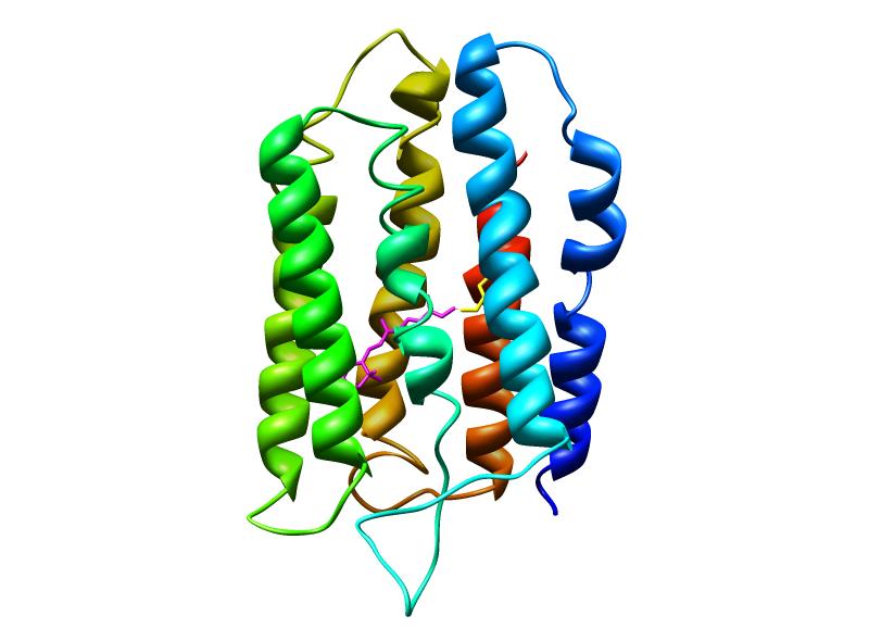
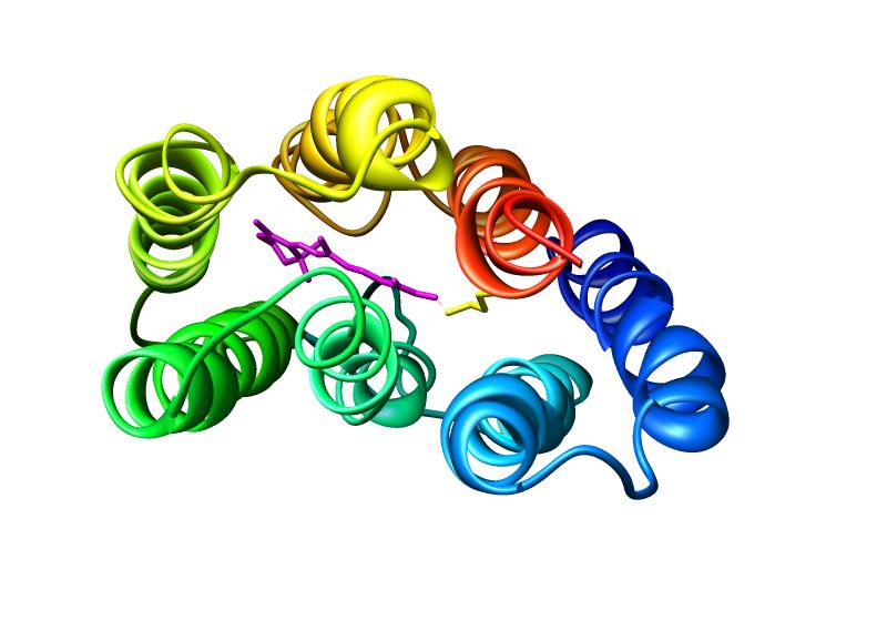
11 Myoglobin Pro Ile Lys Tyr Leu Glu Phe Ile Ser Asp Ala Ile Ile is Val is Ser Lys 103 Bacteriorhodopsin! Schiff base linkage between Lys-216 and retinal pdb code: 1AP9 Leu Ile Val Phe


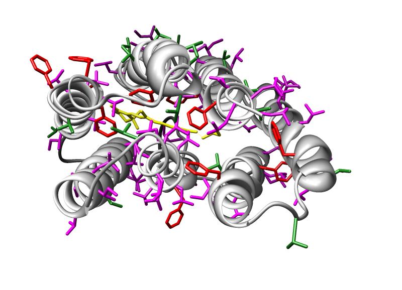
12 elical Bundles: hydrophobic sidechains form an interface between α-helices (de novo protein design) GLY GLU VAL GLU GLU LEU GLU LYS LYS PE LYS GLU LEU TP LYS GLY P AG AG GLY GLU ILE GLU GLU LEU IS LYS LYS PE IS GLU LEU ILE LYS GLY 105 pdb code: 1qp6 g c d f a b e a b c d e f g a b c d e f GLY GLU VAL GLU GLU LEU GLU LYS LYS PE LYS GLU LEU TP LYS GLY P AG AG a b c d e f g a b c d e f g a GLY GLU ILE GLU GLU LEU IS LYS LYS PE IS GLU LEU ILE LYS GLY
13 β-sheets and β-turns parallel anti-parallel crossover loop or turn anti-parallel β-sheet loop or turn 107 β-turn! _ (i+3) (i) (i+2) (i+1) -bond between (i) and (i+1) residues β-turn of Lysozyme (residues: Asn46-Thr47-Asp48-Gly49) (i+3) (i) (i+2) (i+1) (i+1) carbonyl on the opposite side of the sidechains= Type I β-turn β-turn: a region of the protein involving four consecutive residues where the polypeptide! chain folds back on itself by nearly 180. This chain reversal gives proteins a globular! rather than linear structure. (hou & Fasman J. Mol. Biol. 1977, 115, )!


14 β-turn of Lysozyme Typ 53 -Asp 52 -Thr 51 -Ser 50 -Gly 49 -Asp 48 Thr 43 -Asn 44 -Arg Asn 46 -Thr 47 pdb code: 1AZF 109 Anti-parallel β-sheets of lectin pdb code: 2LAL Parallel β-sheets carbonic anhydrase pdb code: 1QM

 ys33-ys42 uman Insulin pdb code: 1XDA hain A: 21 AA s hain B: 30 AA s ys(b)19-ys(a)20 ys6-ys20 ys14-ys31 ys(a)6-ys(a)11")
15 Some amino acids are found more often in certain secondary structures than others. hou, P.F.; Fasman, G.D. Ann ev. Biochem. 1978, 47, α-helix: Met, Glu, Ala, Leu, Gln, Lys, is β-sheet: Thr, Tyr, Phe, Ile, Val, is β-turns: Pro > Asn, Ser, Asp, Gly is: α-helix β-sheet >> β-turn Arg: α-helix β-sheet > β-turn ys: α-helix >> β-sheet > β-turn Trp: β-sheet > α-helix > β-turn 111 Disulfide bonds: covalent structural scaffolds, redox active, reversible EGF pdb code: 3EGF [] 2 ys-s [] ys-s-s-ys (ysteine) ys33-ys42 uman Insulin pdb code: 1XDA hain A: 21 AA s hain B: 30 AA s ys(b)19-ys(a)20 ys6-ys20 ys14-ys31 ys(a)6-ys(a)11 Somatostatin Analog pdb code: 1S ys(b)7-ys(a)
 psoition stabilizes the β-turn remove disulfide Ph 2 Pro-Phe-D-Trp Phe-Thr-Lys 3 Ph 3 Ph")

16 Somatostatin: Ala-Gly-ys-Lys-Asn-Phe-PheTrp S S ys-ser-thr-phe-thr-lys β-turn reponsible for biological activity Ph D-stereochemistry at the (i+2) psoition stabilizes the β-turn remove disulfide Ph 2 Pro-Phe-D-Trp Phe-Thr-Lys 3 Ph 3 Ph same activity as somatostatin 2 Ph 100x more potent than somatostatin lysozyme pdb code: 1AZF Disulfide bonds ribonuclease pdb code: 1ALF ys64 - ys80 ys26 - ys84 ys40 - ys95 ys76 - ys94 is12 is 119 ys30 - ys115 ys65 - ys72 ys6 - ys127 ys58 - ys
Section Week 3. Junaid Malek, M.D.
 Section Week 3 Junaid Malek, M.D. Biological Polymers DA 4 monomers (building blocks), limited structure (double-helix) RA 4 monomers, greater flexibility, multiple structures Proteins 20 Amino Acids,
The Structure of Enzymes!
 The Structure of Enzymes Levels of Protein Structure 0 order amino acid composition Primary Secondary Motifs Tertiary Domains Quaternary ther sequence repeating structural patterns defined by torsion angles
The Structure of Enzymes!
 The Structure of Enzymes Levels of Protein Structure 0 order amino acid composition Primary Secondary Motifs Tertiary Domains Quaternary ther sequence repeating structural patterns defined by torsion angles
Protein Structure. W. M. Grogan, Ph.D. OBJECTIVES
 Protein Structure W. M. Grogan, Ph.D. OBJECTIVES 1. Describe the structure and characteristic properties of typical proteins. 2. List and describe the four levels of structure found in proteins. 3. Relate
Read more about Pauling and more scientists at: Profiles in Science, The National Library of Medicine, profiles.nlm.nih.gov
 2018 Biochemistry 110 California Institute of Technology Lecture 2: Principles of Protein Structure Linus Pauling (1901-1994) began his studies at Caltech in 1922 and was directed by Arthur Amos oyes to
B O C 4 H 2 O O. NOTE: The reaction proceeds with a carbonium ion stabilized on the C 1 of sugar A.
 hbcse 33 rd International Page 101 hemistry lympiad Preparatory 05/02/01 Problems d. In the hydrolysis of the glycosidic bond, the glycosidic bridge oxygen goes with 4 of the sugar B. n cleavage, 18 from
Physiochemical Properties of Residues
 Physiochemical Properties of Residues Various Sources C N Cα R Slide 1 Conformational Propensities Conformational Propensity is the frequency in which a residue adopts a given conformation (in a polypeptide)
Peptides And Proteins
 Kevin Burgess, May 3, 2017 1 Peptides And Proteins from chapter(s) in the recommended text A. Introduction B. omenclature And Conventions by amide bonds. on the left, right. 2 -terminal C-terminal triglycine
Model Mélange. Physical Models of Peptides and Proteins
 Model Mélange Physical Models of Peptides and Proteins In the Model Mélange activity, you will visit four different stations each featuring a variety of different physical models of peptides or proteins.
Introduction to Comparative Protein Modeling. Chapter 4 Part I
 Introduction to Comparative Protein Modeling Chapter 4 Part I 1 Information on Proteins Each modeling study depends on the quality of the known experimental data. Basis of the model Search in the literature
Secondary Structure. Bioch/BIMS 503 Lecture 2. Structure and Function of Proteins. Further Reading. Φ, Ψ angles alone determine protein structure
 Bioch/BIMS 503 Lecture 2 Structure and Function of Proteins August 28, 2008 Robert Nakamoto rkn3c@virginia.edu 2-0279 Secondary Structure Φ Ψ angles determine protein structure Φ Ψ angles are restricted
Basic Principles of Protein Structures
 Basic Principles of Protein Structures Proteins Proteins: The Molecule of Life Proteins: Building Blocks Proteins: Secondary Structures Proteins: Tertiary and Quartenary Structure Proteins: Geometry Proteins
PROTEIN STRUCTURE AMINO ACIDS H R. Zwitterion (dipolar ion) CO 2 H. PEPTIDES Formal reactions showing formation of peptide bond by dehydration:
 CO 2 H. PEPTIDES Formal reactions showing formation of peptide bond by dehydration:") PTEI STUTUE ydrolysis of proteins with aqueous acid or base yields a mixture of free amino acids. Each type of protein yields a characteristic mixture of the ~ 20 amino acids. AMI AIDS Zwitterion (dipolar
PTEI STUTUE ydrolysis of proteins with aqueous acid or base yields a mixture of free amino acids. Each type of protein yields a characteristic mixture of the ~ 20 amino acids. AMI AIDS Zwitterion (dipolar
Packing of Secondary Structures
 7.88 Lecture Notes - 4 7.24/7.88J/5.48J The Protein Folding and Human Disease Professor Gossard Retrieving, Viewing Protein Structures from the Protein Data Base Helix helix packing Packing of Secondary
Major Types of Association of Proteins with Cell Membranes. From Alberts et al
 Major Types of Association of Proteins with Cell Membranes From Alberts et al Proteins Are Polymers of Amino Acids Peptide Bond Formation Amino Acid central carbon atom to which are attached amino group
Lecture 2 and 3: Review of forces (ctd.) and elementary statistical mechanics. Contributions to protein stability
 and elementary statistical mechanics. Contributions to protein stability") Lecture 2 and 3: Review of forces (ctd.) and elementary statistical mechanics. Contributions to protein stability Part I. Review of forces Covalent bonds Non-covalent Interactions: Van der Waals Interactions
Lecture 2 and 3: Review of forces (ctd.) and elementary statistical mechanics. Contributions to protein stability Part I. Review of forces Covalent bonds Non-covalent Interactions: Van der Waals Interactions
BIRKBECK COLLEGE (University of London)
") BIRKBECK COLLEGE (University of London) SCHOOL OF BIOLOGICAL SCIENCES M.Sc. EXAMINATION FOR INTERNAL STUDENTS ON: Postgraduate Certificate in Principles of Protein Structure MSc Structural Molecular Biology
BIRKBECK COLLEGE (University of London) SCHOOL OF BIOLOGICAL SCIENCES M.Sc. EXAMINATION FOR INTERNAL STUDENTS ON: Postgraduate Certificate in Principles of Protein Structure MSc Structural Molecular Biology
Properties of amino acids in proteins
 Properties of amino acids in proteins one of the primary roles of DNA (but not the only one!) is to code for proteins A typical bacterium builds thousands types of proteins, all from ~20 amino acids repeated
Biological Macromolecules
 Introduction for Chem 493 Chemistry of Biological Macromolecules Dr. L. Luyt January 2008 Dr. L. Luyt Chem 493-2008 1 Biological macromolecules are the molecules of life allow for organization serve a
Protein Structure Marianne Øksnes Dalheim, PhD candidate Biopolymers, TBT4135, Autumn 2013
 Protein Structure Marianne Øksnes Dalheim, PhD candidate Biopolymers, TBT4135, Autumn 2013 The presentation is based on the presentation by Professor Alexander Dikiy, which is given in the course compedium:
Supersecondary Structures (structural motifs)
") Supersecondary Structures (structural motifs) Various Sources Slide 1 Supersecondary Structures (Motifs) Supersecondary Structures (Motifs): : Combinations of secondary structures in specific geometric
Supersecondary Structures (structural motifs) Various Sources Slide 1 Supersecondary Structures (Motifs) Supersecondary Structures (Motifs): : Combinations of secondary structures in specific geometric
titin, has 35,213 amino acid residues (the human version of titin is smaller, with only 34,350 residues in the full length protein).
.") Introduction to Protein Structure Proteins are large heteropolymers usually comprised of 50 2500 monomer units, although larger proteins are observed 8. The monomer units of proteins are amino acids. Proteins
Introduction to Protein Structure Proteins are large heteropolymers usually comprised of 50 2500 monomer units, although larger proteins are observed 8. The monomer units of proteins are amino acids. Proteins
Chemistry Chapter 22
 hemistry 2100 hapter 22 Proteins Proteins serve many functions, including the following. 1. Structure: ollagen and keratin are the chief constituents of skin, bone, hair, and nails. 2. atalysts: Virtually
1. What is an ångstrom unit, and why is it used to describe molecular structures?
 1. What is an ångstrom unit, and why is it used to describe molecular structures? The ångstrom unit is a unit of distance suitable for measuring atomic scale objects. 1 ångstrom (Å) = 1 10-10 m. The diameter
What makes a good graphene-binding peptide? Adsorption of amino acids and peptides at aqueous graphene interfaces: Electronic Supplementary
 Electronic Supplementary Material (ESI) for Journal of Materials Chemistry B. This journal is The Royal Society of Chemistry 21 What makes a good graphene-binding peptide? Adsorption of amino acids and
Protein Structures: Experiments and Modeling. Patrice Koehl
 Protein Structures: Experiments and Modeling Patrice Koehl Structural Bioinformatics: Proteins Proteins: Sources of Structure Information Proteins: Homology Modeling Proteins: Ab initio prediction Proteins:
12/6/12. Dr. Sanjeeva Srivastava IIT Bombay. Primary Structure. Secondary Structure. Tertiary Structure. Quaternary Structure.
 Dr. anjeeva rivastava Primary tructure econdary tructure Tertiary tructure Quaternary tructure Amino acid residues α Helix Polypeptide chain Assembled subunits 2 1 Amino acid sequence determines 3-D structure
Details of Protein Structure
 Details of Protein Structure Function, evolution & experimental methods Thomas Blicher, Center for Biological Sequence Analysis Anne Mølgaard, Kemisk Institut, Københavns Universitet Learning Objectives
LS1a Fall 2014 Problem Set #2 Due Monday 10/6 at 6 pm in the drop boxes on the Science Center 2 nd Floor
 LS1a Fall 2014 Problem Set #2 Due Monday 10/6 at 6 pm in the drop boxes on the Science Center 2 nd Floor Note: Adequate space is given for each answer. Questions that require a brief explanation should
Exam I Answer Key: Summer 2006, Semester C
 1. Which of the following tripeptides would migrate most rapidly towards the negative electrode if electrophoresis is carried out at ph 3.0? a. gly-gly-gly b. glu-glu-asp c. lys-glu-lys d. val-asn-lys
Protein Struktur (optional, flexible)
") Protein Struktur (optional, flexible) 22/10/2009 [ 1 ] Andrew Torda, Wintersemester 2009 / 2010, AST nur für Informatiker, Mathematiker,.. 26 kt, 3 ov 2009 Proteins - who cares? 22/10/2009 [ 2 ] Most important
Protein Struktur (optional, flexible) 22/10/2009 [ 1 ] Andrew Torda, Wintersemester 2009 / 2010, AST nur für Informatiker, Mathematiker,.. 26 kt, 3 ov 2009 Proteins - who cares? 22/10/2009 [ 2 ] Most important
7.012 Problem Set 1 Solutions
 ame TA Section 7.012 Problem Set 1 Solutions Your answers to this problem set must be inserted into the large wooden box on wheels outside 68120 by 4:30 PM, Thursday, September 15. Problem sets will not
Lecture 11: Protein Folding & Stability
 Structure - Function Protein Folding: What we know Lecture 11: Protein Folding & Stability 1). Amino acid sequence dictates structure. 2). The native structure represents the lowest energy state for a
Protein Folding & Stability. Lecture 11: Margaret A. Daugherty. Fall Protein Folding: What we know. Protein Folding
 Lecture 11: Protein Folding & Stability Margaret A. Daugherty Fall 2003 Structure - Function Protein Folding: What we know 1). Amino acid sequence dictates structure. 2). The native structure represents
Protein Folding & Stability. Lecture 11: Margaret A. Daugherty. Fall How do we go from an unfolded polypeptide chain to a
 Lecture 11: Protein Folding & Stability Margaret A. Daugherty Fall 2004 How do we go from an unfolded polypeptide chain to a compact folded protein? (Folding of thioredoxin, F. Richards) Structure - Function
Basics of protein structure
 Today: 1. Projects a. Requirements: i. Critical review of one paper ii. At least one computational result b. Noon, Dec. 3 rd written report and oral presentation are due; submit via email to bphys101@fas.harvard.edu
Objective: Students will be able identify peptide bonds in proteins and describe the overall reaction between amino acids that create peptide bonds.
 Scott Seiple AP Biology Lesson Plan Lesson: Primary and Secondary Structure of Proteins Purpose:. To understand how amino acids can react to form peptides through peptide bonds.. Students will be able
Biochemistry Quiz Review 1I. 1. Of the 20 standard amino acids, only is not optically active. The reason is that its side chain.
 Biochemistry Quiz Review 1I A general note: Short answer questions are just that, short. Writing a paragraph filled with every term you can remember from class won t improve your answer just answer clearly,
Chem. 27 Section 1 Conformational Analysis Week of Feb. 6, TF: Walter E. Kowtoniuk Mallinckrodt 303 Liu Laboratory
 Chem. 27 Section 1 Conformational Analysis TF: Walter E. Kowtoniuk wekowton@fas.harvard.edu Mallinckrodt 303 Liu Laboratory ffice hours are: Monday and Wednesday 3:00-4:00pm in Mallinckrodt 303 Course
Final Chem 4511/6501 Spring 2011 May 5, 2011 b Name
 Key 1) [10 points] In RNA, G commonly forms a wobble pair with U. a) Draw a G-U wobble base pair, include riboses and 5 phosphates. b) Label the major groove and the minor groove. c) Label the atoms of
Solutions and Non-Covalent Binding Forces
 Chapter 3 Solutions and Non-Covalent Binding Forces 3.1 Solvent and solution properties Molecules stick together using the following forces: dipole-dipole, dipole-induced dipole, hydrogen bond, van der
Problem Set 1
 2006 7.012 Problem Set 1 Due before 5 PM on FRIDAY, September 15, 2006. Turn answers in to the box outside of 68-120. PLEASE WRITE YOUR ANSWERS ON THIS PRINTOUT. 1. For each of the following parts, pick
Protein Structure Bioinformatics Introduction
 1 Swiss Institute of Bioinformatics Protein Structure Bioinformatics Introduction Basel, 27. September 2004 Torsten Schwede Biozentrum - Universität Basel Swiss Institute of Bioinformatics Klingelbergstr
Sequential resonance assignments in (small) proteins: homonuclear method 2º structure determination
 proteins: homonuclear method 2º structure determination") Lecture 9 M230 Feigon Sequential resonance assignments in (small) proteins: homonuclear method 2º structure determination Reading resources v Roberts NMR of Macromolecules, Chap 4 by Christina Redfield
Lecture 9 M230 Feigon Sequential resonance assignments in (small) proteins: homonuclear method 2º structure determination Reading resources v Roberts NMR of Macromolecules, Chap 4 by Christina Redfield
Biomolecules: lecture 10
 Biomolecules: lecture 10 - understanding in detail how protein 3D structures form - realize that protein molecules are not static wire models but instead dynamic, where in principle every atom moves (yet
Introduction to Protein Folding
 Introduction to Protein Folding Chapter 4 Proteins: Three Dimensional Structure and Function Conformation - three dimensional shape Native conformation - each protein folds into a single stable shape (physiological
7.012 Problem Set 1. i) What are two main differences between prokaryotic cells and eukaryotic cells?
 What are two main differences between prokaryotic cells and eukaryotic cells?") ame 7.01 Problem Set 1 Section Question 1 a) What are the four major types of biological molecules discussed in lecture? Give one important function of each type of biological molecule in the cell? b)
ame 7.01 Problem Set 1 Section Question 1 a) What are the four major types of biological molecules discussed in lecture? Give one important function of each type of biological molecule in the cell? b)
Using Higher Calculus to Study Biologically Important Molecules Julie C. Mitchell
 Using Higher Calculus to Study Biologically Important Molecules Julie C. Mitchell Mathematics and Biochemistry University of Wisconsin - Madison 0 There Are Many Kinds Of Proteins The word protein comes
Protein Dynamics. The space-filling structures of myoglobin and hemoglobin show that there are no pathways for O 2 to reach the heme iron.
 Protein Dynamics The space-filling structures of myoglobin and hemoglobin show that there are no pathways for O 2 to reach the heme iron. Below is myoglobin hydrated with 350 water molecules. Only a small
Protein Structure. Role of (bio)informatics in drug discovery. Bioinformatics
informatics in drug discovery. Bioinformatics") Bioinformatics Protein Structure Principles & Architecture Marjolein Thunnissen Dep. of Biochemistry & Structural Biology Lund University September 2011 Homology, pattern and 3D structure searches need
Bioinformatics Protein Structure Principles & Architecture Marjolein Thunnissen Dep. of Biochemistry & Structural Biology Lund University September 2011 Homology, pattern and 3D structure searches need
Lecture 26: Polymers: DNA Packing and Protein folding 26.1 Problem Set 4 due today. Reading for Lectures 22 24: PKT Chapter 8 [ ].
![Lecture 26: Polymers: DNA Packing and Protein folding 26.1 Problem Set 4 due today. Reading for Lectures 22 24: PKT Chapter 8 [ ].](/thumbs/84/90810734.jpg "Lecture 26: Polymers: DNA Packing and Protein folding 26.1 Problem Set 4 due today. Reading for Lectures 22 24: PKT Chapter 8 [ ].") Lecture 26: Polymers: DA Packing and Protein folding 26.1 Problem Set 4 due today. eading for Lectures 22 24: PKT hapter 8 DA Packing for Eukaryotes: The packing problem for the larger eukaryotic genomes
Lecture 26: Polymers: DA Packing and Protein folding 26.1 Problem Set 4 due today. eading for Lectures 22 24: PKT hapter 8 DA Packing for Eukaryotes: The packing problem for the larger eukaryotic genomes
Overview. The peptide bond. Page 1
 Overview Secondary structure: the conformation of the peptide backbone The peptide bond, steric implications Steric hindrance and sterically allowed conformations. Ramachandran diagrams Side chain conformations
Secondary and sidechain structures
 Lecture 2 Secondary and sidechain structures James Chou BCMP201 Spring 2008 Images from Petsko & Ringe, Protein Structure and Function. Branden & Tooze, Introduction to Protein Structure. Richardson, J.
Advanced Certificate in Principles in Protein Structure. You will be given a start time with your exam instructions
 BIRKBECK COLLEGE (University of London) Advanced Certificate in Principles in Protein Structure MSc Structural Molecular Biology Date: Thursday, 1st September 2011 Time: 3 hours You will be given a start
Ranjit P. Bahadur Assistant Professor Department of Biotechnology Indian Institute of Technology Kharagpur, India. 1 st November, 2013
 Hydration of protein-rna recognition sites Ranjit P. Bahadur Assistant Professor Department of Biotechnology Indian Institute of Technology Kharagpur, India 1 st November, 2013 Central Dogma of life DNA
Supporting Information
 Supporting Information Micelle-Triggered b-hairpin to a-helix Transition in a 14-Residue Peptide from a Choline-Binding Repeat of the Pneumococcal Autolysin LytA HØctor Zamora-Carreras, [a] Beatriz Maestro,
Introduction to" Protein Structure
 Introduction to" Protein Structure Function, evolution & experimental methods Thomas Blicher, Center for Biological Sequence Analysis Learning Objectives Outline the basic levels of protein structure.
Biomolecules: lecture 9
 Biomolecules: lecture 9 - understanding further why amino acids are the building block for proteins - understanding the chemical properties amino acids bring to proteins - realizing that many proteins
4 Proteins: Structure, Function, Folding W. H. Freeman and Company
 4 Proteins: Structure, Function, Folding 2013 W. H. Freeman and Company CHAPTER 4 Proteins: Structure, Function, Folding Learning goals: Structure and properties of the peptide bond Structural hierarchy
Supplementary Figure 3 a. Structural comparison between the two determined structures for the IL 23:MA12 complex. The overall RMSD between the two
 Supplementary Figure 1. Biopanningg and clone enrichment of Alphabody binders against human IL 23. Positive clones in i phage ELISA with optical density (OD) 3 times higher than background are shown for
Chapter 4: Amino Acids
 Chapter 4: Amino Acids All peptides and polypeptides are polymers of alpha-amino acids. lipid polysaccharide enzyme 1940s 1980s. Lipids membrane 1960s. Polysaccharide Are energy metabolites and many of
Any protein that can be labelled by both procedures must be a transmembrane protein.
 1. What kind of experimental evidence would indicate that a protein crosses from one side of the membrane to the other? Regions of polypeptide part exposed on the outside of the membrane can be probed
Charged amino acids (side-chains)
") Proteins are composed of monomers called amino acids There are 20 different amino acids Amine Group Central ydrocarbon N C C R Group Carboxyl Group ALL amino acids have the exact same structure except
Proteins are composed of monomers called amino acids There are 20 different amino acids Amine Group Central ydrocarbon N C C R Group Carboxyl Group ALL amino acids have the exact same structure except
CHAPTER 29 HW: AMINO ACIDS + PROTEINS
 CAPTER 29 W: AMI ACIDS + PRTEIS For all problems, consult the table of 20 Amino Acids provided in lecture if an amino acid structure is needed; these will be given on exams. Use natural amino acids (L)
Protein Structure. Hierarchy of Protein Structure. Tertiary structure. independently stable structural unit. includes disulfide bonds
 Protein Structure Hierarchy of Protein Structure 2 3 Structural element Primary structure Secondary structure Super-secondary structure Domain Tertiary structure Quaternary structure Description amino
Protein Structure Basics
 Protein Structure Basics Presented by Alison Fraser, Christine Lee, Pradhuman Jhala, Corban Rivera Importance of Proteins Muscle structure depends on protein-protein interactions Transport across membranes
Denaturation and renaturation of proteins
 Denaturation and renaturation of proteins Higher levels of protein structure are formed without covalent bonds. Therefore, they are not as stable as peptide covalent bonds which make protein primary structure
Proton Acidity. (b) For the following reaction, draw the arrowhead properly to indicate the position of the equilibrium: HA + K + B -
 For the following reaction, draw the arrowhead properly to indicate the position of the equilibrium: HA + K + B -") Proton Acidity A01 Given that acid A has a pk a of 15 and acid B has a pk a of 10, then: (a) Which of the two acids is stronger? (b) For the following reaction, draw the arrowhead properly to indicate
Proton Acidity A01 Given that acid A has a pk a of 15 and acid B has a pk a of 10, then: (a) Which of the two acids is stronger? (b) For the following reaction, draw the arrowhead properly to indicate
Protein Struktur. Biologen und Chemiker dürfen mit Handys spielen (leise) go home, go to sleep. wake up at slide 39
 go home, go to sleep. wake up at slide 39") Protein Struktur Biologen und Chemiker dürfen mit Handys spielen (leise) go home, go to sleep wake up at slide 39 Andrew Torda, Wintersemester 2016/ 2017 Andrew Torda 17.10.2016 [ 1 ] Proteins - who cares?
Protein Struktur Biologen und Chemiker dürfen mit Handys spielen (leise) go home, go to sleep wake up at slide 39 Andrew Torda, Wintersemester 2016/ 2017 Andrew Torda 17.10.2016 [ 1 ] Proteins - who cares?
BCH 4053 Exam I Review Spring 2017
 BCH 4053 SI - Spring 2017 Reed BCH 4053 Exam I Review Spring 2017 Chapter 1 1. Calculate G for the reaction A + A P + Q. Assume the following equilibrium concentrations: [A] = 20mM, [Q] = [P] = 40fM. Assume
Molecular Structure Prediction by Global Optimization
 Molecular Structure Prediction by Global Optimization K.A. DILL Department of Pharmaceutical Chemistry, University of California at San Francisco, San Francisco, CA 94118 A.T. PHILLIPS Computer Science
Conformational Geometry of Peptides and Proteins:
 Conformational Geometry of Peptides and Proteins: Before discussing secondary structure, it is important to appreciate the conformational plasticity of proteins. Each residue in a polypeptide has three
NMR parameters intensity chemical shift coupling constants 1D 1 H spectra of nucleic acids and proteins
 Lecture #2 M230 NMR parameters intensity chemical shift coupling constants Juli Feigon 1D 1 H spectra of nucleic acids and proteins NMR Parameters A. Intensity (area) 1D NMR spectrum: integrated intensity
Computer simulations of protein folding with a small number of distance restraints
 Vol. 49 No. 3/2002 683 692 QUARTERLY Computer simulations of protein folding with a small number of distance restraints Andrzej Sikorski 1, Andrzej Kolinski 1,2 and Jeffrey Skolnick 2 1 Department of Chemistry,
Biochemistry,530:,, Introduc5on,to,Structural,Biology, Autumn,Quarter,2015,
 Biochemistry,530:,, Introduc5on,to,Structural,Biology, Autumn,Quarter,2015, Course,Informa5on, BIOC%530% GraduateAlevel,discussion,of,the,structure,,func5on,,and,chemistry,of,proteins,and, nucleic,acids,,control,of,enzyma5c,reac5ons.,please,see,the,course,syllabus,and,
Tamer Barakat. Razi Kittaneh. Mohammed Bio. Diala Abu-Hassan
 14 Tamer Barakat Razi Kittaneh Mohammed Bio Diala Abu-Hassan Protein structure: We already know that when two amino acids bind, a dipeptide is formed which is considered to be an oligopeptide. When more
NH 2. Biochemistry I, Fall Term Sept 9, Lecture 5: Amino Acids & Peptides Assigned reading in Campbell: Chapter
 Biochemistry I, Fall Term Sept 9, 2005 Lecture 5: Amino Acids & Peptides Assigned reading in Campbell: Chapter 3.1-3.4. Key Terms: ptical Activity, Chirality Peptide bond Condensation reaction ydrolysis
Translation. A ribosome, mrna, and trna.
 Translation The basic processes of translation are conserved among prokaryotes and eukaryotes. Prokaryotic Translation A ribosome, mrna, and trna. In the initiation of translation in prokaryotes, the Shine-Dalgarno
Programme Last week s quiz results + Summary Fold recognition Break Exercise: Modelling remote homologues
 Programme 8.00-8.20 Last week s quiz results + Summary 8.20-9.00 Fold recognition 9.00-9.15 Break 9.15-11.20 Exercise: Modelling remote homologues 11.20-11.40 Summary & discussion 11.40-12.00 Quiz 1 Feedback
Biochemistry - I SPRING Mondays and Wednesdays 9:30-10:45 AM (MR-1307) Lectures 3-4. Based on Profs. Kevin Gardner & Reza Khayat
 Lectures 3-4. Based on Profs. Kevin Gardner & Reza Khayat") Biochemistry - I Mondays and Wednesdays 9:30-10:45 AM (MR-1307) SPRING 2017 Lectures 3-4 Based on Profs. Kevin Gardner & Reza Khayat 1 Outline Overview of protein structure Peptide bonds Secondary structure
Biochemistry - I Mondays and Wednesdays 9:30-10:45 AM (MR-1307) SPRING 2017 Lectures 3-4 Based on Profs. Kevin Gardner & Reza Khayat 1 Outline Overview of protein structure Peptide bonds Secondary structure
CHEM J-9 June 2014
 CEM1611 2014-J-9 June 2014 Alanine (ala) and lysine (lys) are two amino acids with the structures given below as Fischer projections. The pk a values of the conjugate acid forms of the different functional
Protein structure. Protein structure. Amino acid residue. Cell communication channel. Bioinformatics Methods
 Cell communication channel Bioinformatics Methods Iosif Vaisman Email: ivaisman@gmu.edu SEQUENCE STRUCTURE DNA Sequence Protein Sequence Protein Structure Protein structure ATGAAATTTGGAAACTTCCTTCTCACTTATCAGCCACCT...
HSQC spectra for three proteins
 HSQC spectra for three proteins SH3 domain from Abp1p Kinase domain from EphB2 apo Calmodulin What do the spectra tell you about the three proteins? HSQC spectra for three proteins Small protein Big protein
The Structure and Functions of Proteins
 Wright State University CORE Scholar Computer Science and Engineering Faculty Publications Computer Science and Engineering 2003 The Structure and Functions of Proteins Dan E. Krane Wright State University
1. Amino Acids and Peptides Structures and Properties
 1. Amino Acids and Peptides Structures and Properties Chemical nature of amino acids The!-amino acids in peptides and proteins (excluding proline) consist of a carboxylic acid ( COOH) and an amino ( NH
Supporting information to: Time-resolved observation of protein allosteric communication. Sebastian Buchenberg, Florian Sittel and Gerhard Stock 1
 Supporting information to: Time-resolved observation of protein allosteric communication Sebastian Buchenberg, Florian Sittel and Gerhard Stock Biomolecular Dynamics, Institute of Physics, Albert Ludwigs
THE UNIVERSITY OF MANITOBA. PAPER NO: 409 LOCATION: Fr. Kennedy Gold Gym PAGE NO: 1 of 6 DEPARTMENT & COURSE NO: CHEM 4630 TIME: 3 HOURS
 PAPER NO: 409 LOCATION: Fr. Kennedy Gold Gym PAGE NO: 1 of 6 DEPARTMENT & COURSE NO: CHEM 4630 TIME: 3 HOURS EXAMINATION: Biochemistry of Proteins EXAMINER: J. O'Neil Section 1: You must answer all of
Supplementary figure 1. Comparison of unbound ogm-csf and ogm-csf as captured in the GIF:GM-CSF complex. Alignment of two copies of unbound ovine
 Supplementary figure 1. Comparison of unbound and as captured in the GIF:GM-CSF complex. Alignment of two copies of unbound ovine GM-CSF (slate) with bound GM-CSF in the GIF:GM-CSF complex (GIF: green,
Amino Acids and Peptides
 Amino Acids Amino Acids and Peptides Amino acid a compound that contains both an amino group and a carboxyl group α-amino acid an amino acid in which the amino group is on the carbon adjacent to the carboxyl
Dental Biochemistry Exam The total number of unique tripeptides that can be produced using all of the common 20 amino acids is
 Exam Questions for Dental Biochemistry Monday August 27, 2007 E.J. Miller 1. The compound shown below is CH 3 -CH 2 OH A. acetoacetate B. acetic acid C. acetaldehyde D. produced by reduction of acetaldehyde
Nanobiotechnology. Place: IOP 1 st Meeting Room Time: 9:30-12:00. Reference: Review Papers. Grade: 40% midterm, 60% final report (oral + written)
") Nanobiotechnology Place: IOP 1 st Meeting Room Time: 9:30-12:00 Reference: Review Papers Grade: 40% midterm, 60% final report (oral + written) Midterm: 5/18 Oral Presentation 1. 20 minutes each person
Nanobiotechnology Place: IOP 1 st Meeting Room Time: 9:30-12:00 Reference: Review Papers Grade: 40% midterm, 60% final report (oral + written) Midterm: 5/18 Oral Presentation 1. 20 minutes each person
Supplementary Information
 1 Supplementary Information Figure S1 The V=0.5 Harker section of an anomalous difference Patterson map calculated using diffraction data from the NNQQNY crystal at 1.3 Å resolution. The position of the
Exam III. Please read through each question carefully, and make sure you provide all of the requested information.
 09-107 onors Chemistry ame Exam III Please read through each question carefully, and make sure you provide all of the requested information. 1. A series of octahedral metal compounds are made from 1 mol
Structure and evolution of the spliceosomal peptidyl-prolyl cistrans isomerase Cwc27
 Acta Cryst. (2014). D70, doi:10.1107/s1399004714021695 Supporting information Volume 70 (2014) Supporting information for article: Structure and evolution of the spliceosomal peptidyl-prolyl cistrans isomerase
Conformational Analysis
 Conformational Analysis C01 3 C C 3 is the most stable by 0.9 kcal/mole C02 K eq = K 1-1 * K 2 = 0.45-1 * 0.048 = 0.11 C04 The intermediate in the reaction of 2 has an unfavorable syn-pentane interaction,
UNIT TWELVE. a, I _,o "' I I I. I I.P. l'o. H-c-c. I ~o I ~ I / H HI oh H...- I II I II 'oh. HO\HO~ I "-oh
 UNT TWELVE PROTENS : PEPTDE BONDNG AND POLYPEPTDES 12 CONCEPTS Many proteins are important in biological structure-for example, the keratin of hair, collagen of skin and leather, and fibroin of silk. Other
Biotechnology of Proteins. The Source of Stability in Proteins (III) Fall 2015
 Fall 2015") Biotechnology of Proteins The Source of Stability in Proteins (III) Fall 2015 Conformational Entropy of Unfolding It is The factor that makes the greatest contribution to stabilization of the unfolded
Biotechnology of Proteins The Source of Stability in Proteins (III) Fall 2015 Conformational Entropy of Unfolding It is The factor that makes the greatest contribution to stabilization of the unfolded
Lecture 10 (10/4/17) Lecture 10 (10/4/17)
 Lecture 10 (10/4/17)") Lecture 10 (10/4/17) Reading: Ch4; 125, 138-141, 141-142 Problems: Ch4 (text); 7, 9, 11 Ch4 (study guide); 1, 2 NEXT Reading: Ch4; 125, 132-136 (structure determination) Ch4; 12-130 (Collagen) Problems:
Lecture 10 (10/4/17) Reading: Ch4; 125, 138-141, 141-142 Problems: Ch4 (text); 7, 9, 11 Ch4 (study guide); 1, 2 NEXT Reading: Ch4; 125, 132-136 (structure determination) Ch4; 12-130 (Collagen) Problems:
Examples of Protein Modeling. Protein Modeling. Primary Structure. Protein Structure Description. Protein Sequence Sources. Importing Sequences to MOE
 Examples of Protein Modeling Protein Modeling Visualization Examination of an experimental structure to gain insight about a research question Dynamics To examine the dynamics of protein structures To
A) at equilibrium B) endergonic C) endothermic D) exergonic E) exothermic.
 at equilibrium B) endergonic C) endothermic D) exergonic E) exothermic.") CHEM 2770: Elements of Biochemistry Mid Term EXAMINATION VERSION A Date: October 29, 2014 Instructor: H. Perreault Location: 172 Schultz Time: 4 or 6 pm. Duration: 1 hour Instructions Please mark the Answer
CHEM 2770: Elements of Biochemistry Mid Term EXAMINATION VERSION A Date: October 29, 2014 Instructor: H. Perreault Location: 172 Schultz Time: 4 or 6 pm. Duration: 1 hour Instructions Please mark the Answer