Secondary and sidechain structures
|
|
|
- Roland Gordon
- 5 years ago
- Views:
Transcription
1 Lecture 2 Secondary and sidechain structures James Chou BCMP201 Spring 2008
2 Images from Petsko & Ringe, Protein Structure and Function. Branden & Tooze, Introduction to Protein Structure. Richardson, J. S. & Richardson, D. C. Principles and Patterns of Protein Conformation.
3 IUPAC definition of dihedral angles Definition of φ (AB-CD) dihedral angle A A B C D +! D B C φ (AB-CD) is + if clockwise - if counterclockwise #! " 360 if! >180! = $ %! if! < "180

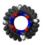
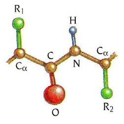
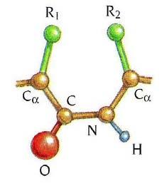
4 Polypeptide structure can be described by backbone dihedral angles Variable backbone dihedrals '! i C i"1 " N i " C i # "C i '! i N i " C i # " C i ' " N i +1 Peptide plane dihedral! i,i +1 C i " # C i ' " # N i +1 # C i +1 ω tends to be planar due to delocalization of the C π electron and the nitrogen lone pair.! =180 0 ±10 0

5 Secondary structures are local backbone structures with repeating φ and ψ Common examples: α helix β strands The Ramachandran plot, or ψ vs. φ plot, is the best way to view secondary structures in terms of the backbone dihedral angles.
6 Regular α helix right-handed! = "60 0, # = "40 0

7 α helix! = "60 0, # = "40 0 right handed, clockwise looking down from the N-terminus H-bond between O of residue i and HN of residue i+4 N 3.6 residues 5.4 A H-bond C Pauling et al., PNAS, 1951


8 Why are α helices right-handed in nature? right handed left handed HB of Arg7 O of Arg7 O of Arg7 HB of Arg7 less steric collision more steric collision

9 Some residues have the left-handed helical φ and ψ left-handed! = +60 0, " = +40 0
10 Glycines frequently have positive φ angles Take home question Why do glycines often have the left-handed dihedrals! = +60, " = +40?
 Helix dipole can coordinate phosphate or sulphate binding")
11 Helix electric dipole moment ~ 3.5 Debye Debye =10!18 esu cm e! 1 angs = 4.8!10 "18 esu cm Wada, Adv. in Biophysics, 9, 1-63 (1976) Helix dipole can coordinate phosphate or sulphate binding
 SO 4")
12 Example: the sulphate-binding protein (PDB code: 1SBP) SO 4 -


13 A single helix in solution is not stable. It must be stabilized by packing with other structural elements. Examples of stabilization of helix by helix-helix packing H-bond energy ~ 7 kj/mol, similar to the thermal energy in water. Thus H-bonds are not enough to keep a single helix stably locked.

14 Representation of helix by the helix wheel

15 Other unusual helical structures right-handed 3 10 helix! = "49 0, # = "26 0

!")


!")
16 3 10 helix 3 residues per turn H-bond (i,i+3)!," = #49,#26 α helix 3.6 residues per turn H-bond (i,i+4)!," = #60,#40 π helix 4.1 residues per turn H-bond (i,i+5)!," = #55,#70

17 Beginning and end of an α helix N-cap: The first residue of an α helix has non-helical Φ,Ψ dihedral angles, but participates, through its main-chain C=O group, in intra-helical hydrogen bonds. The side chain of the N-cap residue frequently accepts a hydrogen bond from the mainchain NH of the N3 residue. C-cap: The last residue of an α-helix has non-helical Φ,Ψ dihedral angles, but participates, through its main-chain N-H group, in intra-helical hydrogen bonds. The C-cap residue is often in the left-handed 3 10 helix conformation. Take home question What are the more likely amino acids for the N-cap? How about the C-cap?
18 ! = "135 0, # = β strands
19 β strands are NOT fully extended polypeptide chains Fully extended peptide! = "180 0, # = Anti-parallel β strands are slightly more stable than parallel β strands. 6 Å! = "140 0, # = 135 0! = "120 0, # = 115 0

20 Example: thioredoxin from E. coli


21 A single sheet in solution is also not stable. It must be stabilized by packing with helices or other sheets. dihydrofolate reductase V domain of Ig light chain retinol-binding protein
22 Non-repetitive structure: turns type I, most common type II, 2nd most common α helix 3 10 helix poly-pro Gly, left-handed 3 10 helix H-bond H-bond

23 Turns in antiparallel β strands type I type II


24 Ca trace of ubiquitin Protein sidechain conformation


25 not staggered staggered Example: valine χ 1 trans 180 +gauche 60 -gauche -60
26 Definition of sidechain χ 1 rotamer Regular, Ser Val, Ile, Thr -60 Hα -60 Hα CA-CB bond Hβ 2 C Hβ 1 Cγ / Oγ N Hβ C Cγ2 Cγ1/ Oγ1 N +60 Hα +60 Hα Hβ 1 Hβ 2 Cγ2 Hβ C N C N Cγ / Oγ Cγ1/ Oγ1 180 Hα 180 Hα Cγ / Oγ Hβ 1 Cγ1/ Oγ1 Cγ2 C N C N Hβ 2 Hβ
.! 1 = 180,! 2 = 180, 0.00 kj/mol! 1 = 68,!")
27 Sidechain conformation is dependent on backbone secondary structures due mainly to steric hindrance. Example of steric hindrance in determining the conformation of pentane (Dunbrack & Karplus, Nat. Struct. Biol., 1994).! 1 = 180,! 2 = 180, 0.00 kj/mol! 1 = 68,! 2 = 177, 3.6 kj/mol! 1 = 65,! 2 = 65, 7.5 kj/mol! 1 = 78,! 2 = 84, 14.9 kj/mol
28 Different values of φ and ψ yield different amount of steric hindrance at a given χ 1. Dunbrack & Karplus, Nat. Struct. Biol., 1994
29 (φ,ψ) dependent χ 1 preference for Valine Valine χ 1 = 180 Valine χ 1 = -60
30 (φ,ψ) dependent χ 1 preference for Isoleucine Isoleucine χ 1 = 180 Isoleucine χ 1 = -60
31 Effect of special residues on the backbone structure Gly often terminates an α helix, ~1/3 of all helices finish with Gly N- and C-terminal ends of the helix Gly C-cap
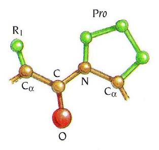
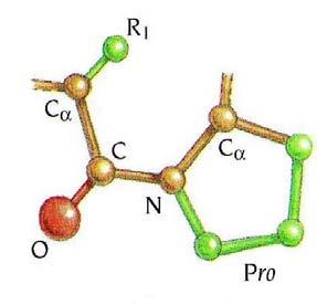
32 Proline also causes distortion or termination of helices N- and C-terminal ends of the helix Note: the closed ring constraints φ to be near -60
33 Proline causes a kink in α helix Proline in tight turns preceded by trans peptides No H-bond
34 cis - trans isomerization of proline X-X peptide 99.9% trans 0.1% cis X-Pro peptide ~70% trans ~30% cis
35 cis - trans isomerization of Proline in a turn


36 Polyproline helices type I!," = #75, residues per turn, right-handed, cis type II!," = #75, residues per turn, left-handed, trans collagen (GPP) n
Overview. The peptide bond. Page 1
 Overview Secondary structure: the conformation of the peptide backbone The peptide bond, steric implications Steric hindrance and sterically allowed conformations. Ramachandran diagrams Side chain conformations
Conformational Geometry of Peptides and Proteins:
 Conformational Geometry of Peptides and Proteins: Before discussing secondary structure, it is important to appreciate the conformational plasticity of proteins. Each residue in a polypeptide has three
titin, has 35,213 amino acid residues (the human version of titin is smaller, with only 34,350 residues in the full length protein).
.") Introduction to Protein Structure Proteins are large heteropolymers usually comprised of 50 2500 monomer units, although larger proteins are observed 8. The monomer units of proteins are amino acids. Proteins
Introduction to Protein Structure Proteins are large heteropolymers usually comprised of 50 2500 monomer units, although larger proteins are observed 8. The monomer units of proteins are amino acids. Proteins
Secondary Structure. Bioch/BIMS 503 Lecture 2. Structure and Function of Proteins. Further Reading. Φ, Ψ angles alone determine protein structure
 Bioch/BIMS 503 Lecture 2 Structure and Function of Proteins August 28, 2008 Robert Nakamoto rkn3c@virginia.edu 2-0279 Secondary Structure Φ Ψ angles determine protein structure Φ Ψ angles are restricted
Introduction to Comparative Protein Modeling. Chapter 4 Part I
 Introduction to Comparative Protein Modeling Chapter 4 Part I 1 Information on Proteins Each modeling study depends on the quality of the known experimental data. Basis of the model Search in the literature
Physiochemical Properties of Residues
 Physiochemical Properties of Residues Various Sources C N Cα R Slide 1 Conformational Propensities Conformational Propensity is the frequency in which a residue adopts a given conformation (in a polypeptide)
Peptides And Proteins
 Kevin Burgess, May 3, 2017 1 Peptides And Proteins from chapter(s) in the recommended text A. Introduction B. omenclature And Conventions by amide bonds. on the left, right. 2 -terminal C-terminal triglycine
Outline. Levels of Protein Structure. Primary (1 ) Structure. Lecture 6:Protein Architecture II: Secondary Structure or From peptides to proteins
 Structure. Lecture 6:Protein Architecture II: Secondary Structure or From peptides to proteins") Lecture 6:Protein Architecture II: Secondary Structure or From peptides to proteins Margaret Daugherty Fall 2004 Outline Four levels of structure are used to describe proteins; Alpha helices and beta sheets
Lecture 6:Protein Architecture II: Secondary Structure or From peptides to proteins Margaret Daugherty Fall 2004 Outline Four levels of structure are used to describe proteins; Alpha helices and beta sheets
HOMOLOGY MODELING. The sequence alignment and template structure are then used to produce a structural model of the target.
 HOMOLOGY MODELING Homology modeling, also known as comparative modeling of protein refers to constructing an atomic-resolution model of the "target" protein from its amino acid sequence and an experimental
Biomolecules: lecture 10
 Biomolecules: lecture 10 - understanding in detail how protein 3D structures form - realize that protein molecules are not static wire models but instead dynamic, where in principle every atom moves (yet
Outline. Levels of Protein Structure. Primary (1 ) Structure. Lecture 6:Protein Architecture II: Secondary Structure or From peptides to proteins
 Structure. Lecture 6:Protein Architecture II: Secondary Structure or From peptides to proteins") Lecture 6:Protein Architecture II: Secondary Structure or From peptides to proteins Margaret Daugherty Fall 2003 Outline Four levels of structure are used to describe proteins; Alpha helices and beta sheets
Lecture 6:Protein Architecture II: Secondary Structure or From peptides to proteins Margaret Daugherty Fall 2003 Outline Four levels of structure are used to describe proteins; Alpha helices and beta sheets
Chem. 27 Section 1 Conformational Analysis Week of Feb. 6, TF: Walter E. Kowtoniuk Mallinckrodt 303 Liu Laboratory
 Chem. 27 Section 1 Conformational Analysis TF: Walter E. Kowtoniuk wekowton@fas.harvard.edu Mallinckrodt 303 Liu Laboratory ffice hours are: Monday and Wednesday 3:00-4:00pm in Mallinckrodt 303 Course
Protein Structure Bioinformatics Introduction
 1 Swiss Institute of Bioinformatics Protein Structure Bioinformatics Introduction Basel, 27. September 2004 Torsten Schwede Biozentrum - Universität Basel Swiss Institute of Bioinformatics Klingelbergstr
Read more about Pauling and more scientists at: Profiles in Science, The National Library of Medicine, profiles.nlm.nih.gov
 2018 Biochemistry 110 California Institute of Technology Lecture 2: Principles of Protein Structure Linus Pauling (1901-1994) began his studies at Caltech in 1922 and was directed by Arthur Amos oyes to
Useful background reading
 Overview of lecture * General comment on peptide bond * Discussion of backbone dihedral angles * Discussion of Ramachandran plots * Description of helix types. * Description of structures * NMR patterns
Announcements. Primary (1 ) Structure. Lecture 7 & 8: PROTEIN ARCHITECTURE IV: Tertiary and Quaternary Structure
 Structure. Lecture 7 & 8: PROTEIN ARCHITECTURE IV: Tertiary and Quaternary Structure") Announcements TA Office Hours: Brian Eckenroth Monday 3-4 pm Thursday 11 am-12 pm Lecture 7 & 8: PROTEIN ARCHITECTURE IV: Tertiary and Quaternary Structure Margaret Daugherty Fall 2003 Homework II posted
Announcements TA Office Hours: Brian Eckenroth Monday 3-4 pm Thursday 11 am-12 pm Lecture 7 & 8: PROTEIN ARCHITECTURE IV: Tertiary and Quaternary Structure Margaret Daugherty Fall 2003 Homework II posted
B O C 4 H 2 O O. NOTE: The reaction proceeds with a carbonium ion stabilized on the C 1 of sugar A.
 hbcse 33 rd International Page 101 hemistry lympiad Preparatory 05/02/01 Problems d. In the hydrolysis of the glycosidic bond, the glycosidic bridge oxygen goes with 4 of the sugar B. n cleavage, 18 from
Protein Structure Basics
 Protein Structure Basics Presented by Alison Fraser, Christine Lee, Pradhuman Jhala, Corban Rivera Importance of Proteins Muscle structure depends on protein-protein interactions Transport across membranes
Solutions In each case, the chirality center has the R configuration
 CAPTER 25 669 Solutions 25.1. In each case, the chirality center has the R configuration. C C 2 2 C 3 C(C 3 ) 2 D-Alanine D-Valine 25.2. 2 2 S 2 d) 2 25.3. Pro,, Trp, Tyr, and is, Trp, Tyr, and is Arg,
Protein Structure. W. M. Grogan, Ph.D. OBJECTIVES
 Protein Structure W. M. Grogan, Ph.D. OBJECTIVES 1. Describe the structure and characteristic properties of typical proteins. 2. List and describe the four levels of structure found in proteins. 3. Relate
Molecular Modelling. part of Bioinformatik von RNA- und Proteinstrukturen. Sonja Prohaska. Leipzig, SS Computational EvoDevo University Leipzig
 part of Bioinformatik von RNA- und Proteinstrukturen Computational EvoDevo University Leipzig Leipzig, SS 2011 Protein Structure levels or organization Primary structure: sequence of amino acids (from
Supporting information to: Time-resolved observation of protein allosteric communication. Sebastian Buchenberg, Florian Sittel and Gerhard Stock 1
 Supporting information to: Time-resolved observation of protein allosteric communication Sebastian Buchenberg, Florian Sittel and Gerhard Stock Biomolecular Dynamics, Institute of Physics, Albert Ludwigs
Properties of amino acids in proteins
 Properties of amino acids in proteins one of the primary roles of DNA (but not the only one!) is to code for proteins A typical bacterium builds thousands types of proteins, all from ~20 amino acids repeated
Lecture 26: Polymers: DNA Packing and Protein folding 26.1 Problem Set 4 due today. Reading for Lectures 22 24: PKT Chapter 8 [ ].
![Lecture 26: Polymers: DNA Packing and Protein folding 26.1 Problem Set 4 due today. Reading for Lectures 22 24: PKT Chapter 8 [ ].](/thumbs/84/90810734.jpg "Lecture 26: Polymers: DNA Packing and Protein folding 26.1 Problem Set 4 due today. Reading for Lectures 22 24: PKT Chapter 8 [ ].") Lecture 26: Polymers: DA Packing and Protein folding 26.1 Problem Set 4 due today. eading for Lectures 22 24: PKT hapter 8 DA Packing for Eukaryotes: The packing problem for the larger eukaryotic genomes
Lecture 26: Polymers: DA Packing and Protein folding 26.1 Problem Set 4 due today. eading for Lectures 22 24: PKT hapter 8 DA Packing for Eukaryotes: The packing problem for the larger eukaryotic genomes
Protein Struktur (optional, flexible)
") Protein Struktur (optional, flexible) 22/10/2009 [ 1 ] Andrew Torda, Wintersemester 2009 / 2010, AST nur für Informatiker, Mathematiker,.. 26 kt, 3 ov 2009 Proteins - who cares? 22/10/2009 [ 2 ] Most important
Protein Struktur (optional, flexible) 22/10/2009 [ 1 ] Andrew Torda, Wintersemester 2009 / 2010, AST nur für Informatiker, Mathematiker,.. 26 kt, 3 ov 2009 Proteins - who cares? 22/10/2009 [ 2 ] Most important
PROTEIN STRUCTURE AMINO ACIDS H R. Zwitterion (dipolar ion) CO 2 H. PEPTIDES Formal reactions showing formation of peptide bond by dehydration:
 CO 2 H. PEPTIDES Formal reactions showing formation of peptide bond by dehydration:") PTEI STUTUE ydrolysis of proteins with aqueous acid or base yields a mixture of free amino acids. Each type of protein yields a characteristic mixture of the ~ 20 amino acids. AMI AIDS Zwitterion (dipolar
PTEI STUTUE ydrolysis of proteins with aqueous acid or base yields a mixture of free amino acids. Each type of protein yields a characteristic mixture of the ~ 20 amino acids. AMI AIDS Zwitterion (dipolar
Structure Determination by NMR Spectroscopy #3-1
 Structure Determination by NMR Spectroscopy #3-1 3. Computational methods in NMR-spectroscopy 3.1 Nomenclature of proteins and nucleic acids 3.1.1. Classification of structural relationships between molecules
Protein Structure. Hierarchy of Protein Structure. Tertiary structure. independently stable structural unit. includes disulfide bonds
 Protein Structure Hierarchy of Protein Structure 2 3 Structural element Primary structure Secondary structure Super-secondary structure Domain Tertiary structure Quaternary structure Description amino
Protein Structure. Role of (bio)informatics in drug discovery. Bioinformatics
informatics in drug discovery. Bioinformatics") Bioinformatics Protein Structure Principles & Architecture Marjolein Thunnissen Dep. of Biochemistry & Structural Biology Lund University September 2011 Homology, pattern and 3D structure searches need
Bioinformatics Protein Structure Principles & Architecture Marjolein Thunnissen Dep. of Biochemistry & Structural Biology Lund University September 2011 Homology, pattern and 3D structure searches need
Conformational Analysis
 Conformational Analysis C01 3 C C 3 is the most stable by 0.9 kcal/mole C02 K eq = K 1-1 * K 2 = 0.45-1 * 0.048 = 0.11 C04 The intermediate in the reaction of 2 has an unfavorable syn-pentane interaction,
Protein Structure and Function. Protein Architecture:
 BCHS 6229 Protein Structure and Function Lecture 2 (October 13, 2011) Protein Architecture: Symmetry relationships and protein structure Primary & Secondary Structure Motifs & Super-secondary Structure
Section Week 3. Junaid Malek, M.D.
 Section Week 3 Junaid Malek, M.D. Biological Polymers DA 4 monomers (building blocks), limited structure (double-helix) RA 4 monomers, greater flexibility, multiple structures Proteins 20 Amino Acids,
Model Mélange. Physical Models of Peptides and Proteins
 Model Mélange Physical Models of Peptides and Proteins In the Model Mélange activity, you will visit four different stations each featuring a variety of different physical models of peptides or proteins.
Central Dogma. modifications genome transcriptome proteome
 entral Dogma DA ma protein post-translational modifications genome transcriptome proteome 83 ierarchy of Protein Structure 20 Amino Acids There are 20 n possible sequences for a protein of n residues!
Supersecondary Structures (structural motifs)
") Supersecondary Structures (structural motifs) Various Sources Slide 1 Supersecondary Structures (Motifs) Supersecondary Structures (Motifs): : Combinations of secondary structures in specific geometric
Supersecondary Structures (structural motifs) Various Sources Slide 1 Supersecondary Structures (Motifs) Supersecondary Structures (Motifs): : Combinations of secondary structures in specific geometric
Objective: Students will be able identify peptide bonds in proteins and describe the overall reaction between amino acids that create peptide bonds.
 Scott Seiple AP Biology Lesson Plan Lesson: Primary and Secondary Structure of Proteins Purpose:. To understand how amino acids can react to form peptides through peptide bonds.. Students will be able
Introduction to Protein Folding
 Introduction to Protein Folding Chapter 4 Proteins: Three Dimensional Structure and Function Conformation - three dimensional shape Native conformation - each protein folds into a single stable shape (physiological
Sequential resonance assignments in (small) proteins: homonuclear method 2º structure determination
 proteins: homonuclear method 2º structure determination") Lecture 9 M230 Feigon Sequential resonance assignments in (small) proteins: homonuclear method 2º structure determination Reading resources v Roberts NMR of Macromolecules, Chap 4 by Christina Redfield
Lecture 9 M230 Feigon Sequential resonance assignments in (small) proteins: homonuclear method 2º structure determination Reading resources v Roberts NMR of Macromolecules, Chap 4 by Christina Redfield
Major Types of Association of Proteins with Cell Membranes. From Alberts et al
 Major Types of Association of Proteins with Cell Membranes From Alberts et al Proteins Are Polymers of Amino Acids Peptide Bond Formation Amino Acid central carbon atom to which are attached amino group
The Structure and Functions of Proteins
 Wright State University CORE Scholar Computer Science and Engineering Faculty Publications Computer Science and Engineering 2003 The Structure and Functions of Proteins Dan E. Krane Wright State University
Problem Set 1
 2006 7.012 Problem Set 1 Due before 5 PM on FRIDAY, September 15, 2006. Turn answers in to the box outside of 68-120. PLEASE WRITE YOUR ANSWERS ON THIS PRINTOUT. 1. For each of the following parts, pick
Protein Data Bank Contents Guide: Atomic Coordinate Entry Format Description. Version Document Published by the wwpdb
 Protein Data Bank Contents Guide: Atomic Coordinate Entry Format Description Version 3.30 Document Published by the wwpdb This format complies with the PDB Exchange Dictionary (PDBx) http://mmcif.pdb.org/dictionaries/mmcif_pdbx.dic/index/index.html.
BCH 4053 Spring 2003 Chapter 6 Lecture Notes
 BCH 4053 Spring 2003 Chapter 6 Lecture Notes 1 CHAPTER 6 Proteins: Secondary, Tertiary, and Quaternary Structure 2 Levels of Protein Structure Primary (sequence) Secondary (ordered structure along peptide
Supplementary Information
 1 Supplementary Information Figure S1 The V=0.5 Harker section of an anomalous difference Patterson map calculated using diffraction data from the NNQQNY crystal at 1.3 Å resolution. The position of the
The Anatomy & Taxonomy of Protein Structure
 The Anatomy & Taxonomy of Protein Structure by Jane S. Richardson II. Basic Elements Of Protein Structure A. Helices The α-helix is the classic element of protein structure. A single α-helix can order
BCMP 201 Protein biochemistry
 BCMP 201 Protein biochemistry BCMP 201 Protein biochemistry with emphasis on the interrelated roles of protein structure, catalytic activity, and macromolecular interactions in biological processes. The
Protein Structure Marianne Øksnes Dalheim, PhD candidate Biopolymers, TBT4135, Autumn 2013
 Protein Structure Marianne Øksnes Dalheim, PhD candidate Biopolymers, TBT4135, Autumn 2013 The presentation is based on the presentation by Professor Alexander Dikiy, which is given in the course compedium:
1. What is an ångstrom unit, and why is it used to describe molecular structures?
 1. What is an ångstrom unit, and why is it used to describe molecular structures? The ångstrom unit is a unit of distance suitable for measuring atomic scale objects. 1 ångstrom (Å) = 1 10-10 m. The diameter
4 Proteins: Structure, Function, Folding W. H. Freeman and Company
 4 Proteins: Structure, Function, Folding 2013 W. H. Freeman and Company CHAPTER 4 Proteins: Structure, Function, Folding Learning goals: Structure and properties of the peptide bond Structural hierarchy
Protein structure. Protein structure. Amino acid residue. Cell communication channel. Bioinformatics Methods
 Cell communication channel Bioinformatics Methods Iosif Vaisman Email: ivaisman@gmu.edu SEQUENCE STRUCTURE DNA Sequence Protein Sequence Protein Structure Protein structure ATGAAATTTGGAAACTTCCTTCTCACTTATCAGCCACCT...
Basic Principles of Protein Structures
 Basic Principles of Protein Structures Proteins Proteins: The Molecule of Life Proteins: Building Blocks Proteins: Secondary Structures Proteins: Tertiary and Quartenary Structure Proteins: Geometry Proteins
Biochemistry: Concepts and Connections
 Biochemistry: Concepts and Connections Dean R. Appling Spencer J. Anthony-Cahill Christopher K. Mathews Chapter 6 The Three Dimensional Structure of Proteins Cartoon representation of myoglobin, showing
Packing of Secondary Structures
 7.88 Lecture Notes - 4 7.24/7.88J/5.48J The Protein Folding and Human Disease Professor Gossard Retrieving, Viewing Protein Structures from the Protein Data Base Helix helix packing Packing of Secondary
Protein Struktur. Biologen und Chemiker dürfen mit Handys spielen (leise) go home, go to sleep. wake up at slide 39
 go home, go to sleep. wake up at slide 39") Protein Struktur Biologen und Chemiker dürfen mit Handys spielen (leise) go home, go to sleep wake up at slide 39 Andrew Torda, Wintersemester 2016/ 2017 Andrew Torda 17.10.2016 [ 1 ] Proteins - who cares?
Protein Struktur Biologen und Chemiker dürfen mit Handys spielen (leise) go home, go to sleep wake up at slide 39 Andrew Torda, Wintersemester 2016/ 2017 Andrew Torda 17.10.2016 [ 1 ] Proteins - who cares?
THE UNIVERSITY OF MANITOBA. PAPER NO: 409 LOCATION: Fr. Kennedy Gold Gym PAGE NO: 1 of 6 DEPARTMENT & COURSE NO: CHEM 4630 TIME: 3 HOURS
 PAPER NO: 409 LOCATION: Fr. Kennedy Gold Gym PAGE NO: 1 of 6 DEPARTMENT & COURSE NO: CHEM 4630 TIME: 3 HOURS EXAMINATION: Biochemistry of Proteins EXAMINER: J. O'Neil Section 1: You must answer all of
Biophysical Society On-line Textbook
 Biophysical Society On-line Textbook PROTEINS CHAPTER 1. PROTEIN STRUCTURE Section 1. Primary structure, secondary motifs, tertiary architecture, and quaternary organization Jannette Carey* and Vanessa
Part II => PROTEINS and ENZYMES. 2.3 PROTEIN STRUCTURE 2.3a Secondary Structure 2.3b Tertiary Structure 2.3c Quaternary Structure
 Part II => PROTEINS and ENZYMES 2.3 PROTEIN STRUCTURE 2.3a Secondary Structure 2.3b Tertiary Structure 2.3c Quaternary Structure Section 2.3a: Secondary Structure Synopsis 2.3a - Secondary structure refers
Biochemistry - I SPRING Mondays and Wednesdays 9:30-10:45 AM (MR-1307) Lectures 3-4. Based on Profs. Kevin Gardner & Reza Khayat
 Lectures 3-4. Based on Profs. Kevin Gardner & Reza Khayat") Biochemistry - I Mondays and Wednesdays 9:30-10:45 AM (MR-1307) SPRING 2017 Lectures 3-4 Based on Profs. Kevin Gardner & Reza Khayat 1 Outline Overview of protein structure Peptide bonds Secondary structure
Biochemistry - I Mondays and Wednesdays 9:30-10:45 AM (MR-1307) SPRING 2017 Lectures 3-4 Based on Profs. Kevin Gardner & Reza Khayat 1 Outline Overview of protein structure Peptide bonds Secondary structure
Protein Structure & Motifs
 & Motifs Biochemistry 201 Molecular Biology January 12, 2000 Doug Brutlag Introduction Proteins are more flexible than nucleic acids in structure because of both the larger number of types of residues
Ramachandran Plot. 4ysz Phi (degrees) Plot statistics
 Plot statistics") B Ramachandran Plot ~b b 135 b ~b ~l l Psi (degrees) 5-5 a A ~a L - -135 SER HIS (F) 59 (G) SER (B) ~b b LYS ASP ASP 315 13 13 (A) (F) (B) LYS ALA ALA 315 173 (E) 173 (E)(A) ~p p ~b - -135 - -5 5 135 (degrees)
B Ramachandran Plot ~b b 135 b ~b ~l l Psi (degrees) 5-5 a A ~a L - -135 SER HIS (F) 59 (G) SER (B) ~b b LYS ASP ASP 315 13 13 (A) (F) (B) LYS ALA ALA 315 173 (E) 173 (E)(A) ~p p ~b - -135 - -5 5 135 (degrees)
HSQC spectra for three proteins
 HSQC spectra for three proteins SH3 domain from Abp1p Kinase domain from EphB2 apo Calmodulin What do the spectra tell you about the three proteins? HSQC spectra for three proteins Small protein Big protein
Resonance assignments in proteins. Christina Redfield
 Resonance assignments in proteins Christina Redfield 1. Introduction The assignment of resonances in the complex NMR spectrum of a protein is the first step in any study of protein structure, function
Structure and evolution of the spliceosomal peptidyl-prolyl cistrans isomerase Cwc27
 Acta Cryst. (2014). D70, doi:10.1107/s1399004714021695 Supporting information Volume 70 (2014) Supporting information for article: Structure and evolution of the spliceosomal peptidyl-prolyl cistrans isomerase
The Structure of Enzymes!
 The Structure of Enzymes Levels of Protein Structure 0 order amino acid composition Primary Secondary Motifs Tertiary Domains Quaternary ther sequence repeating structural patterns defined by torsion angles
The Structure of Enzymes!
 The Structure of Enzymes Levels of Protein Structure 0 order amino acid composition Primary Secondary Motifs Tertiary Domains Quaternary ther sequence repeating structural patterns defined by torsion angles
Supporting Information
 Supporting Information Micelle-Triggered b-hairpin to a-helix Transition in a 14-Residue Peptide from a Choline-Binding Repeat of the Pneumococcal Autolysin LytA HØctor Zamora-Carreras, [a] Beatriz Maestro,
What makes a good graphene-binding peptide? Adsorption of amino acids and peptides at aqueous graphene interfaces: Electronic Supplementary
 Electronic Supplementary Material (ESI) for Journal of Materials Chemistry B. This journal is The Royal Society of Chemistry 21 What makes a good graphene-binding peptide? Adsorption of amino acids and
Computational Protein Design
 11 Computational Protein Design This chapter introduces the automated protein design and experimental validation of a novel designed sequence, as described in Dahiyat and Mayo [1]. 11.1 Introduction Given
LS1a Fall 2014 Problem Set #2 Due Monday 10/6 at 6 pm in the drop boxes on the Science Center 2 nd Floor
 LS1a Fall 2014 Problem Set #2 Due Monday 10/6 at 6 pm in the drop boxes on the Science Center 2 nd Floor Note: Adequate space is given for each answer. Questions that require a brief explanation should
Examples of Protein Modeling. Protein Modeling. Primary Structure. Protein Structure Description. Protein Sequence Sources. Importing Sequences to MOE
 Examples of Protein Modeling Protein Modeling Visualization Examination of an experimental structure to gain insight about a research question Dynamics To examine the dynamics of protein structures To
Bioinformatics. Macromolecular structure
 Bioinformatics Macromolecular structure Contents Determination of protein structure Structure databases Secondary structure elements (SSE) Tertiary structure Structure analysis Structure alignment Domain
The Anatomy & Taxonomy of Protein Structure
 The Anatomy & Taxonomy of Protein Structure by Jane S. Richardson I. BACKGROUND A. Introduction X-Ray crystallography is a technically sophisticated but conceptually simple-minded method with the great
Protein Dynamics. The space-filling structures of myoglobin and hemoglobin show that there are no pathways for O 2 to reach the heme iron.
 Protein Dynamics The space-filling structures of myoglobin and hemoglobin show that there are no pathways for O 2 to reach the heme iron. Below is myoglobin hydrated with 350 water molecules. Only a small
A CONFORMATIONAL STUDY OF PROLINE DERIVATIVES. M.E. Kamwaya * and F.N. Ngassapa
 , 199-206. ISSN 1011-3924 Printed in Ethiopia 2002 Chemical Society of Ethiopia A CONFORMATIONAL STUDY OF PROLINE DERIVATIVES M.E. Kamwaya * and F.N. Ngassapa Chemistry Department, University of Dar es
Protein Structure Refinement Using 13 C α Chemical. Shift Tensors. Benjamin J. Wylie, Charles D. Schwieters, Eric Oldfield and Chad M.
 Protein Structure Refinement Using 13 C α Chemical Shift Tensors Benjamin J. Wylie, Charles D. Schwieters, Eric Oldfield and Chad M. Rienstra * Department of Chemistry, University of Illinois at Urbana-Champaign,
NMR parameters intensity chemical shift coupling constants 1D 1 H spectra of nucleic acids and proteins
 Lecture #2 M230 NMR parameters intensity chemical shift coupling constants Juli Feigon 1D 1 H spectra of nucleic acids and proteins NMR Parameters A. Intensity (area) 1D NMR spectrum: integrated intensity
NMR, X-ray Diffraction, Protein Structure, and RasMol
 NMR, X-ray Diffraction, Protein Structure, and RasMol Introduction So far we have been mostly concerned with the proteins themselves. The techniques (NMR or X-ray diffraction) used to determine a structure
Biomolecules: lecture 9
 Biomolecules: lecture 9 - understanding further why amino acids are the building block for proteins - understanding the chemical properties amino acids bring to proteins - realizing that many proteins
Introduction to" Protein Structure
 Introduction to" Protein Structure Function, evolution & experimental methods Thomas Blicher, Center for Biological Sequence Analysis Learning Objectives Outline the basic levels of protein structure.
Figure 1. Molecules geometries of 5021 and Each neutral group in CHARMM topology was grouped in dash circle.
 Project I Chemistry 8021, Spring 2005/2/23 This document was turned in by a student as a homework paper. 1. Methods First, the cartesian coordinates of 5021 and 8021 molecules (Fig. 1) are generated, in
Ramachandran and his Map
 Ramachandran and his Map C Ramakrishnan Introduction C Ramakrishnan, retired professor from Molecular Biophysics Unit, Indian Institute of Science, Bangalore, has been associated with Professor G N Ramachandran
Using Higher Calculus to Study Biologically Important Molecules Julie C. Mitchell
 Using Higher Calculus to Study Biologically Important Molecules Julie C. Mitchell Mathematics and Biochemistry University of Wisconsin - Madison 0 There Are Many Kinds Of Proteins The word protein comes
BSc and MSc Degree Examinations
 Examination Candidate Number: Desk Number: BSc and MSc Degree Examinations 2018-9 Department : BIOLOGY Title of Exam: Molecular Biology and Biochemistry Part I Time Allowed: 1 hour and 30 minutes Marking
Report of protein analysis
 Report of protein analysis By the WHAT IF program 2010-09-19 1 Introduction what check is the name of the validation option in what if. It doesn t matter whether you use the what check program or the what
Molecular Structure Prediction by Global Optimization
 Molecular Structure Prediction by Global Optimization K.A. DILL Department of Pharmaceutical Chemistry, University of California at San Francisco, San Francisco, CA 94118 A.T. PHILLIPS Computer Science
Exam I Answer Key: Summer 2006, Semester C
 1. Which of the following tripeptides would migrate most rapidly towards the negative electrode if electrophoresis is carried out at ph 3.0? a. gly-gly-gly b. glu-glu-asp c. lys-glu-lys d. val-asn-lys
Proton Acidity. (b) For the following reaction, draw the arrowhead properly to indicate the position of the equilibrium: HA + K + B -
 For the following reaction, draw the arrowhead properly to indicate the position of the equilibrium: HA + K + B -") Proton Acidity A01 Given that acid A has a pk a of 15 and acid B has a pk a of 10, then: (a) Which of the two acids is stronger? (b) For the following reaction, draw the arrowhead properly to indicate
Proton Acidity A01 Given that acid A has a pk a of 15 and acid B has a pk a of 10, then: (a) Which of the two acids is stronger? (b) For the following reaction, draw the arrowhead properly to indicate
CHAPTER 29 HW: AMINO ACIDS + PROTEINS
 CAPTER 29 W: AMI ACIDS + PRTEIS For all problems, consult the table of 20 Amino Acids provided in lecture if an amino acid structure is needed; these will be given on exams. Use natural amino acids (L)
Biochemistry,530:,, Introduc5on,to,Structural,Biology, Autumn,Quarter,2015,
 Biochemistry,530:,, Introduc5on,to,Structural,Biology, Autumn,Quarter,2015, Course,Informa5on, BIOC%530% GraduateAlevel,discussion,of,the,structure,,func5on,,and,chemistry,of,proteins,and, nucleic,acids,,control,of,enzyma5c,reac5ons.,please,see,the,course,syllabus,and,
Report of protein analysis
 Report of protein analysis By the WHAT IF program 2010-09-19 1 Introduction what check is the name of the validation option in what if. It doesn t matter whether you use the what check program or the what
B. β Structure. All contents of this document, unless otherwise noted, are David C. & Jane S. Richardson. All Rights Reserved.
 B. β Structure The other major structural element found in globular proteins is the β sheet. Historically, it was first observed as the β, or extended, form of keratin fibers. An approximate understanding
F. Piazza Center for Molecular Biophysics and University of Orléans, France. Selected topic in Physical Biology. Lecture 1
 Zhou Pei-Yuan Centre for Applied Mathematics, Tsinghua University November 2013 F. Piazza Center for Molecular Biophysics and University of Orléans, France Selected topic in Physical Biology Lecture 1
Dihedral Angles. Homayoun Valafar. Department of Computer Science and Engineering, USC 02/03/10 CSCE 769
 Dihedral Angles Homayoun Valafar Department of Computer Science and Engineering, USC The precise definition of a dihedral or torsion angle can be found in spatial geometry Angle between to planes Dihedral
3. Solutions W = N!/(N A!N B!) (3.1) Using Stirling s approximation ln(n!) = NlnN N: ΔS mix = k (N A lnn + N B lnn N A lnn A N B lnn B ) (3.
 (3.1) Using Stirling s approximation ln(n!) = NlnN N: ΔS mix = k (N A lnn + N B lnn N A lnn A N B lnn B ) (3.") 3. Solutions Many biological processes occur between molecules in aqueous solution. In addition, many protein and nucleic acid molecules adopt three-dimensional structure ( fold ) in aqueous solution.
3. Solutions Many biological processes occur between molecules in aqueous solution. In addition, many protein and nucleic acid molecules adopt three-dimensional structure ( fold ) in aqueous solution.
Short peptides, too short to form any structure that is stabilized
 Origin of the neighboring residue effect on peptide backbone conformation Franc Avbelj and Robert L. Baldwin National Institute of Chemistry, Hajdrihova 19, Ljubljana Sl 1115, Slovenia; and Department
April, The energy functions include:
 REDUX A collection of Python scripts for torsion angle Monte Carlo protein molecular simulations and analysis The program is based on unified residue peptide model and is designed for more efficient exploration
 Title Energy Calculations on Di- and Poly on Polymer Chemistry IX) Author(s) Nishikawa, Ken; Ooi, Tatsuo Citation Bulletin of the Institute for Chemi University (1972), 50(2): 94-106 Issue Date 1972-06-30
Lecture 10 (10/4/17) Lecture 10 (10/4/17)
 Lecture 10 (10/4/17)") Lecture 10 (10/4/17) Reading: Ch4; 125, 138-141, 141-142 Problems: Ch4 (text); 7, 9, 11 Ch4 (study guide); 1, 2 NEXT Reading: Ch4; 125, 132-136 (structure determination) Ch4; 12-130 (Collagen) Problems:
Lecture 10 (10/4/17) Reading: Ch4; 125, 138-141, 141-142 Problems: Ch4 (text); 7, 9, 11 Ch4 (study guide); 1, 2 NEXT Reading: Ch4; 125, 132-136 (structure determination) Ch4; 12-130 (Collagen) Problems:
Lecture 2 and 3: Review of forces (ctd.) and elementary statistical mechanics. Contributions to protein stability
 and elementary statistical mechanics. Contributions to protein stability") Lecture 2 and 3: Review of forces (ctd.) and elementary statistical mechanics. Contributions to protein stability Part I. Review of forces Covalent bonds Non-covalent Interactions: Van der Waals Interactions
Lecture 2 and 3: Review of forces (ctd.) and elementary statistical mechanics. Contributions to protein stability Part I. Review of forces Covalent bonds Non-covalent Interactions: Van der Waals Interactions
Introduction solution NMR
 2 NMR journey Introduction solution NMR Alexandre Bonvin Bijvoet Center for Biomolecular Research with thanks to Dr. Klaartje Houben EMBO Global Exchange course, IHEP, Beijing April 28 - May 5, 20 3 Topics