Biochemistry 9001 Protein structure determination by 3D Electron Microscopy
|
|
|
- Kevin Ward
- 6 years ago
- Views:
Transcription


1 Biochemistry 9001 Protein structure determination by 3D Electron Microscopy Tommi A. White, Ph.D. Associate Director, Electron Microscopy Core Facility Assistant Research Professor, Biochemistry
2 Goals of this course Introduce and familiarize you to concepts related to 3D Electron Microscopy and protein structure determination Provide hands on training and techniques to assist you in protein structure determination using electron microscopy
3 Instructors Tommi A. White, Ph.D. Ph.D. Biochemistry, University of Missouri Jack Tanner Structural Studies of Bacterial Proline Catabolic Enzymes Post-doctoral Fellowship, National Cancer Institute at NIH Sriram Subramaniam Cryo- Electron Tomography of SIV Envelope Glycoproteins Associate Director, Electron Microscopy Core 2012-present Assistant Research Professor in Biochemistry 2012-present Narahari Akkaladevi, Ph.D. Ph.D. Biochemistry, Montana State Post-doctoral Fellowship, Kansas University Medical Center Mark Fisher Structural Studies of Anthrax Toxin Pores in Nanodiscs Post-doctoral Fellowship, University of Missouri Gerald Hazelbauer Structural Studies of Chemoreceptors in Nanodiscs
4 Course structure 5 lectures 5 labs & lab reports 2 weeks for individual projects Project Abstract Project Presentation Project report
5 Lectures Mondays 10:30 am noon Theory, examples from literature and EMC
6 Labs 2 hours One-on-one individual time Monday, Tuesday, Wednesday Hands on applications, demonstration and Labs

7 Electron Microscopy Core (EMC) Hosted by Office of Research Early 90 s had electron microscopes in Life Sciences Geological Sciences CAFNR Materials Spec Prep FEI Quanta 600 SEM Hitachi School of Medicine S-4700 SEM Veterinary medicine JEOL 1400 TEM Cryo Prep/ Mechanical Fixation Connawa 1995 combined into Electron Embedding Microscopy y Core Office Hosted by Veterinary Pathology Specimen Preparatio Office Located in Veterinary n: Medicine Building Microtome FEI Tecnai F30 TEM Stairs Veterinar y Medicine Building
8 Projects Project consultation before July 13 th, 2015 Project Abstract 3 slides, July 13 th, 2015 Project Data Collection/Processing July 13 th -July 26 th, 2014 Project Presentation 15 min, July 27 th, 2015 Project Report 2 pages, due by July 31 st, 2015

9 Computation Apply for lewis account edu/user_guide.html UMBC Account Request Form and send to me by 6/15/2015 Purchase a thumb drive by 6/22/2015 USB3 16 GB
10 Why do we need 3D?

11 Frank, 2006

12 Williams & Carter, 1996
13 What is 3DEM?
14 What is 3DEM? 3-Dimensional Electron Microscopy Using electron microscopy to determine a structure in 3- dimensions Proteins are embedded to maintain their shape Heavy metal stain Sugar (trehalose) Ice (non-crystalline) Examples: Single Particle Reconstruction (Single Particle Analysis) Random Conical Tilt Electron Tomography Electron Crystallography Serial Block Face Imaging
 resolution from thirty thousand cryo-em particles (2013) Xiao-chen Bai, Israel S Fernandez, Greg")
15 3DEM Examples: Single Particle Reconstruction Ribosome structures to near-atomic (4.1 Å) resolution from thirty thousand cryo-em particles (2013) Xiao-chen Bai, Israel S Fernandez, Greg McMullan, Sjors HW Scheres

16 3DEM Examples: Cryo-electron Tomography

17 3DEM Examples: Serial Block Face imaging

18 3DEM Examples: Single Particle Reconstruction
19 Why electrons?

20 Subramaniam 2005 Curr Opin Microbiol.
21 Serial Block Face Imaging Tomography Single Particle Analysis Subramaniam 2005 Curr Opin Microbiol., modified TAW

22 Electrons Involved in electricity, magnetism, thermal conductivity A moving electron creates a magnetic field Wave-particle duality Small particle 9 x grams Negatively charged 1.6 x Coulomb Higher wavelength ~ kev 90% the speed of light 270,000 km/s Easily accelerated resting energy MeV Williams & Carter, 1996
23 Single Particle Reconstruction quantitative way of determining the structure of macromolecules from micrographs, showing them as a collection of isolated, unattached particles Uses not one, but thousands of macromolecules Multiple orientations or Views in 2D projections Group same views together, align and average Reconstruct 3D from multiple 2D views Frank, 2009, QrevBiophys
24 Comparison to X-ray Crystallography To determine the structure factors, need both amplitudes phase Phase problem In diffraction patterns, you can only measure the intensity/amplitudes of the planes Phase measuring diffractometer - W. Hoppe, 1983 With TEM images, we measure both amplitude and phase
25 Transmission Electron Microscopy (TEM)

26 Transmission Electron Microscopy (TEM)
 Objective Aperture (contrast) Projection Lens (3) Fluorescent Screen Digital Camera (CCD) F.")
27 Filament (LaB6, FEG) Condenser Lenses (C1 beam size,c2 intensity) Condenser Aperture (reduces scattering) Objective Lens (upper/lower pole piece) Objective Aperture (contrast) Projection Lens (3) Fluorescent Screen Digital Camera (CCD) F. dekok, 2012
28 Vacuum Electron microscopes have high vacuums why? Electrons are very easily scattered Want to control their trajectories Challenge for biological specimens Most are hydrated (esp. proteins) How to work around?
29 Vacuum: Pumps Type Range (mbar) Use Mechanical atm roughing, backing Oil Diffusion main, column Turbomolecular main, column Ion column, gun
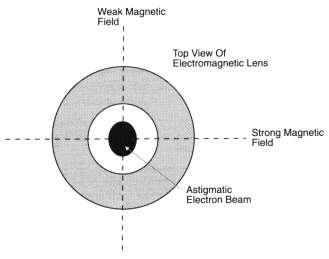
30 Filaments Lanthanum Hexaboride Schottky Field Emitter
 Crossover =")

31 Field Emission Gun: Types LaB6 JEOL JEM 1400 Lanthanum Hexaboride Vacuum (10-4 ) Crossover = 10 micron $1K Schottky FEG FEI Tecnai F30 Twin Zirconium Oxide High Vacuum < micron $12K W&C p.74
32 Spatial Coherence Perfect spatial coherence would be all electrons emit from the same point Smaller tips more coherent Better phase contrast Better resolution Which tip has better spatial coherence?
 or ZrO 2 W&C p.")
33 Field Emission Gun (FEG) Source of electrons = the gun Provides large stable current in a small electron beam Source determines resolution Tungsten (W) or ZrO 2 W&C p. 80
34 Field Emission Gun: How does it work? Produces electrons upon application of a large electric potential FEG = cathode (-300 kv) Anode 1 = extraction voltage Positive by a few kv Intense electric field Electrons tunnel out Anode 2 = accelerates electrons TEM = kv Produces a refined electrostatic lens for crossover W&C p.80
35 Magnify/Demagnify Focus Illumination Lenses: purpose


36 Lenses: what are they? Electromagnets Vary current using a coil around a soft iron core Affects resultant magnetic field W&C p.97

37 Lenses W&C p.93
38 Lens Abberations Spherical Chromatic


39 Lens Abberations: Astigmatism
40 Lens abberations: Astigmatism Astigmatism Corrected Astigmatism
41 Apertures Strip of metal 4 Holes drilled with decreasing size Limits undesirable scattering, leading to noise in images.

42 Digital Camera
43 Detector Quantum Efficiency Metric to compare efficiency of detection Where S = signal (electrons) N = Noise (background) A perfect detector would have a DQE of 1 Detectors have <1

44 Charge Coupled Device (CCD) Metal-insulator-silicon devices that store charge Each pixel in an array is an individual capacitor Isolated from each other Collect charge in the well Proportional to incident radiation intensity Our CCDs JEOL = 2K x 2K F30 = 4K x 4K W&C p.120

45 Renaissance in imaging Direct electron Detection Revolutionizing cryo-em Take movies and compensate for drift over course of acquisition
46 Dose


47 TEM Contrast mechanisms Thickness Mass Diffraction
48 Beam-specimen interactions Elastic Scattering No Energy loss High angle (>5 o ) Interactions with nucleus or inner valence electrons Inelastic Scattering Energy Loss Low angle <5 o Interactions with outer valence electrons Lost energy as X-rays Plasmons Beam damage Heat/bubbling
Protein structure determination by 3D Electron Microscopy
 Biochemistry 9001 Protein structure determination by 3D Electron Microscopy Tommi A. White, Ph.D. Director, Electron Microscopy Core Facility Assistant Research Professor, Biochemistry Goals of this course
Electron microscopy in molecular cell biology I
 Electron microscopy in molecular cell biology I Electron optics and image formation Werner Kühlbrandt Max Planck Institute of Biophysics chemistry biology Objects of interest Galaxy 10 6 light years 10
Transmission Electron Microscopy
 L. Reimer H. Kohl Transmission Electron Microscopy Physics of Image Formation Fifth Edition el Springer Contents 1 Introduction... 1 1.1 Transmission Electron Microscopy... 1 1.1.1 Conventional Transmission
= 6 (1/ nm) So what is probability of finding electron tunneled into a barrier 3 ev high?
 So what is probability of finding electron tunneled into a barrier 3 ev high?") STM STM With a scanning tunneling microscope, images of surfaces with atomic resolution can be readily obtained. An STM uses quantum tunneling of electrons to map the density of electrons on the surface
STM STM With a scanning tunneling microscope, images of surfaces with atomic resolution can be readily obtained. An STM uses quantum tunneling of electrons to map the density of electrons on the surface
sin" =1.22 # D "l =1.22 f# D I: In introduction to molecular electron microscopy - Imaging macromolecular assemblies
 I: In introduction to molecular electron microscopy - Imaging macromolecular assemblies Yifan Cheng Department of Biochemistry & Biophysics office: GH-S472D; email: ycheng@ucsf.edu 2/20/2015 - Introduction
History of 3D Electron Microscopy and Helical Reconstruction
 T H E U N I V E R S I T Y of T E X A S S C H O O L O F H E A L T H I N F O R M A T I O N S C I E N C E S A T H O U S T O N History of 3D Electron Microscopy and Helical Reconstruction For students of HI
Electron Microprobe Analysis 1 Nilanjan Chatterjee, Ph.D. Principal Research Scientist
 12.141 Electron Microprobe Analysis 1 Nilanjan Chatterjee, Ph.D. Principal Research Scientist Massachusetts Institute of Technology Electron Microprobe Facility Department of Earth, Atmospheric and Planetary
Electron Microprobe Analysis 1 Nilanjan Chatterjee, Ph.D. Principal Research Scientist
 12.141 Electron Microprobe Analysis 1 Nilanjan Chatterjee, Ph.D. Principal Research Scientist Massachusetts Institute of Technology Electron Microprobe Facility Department of Earth, Atmospheric and Planetary
object objective lens eyepiece lens
 Advancing Physics G495 June 2015 SET #1 ANSWERS Field and Particle Pictures Seeing with electrons The compound optical microscope Q1. Before attempting this question it may be helpful to review ray diagram
MSE 321 Structural Characterization
 Optical Microscope Plan Lenses In an "ideal" single-element lens system all planar wave fronts are focused to a point at distance f from the lens; therefore: Image near the optical axis will be in perfect
Lecture CIMST Winter School. 1. What can you see by TEM?
 Lecture CIMST Winter School Cryo-electron microscopy and tomography of biological macromolecules 20.1.2011 9:00-9:45 in Y03G91 Dr. Takashi Ishikawa OFLB/005 Tel: 056 310 4217 e-mail: takashi.ishikawa@psi.ch
Basic structure of SEM
 Table of contents Basis structure of SEM SEM imaging modes Comparison of ordinary SEM and FESEM Electron behavior Electron matter interaction o Elastic interaction o Inelastic interaction o Interaction
Introduction to Electron Microscopy Andres Kaech. Instrumentation
 Center for Microscopy and Image Analysis Introduction to Electron Microscopy Andres Kaech Instrumentation The types of electron microscopes Transmission electron microscope (TEM) Scanning electron microscope
Electron Microscopy. SEM = Scanning Electron Microscopy TEM = Transmission Electron Microscopy. E. coli, William E. Bentley, Maryland, USA
 Electron Microscopy Transmission Electron Microscopy SEM = Scanning Electron Microscopy TEM = Transmission Electron Microscopy Sara Henriksson, UCEM 2017-02-16 E. coli, William E. Bentley, Maryland, USA
AP5301/ Name the major parts of an optical microscope and state their functions.
 Review Problems on Optical Microscopy AP5301/8301-2015 1. Name the major parts of an optical microscope and state their functions. 2. Compare the focal lengths of two glass converging lenses, one with
tip conducting surface
 PhysicsAndMathsTutor.com 1 1. The diagram shows the tip of a scanning tunnelling microscope (STM) above a conducting surface. The tip is at a potential of 1.0 V relative to the surface. If the tip is sufficiently
Molecular electron microscopy
 Molecular electron microscopy - Imaging macromolecular assemblies Yifan Cheng Department of Biochemistry & Biophysics office: GH-S427B; email: ycheng@ucsf.edu 2/22/2013 - Introduction of Molecular Microscopy:
Scanning Electron Microscopy
 Scanning Electron Microscopy Field emitting tip Grid 2kV 100kV Anode ZEISS SUPRA Variable Pressure FESEM Dr Heath Bagshaw CMA bagshawh@tcd.ie Why use an SEM? Fig 1. Examples of features resolvable using
Invited Lecture. "Different Aspects of Electron Microscopy. Sardar Vallabhbhai National Institute of Technology, Surat. Deepak Rajput & S.K.
 Invited Lecture on "Different Aspects of Electron Microscopy at Sardar Vallabhbhai National Institute of Technology, Surat Deepak Rajput & S.K. Tiwary R&D and Product Development Essar Steel Limited Abstract
CHEM 681 Seminar Mingqi Zhao April 20, 1998 Room 2104, 4:00 p.m. High Resolution Transmission Electron Microscopy: theories and applications
 CHEM 681 Seminar Mingqi Zhao April 20, 1998 Room 2104, 4:00 p.m. High Resolution Transmission Electron Microscopy: theories and applications In materials science, people are always interested in viewing
MSE 321 Structural Characterization
 Auger Spectroscopy Auger Electron Spectroscopy (AES) Scanning Auger Microscopy (SAM) Incident Electron Ejected Electron Auger Electron Initial State Intermediate State Final State Physical Electronics
Modern Optical Spectroscopy
 Modern Optical Spectroscopy X-Ray Microanalysis Shu-Ping Lin, Ph.D. Institute of Biomedical Engineering E-mail: splin@dragon.nchu.edu.tw Website: http://web.nchu.edu.tw/pweb/users/splin/ Backscattered
SEM Doctoral Course MS-636. April 11-13, 2016
 Thomas LaGrange, Ph.D. Faculty Lecturer and Senior Staff Scientist Electron Sources, Optics and Detectors SEM Doctoral Course MS-636 April 11-13, 2016 Summary Electron propagation is only possible through
High-Resolution. Transmission. Electron Microscopy
 Part 4 High-Resolution Transmission Electron Microscopy 186 Significance high-resolution transmission electron microscopy (HRTEM): resolve object details smaller than 1nm (10 9 m) image the interior of
Part I Basics and Methods
 j1 Part I Basics and Methods In-situ Electron Microscopy: Applications in Physics, Chemistry and Materials Science, First Edition. Edited by Gerhard Dehm, James M. Howe, and Josef Zweck. Ó 2012 Wiley-VCH
MSE 321 Structural Characterization
 Auger Spectroscopy Auger Electron Spectroscopy (AES) Scanning Auger Microscopy (SAM) Incident Electron Ejected Electron Auger Electron Initial State Intermediate State Final State Physical Electronics
Chapter 9. Electron mean free path Microscopy principles of SEM, TEM, LEEM
 Chapter 9 Electron mean free path Microscopy principles of SEM, TEM, LEEM 9.1 Electron Mean Free Path 9. Scanning Electron Microscopy (SEM) -SEM design; Secondary electron imaging; Backscattered electron
Practical course in scanning electron microscopy
 Practical course in scanning electron microscopy Fortgeschrittenen Praktikum an der Technischen Universität München Wintersemester 2017/2018 Table of contents 1. Introduction 3 2. Formation of an electron
Part II: Thin Film Characterization
 Part II: Thin Film Characterization General details of thin film characterization instruments 1. Introduction to Thin Film Characterization Techniques 2. Structural characterization: SEM, TEM, AFM, STM
The illumination source: the electron beam
 The SEM Column The illumination source: the electron beam The probe of the electron microscope is an electron beam with very high and stable energy (10-100 kev) in order to get images with high resolution.
Praktikum zur. Materialanalytik
 Praktikum zur Materialanalytik Energy Dispersive X-ray Spectroscopy B513 Stand: 19.10.2016 Contents 1 Introduction... 2 2. Fundamental Physics and Notation... 3 2.1. Alignments of the microscope... 3 2.2.
Basic Principles Brief history of EM
 SIR WILLIAM DUNN SCHOOL OF PATHOLOGY Basic Principles of Electron Microscopy (EM) Dr Errin Johnson Head of the Dunn School EM Facility Basic Principles Brief history of EM 1873 Hermann von Helmholtz &
Nano-Microscopy. Lecture 2. Scanning and Transmission Electron Microscopies: Principles. Pavel Zinin HIGP, University of Hawaii, Honolulu, USA
 GG 711: Advanced Techniques in Geophysics and Materials Science Nano-Microscopy. Lecture 2 Scanning and Transmission Electron Microscopies: Principles Pavel Zinin HIGP, University of Hawaii, Honolulu,
Analytical Methods for Materials
 Analytical Methods for Materials Lesson 21 Electron Microscopy and X-ray Spectroscopy Suggested Reading Leng, Chapter 3, pp. 83-126; Chapter 4, pp. 127-160; Chapter 6, pp. 191-219 P.J. Goodhew, J. Humphreys
The Basic of Transmission Electron Microscope. Text book: Transmission electron microscopy by David B Williams & C. Barry Carter.
 The Basic of Transmission Electron Microscope Text book: Transmission electron microscopy by David B Williams & C. Barry Carter. 2009, Springer Background survey http://presemo.aalto.fi/tem1 Microscopy
Chapter 2 Structure and Imaging of a Transmission Electron Microscope (TEM)
") Chapter 2 Structure and Imaging of a Transmission Electron Microscope (TEM) In this chapter, we overview the structure of a transmission electron microscope (TEM) for nanoimaging, and mathematical descriptions
Chapter 2 Structure and Imaging of a Transmission Electron Microscope (TEM) In this chapter, we overview the structure of a transmission electron microscope (TEM) for nanoimaging, and mathematical descriptions
M2 TP. Low-Energy Electron Diffraction (LEED)
") M2 TP Low-Energy Electron Diffraction (LEED) Guide for report preparation I. Introduction: Elastic scattering or diffraction of electrons is the standard technique in surface science for obtaining structural
M2 TP Low-Energy Electron Diffraction (LEED) Guide for report preparation I. Introduction: Elastic scattering or diffraction of electrons is the standard technique in surface science for obtaining structural
Why microscopy?
 Electron Microscopy Why microscopy? http://www.cellsalive.com/howbig.htm 2 Microscopes are used as magnifying tools (although not exclusively as will see later on). The resolution of the human eye is limited
CIMST Summer School Cryo-electron microscopy for structural biology 11 September, 2014 Dr. Takashi Ishikawa
 CIMST Summer School Cryo-electron microscopy for structural biology 11 September, 2014 Dr. Takashi Ishikawa Tel: 056 310 4217 e-mail: takashi.ishikawa@psi.ch Lab webpage: http://www.psi.ch/lbr/takashi-ishikawa
CHARACTERIZATION of NANOMATERIALS KHP
 CHARACTERIZATION of NANOMATERIALS Overview of the most common nanocharacterization techniques MAIN CHARACTERIZATION TECHNIQUES: 1.Transmission Electron Microscope (TEM) 2. Scanning Electron Microscope
Imaging Methods: Scanning Force Microscopy (SFM / AFM)
") Imaging Methods: Scanning Force Microscopy (SFM / AFM) The atomic force microscope (AFM) probes the surface of a sample with a sharp tip, a couple of microns long and often less than 100 Å in diameter.
Imaging Methods: Scanning Force Microscopy (SFM / AFM) The atomic force microscope (AFM) probes the surface of a sample with a sharp tip, a couple of microns long and often less than 100 Å in diameter.
h p λ = mν Back to de Broglie and the electron as a wave you will learn more about this Equation in CHEM* 2060
 Back to de Broglie and the electron as a wave λ = mν h = h p you will learn more about this Equation in CHEM* 2060 We will soon see that the energies (speed for now if you like) of the electrons in the
Transmission Electron Microscope. Experimental Instruction
 Transmission Electron Microscope Experimental Instruction In advanced practical course [F-Praktikum] Date: April 2017 Contents 1 Task 3 2 Theoretical Basics 3 2.1 Bragg Diffraction......................................
Transmission Electron Microscopy
 Transmission Electron Microscopy Fu-Rong Chen Transmission Electron Microscopy David B. Williams C. Barry Carter Background:Solid State Physics Materials Science 1.1 Why Electron Microscope? 1.1 Why Electron
QUANTUM PHYSICS. Limitation: This law holds well only for the short wavelength and not for the longer wavelength. Raleigh Jean s Law:
 Black body: A perfect black body is one which absorbs all the radiation of heat falling on it and emits all the radiation when heated in an isothermal enclosure. The heat radiation emitted by the black
Interactions with Matter
 Manetic Lenses Manetic fields can displace electrons Manetic field can be produced by passin an electrical current throuh coils of wire Manetic field strenth can be increased by usin a soft ferromanetic
SEM Optics and Application to Current Research
 SEM Optics and Application to Current Research Azure Avery May 28, 2008 1 Introduction 1.1 History The optical microscope was invented in the early 17th century. Although revolutionary, the earliest microscopes
SEM stands for Scanning Electron Microscopy. The earliest known work describing
 1. HISTORY ABOUT SEM SEM stands for Scanning Electron Microscopy. The earliest known work describing the concept of a Scanning Electron Microscope was by M. Knoll (1935) who, along with other pioneers
Gaetano L Episcopo. Scanning Electron Microscopy Focus Ion Beam and. Pulsed Plasma Deposition
 Gaetano L Episcopo Scanning Electron Microscopy Focus Ion Beam and Pulsed Plasma Deposition Hystorical background Scientific discoveries 1897: J. Thomson discovers the electron. 1924: L. de Broglie propose
Chapter 10: Wave Properties of Particles
 Chapter 10: Wave Properties of Particles Particles such as electrons may demonstrate wave properties under certain conditions. The electron microscope uses these properties to produce magnified images
Electron Microscopy (TEM and SEM)
") 7 Electron Microscopy (TEM and SEM) Paul Verkade Wolfson Bioimaging Facility, Physiology & Pharmacology and Biochemistry, University of Bristol, UK 7.1 Basic how-to-do and why-do section 7.1.1 Electron
7 Electron Microscopy (TEM and SEM) Paul Verkade Wolfson Bioimaging Facility, Physiology & Pharmacology and Biochemistry, University of Bristol, UK 7.1 Basic how-to-do and why-do section 7.1.1 Electron
MT Electron microscopy Scanning electron microscopy and electron probe microanalysis
 MT-0.6026 Electron microscopy Scanning electron microscopy and electron probe microanalysis Eero Haimi Research Manager Outline 1. Introduction Basics of scanning electron microscopy (SEM) and electron
Chapter 2 Instrumentation for Analytical Electron Microscopy Lecture 5. Chapter 2 CHEM 793, 2011 Fall 1
 Chater Instrumentation for Analytical Electron Microscoy Lecture 5 Chater CHEM 793, 011 Fall 1 Outline Electron Sources (Electron Guns) Thermionic: LaB 6 or W Field emission gun: cold or Schottky Lenses
Transmission Electron Microscopy: A Textbook For Materials Science (4-Vol Set) By C. Barry Carter, David B. Williams
 By C. Barry Carter, David B. Williams") Transmission Electron Microscopy: A Textbook For Materials Science (4-Vol Set) By C. Barry Carter, David B. Williams If you are searched for the ebook Transmission Electron Microscopy: A Textbook for Materials
Transmission Electron Microscopy: A Textbook For Materials Science (4-Vol Set) By C. Barry Carter, David B. Williams If you are searched for the ebook Transmission Electron Microscopy: A Textbook for Materials
Chemical Analysis in TEM: XEDS, EELS and EFTEM. HRTEM PhD course Lecture 5
 Chemical Analysis in TEM: XEDS, EELS and EFTEM HRTEM PhD course Lecture 5 1 Part IV Subject Chapter Prio x-ray spectrometry 32 1 Spectra and mapping 33 2 Qualitative XEDS 34 1 Quantitative XEDS 35.1-35.4
Microscopy, Staining, and Classification
 PowerPoint Lecture Presentations prepared by Mindy Miller-Kittrell, North Carolina State University C H A P T E R 4 Microscopy, Staining, and Classification Microscopy Light Microscopy 1) Bright-field
Weak-Beam Dark-Field Technique
 Basic Idea recall bright-field contrast of dislocations: specimen close to Bragg condition, s î 0 Weak-Beam Dark-Field Technique near the dislocation core, some planes curved to s = 0 ) strong Bragg reflection
April 10th-12th, 2017
 Thomas LaGrange, Ph.D. Faculty Lecturer and Senior Staff Scientist Introduction: Basics of Transmission Electron Microscopy (TEM) TEM Doctoral Course MS-637 April 10th-12th, 2017 Outline 1. What is microcopy?
MEMS Metrology. Prof. Tianhong Cui ME 8254
 MEMS Metrology Prof. Tianhong Cui ME 8254 What is metrology? Metrology It is the science of weights and measures Refers primarily to the measurements of length, weight, time, etc. Mensuration- A branch
A Brief Introduction to Medical Imaging. Outline
 A Brief Introduction to Medical Imaging Outline General Goals Linear Imaging Systems An Example, The Pin Hole Camera Radiations and Their Interactions with Matter Coherent vs. Incoherent Imaging Length
Quantum Interference and Duality
 Quantum Interference and Duality Kiyohide NOMURA Department of Physics December 21, 2016 1 / 49 Quantum Physics(Mechanics) Basic notion of Quantum Physics: Wave-Particle Duality Light (electromagnetic
Basic physics Questions
 Chapter1 Basic physics Questions S. Ilyas 1. Which of the following statements regarding protons are correct? a. They have a negative charge b. They are equal to the number of electrons in a non-ionized
What can we see with a transmission electron microscope?
 What can we see with a transmission electron microscope? β-galactosidase Subramaniam Lab o.lambert@cbmn.u-bordeaux.fr Renafobis 2017 1 Film or camera Renafobis 2017 2 1897 J.J. Thomson Electron discovery
Transmission Electron Microscopy. Part #1 Diffraction Conventional Imaging
 Transmission Electron Microscopy Part #1 Diffraction Conventional Imaging Nicolas Menguy Institut de Minéralogie, de Physique des Matériaux et de Cosmochimie Outline Part 1 : Conventional TEM - Transmission
History of electron microscopy
 History of electron microscopy C9940 3-Dimensional Transmission Electron Microscopy S1007 Doing structural biology with the electron microscope February 16, 2015 Syllabus Week Date Instructor Topic 1 02/16
Electron beam scanning
 Electron beam scanning The Electron beam scanning operates through an electro-optical system which has the task of deflecting the beam Synchronously with cathode ray tube which create the image, beam moves
November 30th -December 2 nd, st 2nd 3rd. 8:15 7)HRTEM 10) TEM imaging and diffraction examples. 9:15 8)HRTEM 10) Diffraction going further
HRTEM 10) TEM imaging and diffraction examples. 9:15 8)HRTEM 10) Diffraction going further") Thomas LaGrange, Ph.D. Faculty and Staff Scientist Introduction: Basics of Transmission Electron Microscopy (TEM) TEM Doctoral Course MS-637 November 30th -December 2 nd, 2015 Planning MSE-637 TEM -basics
Thomas LaGrange, Ph.D. Faculty and Staff Scientist Introduction: Basics of Transmission Electron Microscopy (TEM) TEM Doctoral Course MS-637 November 30th -December 2 nd, 2015 Planning MSE-637 TEM -basics
Energy-Filtering. Transmission. Electron Microscopy
 Part 3 Energy-Filtering Transmission Electron Microscopy 92 Energy-Filtering TEM Principle of EFTEM expose specimen to mono-energetic electron radiation inelastic scattering in the specimen poly-energetic
Ecole Franco-Roumaine : Magnétisme des systèmes nanoscopiques et structures hybrides - Brasov, Modern Analytical Microscopic Tools
 1. Introduction Solid Surfaces Analysis Group, Institute of Physics, Chemnitz University of Technology, Germany 2. Limitations of Conventional Optical Microscopy 3. Electron Microscopies Transmission Electron
Photon Instrumentation. First Mexican Particle Accelerator School Guanajuato Oct 6, 2011
 Photon Instrumentation First Mexican Particle Accelerator School Guanajuato Oct 6, 2011 Outline The Electromagnetic Spectrum Photon Detection Interaction of Photons with Matter Photoelectric Effect Compton
Scanning Electron Microscopy & Ancillary Techniques
 Scanning Electron Microscopy & Ancillary Techniques By Pablo G. Caceres-Valencia The prototype of the first Stereoscan supplied by the Cambridge Instrument Company to the dupont Company, U.S.A. (1965)
The Tecnai Arctica (TEM-9) Cryo-EM workflow. Contact: Svetla Stoilova-McPhie, PhD Advanced bioimaging scientist
 Cryo-EM workflow. Contact: Svetla Stoilova-McPhie, PhD Advanced bioimaging scientist") The Tecnai Arctica (TEM-9) Cryo-EM workflow Contact: Svetla Stoilova-McPhie, PhD Advanced bioimaging scientist stoilovamcphie@fas.harvard.edu The Arctica Cryo-EM training chart Cryo-TEM training involves
The Tecnai Arctica (TEM-9) Cryo-EM workflow Contact: Svetla Stoilova-McPhie, PhD Advanced bioimaging scientist stoilovamcphie@fas.harvard.edu The Arctica Cryo-EM training chart Cryo-TEM training involves
IMAGING DIFFRACTION SPECTROSCOPY
 TEM Techniques TEM/STEM IMAGING DIFFRACTION SPECTROSCOPY Amplitude contrast (diffracion contrast) Phase contrast (highresolution imaging) Selected area diffraction Energy dispersive X-ray spectroscopy
Conventional Transmission Electron Microscopy. Introduction. Text Books. Text Books. EMSE-509 CWRU Frank Ernst
 Text Books Conventional Transmission Electron Microscopy EMSE-509 CWRU Frank Ernst D. B. Williams and C. B. Carter: Transmission Electron Microscopy, New York: Plenum Press (1996). L. Reimer: Transmission
6. Analytical Electron Microscopy
 Physical Principles of Electron Microscopy 6. Analytical Electron Microscopy Ray Egerton University of Alberta and National Institute of Nanotechnology Edmonton, Canada www.tem-eels.ca regerton@ualberta.ca
Electron Microscopy I
 Characterization of Catalysts and Surfaces Characterization Techniques in Heterogeneous Catalysis Electron Microscopy I Introduction Properties of electrons Electron-matter interactions and their applications
Auger Electron Spectroscopy (AES) Prof. Paul K. Chu
 Prof. Paul K. Chu") Auger Electron Spectroscopy (AES) Prof. Paul K. Chu Auger Electron Spectroscopy Introduction Principles Instrumentation Qualitative analysis Quantitative analysis Depth profiling Mapping Examples The Auger
Auger Electron Spectroscopy (AES) Prof. Paul K. Chu Auger Electron Spectroscopy Introduction Principles Instrumentation Qualitative analysis Quantitative analysis Depth profiling Mapping Examples The Auger
Scanning Electron Microscopy
 Scanning Electron Microscopy Amanpreet Kaur 1 www.reading.ac.uk/emlab Scanning Electron Microscopy What is scanning electron microscopy? Basic features of conventional SEM Limitations of conventional SEM
From electron crystallography to single particle cryoem
 From electron crystallography to single particle cryoem Nobel Lectures in Chemistry 8 th December 2017 Richard Henderson From Baker & Henderson (2001) Int.Tab.Cryst.Vol.F, on-line (2006), revised (2011)
Particles and Waves Particles Waves
 Particles and Waves Particles Discrete and occupy space Exist in only one location at a time Position and velocity can be determined with infinite accuracy Interact by collisions, scattering. Waves Extended,
Low Vacuum Scanning Electron Microscopy and Microanalysis
 Low Vacuum Scanning Electron Microscopy and Microanalysis Principles and Practice of Variable Pressure/Environmental Scanning Electron Microscopy (VP-ESEM), Debbie J Stokes, John Wiley&Sons 2008 Several
KMÜ 396 MATERIALS SCIENCE AND TECH. I PRESENTATION ELECTRON ENERGY LOSS SPECTROSCOPY (EELS) TUĞÇE SEZGİN
 TUĞÇE SEZGİN") KMÜ 396 MATERIALS SCIENCE AND TECH. I PRESENTATION ELECTRON ENERGY LOSS SPECTROSCOPY (EELS) TUĞÇE SEZGİN 20970725 HACETTEPE UNIVERSITY DEPARTMENT OF CHEMICAL ENGINEERING, SPRING 2011,APRIL,ANKARA CONTENTS
KMÜ 396 MATERIALS SCIENCE AND TECH. I PRESENTATION ELECTRON ENERGY LOSS SPECTROSCOPY (EELS) TUĞÇE SEZGİN 20970725 HACETTEPE UNIVERSITY DEPARTMENT OF CHEMICAL ENGINEERING, SPRING 2011,APRIL,ANKARA CONTENTS
PHYS 3313 Section 001 Lecture #7
 PHYS 3313 Section 001 Lecture #7 Photoelectric Effect Compton Effect Pair production/pair annihilation PHYS 3313-001, Fall 1 Reading assignments: CH3.9 Announcements Homework #2 CH3 end of the chapter
stands for Transmission Electron (Microscope/Microscopy) Q: Why use electrons instead of light for imaging nanomaterials?
 Q: Why use electrons instead of light for imaging nanomaterials?") What is TEM? stands for Transmission Electron (Microscope/Microscopy) Q: Why use electrons instead of light for imaging nanomaterials? A: 1) Shorter wavelength () Higher resolution ) Wavelength determined
What is TEM? stands for Transmission Electron (Microscope/Microscopy) Q: Why use electrons instead of light for imaging nanomaterials? A: 1) Shorter wavelength () Higher resolution ) Wavelength determined
4. Inelastic Scattering
 1 4. Inelastic Scattering Some inelastic scattering processes A vast range of inelastic scattering processes can occur during illumination of a specimen with a highenergy electron beam. In principle, many
HOW TO APPROACH SCANNING ELECTRON MICROSCOPY AND ENERGY DISPERSIVE SPECTROSCOPY ANALYSIS. SCSAM Short Course Amir Avishai
 HOW TO APPROACH SCANNING ELECTRON MICROSCOPY AND ENERGY DISPERSIVE SPECTROSCOPY ANALYSIS SCSAM Short Course Amir Avishai RESEARCH QUESTIONS Sea Shell Cast Iron EDS+SE Fe Cr C Objective Ability to ask the
Electron Microprobe Analysis and Scanning Electron Microscopy
 Electron Microprobe Analysis and Scanning Electron Microscopy Electron microprobe analysis (EMPA) Analytical technique in which a beam of electrons is focused on a sample surface, producing X-rays from
Astronomy 203 practice final examination
 Astronomy 203 practice final examination Fall 1999 If this were a real, in-class examination, you would be reminded here of the exam rules, which are as follows: You may consult only one page of formulas
Chemistry Instrumental Analysis Lecture 19 Chapter 12. Chem 4631
 Chemistry 4631 Instrumental Analysis Lecture 19 Chapter 12 There are three major techniques used for elemental analysis: Optical spectrometry Mass spectrometry X-ray spectrometry X-ray Techniques include:
I live in this atom, with my other electron brothers
 Hello, my name is Electron, John Electron. I am going to tell you how my work is in an electron microscope. I live in this atom, with my other electron brothers The filament crowns the column of the electron
CBE Science of Engineering Materials. Scanning Electron Microscopy (SEM)
") CBE 30361 Science of Engineering Materials Scanning Electron Microscopy (SEM) Scale of Structure Organization Units: micrometer = 10-6 m = 1µm nanometer= 10-9 m = 1nm Angstrom = 10-10 m = 1Å A hair is
CBE 30361 Science of Engineering Materials Scanning Electron Microscopy (SEM) Scale of Structure Organization Units: micrometer = 10-6 m = 1µm nanometer= 10-9 m = 1nm Angstrom = 10-10 m = 1Å A hair is
Modern physics ideas are strange! L 36 Modern Physics [2] The Photon Concept. How are x-rays produced? The uncertainty principle
![Modern physics ideas are strange! L 36 Modern Physics [2] The Photon Concept. How are x-rays produced? The uncertainty principle](/thumbs/88/117098787.jpg "Modern physics ideas are strange! L 36 Modern Physics [2] The Photon Concept. How are x-rays produced? The uncertainty principle") L 36 Modern Physics [2] X-rays & gamma rays How lasers work Medical applications of lasers Applications of high power lasers Medical imaging techniques CAT scans MRI s Modern physics ideas are strange!
L 36 Modern Physics [2] X-rays & gamma rays How lasers work Medical applications of lasers Applications of high power lasers Medical imaging techniques CAT scans MRI s Modern physics ideas are strange!
The Photon Concept. Modern Physics [2] How are x-rays produced? Gamma rays. X-ray and gamma ray photons. X-rays & gamma rays How lasers work
![The Photon Concept. Modern Physics [2] How are x-rays produced? Gamma rays. X-ray and gamma ray photons. X-rays & gamma rays How lasers work](/thumbs/75/72921848.jpg "The Photon Concept. Modern Physics [2] How are x-rays produced? Gamma rays. X-ray and gamma ray photons. X-rays & gamma rays How lasers work") Modern Physics [2] X-rays & gamma rays How lasers work Medical applications of lasers Applications of high power lasers Medical imaging techniques CAT scans MRI s The Photon Concept a beam of light waves
Modern Physics [2] X-rays & gamma rays How lasers work Medical applications of lasers Applications of high power lasers Medical imaging techniques CAT scans MRI s The Photon Concept a beam of light waves
Everhart-Thornley detector
 SEI Detector Everhart-Thornley detector Microscope chamber wall Faraday cage Scintillator Electrons in Light pipe Photomultiplier Electrical signal out Screen Quartz window +200 V +10 kv Always contains
Basic principles of x-ray production
 Production of X-Rays part 1 George Starkschall, Ph.D. Lecture Objectives Identify what is needed to produce x-rays Describe how a diagnostic x-ray tube produces x-rays Describe the types of interactions
X-RAY PRODUCTION. Prepared by:- EN KAMARUL AMIN BIN ABDULLAH
 X-RAY PRODUCTION Prepared by:- EN KAMARUL AMIN BIN ABDULLAH OBJECTIVES Discuss the process of x-ray being produced (conditions) Explain the principles of energy conversion in x-ray production (how energy
The Wave Nature of Matter *
 OpenStax-CNX module: m42576 1 The Wave Nature of Matter * OpenStax This work is produced by OpenStax-CNX and licensed under the Creative Commons Attribution License 3.0 Abstract Describe the Davisson-Germer
Introduction to electron microscopes: electron optics, interactions and signals
 Introduction to electron microscopes: electron optics, interactions and signals J.L. Lábár 1 Research Institute for Technical Physics and Materials Science, H-1121, Budapest, Konkoly- Thege u. 29-33, Hungary
SOLID STATE PHYSICS PHY F341. Dr. Manjuladevi.V Associate Professor Department of Physics BITS Pilani
 SOLID STATE PHYSICS PHY F341 Dr. Manjuladevi.V Associate Professor Department of Physics BITS Pilani 333031 manjula@bits-pilani.ac.in Characterization techniques SEM AFM STM BAM Outline What can we use
Assessment of the Azimuthal Homogeneity of the Neutral Gas in a Hall Effect Thruster using Electron Beam Fluorescence
 Assessment of the Azimuthal Homogeneity of the Neutral Gas in a Hall Effect Thruster using Electron Beam Fluorescence IEPC-2015-91059 / ISTS-2015-b-91059 Presented at Joint Conference of 30th International