Structure and mechanism of an intramembrane liponucleotide synthetase central for phospholipid biosynthesis
|
|
|
- Clement Hood
- 5 years ago
- Views:
Transcription
1 Structure and mechanism of an intramembrane liponucleotide synthetase central for phospholipid biosynthesis Xiuying Liu 1,3, Yan Yin 1,2,3, Jinjun Wu 1 and Zhenfeng Liu 1 1 National Laboratory of Biomacromolecules, Institute of Biophysics, Chinese Academy of Sciences, 15 Datun Road, Chaoyang District, Beijing, People s Republic of China. 2 University of Chinese Academy of Sciences, Beijing, People s Republic of China. 3 These authors contributed equally to the project. Supplementary Figures 1-10 and Table 1. 1
 Prokaryotic phospholipid biosynthesis pathways mediated by CDP-DAG.")
2 Supplementary Figure 1 The CDP-DAG mediated phospholipid biosynthesis and recycling pathways in prokaryotic and eukaryotic organisms. (a) Prokaryotic phospholipid biosynthesis pathways mediated by CDP-DAG. ACP, acyl carrier protein; PlsY/PlsB, Glycerol-3-phosphate acyltransferase; PlsC, 1-acyl-sn-glycerol-3-phosphate acyltransferase; Cds: CDP-DAG synthetase, encoded CdsA gene in prokaryotes (or by CDS genes in eukaryotes). PssA, bacterial phosphatidylserine synthase; Psd, phosphatidylserine decarboxylase; PgsA, phosphatidylglycerol phosphate (PGP) synthase; PgsA*, The pgsa gene in M. smegmatis encodes the phosphatidylinositol synthase enzyme. PgpA/B/C, PGP phosphatases; Cls, cardiolipin synthase. (b) Eukaryotic phospholipid biosynthesis and 2
3 recycling pathways mediated by CDP-DAG. GPAT, Glycerol-3-phosphate acyltransferase; AGPAT, 1-acylglycerol-3-phosphate-O-acyltransferase; PI kinase, phosphatidylinositol kinase; PIP kinase, phosphatidylinositol phosphate kinase; PLC, phospholipase C; DAGK, diacylglycerol kinase; PGS, PGP synthase; PSS, phosphatidylserine synthase found in Saccharomyces cerevisiae and some plants (such as wheat) with function similar to that of bacterial PssA; PSD, phosphatidylserine decarboxylase. 3
 Zoom-in views of the red (b) and blue-box (c) shaded interfacial regions shown in a, respectively.")
4 Supplementary Figure 2 Role of methylmercury in stabilizing the crystal packing interfaces between adjacent TmCdsA molecules. (a) Overview of the crystal packing of TmCdsA. (b and c) Zoom-in views of the red (b) and blue-box (c) shaded interfacial regions shown in a, respectively. Mercury atoms are shown as purple spheres and the cysteine residues involved in binding methylmercury groups are presented as stick models. 4
 or along the membrane normal from periplasmic side (b).")
5 Supplementary Figure 3 The experimental electron density maps of TmCdsA. (a, b) The initial electron density map at 3.8 Å resolution output by the Phenix Autosol program with Autobuild model superposed on the map. The map is viewed along the membrane plane (a) or along the membrane normal from periplasmic side (b). (c, d) Anomalous difference Fourier maps of various mercury derivatives used as site-specific labels for the verification of sequence registration during structural model building and refinement. 5
 Top view along the membrane normal.")
6 Supplementary Figure 4 Superposition of the overall structures of S200C/S223C (inactive) and S200C/S258C (active) mutants of TmCdsA. (a) Side view along the membrane plane. Color codes: blue, S200C/S223C; orange, S200C/S258C. Stereo images are presented. (b) Top view along the membrane normal. (c) A zoom-in view of the engineered C223 and C258 sites in the two structures. The two cysteine residues are highlighted as sphere models. The red arrows show the slight differences between the two structures. 6
 Crosslinker concentration and time-dependent crosslinking kinetics of wild type TmCdsA, S200C/S258C and S200C/S223C mutants. GA, glutaraldehyde.")
7 Supplementary Figure 5 The oligomeric states of TmCdsA and mutants in solution. (a, b) Crosslinker concentration and time-dependent crosslinking kinetics of wild type TmCdsA, S200C/S258C and S200C/S223C mutants. GA, glutaraldehyde. (c, d, e) Size-exclusion chromatography with multi-angle light-scattering (SEC-MALS) measurements on the protein masses of wild type TmCdsA (c), S200C/S258C (d) and S200C/S223C (e) mutants. 7
8 8

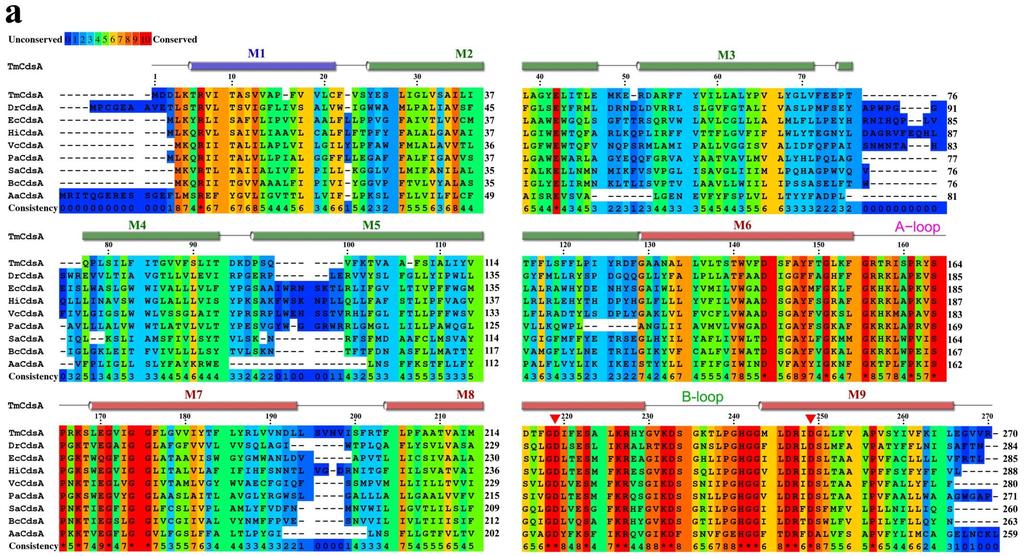
9 Supplementary Figure 6 Alignment of the amino acid sequences of TmCdsA and other Cds homologs. (a) TmCdsA aligned with various prokaryotic Cds homologs. The different background color codes indicate the varying degree of amino acid residue conservation among the Cds homologs, with the red color highlighting identical residues and blue color denoting unconserved residues. The regions covering transmembrane M1-M9 helices are labeled above the sequence as blue, green and red cylinders. The domain-specific color codes of the transmembrane helices 9
10 are consistent with those shown in Figure 3c, namely blue for NTD, green for MD and red for CTD. The solid red triangles mark the two highly conserved Asp residues (D219 and D249) that are directly involved in binding the Mg 2+ -K + di-metal center and essential for the enzyme activity. Abbreviation of species names: Tm, Thermotoga maritima; Dr, Deinococcus radiodurans; Ec, Escherichia coli; Hi, Haemophilus influenzae; Vc, Vibrio cholerae; Pa, Pseudomonas aeruginosa; Sa, Staphylococcus aureus; Bc, Bacillus cereus; Aa, Aquifex aeolicus. The amino acid sequence of TmCdsA shares 27% identity or 49% similarity with that of EcCdsA which was originally purified and characterized in an early biochemical study 1. (b) TmCdsA aligned with various eukaryotic Cds homologs. Sc, Saccharomyces cerevisiae; Da, Danio rerio; Hs, Homo sapiens; Dm, Drosophila melanogaster. The DmCds1 and DaCds2 are the two eukaryotic homologs reported to have essential roles in the recycling of phosphoinositide during signal transduction 2,3. 10
 ions bound to TmCdsA. (b) Isomorphous and anomalous difference Fourier peaks of Mn 2+ and Cs + ion bound to TmCdsA. The F Mn -F Mg map in blue is contoured at +5.")
11 Supplementary Figure 7 Probing the active site of TmCdsA with heavy surrogates of K + and Mg 2+ ions. (a) Anomalous difference Fourier peaks of Tl + (red, +5.0 level) and Ba 2+ (green, +6.0 level) ions bound to TmCdsA. (b) Isomorphous and anomalous difference Fourier peaks of Mn 2+ and Cs + ion bound to TmCdsA. The F Mn -F Mg map in blue is contoured at +5.5 level, while the Mn-anomalous difference map in purple is at +4.0 level. The F Cs -F K map in orange and the Cs-anomalous difference map in cyan are both contoured at +6.0 level. 11
12 Supplementary Figure 8 Rb + and Mn 2+ ions simultaneously bind to the active site of TmCdsA. The F Rb+Mn -F K+Mn isomorphous difference map in blue is contoured at +3.0 level. The anomalous difference map in purple using data collected at Å wavelength is contoured at +3.5 level. Only Mn 2+ instead of Rb + produces detectable anomalous signal under this wavelength. Both maps were computed to 4.5 Å resolution. The silver bullet models indicate the refined positions of Mg 2+ and K + ions in the structure of S200C/S223C crystal. The peaks of Rb + and Mn 2+ ions slightly deviate from the positions of K + and Mg 2+ site due to minor changes of the unit cell dimensions in the derivatized crystal. 12
13 Supplementary Figure 9 Analyses on the mono-dispersity of the wild-type, EDTA-treated TmCdsA proteins and the various mutants used for the activity assay. (a, b and c) Gel filtration (superdex /300 GL) profiles of the EDTA-treated TmCdsA and various mutants used for activity assay shown in Fig. 4e. For comparison, the elution profile of the wild-type protein is included in all three panels. (d) Dynamic light-scattering analyses on the main peak fractions (at around 12.8 ml) eluted from the gel filtration column for further verification of sample mono-dispersity. R h (nm) represents the hydrodynamic radius of the particles in solution. 13
14 Supplementary Figure 10 Substrate-dependent kinetic analyses of TmCdsA in the presence of 200 mm KCl and 2 mm MgCl 2. (a) CTP-dependent kinetic data measured in the presence of 2 mm DOPA. Each individual data points are plotted as mean value ± standard error (SEM as indicated by the error bars. n=4 For the data points with [CTP] = 0.5, 1.0, 2.0 and 3.0 mm, while the other data points were measured with n=3 instead). For those data points with small errors, the error bars are buried within the symbols. (b) PA-dependent kinetic data measured in the presence of 4 mm CTP. For the data points with [PA] = 0.75, 1 and 1.5 mm, n=4, while the others are with n=3 instead. The kinetic parameters of the fitted curves are summarized in Supplementary Table 1. 14
15 15
16 Supplementary Table 1 Kinetic parameters of TmCdsA and EcCdsA Enzyme CTP Dependence V max K half /K m Hill coefficient PA dependence V max K half /K m Hill coefficient TmCdsA 1.52 ± 0.11 K half = 1.82 ± ± ± 0.34 K half = 0.22 ± ± 0.4 EcCdsA 55.0 K m = 0.58 ± 0.16 N/A 55.0 K m = 0.28 ± 0.05 N/A The parameters are estimated by using the GraphPad Prism 6 software to fit the plots of initial velocity versus substrate (CTP or PA) concentration shown in Supplementary Figure 10. For CTP-dependent and PA-dependent kinetic data of TmCdsA, the allosteric sigmoidal model ((Y = V max *X h /(K half h + X h ); h = Hill coefficient) is applied for data fitting. The error presented is ± S. E. of the fit. Units for V max are mol of CDP-DAG produced per min per mg enzyme. The unit for K half or K m is mm. Data reported in the literature 1. Supplementary References: 1. Sparrow, C. P. & Raetz, C. R. Purification and properties of the membrane-bound CDP-diglyceride synthetase from Escherichia coli. J. Biol. Chem. 260, (1985). 2. Wu, L., Niemeyer, B., Colley, N., Socolich, M. & Zuker, C. S. Regulation of PLC-mediated signalling in vivo by CDP-diacylglycerol synthase. Nature 373, (1995). 3. Pan, W. et al. CDP-diacylglycerol synthetase-controlled phosphoinositide availability limits VEGFA signaling and vascular morphogenesis. Blood 120, (2012). 16
SUPPLEMENTARY INFORMATION
 Supplementary Table 1: Amplitudes of three current levels. Level 0 (pa) Level 1 (pa) Level 2 (pa) TrkA- TrkH WT 200 K 0.01 ± 0.01 9.5 ± 0.01 18.7 ± 0.03 200 Na * 0.001 ± 0.01 3.9 ± 0.01 12.5 ± 0.03 200
SUPPLEMENTARY INFORMATION
 Supplementary Table 1: Data collection, phasing and refinement statistics ChbC/Ta 6 Br 12 Native ChbC Data collection Space group P4 3 2 1 2 P4 3 2 1 2 Cell dimensions a, c (Å) 132.75, 453.57 132.81, 452.95
SUPPLEMENTARY INFORMATION
 doi:10.1038/nature12045 Supplementary Table 1 Data collection and refinement statistics. Native Pt-SAD X-ray source SSRF BL17U SPring-8 BL41XU Wavelength (Å) 0.97947 1.07171 Space group P2 1 2 1 2 1 P2
SUPPLEMENTARY INFORMATION
 SUPPLEMENTARY INFORMATION doi:10.1038/nature11524 Supplementary discussion Functional analysis of the sugar porter family (SP) signature motifs. As seen in Fig. 5c, single point mutation of the conserved
Supplementary Figure 1. Biochemical and sequence alignment analyses the
 Supplementary Figure 1. Biochemical and sequence alignment analyses the interaction of OPTN and TBK1. (a) Analytical gel filtration chromatography analysis of the interaction between TBK1 CTD and OPTN(1-119).
Nature Structural & Molecular Biology: doi: /nsmb Supplementary Figure 1
 Supplementary Figure 1 Crystallization. a, Crystallization constructs of the ET B receptor are shown, with all of the modifications to the human wild-type the ET B receptor indicated. Residues interacting
Supplementary Figure 1 Crystal contacts in COP apo structure (PDB code 3S0R)
") Supplementary Figure 1 Crystal contacts in COP apo structure (PDB code 3S0R) Shown in cyan and green are two adjacent tetramers from the crystallographic lattice of COP, forming the only unique inter-tetramer
Supplementary Figure 1 Crystal contacts in COP apo structure (PDB code 3S0R) Shown in cyan and green are two adjacent tetramers from the crystallographic lattice of COP, forming the only unique inter-tetramer
SUPPLEMENTARY INFORMATION
 doi:10.1038/nature11085 Supplementary Tables: Supplementary Table 1. Summary of crystallographic and structure refinement data Structure BRIL-NOP receptor Data collection Number of crystals 23 Space group
Cryo-EM data collection, refinement and validation statistics
 1 Table S1 Cryo-EM data collection, refinement and validation statistics Data collection and processing CPSF-160 WDR33 (EMDB-7114) (PDB 6BM0) CPSF-160 WDR33 (EMDB-7113) (PDB 6BLY) CPSF-160 WDR33 CPSF-30
SUPPLEMENTARY INFORMATION
 Fig. 1 Influences of crystal lattice contacts on Pol η structures. a. The dominant lattice contact between two hpol η molecules (silver and gold) in the type 1 crystals. b. A close-up view of the hydrophobic
SUPPLEMENTARY INFORMATION
 Data collection Supplementary Table 1 Statistics of data collection, phasing and refinement Native Se-MAD Space group P2 1 2 1 2 1 P2 1 2 1 2 1 Cell dimensions a, b, c (Å) 50.4, 94.2, 115.4 49.8, 94.2,
Supplementary figure 1 Application of tmfret in LeuT. (a) To assess the feasibility of using tmfret for distance-dependent measurements in LeuT, a
 To assess the feasibility of using tmfret for distance-dependent measurements in LeuT, a") Supplementary figure 1 Application of tmfret in LeuT. (a) To assess the feasibility of using tmfret for distance-dependent measurements in LeuT, a series of tmfret-pairs comprised of single cysteine mutants
Supplementary figure 1 Application of tmfret in LeuT. (a) To assess the feasibility of using tmfret for distance-dependent measurements in LeuT, a series of tmfret-pairs comprised of single cysteine mutants
Supporting Information
 Supporting Information Control of methylglyoxal synthesis in Bacillus subtilis: Structural basis for the regulatory interaction of the methylglyoxal synthase MgsA with the carbon flux regulator Crh Achim
SUPPLEMENTARY INFORMATION
 SUPPLEMENTARY INFORMATION Structure of human carbamoyl phosphate synthetase: deciphering the on/off switch of human ureagenesis Sergio de Cima, Luis M. Polo, Carmen Díez-Fernández, Ana I. Martínez, Javier
Supplemental Data SUPPLEMENTAL FIGURES
 Supplemental Data CRYSTAL STRUCTURE OF THE MG.ADP-INHIBITED STATE OF THE YEAST F 1 C 10 ATP SYNTHASE Alain Dautant*, Jean Velours and Marie-France Giraud* From Université Bordeaux 2, CNRS; Institut de
Full-length GlpG sequence was generated by PCR from E. coli genomic DNA. (with two sequence variations, D51E/L52V, from the gene bank entry aac28166),
,") Supplementary Methods Protein expression and purification Full-length GlpG sequence was generated by PCR from E. coli genomic DNA (with two sequence variations, D51E/L52V, from the gene bank entry aac28166),
Supplementary Methods Protein expression and purification Full-length GlpG sequence was generated by PCR from E. coli genomic DNA (with two sequence variations, D51E/L52V, from the gene bank entry aac28166),
SUPPLEMENTARY INFORMATION
 Supplementary materials Figure S1 Fusion protein of Sulfolobus solfataricus SRP54 and a signal peptide. a, Expression vector for the fusion protein. The signal peptide of yeast dipeptidyl aminopeptidase
Acta Cryst. (2014). D70, doi: /s
. D70, doi: /s") Acta Cryst. (2014). D70, doi:10.1107/s1399004714021166 Supporting information Volume 70 (2014) Supporting information for article: Elucidation of the bicarbonate binding site and insights into the carboxylation
Acta Cryst. (2014). D70, doi:10.1107/s1399004714021166 Supporting information Volume 70 (2014) Supporting information for article: Elucidation of the bicarbonate binding site and insights into the carboxylation
Table S1. Overview of used PDZK1 constructs and their binding affinities to peptides. Related to figure 1.
 Table S1. Overview of used PDZK1 constructs and their binding affinities to peptides. Related to figure 1. PDZK1 constru cts Amino acids MW [kda] KD [μm] PEPT2-CT- FITC KD [μm] NHE3-CT- FITC KD [μm] PDZK1-CT-
THE CRYSTAL STRUCTURE OF THE SGT1-SKP1 COMPLEX: THE LINK BETWEEN
 THE CRYSTAL STRUCTURE OF THE SGT1-SKP1 COMPLEX: THE LINK BETWEEN HSP90 AND BOTH SCF E3 UBIQUITIN LIGASES AND KINETOCHORES Oliver Willhoft, Richard Kerr, Dipali Patel, Wenjuan Zhang, Caezar Al-Jassar, Tina
Supplementary materials. Crystal structure of the carboxyltransferase domain. of acetyl coenzyme A carboxylase. Department of Biological Sciences
 Supplementary materials Crystal structure of the carboxyltransferase domain of acetyl coenzyme A carboxylase Hailong Zhang, Zhiru Yang, 1 Yang Shen, 1 Liang Tong Department of Biological Sciences Columbia
SUPPLEMENTARY INFORMATION
 Table of Contents Page Supplementary Table 1. Diffraction data collection statistics 2 Supplementary Table 2. Crystallographic refinement statistics 3 Supplementary Fig. 1. casic1mfc packing in the R3
SUPPLEMENTARY INFORMATION
 www.nature.com/nature 1 Figure S1 Sequence alignment. a Structure based alignment of the plgic of E. chrysanthemi (ELIC), the acetylcholine binding protein from the snail Lymnea stagnalis (AchBP, PDB code
Nature Structural & Molecular Biology: doi: /nsmb Supplementary Figure 1
 Supplementary Figure 1 Chemical structure of LPS and LPS biogenesis in Gram-negative bacteria. a. Chemical structure of LPS. LPS molecule consists of Lipid A, core oligosaccharide and O-antigen. The polar
SUPPLEMENTARY INFORMATION
 doi:10.1038/nature11054 Supplementary Fig. 1 Sequence alignment of Na v Rh with NaChBac, Na v Ab, and eukaryotic Na v and Ca v homologs. Secondary structural elements of Na v Rh are indicated above the
Supplementary Information. The protease GtgE from Salmonella exclusively targets. inactive Rab GTPases
 Supplementary Information The protease GtgE from Salmonella exclusively targets inactive Rab GTPases Table of Contents Supplementary Figures... 2 Supplementary Figure 1... 2 Supplementary Figure 2... 3
Structure and RNA-binding properties. of the Not1 Not2 Not5 module of the yeast Ccr4 Not complex
 Structure and RNA-binding properties of the Not1 Not2 Not5 module of the yeast Ccr4 Not complex Varun Bhaskar 1, Vladimir Roudko 2,3, Jerome Basquin 1, Kundan Sharma 4, Henning Urlaub 4, Bertrand Seraphin
Supplementary Information. Structural basis for precursor protein-directed ribosomal peptide macrocyclization
 Supplementary Information Structural basis for precursor protein-directed ribosomal peptide macrocyclization Kunhua Li 1,3, Heather L. Condurso 1,3, Gengnan Li 1, Yousong Ding 2 and Steven D. Bruner 1*
SUPPLEMENTARY INFORMATION. doi: /nature07461
 Figure S1 Electrophysiology. a ph-activation of. Two-electrode voltage clamp recordings of Xenopus oocytes expressing in comparison to waterinjected oocytes. Currents were recorded at 40 mv. The ph of
Serine-7 but not serine-5 phosphorylation primes RNA polymerase II CTD for P-TEFb recognition
 Supplementary Information to Serine-7 but not serine-5 phosphorylation primes RNA polymerase II CTD for P-TEFb recognition Nadine Czudnochowski 1,2, *, Christian A. Bösken 1, * & Matthias Geyer 1 1 Max-Planck-Institut
Structural insights into energy regulation of light-harvesting complex from spinach CP29
 SUPPLEMENTARY INFORMATION Structural insights into energy regulation of light-harvesting complex from spinach CP29 Xiaowei Pan 1, Mei Li 1, Tao Wan 1,2, Longfei Wang 1,2, Chenjun Jia 1,2, Zhiqiang Hou
SUPPLEMENTARY INFORMATION
 Supplementary Results DNA binding property of the SRA domain was examined by an electrophoresis mobility shift assay (EMSA) using synthesized 12-bp oligonucleotide duplexes containing unmodified, hemi-methylated,
SUPPLEMENTARY INFORMATION
 Dph2 SeMet (iron-free) # Dph2 (iron-free) Dph2-[4Fe-4S] Data collection Space group P2 1 2 1 2 1 P2 1 2 1 2 1 P2 1 2 1 2 1 Cell dimensions a, b, c (Å) 58.26, 82.08, 160.42 58.74, 81.87, 160.01 55.70, 80.53,
Analysis of nucleotide binding to p97 reveals the properties of a tandem AAA hexameric ATPase
 SUPPLEMENTARY INFORMATION Analysis of nucleotide binding to p97 reveals the properties of a tandem AAA hexameric ATPase Louise C Briggs, Geoff S Baldwin, Non Miyata, Hisao Kondo, Xiaodong Zhang, Paul S
Nature Structural & Molecular Biology: doi: /nsmb Supplementary Figure 1
 Supplementary Figure 1 Identification of the ScDcp2 minimal region interacting with both ScDcp1 and the ScEdc3 LSm domain. Pull-down experiment of untagged ScEdc3 LSm with various ScDcp1-Dcp2-His 6 fragments.
Diphthamide biosynthesis requires a radical iron-sulfur enzyme. Pennsylvania State University, University Park, Pennsylvania 16802, USA
 Diphthamide biosynthesis requires a radical iron-sulfur enzyme Yang Zhang, 1,4 Xuling Zhu, 1,4 Andrew T. Torelli, 1 Michael Lee, 2 Boris Dzikovski, 1 Rachel Koralewski, 1 Eileen Wang, 1 Jack Freed, 1 Carsten
FW 1 CDR 1 FW 2 CDR 2
 Supplementary Figure 1 Supplementary Figure 1: Interface of the E9:Fas structure. The two interfaces formed by V H and V L of E9 with Fas are shown in stereo. The Fas receptor is represented as a surface
Supplementary Figure 1. Aligned sequences of yeast IDH1 (top) and IDH2 (bottom) with isocitrate
 and IDH2 (bottom) with isocitrate") SUPPLEMENTARY FIGURE LEGENDS Supplementary Figure 1. Aligned sequences of yeast IDH1 (top) and IDH2 (bottom) with isocitrate dehydrogenase from Escherichia coli [ICD, pdb 1PB1, Mesecar, A. D., and Koshland,
SUPPLEMENTARY FIGURE LEGENDS Supplementary Figure 1. Aligned sequences of yeast IDH1 (top) and IDH2 (bottom) with isocitrate dehydrogenase from Escherichia coli [ICD, pdb 1PB1, Mesecar, A. D., and Koshland,
Genome Annotation. Bioinformatics and Computational Biology. Genome sequencing Assembly. Gene prediction. Protein targeting.
 Genome Annotation Bioinformatics and Computational Biology Genome Annotation Frank Oliver Glöckner 1 Genome Analysis Roadmap Genome sequencing Assembly Gene prediction Protein targeting trna prediction
ml. ph 7.5 ph 6.5 ph 5.5 ph 4.5. β 2 AR-Gs complex + GDP β 2 AR-Gs complex + GTPγS
 a UV28 absorption (mau) 9 8 7 5 3 β 2 AR-Gs complex β 2 AR-Gs complex + GDP β 2 AR-Gs complex + GTPγS β 2 AR-Gs complex dissociated complex excess nucleotides b 9 8 7 5 3 β 2 AR-Gs complex β 2 AR-Gs complex
Structure and Function of Neisseria gonorrhoeae MtrF Illuminates a Class of Antimetabolite Efflux Pumps
 Cell Reports Supplemental Information Structure and Function of Neisseria gonorrhoeae MtrF Illuminates a Class of Antimetabolite Efflux Pumps Chih-Chia Su, Jani Reddy Bolla, Nitin Kumar, Abhijith Radhakrishnan,
Structure, mechanism and ensemble formation of the Alkylhydroperoxide Reductase subunits. AhpC and AhpF from Escherichia coli
 Structure, mechanism and ensemble formation of the Alkylhydroperoxide Reductase subunits AhpC and AhpF from Escherichia coli Phat Vinh Dip 1,#, Neelagandan Kamariah 2,#, Malathy Sony Subramanian Manimekalai
SUPPLEMENTARY INFORMATION
 SUPPLEMENTARY INFORMATION doi:10.1038/nature11744 Supplementary Table 1. Crystallographic data collection and refinement statistics. Wild-type Se-Met-BcsA-B SmCl 3 -soaked EMTS-soaked Data collection Space
Table 1. Crystallographic data collection, phasing and refinement statistics. Native Hg soaked Mn soaked 1 Mn soaked 2
 Table 1. Crystallographic data collection, phasing and refinement statistics Native Hg soaked Mn soaked 1 Mn soaked 2 Data collection Space group P2 1 2 1 2 1 P2 1 2 1 2 1 P2 1 2 1 2 1 P2 1 2 1 2 1 Cell
SUPPLEMENTARY INFORMATION
 doi:1.138/nature1737 Supplementary Table 1 variant Description FSEC - 2B12 a FSEC - 6A1 a K d (leucine) c Leucine uptake e K (wild-type like) K (Y18F) K (TS) K (TSY) K288A mutant, lipid facing side chain
for Molecular Biology and Neuroscience and Institute of Medical Microbiology, Rikshospitalet-Radiumhospitalet
 SUPPLEMENTARY INFORMATION TO Structural basis for enzymatic excision of N -methyladenine and N 3 -methylcytosine from DNA Ingar Leiros,5, Marivi P. Nabong 2,3,5, Kristin Grøsvik 3, Jeanette Ringvoll 2,
T H E J O U R N A L O F G E N E R A L P H Y S I O L O G Y. jgp
 S u p p l e m e n ta l m at e r i a l jgp Lee et al., http://www.jgp.org/cgi/content/full/jgp.201411219/dc1 T H E J O U R N A L O F G E N E R A L P H Y S I O L O G Y S u p p l e m e n ta l D I S C U S
Acta Crystallographica Section D
 Supporting information Acta Crystallographica Section D Volume 70 (2014) Supporting information for article: Structural characterization of the virulence factor Nuclease A from Streptococcus agalactiae
SUPPLEMENTARY INFORMATION
 doi:10.108/nature11899 Supplementar Table 1. Data collection and refinement statistics (+TPMP, native) (-TPMP, native) (+TPMP, recombinant) (MgCl ) (MgSO ) Data collection Space group C P 1 C P 1 1 P 1
Inhibition of S532C by MTSET at intracellular ph 6.8 indicates accessibility in the closed
 Supplementary Text Inhibition of S532C by MTSET at intracellular ph 6.8 indicates accessibility in the closed state It is difficult to examine accessibility of cysteine-substituted mutants in the fully
2 Genome evolution: gene fusion versus gene fission
 2 Genome evolution: gene fusion versus gene fission Berend Snel, Peer Bork and Martijn A. Huynen Trends in Genetics 16 (2000) 9-11 13 Chapter 2 Introduction With the advent of complete genome sequencing,
Supporting Information
 Supporting Information Ottmann et al. 10.1073/pnas.0907587106 Fig. S1. Primary structure alignment of SBT3 with C5 peptidase from Streptococcus pyogenes. The Matchmaker tool in UCSF Chimera (http:// www.cgl.ucsf.edu/chimera)
Biophysics 490M Project
 Biophysics 490M Project Dan Han Department of Biochemistry Structure Exploration of aa 3 -type Cytochrome c Oxidase from Rhodobacter sphaeroides I. Introduction: All organisms need energy to live. They
Structural characterization of NiV N 0 P in solution and in crystal.
 Supplementary Figure 1 Structural characterization of NiV N 0 P in solution and in crystal. (a) SAXS analysis of the N 32-383 0 -P 50 complex. The Guinier plot for complex concentrations of 0.55, 1.1,
Cks1 CDK1 CDK1 CDK1 CKS1. are ice- lobe. conserved. conserved
 Cks1 d CKS1 Supplementary Figure 1 The -Cks1 crystal lattice. (a) Schematic of the - Cks1 crystal lattice. -Cks1 crystallizes in a lattice that contains c 4 copies of the t - Cks1 dimer in the crystallographic
The Fic protein Doc uses an inverted substrate to phosphorylate and. inactivate EF-Tu
 The Fic protein Doc uses an inverted substrate to phosphorylate and inactivate EF-Tu Daniel Castro-Roa 1, Abel Garcia-Pino 2,3 *, Steven De Gieter 2,3, Nico A.J. van Nuland 2,3, Remy Loris 2,3, Nikolay
Supplementary Figures
 1 Supplementary Figures Supplementary Figure 1 Type I FGFR1 inhibitors (a) Chemical structures of a pyrazolylaminopyrimidine inhibitor (henceforth referred to as PAPI; PDB-code of the FGFR1-PAPI complex:
Nature Structural & Molecular Biology: doi: /nsmb Supplementary Figure 1. Different crystal forms obtained for Sky
 Supplementary Figure 1 Different crystal forms obtained for Sky 1 353. (a) Crystal form 1 obtained in the presence of 20% PEG 3350 and 0.2 M ammonium citrate tribasic ph 7.0. (b) Crystal form 1 of the
Expanded View Figures
 The EMBO Journal Structure of a Dm peptide bound to the OT module Tobias Raisch et al Expanded View Figures A Hs Dm 262 297 685 8 HEAT HEAT MIF4G 9BD 1SHD 761 91 193 169 1152 1317 16 1376 1467 HEAT HEAT
Rho1 binding site PtdIns(4,5)P2 binding site Both sites
P2 binding site Both sites") localization Mutation site DMSO LatB WT F77A I115A I131A K134A Rho1 binding site PtdIns(4,5)P2 binding site Both sites E186A E199A N201A R84A-E186A-E199A L131A-K136A-E186A L131A-E186A-E199A K136A-E186A-E199A
localization Mutation site DMSO LatB WT F77A I115A I131A K134A Rho1 binding site PtdIns(4,5)P2 binding site Both sites E186A E199A N201A R84A-E186A-E199A L131A-K136A-E186A L131A-E186A-E199A K136A-E186A-E199A
Nature Structural & Molecular Biology: doi: /nsmb Supplementary Figure 1
 Supplementary Figure 1 Zn 2+ -binding sites in USP18. (a) The two molecules of USP18 present in the asymmetric unit are shown. Chain A is shown in blue, chain B in green. Bound Zn 2+ ions are shown as
SI Text S1 Solution Scattering Data Collection and Analysis. SI references
 SI Text S1 Solution Scattering Data Collection and Analysis. The X-ray photon energy was set to 8 kev. The PILATUS hybrid pixel array detector (RIGAKU) was positioned at a distance of 606 mm from the sample.
Nature Structural and Molecular Biology: doi: /nsmb.2783
 Supplementary Figure 1: Crystallized chimera construct (mhv1cc). (a) Sequence alignment between mhv1cc and other VSDs. These sequences (mhv1cc, Kv1.2 Kv2.1; shaker family voltage gated potassium channel
Structural insights into WcbI, a novel polysaccharide-biosynthesis enzyme
 Volume 1 (2014) Supporting information for article: Structural insights into WcbI, a novel polysaccharide-biosynthesis enzyme Mirella Vivoli, Emily Ayres, Edward Beaumont, Michail N. Isupov and Nicholas
Supplementary figure 1. Comparison of unbound ogm-csf and ogm-csf as captured in the GIF:GM-CSF complex. Alignment of two copies of unbound ovine
 Supplementary figure 1. Comparison of unbound and as captured in the GIF:GM-CSF complex. Alignment of two copies of unbound ovine GM-CSF (slate) with bound GM-CSF in the GIF:GM-CSF complex (GIF: green,
Supporting Information
 Electronic Supplementary Material (ESI) for Physical Chemistry Chemical Physics. This journal is the Owner Societies 2016 Supporting Information Lipid molecules can induce an opening of membrane-facing
Supplementary Figure 1 Schematic overview of ASTNs in neuronal migration. (a) Schematic of roles played by ASTNs 1 and 2. ASTN-1-mediated adhesions
 Schematic of roles played by ASTNs 1 and 2. ASTN-1-mediated adhesions") Supplementary Figure 1 Schematic overview of ASTNs in neuronal migration. (a) Schematic of roles played by ASTNs 1 and 2. ASTN-1-mediated adhesions undergo endocytosis into clathrin-coated vesicles dependent
Supplementary Figure 1 Schematic overview of ASTNs in neuronal migration. (a) Schematic of roles played by ASTNs 1 and 2. ASTN-1-mediated adhesions undergo endocytosis into clathrin-coated vesicles dependent
BIOCHEMISTRY and MOLECULAR BIOLOGY INTERNATIONAL. Qing-Xi Chen ~, and Hai-Meng Zhou*
 Vol. 46, No. 2, October 1998 BOCHEMSTRY and MOLECULAR BOLOGY NTERNATONAL Pages 225-231 An Essential Lysine Residue of Green Crab (Scylla Serrata) Alkaline Phosphatase Qing-Xi Chen ~, and Hai-Meng Zhou*
Supplemental Information. Tunnel Formation Inferred from the I-Form. Structures of the Proton-Driven. Protein Secretion Motor SecDF
 Cell Reports, Volume 19 Supplemental Information Tunnel Formation Inferred from the I-Form Structures of the Proton-riven Protein Secretion Motor SecF rata Furukawa, Kunihito Yoshikaie, Takaharu Mori,
Nature Structural and Molecular Biology: doi: /nsmb Supplementary Figure 1. Definition and assessment of ciap1 constructs.
 Supplementary Figure 1 Definition and assessment of ciap1 constructs. (a) ciap1 constructs used in this study are shown as primary structure schematics with domains colored as in the main text. Mutations
SUPPLEMENTARY INFORMATION
 Supplementary Table S1 Kinetic Analyses of the AMSH-LP mutants AMSH-LP K M (μm) k cat x 10-3 (s -1 ) WT 71.8 ± 6.3 860 ± 65.4 T353A 76.8 ± 11.7 46.3 ± 3.7 F355A 58.9 ± 10.4 5.33 ± 0.30 proximal S358A 75.1
Nature Structural and Molecular Biology: doi: /nsmb Supplementary Figure 1. MhsT and LeuT architecture.
 Supplementary Figure 1 MhsT and LeuT architecture. a, Cartoon structure representation and topology diagram for MhsT in the occluded inward-facing state and b, LeuT in the occluded outward-facing state
Crystal lattice Real Space. Reflections Reciprocal Space. I. Solving Phases II. Model Building for CHEM 645. Purified Protein. Build model.
 I. Solving Phases II. Model Building for CHEM 645 Purified Protein Solve Phase Build model and refine Crystal lattice Real Space Reflections Reciprocal Space ρ (x, y, z) pronounced rho F hkl 2 I F (h,
Regulation and signaling. Overview. Control of gene expression. Cells need to regulate the amounts of different proteins they express, depending on
 Regulation and signaling Overview Cells need to regulate the amounts of different proteins they express, depending on cell development (skin vs liver cell) cell stage environmental conditions (food, temperature,
pyridoxal phosphate synthase
 Supplementary Information 13 C-NMR snapshots of the complex reaction coordinate of pyridoxal phosphate synthase Jeremiah W. Hanes, Ivan Keresztes, and Tadhg P. Begley * Department of Chemistry and Chemical
SUPPLEMENTARY FIGURES
 SUPPLEMENTARY FIGURES Supplementary Figure 1 Protein sequence alignment of Vibrionaceae with either a 40-residue insertion or a 44-residue insertion. Identical residues are indicated by red background.
Supplementary Figure 1. SDS-PAGE analysis of GFP oligomer variants with different linkers. Oligomer mixtures were applied to a PAGE gel containing
 Supplementary Figure 1. SDS-PAGE analysis of GFP oligomer variants with different linkers. Oligomer mixtures were applied to a PAGE gel containing 0.1% SDS without boiling. The gel was analyzed by a fluorescent
Structural insights into Aspergillus fumigatus lectin specificity - AFL binding sites are functionally non-equivalent
 Acta Cryst. (2015). D71, doi:10.1107/s1399004714026595 Supporting information Volume 71 (2015) Supporting information for article: Structural insights into Aspergillus fumigatus lectin specificity - AFL
type GroEL-GroES complex. Crystals were grown in buffer D (100 mm HEPES, ph 7.5,
 Supplementary Material Supplementary Materials and Methods Structure Determination of SR1-GroES-ADP AlF x SR1-GroES-ADP AlF x was purified as described in Materials and Methods for the wild type GroEL-GroES
SUPPLEMENTARY INFORMATION. Pistol Ribozyme Adopts a Pseudoknot Fold. Facilitating Site-specific In-line Cleavage
 UPPLEMENTAY INFMATIN Pistol ibozyme Adopts a Pseudoknot Fold Facilitating ite-specific In-line Cleavage Aiming en 1,2,4, Nikola Vušurović 3,4, Jennifer Gebetsberger 3, Pu Gao 2, Michael Juen 3, Christoph
The Riboswitch is functionally separated into the ligand binding APTAMER and the decision-making EXPRESSION PLATFORM
 The Riboswitch is functionally separated into the ligand binding APTAMER and the decision-making EXPRESSION PLATFORM Purine riboswitch TPP riboswitch SAM riboswitch glms ribozyme In-line probing is used
Tellurite resistance protein/ethidium efflux transporter/ proflavin transporter. Putative inner membrane protein: function unknown
 Additional file 1. Table S1 and Figures S1-4 of Zhang et al. High-level production of membrane proteins in E. coli BL21(DE3) by omitting the inducer IPTG Table S1. Properties of the membrane proteins used
Supporting Information
 Electronic Supplementary Material (ESI) for Dalton Transactions. This journal is The Royal Society of Chemistry 2017 Supporting Information Application of the dimensional reduction formalism to Pb 9- xba
Supplementary Information for
 Supplementary Information for Structural basis for the inhibition of Mycobacterium tuberculosis L,D-transpeptidase by meropenem, a drug effective against extensively drug-resistant strains Hyoun Sook Kim
BIOINFORMATICS LAB AP BIOLOGY
 BIOINFORMATICS LAB AP BIOLOGY Bioinformatics is the science of collecting and analyzing complex biological data. Bioinformatics combines computer science, statistics and biology to allow scientists to
Three types of RNA polymerase in eukaryotic nuclei
 Three types of RNA polymerase in eukaryotic nuclei Type Location RNA synthesized Effect of α-amanitin I Nucleolus Pre-rRNA for 18,.8 and 8S rrnas Insensitive II Nucleoplasm Pre-mRNA, some snrnas Sensitive
Crystal Structure of Fibroblast Growth Factor 9 (FGF9) Reveals Regions. Implicated in Dimerization and Autoinhibition
 Reveals Regions. Implicated in Dimerization and Autoinhibition") JBC Papers in Press. Published on November 1, 2000 as Manuscript M006502200 Crystal Structure of Fibroblast Growth Factor 9 (FGF9) Reveals Regions Implicated in Dimerization and Autoinhibition 1 Copyright
JBC Papers in Press. Published on November 1, 2000 as Manuscript M006502200 Crystal Structure of Fibroblast Growth Factor 9 (FGF9) Reveals Regions Implicated in Dimerization and Autoinhibition 1 Copyright
Sensitive NMR Approach for Determining the Binding Mode of Tightly Binding Ligand Molecules to Protein Targets
 Supporting information Sensitive NMR Approach for Determining the Binding Mode of Tightly Binding Ligand Molecules to Protein Targets Wan-Na Chen, Christoph Nitsche, Kala Bharath Pilla, Bim Graham, Thomas
Figure 2. Amino acid sequence alignment of L-carbamoylases. A BLAST search was conducted with BsLcar sequence, using the UNIREF100 sequence cluster
 Figure 1. A) Simulated MIT MAP at 1.2 σ contours (blue), generated by shaking the coordinates using PDBSET from the CCP4 program suite [1], removing cacodylate molecule from the model, and refining 5 cycles
Transmembrane Domains (TMDs) of ABC transporters
 of ABC transporters") Transmembrane Domains (TMDs) of ABC transporters Most ABC transporters contain heterodimeric TMDs (e.g. HisMQ, MalFG) TMDs show only limited sequence homology (high diversity) High degree of conservation
Transmembrane Domains (TMDs) of ABC transporters Most ABC transporters contain heterodimeric TMDs (e.g. HisMQ, MalFG) TMDs show only limited sequence homology (high diversity) High degree of conservation
Regulació electrostàtica de canals microfluídics i porus biològics. Jordi Faraudo Institut de Ciència de Materials de Barcelona
 Regulació electrostàtica de canals microfluídics i porus biològics Jordi Faraudo Institut de Ciència de Materials de Barcelona A few (interesting?) examples of nanofluidic devices Electrostatic regulation
SUPPLEMENTARY INFORMATION
 Figure S1. Secondary structure of CAP (in the camp 2 -bound state) 10. α-helices are shown as cylinders and β- strands as arrows. Labeling of secondary structure is indicated. CDB, DBD and the hinge are
RNA Polymerase I Contains a TFIIF-Related DNA-Binding Subcomplex
 Molecular Cell, Volume 39 Supplemental Information RNA Polymerase I Contains a TFIIFRelated DNABinding Subcomplex Sebastian R. Geiger, Kristina Lorenzen, Amelie Schreieck, Patrizia Hanecker, Dirk Kostrewa,
SUPPLEMENTARY INFORMATION
 Supplementary Figure 1: The HpUreI crystal used for collection of native diffraction data. The crystal belongs to spacegroup P4 2 2 1 2 and has an approximate maximal dimension of 0.25 mm. Supplementary
Supplementary Materials for
 advances.sciencemag.org/cgi/content/full/3/4/e1600663/dc1 Supplementary Materials for A dynamic hydrophobic core orchestrates allostery in protein kinases Jonggul Kim, Lalima G. Ahuja, Fa-An Chao, Youlin
Supplemental Information. Molecular Basis of Spectral Diversity. in Near-Infrared Phytochrome-Based. Fluorescent Proteins
 Chemistry & Biology, Volume 22 Supplemental Information Molecular Basis of Spectral Diversity in Near-Infrared Phytochrome-Based Fluorescent Proteins Daria M. Shcherbakova, Mikhail Baloban, Sergei Pletnev,
Types of biological networks. I. Intra-cellurar networks
 Types of biological networks I. Intra-cellurar networks 1 Some intra-cellular networks: 1. Metabolic networks 2. Transcriptional regulation networks 3. Cell signalling networks 4. Protein-protein interaction
Supporting Information
 Supporting Information Oxaliplatin binding to human copper chaperone Atox1 and protein dimerization Benny D. Belviso, 1 Angela Galliani, 2 Alessia Lasorsa, 2 Valentina Mirabelli, 1,3 Rocco Caliandro, 1
Bahnson Biochemistry Cume, April 8, 2006 The Structural Biology of Signal Transduction
 Name page 1 of 6 Bahnson Biochemistry Cume, April 8, 2006 The Structural Biology of Signal Transduction Part I. The ion Ca 2+ can function as a 2 nd messenger. Pick a specific signal transduction pathway
Supplementary Figure 1 Crystal packing of ClR and electron density maps. Crystal packing of type A crystal (a) and type B crystal (b).
 and type B crystal (b).") Supplementary Figure 1 Crystal packing of ClR and electron density maps. Crystal packing of type A crystal (a) and type B crystal (b). Crystal contacts at B-C loop are magnified and stereo view of A-weighted
Supplementary Figure 1 Crystal packing of ClR and electron density maps. Crystal packing of type A crystal (a) and type B crystal (b). Crystal contacts at B-C loop are magnified and stereo view of A-weighted