Full-length GlpG sequence was generated by PCR from E. coli genomic DNA. (with two sequence variations, D51E/L52V, from the gene bank entry aac28166),
|
|
|
- Norman Morris
- 5 years ago
- Views:
Transcription
1 Supplementary Methods Protein expression and purification Full-length GlpG sequence was generated by PCR from E. coli genomic DNA (with two sequence variations, D51E/L52V, from the gene bank entry aac28166), and cloned into pet-41b(+) in frame with a C-terminal octahistidine tag (Novagen, Inc.). The recombinant protein was expressed in E. coli. BL21(DE3) cells by induction at an O.D. 600nm value of 0.6 with 0.2mM isopropyl-β-dthiogalactopyranoside (IPTG). After overnight growth at 22 o C, cells were harvested, homogenized and lysed by French Press in a 50mM phosphate buffer (ph 7.0) with 300mM NaCl, 0.1mM phenylmethylsulphonyl fluoride (PMSF), 2µg/ml pepstatin and EDTA-free protease inhibitor cocktail (Roche, Inc.). The membrane protein was extracted, by directly adding, 0.1g per gram of cells, solid decylmaltoside (DM) (Anatrace, Inc.) to the lysate, for three hours at room temperature. Unlysed cells and cell debris were discarded by centrifugation. The resulting supernatant was applied onto a Talon Co(2+) affinity column (Clontech, Inc.), and eluted with 5mM DM, 50mM phosphate buffer (ph 7.0), 300mM NaCl and 200mM imidazole. The yield of the octahistidine-tagged protein was approximately 1mg per gram of cells, as determined by the Bradford method. The purified protein was incubated with α-chymotrypsin (Sigma, Inc.), 0.5 unit enzyme per mg membrane protein, at room temperature for two days, which removed the octahistidine tag and the N-terminal soluble domain of GlpG (Supplementary Fig. 2a). N-terminal sequencing indicated that the resulting core 1
2 domain started from Ala-87 or Arg-90. The truncated protein was further purified on a Superdex G-200 column (Amersham Biosciences, Inc.) in 5mM DM, 50mM Tris-HCl (ph 7.6) and 150mM NaCl (Supplementary Fig. 2b). Seleno-methionine substituted protein was purified similarly, with the exception that 5mM β- mercaptoethanol was included in all solutions. Proteolytic activity of GlpG Detergent-solubilized GlpG is capable of cleaving dye-labeled casein, causing an increase of fluorescence intensity 21. The mechanism of this reaction, and whether or not it truly mimics that of intramembrane proteolysis in vivo, are not understood at this time. Despite these limitations, we have used this assay to examine the proteolytic activity of GlpG core domain in the detergent used for crystallization. To measure the overall increase of fluorescence intensity (Supplementary Fig. 2c), 5µg BODIPY FL casein (Invitrogen, Inc.) was mixed at 37 C with 1.6µM enzyme in a 0.2ml assay buffer containing 50mM Tris-HCl (ph 8.1), 16mM NG, 7.5% glycerol, 10mM β-mercaptoethanol and 100mM KCl. Fluorescence emission at 513nm was measured at 37 C with an excitation wavelength of 503nm using a Tecan Safire multi-detection microplate reader. To directly demonstrate that the increase of fluorescence intensity was due to proteolysis of the dye-labeled substrate (Supplementary Fig. 2d), the reaction mixture was resolved by SDS-PAGE and visualized on a BioImaging Systems (UVP, Inc.). Both methods showed that the core domain was less active than the full-length protein, which raised the possibility that removing the N-terminal 2
3 soluble domain could have slightly disturbed the structure, or modified the property, of the transmembrane core region. Crystallographic analysis. GlpG crystallized in space group R32. Protein occupied ~40% of the crystal. The electron density map based on experimental phases obtained from multiwavelength anomalous dispersion (MAD) clearly defined the conformation for most of the polypeptide (Supplementary Fig. 3a). We have refined the model to an R free value of 25%. The final model included residues 91 to 272 of GlpG, 12 detergent and 31 water molecules (Supplementary Fig. 4). Since 86 N- terminal residues of GlpG were removed by chymotrypsin, only 4 N-terminal residues of the core domain were disordered in the crystal. The exact chymotrypsin cutting site near the C-terminus was not known. However, since clear electron density could be seen up to Ala-272, only 4 C-terminal residues of GlpG were missing from the model. The electron density for the protein main chain contained no breaks. The side chain of a surface glutamine (Gln-220) was not visible, probably disordered. Only one residue, Ser-248, appeared in the disallowed region of the Ramachandran plot. This residue is in the cap (L5) region, between two methionines that cover the catalytic diad, which could have a strained conformation in the closed state of the enzyme. 3
4 Supplementary Table S1 Data collection statistics. Data collection Native (X6A) Se-Met (X29) Inflection Peak Remote Space group R32 Cell dimensions (Å) a=110.8 a=110.8 a=110.8 a=110.9 c=127.6 c=127.9 c=128.1 c=128.1 Wavelength (Å) a Resolution (Å) ( ) ( ) ( ) ( ) Observed reflections 195,922 96, , ,559 Unique reflections 17,817 7,628 7,543 5,069 Redundancy a Completeness (%) 99.9 (100.0) (100.0) 99.1 (94.2) 99.9 (99.0) a <I/σ> 11.6 (3.5) 10.8 (2.7) 10.2 (2.9) 8.6 (3.1) a,b R merge (0.414) (0.295) (0.385) (0.424) a Highest resolution shell is shown in parentheses. b R merge = I i - <I> / I i 4
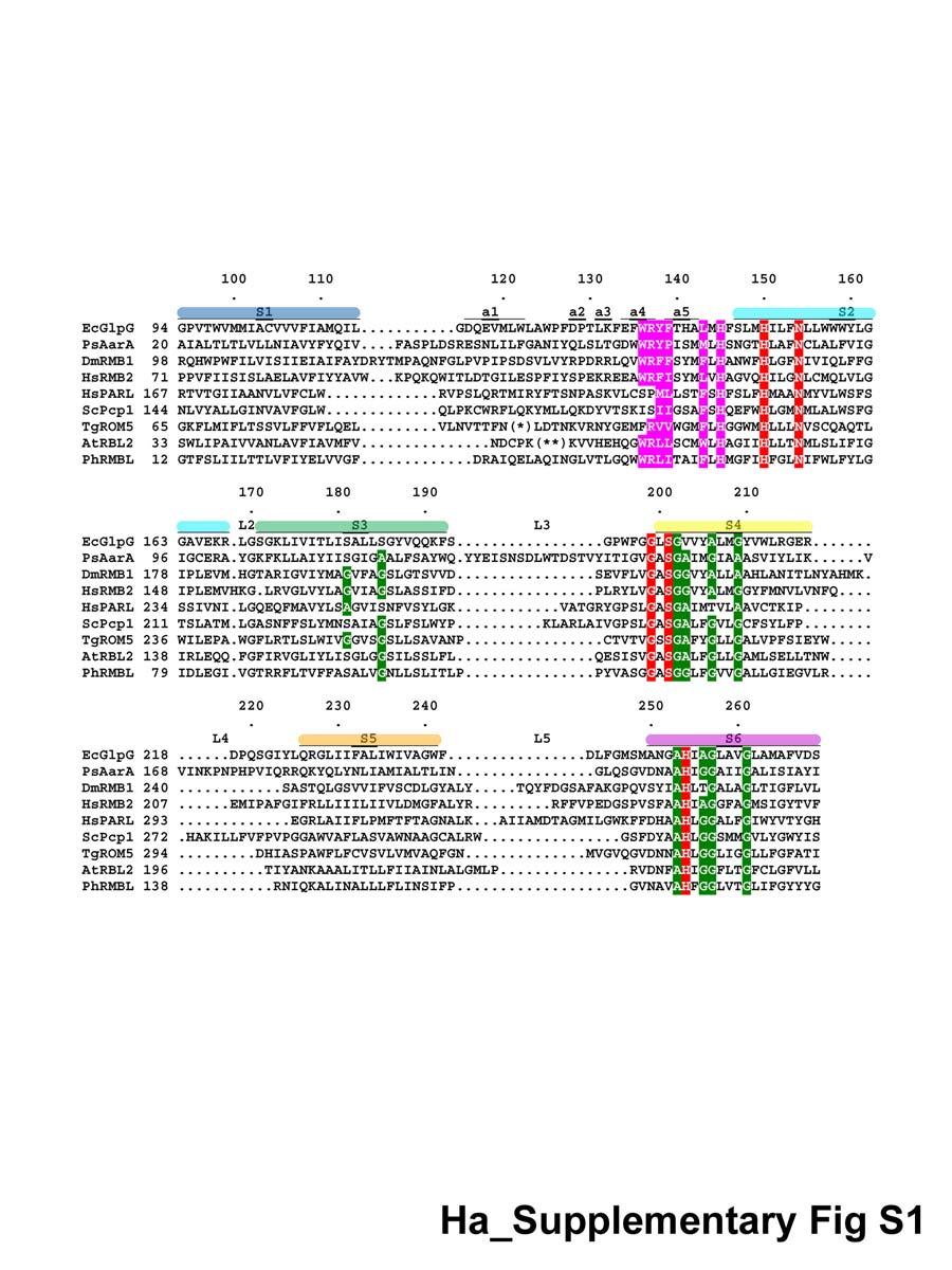
5 Supplementary Figure Legends Figure S1 Structure-based sequence alignment of selected rhomboid proteases. These include E. coli GlpG (EcGlpG), P. stuartii AarA (PsAarA), D. melanogaster rhomboid-1 (DmRMB1), H. sapiens RHBDL2 (HsRMB2), H. sapiens presenilinassociated rhomboid-like (HsPARL), S. cerevisiae Pcp1/Rbd1p (ScPcp1), T. gondii rhomboid-5 (TgROM5), A. thaliana rhomboid-2 (AtRBL2), P. horikoshii rhomboid-like (PhRMBL). Residue numbers above the sequence correspond to those of GlpG; residue numbers for each species are given at the beginning of the line. Colored bars above the sequence indicate the transmembrane helices S1-S6 (same color scheme as that in Fig. 2a). The short helices on L1 are also indicated (a1-a5). The conserved residues of the active site are highlighted in red; residues that function in gating highlighted in pink; residues that mediates strong intramolecular helix-helix association highlighted in green. TgROM5 and AtRBL2 have longer L1 loops than the rest: 100 residues in TgROM5 at (*) and 40 residues in AtRBL2 at (**) are omitted in the alignment. Figure S2 Protein purification and activity. a, Chymotrypsin cut. (Left) SDS- PAGE gel stained by Coomassie. (Right) Western blot by a monoclonal antibody against polyhistidine tag. The sample (-) without chymotrypsin treatment contained a tiny amount of N, only obvious by Western, that was probably derived from the full-length protein (missing the N-terminal region, but containing the core and C-terminal histidine tag). Chymotrypsin trimmed the full-length 5
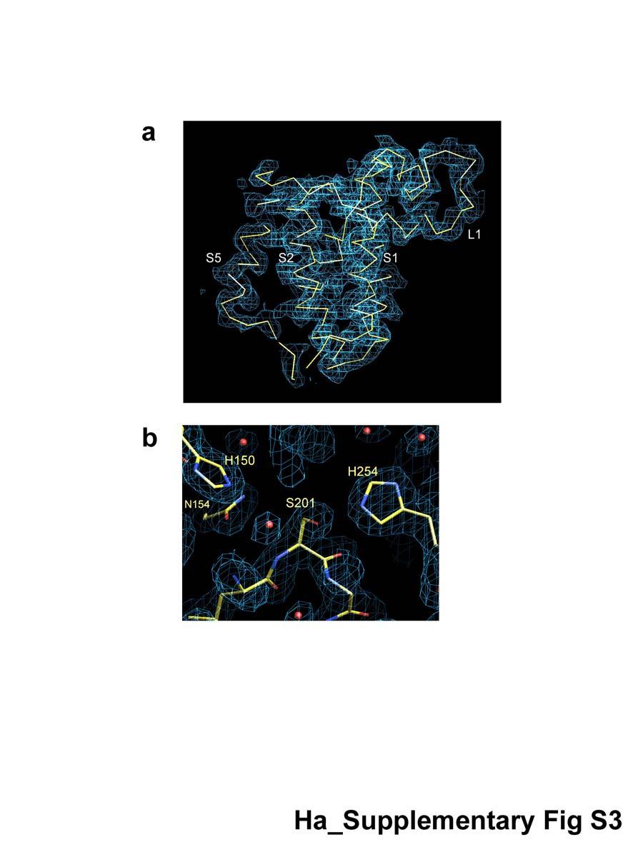
6 protein first to N, then to the core where the histidine tag was also removed. Chymotrypsin did not generate any nicks in the core domain used for crystallization. b, Elution profile of the core domain from a size-exclusion column. The elution volume (13.6ml) corresponds to a molecular weight of 81kDa. c, The proteolytic activity assay. Sixty data points were collected over a period of 2 hours, and measurements were performed in triplicate with multiple readings averaged for each well. Both full-length GlpG (red) and the core domain (green) were active in NG, the detergent used for crystallization. d, The increase of fluorescence was due to proteolysis of the dye-labeled casein (S, substrate), which generated smaller and highly fluorescent peptide fragments (P, products). In this experiment, 5µg BODIPY FL casein was incubated with different amounts of enzyme in a 50µl assay buffer at 37 C for 1 hour. The reaction mixtures were resolved by a 16% Tris-Tricine gel. Figure S3 The electron density maps. a, Experimental map calculated with F obs s and density-modified MAD phases, and contoured at 1.5σ level. The Cα trace of the final GlpG model is also shown (yellow). b, The final 2Fo-Fc map, contoured at 1.5σ level, at the active site of GlpG. The side chains of the four conserved residues, His-150, Asn-154, Ser-201 and His-254, from membrane spanning segments S2, S4 and S6 are shown. The red spheres represent nearby bound water molecules. 6
7 Figure S4 A stereo diagram of the Cα trace of GlpG core domain (the front view). The black dots mark every tenth residue. This illustration, as well as those in Fig. S5, are generated by MOLSCRIPT 47. Figure S5 GlpG trimer in the crystal. a, The front view of the trimer. Bound detergents are shown as ball-and-stick models. b, The top view of the trimer from the extracellular side. 7
8 8
9 9
10 10
11 11
12 12
SUPPLEMENTARY INFORMATION
 SUPPLEMENTARY INFORMATION doi:10.1038/nature11524 Supplementary discussion Functional analysis of the sugar porter family (SP) signature motifs. As seen in Fig. 5c, single point mutation of the conserved
SUPPLEMENTARY INFORMATION
 Supplementary materials Figure S1 Fusion protein of Sulfolobus solfataricus SRP54 and a signal peptide. a, Expression vector for the fusion protein. The signal peptide of yeast dipeptidyl aminopeptidase
Supplementary Information. The protease GtgE from Salmonella exclusively targets. inactive Rab GTPases
 Supplementary Information The protease GtgE from Salmonella exclusively targets inactive Rab GTPases Table of Contents Supplementary Figures... 2 Supplementary Figure 1... 2 Supplementary Figure 2... 3
Table 1. Crystallographic data collection, phasing and refinement statistics. Native Hg soaked Mn soaked 1 Mn soaked 2
 Table 1. Crystallographic data collection, phasing and refinement statistics Native Hg soaked Mn soaked 1 Mn soaked 2 Data collection Space group P2 1 2 1 2 1 P2 1 2 1 2 1 P2 1 2 1 2 1 P2 1 2 1 2 1 Cell
Table S1. Overview of used PDZK1 constructs and their binding affinities to peptides. Related to figure 1.
 Table S1. Overview of used PDZK1 constructs and their binding affinities to peptides. Related to figure 1. PDZK1 constru cts Amino acids MW [kda] KD [μm] PEPT2-CT- FITC KD [μm] NHE3-CT- FITC KD [μm] PDZK1-CT-
SUPPLEMENTARY INFORMATION
 Fig. 1 Influences of crystal lattice contacts on Pol η structures. a. The dominant lattice contact between two hpol η molecules (silver and gold) in the type 1 crystals. b. A close-up view of the hydrophobic
SUPPLEMENTARY INFORMATION
 doi:10.1038/nature11054 Supplementary Fig. 1 Sequence alignment of Na v Rh with NaChBac, Na v Ab, and eukaryotic Na v and Ca v homologs. Secondary structural elements of Na v Rh are indicated above the
SUPPLEMENTARY INFORMATION
 doi:10.1038/nature12045 Supplementary Table 1 Data collection and refinement statistics. Native Pt-SAD X-ray source SSRF BL17U SPring-8 BL41XU Wavelength (Å) 0.97947 1.07171 Space group P2 1 2 1 2 1 P2
SUPPLEMENTARY INFORMATION
 Supplementary Results DNA binding property of the SRA domain was examined by an electrophoresis mobility shift assay (EMSA) using synthesized 12-bp oligonucleotide duplexes containing unmodified, hemi-methylated,
Structure and RNA-binding properties. of the Not1 Not2 Not5 module of the yeast Ccr4 Not complex
 Structure and RNA-binding properties of the Not1 Not2 Not5 module of the yeast Ccr4 Not complex Varun Bhaskar 1, Vladimir Roudko 2,3, Jerome Basquin 1, Kundan Sharma 4, Henning Urlaub 4, Bertrand Seraphin
High-resolution crystal structure of ERAP1 with bound phosphinic transition-state analogue inhibitor
 High-resolution crystal structure of ERAP1 with bound phosphinic transition-state analogue inhibitor Petros Giastas 1, Margarete Neu 2, Paul Rowland 2, and Efstratios Stratikos 1 1 National Center for
SI Text S1 Solution Scattering Data Collection and Analysis. SI references
 SI Text S1 Solution Scattering Data Collection and Analysis. The X-ray photon energy was set to 8 kev. The PILATUS hybrid pixel array detector (RIGAKU) was positioned at a distance of 606 mm from the sample.
Supporting Information
 Supporting Information Self-Assembly of Glutathione S-transferases into Nanowires Wei Zhang, a Quan Luo,* a Lu Miao, a Yushi Bai, a Zeyuan Dong, a Jiayun Xu, a and Junqiu Liu* a a State Key Laboratory
SUPPLEMENTARY INFORMATION
 Supplementary Table 1: Amplitudes of three current levels. Level 0 (pa) Level 1 (pa) Level 2 (pa) TrkA- TrkH WT 200 K 0.01 ± 0.01 9.5 ± 0.01 18.7 ± 0.03 200 Na * 0.001 ± 0.01 3.9 ± 0.01 12.5 ± 0.03 200
SUPPLEMENTARY INFORMATION. doi: /nature07461
 Figure S1 Electrophysiology. a ph-activation of. Two-electrode voltage clamp recordings of Xenopus oocytes expressing in comparison to waterinjected oocytes. Currents were recorded at 40 mv. The ph of
SUPPLEMENTARY INFORMATION
 doi:1.138/nature1737 Supplementary Table 1 variant Description FSEC - 2B12 a FSEC - 6A1 a K d (leucine) c Leucine uptake e K (wild-type like) K (Y18F) K (TS) K (TSY) K288A mutant, lipid facing side chain
Nature Structural & Molecular Biology: doi: /nsmb Supplementary Figure 1
 Supplementary Figure 1 Identification of the ScDcp2 minimal region interacting with both ScDcp1 and the ScEdc3 LSm domain. Pull-down experiment of untagged ScEdc3 LSm with various ScDcp1-Dcp2-His 6 fragments.
Supporting Information
 Protein-Observed Fluorine NMR is a Complementary Ligand Discovery Method to 1 H CPMG Ligand- Observed NMR. Andrew K. Urick, 1,2 Luis Pablo Calle, 3 Juan F. Espinosa, 3 Haitao Hu, 2 * William C. K. Pomerantz
SUPPLEMENTARY INFORMATION
 Data collection Supplementary Table 1 Statistics of data collection, phasing and refinement Native Se-MAD Space group P2 1 2 1 2 1 P2 1 2 1 2 1 Cell dimensions a, b, c (Å) 50.4, 94.2, 115.4 49.8, 94.2,
SUPPLEMENTARY INFORMATION
 Table of Contents Page Supplementary Table 1. Diffraction data collection statistics 2 Supplementary Table 2. Crystallographic refinement statistics 3 Supplementary Fig. 1. casic1mfc packing in the R3
for Molecular Biology and Neuroscience and Institute of Medical Microbiology, Rikshospitalet-Radiumhospitalet
 SUPPLEMENTARY INFORMATION TO Structural basis for enzymatic excision of N -methyladenine and N 3 -methylcytosine from DNA Ingar Leiros,5, Marivi P. Nabong 2,3,5, Kristin Grøsvik 3, Jeanette Ringvoll 2,
SUPPLEMENTARY INFORMATION
 doi:10.1038/nature10458 Active Site Remodeling in the Bifunctional Fructose-1,6- bisphosphate aldolase/phosphatase Juan Du, Rafael F. Say, Wei Lü, Georg Fuchs & Oliver Einsle SUPPLEMENTARY FIGURES Figure
Structure of a bacterial multi-drug ABC transporter
 1 Structure of a bacterial multi-drug ABC transporter Roger J. P. Dawson and Kaspar P. Locher Institute of Molecular Biology and Biophysics, ETH Zurich, 8093 Zurich, Switzerland Supplementary Information
Acta Crystallographica Section D
 Supporting information Acta Crystallographica Section D Volume 70 (2014) Supporting information for article: Structural characterization of the virulence factor Nuclease A from Streptococcus agalactiae
Supporting online material
 Supporting online material Materials and Methods Target proteins All predicted ORFs in the E. coli genome (1) were downloaded from the Colibri data base (2) (http://genolist.pasteur.fr/colibri/). 737 proteins
Supplementary Figure 1. SDS-PAGE analysis of GFP oligomer variants with different linkers. Oligomer mixtures were applied to a PAGE gel containing
 Supplementary Figure 1. SDS-PAGE analysis of GFP oligomer variants with different linkers. Oligomer mixtures were applied to a PAGE gel containing 0.1% SDS without boiling. The gel was analyzed by a fluorescent
SUPPLEMENTARY INFORMATION
 Supplementary Table S1 Kinetic Analyses of the AMSH-LP mutants AMSH-LP K M (μm) k cat x 10-3 (s -1 ) WT 71.8 ± 6.3 860 ± 65.4 T353A 76.8 ± 11.7 46.3 ± 3.7 F355A 58.9 ± 10.4 5.33 ± 0.30 proximal S358A 75.1
ml. ph 7.5 ph 6.5 ph 5.5 ph 4.5. β 2 AR-Gs complex + GDP β 2 AR-Gs complex + GTPγS
 a UV28 absorption (mau) 9 8 7 5 3 β 2 AR-Gs complex β 2 AR-Gs complex + GDP β 2 AR-Gs complex + GTPγS β 2 AR-Gs complex dissociated complex excess nucleotides b 9 8 7 5 3 β 2 AR-Gs complex β 2 AR-Gs complex
SUPPLEMENTARY INFORMATION
 SUPPLEMENTARY INFORMATION doi:10.1038/nature11744 Supplementary Table 1. Crystallographic data collection and refinement statistics. Wild-type Se-Met-BcsA-B SmCl 3 -soaked EMTS-soaked Data collection Space
Serine-7 but not serine-5 phosphorylation primes RNA polymerase II CTD for P-TEFb recognition
 Supplementary Information to Serine-7 but not serine-5 phosphorylation primes RNA polymerase II CTD for P-TEFb recognition Nadine Czudnochowski 1,2, *, Christian A. Bösken 1, * & Matthias Geyer 1 1 Max-Planck-Institut
SUPPLEMENTARY INFORMATION
 Structure of an ABC transporter-binding protein complex Kaspar Hollenstein, Dominik C. Frei, and Kaspar P. Locher Institute of Molecular Biology and Biophysics, ETH Zurich, 8093 Zurich, Switzerland Supplementary
Structure and Function of Neisseria gonorrhoeae MtrF Illuminates a Class of Antimetabolite Efflux Pumps
 Cell Reports Supplemental Information Structure and Function of Neisseria gonorrhoeae MtrF Illuminates a Class of Antimetabolite Efflux Pumps Chih-Chia Su, Jani Reddy Bolla, Nitin Kumar, Abhijith Radhakrishnan,
Supplementary Figure 1. Biochemical and sequence alignment analyses the
 Supplementary Figure 1. Biochemical and sequence alignment analyses the interaction of OPTN and TBK1. (a) Analytical gel filtration chromatography analysis of the interaction between TBK1 CTD and OPTN(1-119).
Supplementary Figure 1 Crystal contacts in COP apo structure (PDB code 3S0R)
") Supplementary Figure 1 Crystal contacts in COP apo structure (PDB code 3S0R) Shown in cyan and green are two adjacent tetramers from the crystallographic lattice of COP, forming the only unique inter-tetramer
Supplementary Figure 1 Crystal contacts in COP apo structure (PDB code 3S0R) Shown in cyan and green are two adjacent tetramers from the crystallographic lattice of COP, forming the only unique inter-tetramer
Supplementary materials. Crystal structure of the carboxyltransferase domain. of acetyl coenzyme A carboxylase. Department of Biological Sciences
 Supplementary materials Crystal structure of the carboxyltransferase domain of acetyl coenzyme A carboxylase Hailong Zhang, Zhiru Yang, 1 Yang Shen, 1 Liang Tong Department of Biological Sciences Columbia
Bacterial protease uses distinct thermodynamic signatures for substrate recognition
 Bacterial protease uses distinct thermodynamic signatures for substrate recognition Gustavo Arruda Bezerra, Yuko Ohara-Nemoto, Irina Cornaciu, Sofiya Fedosyuk, Guillaume Hoffmann, Adam Round, José A. Márquez,
Supporting Information. Chemo-enzymatic Synthesis of Isotopically Labeled Nicotinamide Ribose
 Electronic Supplementary Material (ESI) for Organic & Biomolecular Chemistry. This journal is The Royal Society of Chemistry 2018 Supporting Information Chemo-enzymatic Synthesis of Isotopically Labeled
According to the manufacture s direction (Pierce), RNA and DNA
, RNA and DNA") Supplementary method Electrophoretic Mobility-shift assay (EMSA) According to the manufacture s direction (Pierce), RNA and DNA oligonuleotides were firstly labeled by biotin. TAVb (1pM) was incubated
Supplementary method Electrophoretic Mobility-shift assay (EMSA) According to the manufacture s direction (Pierce), RNA and DNA oligonuleotides were firstly labeled by biotin. TAVb (1pM) was incubated
SUPPLEMENTARY INFORMATION
 doi:10.1038/nature11085 Supplementary Tables: Supplementary Table 1. Summary of crystallographic and structure refinement data Structure BRIL-NOP receptor Data collection Number of crystals 23 Space group
Secondary Structure. Bioch/BIMS 503 Lecture 2. Structure and Function of Proteins. Further Reading. Φ, Ψ angles alone determine protein structure
 Bioch/BIMS 503 Lecture 2 Structure and Function of Proteins August 28, 2008 Robert Nakamoto rkn3c@virginia.edu 2-0279 Secondary Structure Φ Ψ angles determine protein structure Φ Ψ angles are restricted
Supplementary Information
 Supplementary Information The direct role of selenocysteine in [NiFeSe] hydrogenase maturation and catalysis Marta C. Marques a, Cristina Tapia b, Oscar Gutiérrez-Sanz b, Ana Raquel Ramos a, Kimberly L.
SUPPLEMENTARY INFORMATION
 Supplementary Table 1: Data collection, phasing and refinement statistics ChbC/Ta 6 Br 12 Native ChbC Data collection Space group P4 3 2 1 2 P4 3 2 1 2 Cell dimensions a, c (Å) 132.75, 453.57 132.81, 452.95
Supporting Information
 Supporting Information Ottmann et al. 10.1073/pnas.0907587106 Fig. S1. Primary structure alignment of SBT3 with C5 peptidase from Streptococcus pyogenes. The Matchmaker tool in UCSF Chimera (http:// www.cgl.ucsf.edu/chimera)
ydci GTC TGT TTG AAC GCG GGC GAC TGG GCG CGC AAT TAA CGG TGT GTA GGC TGG AGC TGC TTC
 Table S1. DNA primers used in this study. Name ydci P1ydcIkd3 Sequence GTC TGT TTG AAC GCG GGC GAC TGG GCG CGC AAT TAA CGG TGT GTA GGC TGG AGC TGC TTC Kd3ydcIp2 lacz fusion YdcIendP1 YdcItrgP2 GAC AGC
THE CRYSTAL STRUCTURE OF THE SGT1-SKP1 COMPLEX: THE LINK BETWEEN
 THE CRYSTAL STRUCTURE OF THE SGT1-SKP1 COMPLEX: THE LINK BETWEEN HSP90 AND BOTH SCF E3 UBIQUITIN LIGASES AND KINETOCHORES Oliver Willhoft, Richard Kerr, Dipali Patel, Wenjuan Zhang, Caezar Al-Jassar, Tina
SUPPLEMENTARY INFORMATION
 Dph2 SeMet (iron-free) # Dph2 (iron-free) Dph2-[4Fe-4S] Data collection Space group P2 1 2 1 2 1 P2 1 2 1 2 1 P2 1 2 1 2 1 Cell dimensions a, b, c (Å) 58.26, 82.08, 160.42 58.74, 81.87, 160.01 55.70, 80.53,
Resonance Assignment of the RGS Domain of Human RGS10
 Resonance Assignment of the RGS Domain of Human RGS10 Oleg Y. Fedorov 2, Victoria A. Higman 1, Peter Schmieder 1, Martina Leidert 1, Annette Diehl 1, Jonathan Elkins 2, Meera Soundararajan 2, Hartmut Oschkinat
Tellurite resistance protein/ethidium efflux transporter/ proflavin transporter. Putative inner membrane protein: function unknown
 Additional file 1. Table S1 and Figures S1-4 of Zhang et al. High-level production of membrane proteins in E. coli BL21(DE3) by omitting the inducer IPTG Table S1. Properties of the membrane proteins used
Supplementary figure 1 Application of tmfret in LeuT. (a) To assess the feasibility of using tmfret for distance-dependent measurements in LeuT, a
 To assess the feasibility of using tmfret for distance-dependent measurements in LeuT, a") Supplementary figure 1 Application of tmfret in LeuT. (a) To assess the feasibility of using tmfret for distance-dependent measurements in LeuT, a series of tmfret-pairs comprised of single cysteine mutants
Supplementary figure 1 Application of tmfret in LeuT. (a) To assess the feasibility of using tmfret for distance-dependent measurements in LeuT, a series of tmfret-pairs comprised of single cysteine mutants
SUPPLEMENTARY INFORMATION
 Supplementary Figure 1: The HpUreI crystal used for collection of native diffraction data. The crystal belongs to spacegroup P4 2 2 1 2 and has an approximate maximal dimension of 0.25 mm. Supplementary
SUPPLEMENTARY INFORMATION
 doi:10.108/nature11899 Supplementar Table 1. Data collection and refinement statistics (+TPMP, native) (-TPMP, native) (+TPMP, recombinant) (MgCl ) (MgSO ) Data collection Space group C P 1 C P 1 1 P 1
SUPPLEMENTARY FIGURES
 SUPPLEMENTARY FIGURES Supplementary Figure 1 Protein sequence alignment of Vibrionaceae with either a 40-residue insertion or a 44-residue insertion. Identical residues are indicated by red background.
Structural analysis of a rhomboid family intramembrane protease reveals a gating mechanism for substrate entry
 Structural analysis of a rhomboid family intramembrane protease reveals a gating mechanism for substrate entry Zhuoru Wu 1,4, Nieng Yan 1,4, Liang Feng 1,4, Adam Oberstein 1, Hanchi Yan 1, Rosanna P Baker
Supplementary Information. Structural basis for precursor protein-directed ribosomal peptide macrocyclization
 Supplementary Information Structural basis for precursor protein-directed ribosomal peptide macrocyclization Kunhua Li 1,3, Heather L. Condurso 1,3, Gengnan Li 1, Yousong Ding 2 and Steven D. Bruner 1*
Supporting Information
 Supporting Information Structural Analysis of the Binding of Type I, I 1/2, and II Inhibitors to Eph Tyrosine Kinases Jing Dong, *1 Hongtao Zhao, 1 Ting Zhou, 1 Dimitrios Spiliotopoulos, 1 Chitra Rajendran,
SUPPLEMENTARY INFORMATION
 SUPPLEMENTARY INFORMATION doi:10.1038/nature11539 Supplementary Figure 1 Schematic representation of plant (A) and mammalian (B) P 2B -ATPase domain organization. Actuator (A-), nucleotide binding (N-),
Copyright WILEY-VCH Verlag GmbH, D Weinheim, Supporting Information for Angew. Chem. Int. Ed. Z 18050
 Copyright WILEY-VCH Verlag GmbH, D-69451 Weinheim, 2001. Supporting Information for Angew. Chem. Int. Ed. Z 18050 Protein Affinity Labeling Mediated by Genetically Encoded Peptide Tags Frank Amini, Thomas
Biological Sciences 11 Spring Experiment 4. Protein crosslinking
 Biological Sciences 11 Spring 2000 Experiment 4. Protein crosslinking = C - CH 2 - CH 2 - CH 2 - C = H H GA Cl - H 2 N N H 2 Cl - C - CH 2 - CH 2 - CH 2 - CH 2 - CH 2 - CH 2 - C DMS CH 3 CH 3 N - - C -
Supporting Information
 Supporting Information Structural Basis of the Antiproliferative Activity of Largazole, a Depsipeptide Inhibitor of the Histone Deacetylases Kathryn E. Cole 1, Daniel P. Dowling 1,2, Matthew A. Boone 3,
Stabilizing the CH2 domain of an Antibody by Engineering in an Enhanced Aromatic Sequon
 Stabilizing the CH2 domain of an Antibody by Engineering in an Enhanced Aromatic Sequon Wentao Chen,, Leopold Kong, Stephen Connelly, Julia M. Dendle,, Yu Liu,, Ian A. Wilson,#, Evan T. Powers, *, Jeffery
Supplemental Data SUPPLEMENTAL FIGURES
 Supplemental Data CRYSTAL STRUCTURE OF THE MG.ADP-INHIBITED STATE OF THE YEAST F 1 C 10 ATP SYNTHASE Alain Dautant*, Jean Velours and Marie-France Giraud* From Université Bordeaux 2, CNRS; Institut de
Lecture 8 (9/29/17) Lecture 8 (9/29/17)
 Lecture 8 (9/29/17)") Reading: Ch3; 97-102 Lecture 8 (9/29/17) Problems: Ch3 (text); 18, 19, 20, 23 Ch3 (Study guide); 9 Ch4 (Study guide); 3 NEXT Reading: Ch4; 119-122, 125-126, 131-133 Ch4; 123-124, 130-131, 133, 137-138
Reading: Ch3; 97-102 Lecture 8 (9/29/17) Problems: Ch3 (text); 18, 19, 20, 23 Ch3 (Study guide); 9 Ch4 (Study guide); 3 NEXT Reading: Ch4; 119-122, 125-126, 131-133 Ch4; 123-124, 130-131, 133, 137-138
Supplemental Methods. Protein expression and purification
 Supplemental Methods Protein expression and purification The isolated collagen-binding domain of hlair-1, amino acid 22-122, was cloned into pet3xa using introduced BamHI and NotI sites at the 5 and 3
Protein Quantification Kit (Bradford Assay)
") Protein Quantification Kit (Bradford Assay) Booklet Item NO. KTD3002 Product Name Protein Quantification Kit (Bradford Assay) ATTENTION For laboratory research use only. Not for clinical or diagnostic
Protein Quantification Kit (Bradford Assay) Booklet Item NO. KTD3002 Product Name Protein Quantification Kit (Bradford Assay) ATTENTION For laboratory research use only. Not for clinical or diagnostic
Supplementary Information. Cryo-EM Studies of Drp1 Reveal Cardiolipin Interactions that Activate
 Supplementary Information Cryo-EM Studies of Drp1 Reveal Cardiolipin Interactions that Activate the Helical Oligomer Christopher A. Francy 1, 2, 3, Ryan W. Clinton 1, 2, 3, Chris Fröhlich 4, 5, Colleen
Structure and mechanism of an intramembrane liponucleotide synthetase central for phospholipid biosynthesis
 Structure and mechanism of an intramembrane liponucleotide synthetase central for phospholipid biosynthesis Xiuying Liu 1,3, Yan Yin 1,2,3, Jinjun Wu 1 and Zhenfeng Liu 1 1 National Laboratory of Biomacromolecules,
Supplementary Figure 1 Pairing alignments, turns and extensions within the structure of the ribozyme-product complex. (a) The alignment of the G27
 The alignment of the G27") Supplementary Figure 1 Pairing alignments, turns and extensions within the structure of the ribozyme-product complex. (a) The alignment of the G27 A40 non-canonical pair stacked over the A41 (G1-C26) three-base
Supplementary Figure 1 Pairing alignments, turns and extensions within the structure of the ribozyme-product complex. (a) The alignment of the G27 A40 non-canonical pair stacked over the A41 (G1-C26) three-base
Supplementary Materials for
 www.advances.sciencemag.org/cgi/content/full/1/7/e1500263/dc1 Supplementary Materials for Newton s cradle proton relay with amide imidic acid tautomerization in inverting cellulase visualized by neutron
BA, BSc, and MSc Degree Examinations
 Examination Candidate Number: Desk Number: BA, BSc, and MSc Degree Examinations 2017-8 Department : BIOLOGY Title of Exam: Molecular Biology and Biochemistry Part I Time Allowed: 1 hour and 30 minutes
SUPPLEMENTARY INFORMATION
 5 N 4 8 20 22 24 2 28 4 8 20 22 24 2 28 a b 0 9 8 7 H c (kda) 95 0 57 4 28 2 5.5 Precipitate before NMR expt. Supernatant before NMR expt. Precipitate after hrs NMR expt. Supernatant after hrs NMR expt.
type GroEL-GroES complex. Crystals were grown in buffer D (100 mm HEPES, ph 7.5,
 Supplementary Material Supplementary Materials and Methods Structure Determination of SR1-GroES-ADP AlF x SR1-GroES-ADP AlF x was purified as described in Materials and Methods for the wild type GroEL-GroES
Illegitimate translation causes unexpected gene expression from on-target out-of-frame alleles
 Illegitimate translation causes unexpected gene expression from on-target out-of-frame alleles created by CRISPR-Cas9 Shigeru Makino, Ryutaro Fukumura, Yoichi Gondo* Mutagenesis and Genomics Team, RIKEN
SUPPLEMENTARY INFORMATION
 doi:10.1038/nature10244 a O07391_MYCAV/127-243 NLPC_HAEIN/80-181 SPR_SHIFL/79-183 P74160_SYNY3/112-245 O24914_HELPY/301-437 Q51835_PORGI/68-178 DPP6_BACSH/163-263 YKFC_BACSU/185-292 YDHO_ECOLI/153-263
Nitrogenase MoFe protein from Clostridium pasteurianum at 1.08 Å resolution: comparison with the Azotobacter vinelandii MoFe protein
 Acta Cryst. (2015). D71, 274-282, doi:10.1107/s1399004714025243 Supporting information Volume 71 (2015) Supporting information for article: Nitrogenase MoFe protein from Clostridium pasteurianum at 1.08
Supplementary Information to
 Supplementary Information to Wiesner et al.: A change in conformational dynamics underlies the activation of Eph receptor tyrosine kinases Supplementary Material and Methods Cloning and Mutagenesis Site-directed
SUPPLEMENTARY INFORMATION
 DOI: 0.038/NCHEM.795 Oligomerization transforms human APOBEC3G from an efficient enzyme to a slowly dissociating nucleic acid -binding protein Kathy R. Chaurasiya, Micah J. McCauley, Wei Wang, Dominic
Alkaline Phosphatase Labeling Kit-NH2
 Alkaline Phosphatase Labeling Kit-NH2 Catalog Number KA0001 1 Kit Version: 02 Intended for research use only www.abnova.com Table of Contents Introduction... 3 Background... 3 Principle of the Assay...
Supplemental Information. The Mitochondrial Fission Receptor MiD51. Requires ADP as a Cofactor
 Structure, Volume 22 Supplemental Information The Mitochondrial Fission Receptor MiD51 Requires ADP as a Cofactor Oliver C. Losón, Raymond Liu, Michael E. Rome, Shuxia Meng, Jens T. Kaiser, Shu-ou Shan,
SUPPLEMENTARY INFORMATION
 www.nature.com/nature 1 Figure S1 Sequence alignment. a Structure based alignment of the plgic of E. chrysanthemi (ELIC), the acetylcholine binding protein from the snail Lymnea stagnalis (AchBP, PDB code
Structure and evolution of the spliceosomal peptidyl-prolyl cistrans isomerase Cwc27
 Acta Cryst. (2014). D70, doi:10.1107/s1399004714021695 Supporting information Volume 70 (2014) Supporting information for article: Structure and evolution of the spliceosomal peptidyl-prolyl cistrans isomerase
Aggrecanase Activity Assay Kit
 Aggrecanase Activity Assay Kit Catalog Number KA1497 96 assays Version: 03 Intended for research use only www.abnova.com Table of Contents Introduction... 3 Background... 3 Principle of the Assay... 3
TrioMol Isolation Reagent
 TrioMol Isolation Reagent Technical Manual No. 0242 Version 06142007 I Description... 1 II Key Features... 1 III Storage..... 1 IV General Protocol Using Triomol Isolation Reagent 1 V Troubleshooting.
TrioMol Isolation Reagent
 TrioMol Isolation Reagent Technical Manual No. 0242 Version 06142007 I Description... 1 II Key Features... 1 III Storage..... 1 IV General Protocol Using Triomol Isolation Reagent 1 V Troubleshooting.
Cks1 CDK1 CDK1 CDK1 CKS1. are ice- lobe. conserved. conserved
 Cks1 d CKS1 Supplementary Figure 1 The -Cks1 crystal lattice. (a) Schematic of the - Cks1 crystal lattice. -Cks1 crystallizes in a lattice that contains c 4 copies of the t - Cks1 dimer in the crystallographic
NOVABEADS FOOD 1 DNA KIT
 NOVABEADS FOOD 1 DNA KIT NOVABEADS FOOD DNA KIT is the new generation tool in molecular biology techniques and allows DNA isolations from highly processed food products. The method is based on the use
CH 3 CH 2 OH +H 2 O CHO. 2e + 2H + + O 2 H 2 O +HCOOH
 2 4 H CH 3 2e + 2H + + 2 H 2 2 H CH 2 H 2e + 2H + + 2 H 2 2 H +H 2 CH 2e + 2H + + 2 H 2 2 H +HCH Supplemental Figure S. The three-step 4DM reaction, each step requires two reducing equivalents from ADPH
Malachite Green Phosphate Detection Kit Catalog Number: DY996
 Malachite Green Phosphate Detection Kit Catalog Number: DY996 This Malachite Green Phosphate Detection Kit employs a simple, sensitive, reproducible, and non-radioactive method for measuring inorganic
Supplementary Materials for
 www.sciencesignaling.org/cgi/content/full/5/243/ra68/dc1 Supplementary Materials for Superbinder SH2 Domains Act as Antagonists of Cell Signaling Tomonori Kaneko, Haiming Huang, Xuan Cao, Xing Li, Chengjun
Superoxide Dismutase Activity Assay Kit
 Superoxide Dismutase Activity Assay Kit Catalog Number KA0783 100 assays Version: 04 Intended for research use only www.abnova.com Table of Contents Introduction... 3 Background... 3 General Information...
Protein assay. Absorbance Fluorescence Emission Colorimetric detection BIO/MDT 325. Absorbance
 Protein assay Absorbance Fluorescence Emission Colorimetric detection BIO/MDT 325 Absorbance Using A280 to Determine Protein Concentration Determination of protein concentration by measuring absorbance
Supplementary figure 1. Comparison of unbound ogm-csf and ogm-csf as captured in the GIF:GM-CSF complex. Alignment of two copies of unbound ovine
 Supplementary figure 1. Comparison of unbound and as captured in the GIF:GM-CSF complex. Alignment of two copies of unbound ovine GM-CSF (slate) with bound GM-CSF in the GIF:GM-CSF complex (GIF: green,
Nature Structural & Molecular Biology: doi: /nsmb Supplementary Figure 1
 Supplementary Figure 1 Crystallization. a, Crystallization constructs of the ET B receptor are shown, with all of the modifications to the human wild-type the ET B receptor indicated. Residues interacting
RUBIC Buffer Screen For stable, happy proteins From purification all the way through to characterization by NMR, SAXS or Crystallography.
 RUBIC Buffer Screen MD1-96 For stable, happy proteins From purification all the way through to characterization by NMR, SAXS or Crystallography. RUBIC Buffer Screen- designed at the EMBL Hamburg and optimized
Data Sheet. Azide Cy5 RNA T7 Transcription Kit
 Cat. No. Size 1. Description PP-501-Cy5 10 reactions à 40 µl For in vitro use only Quality guaranteed for 12 months Store all components at -20 C. Avoid freeze and thaw cycles. DBCO-Sulfo-Cy5 must be stored
SUPPLEMENTARY INFORMATION
 doi:10.1038/nature10955 Supplementary Figures Supplementary Figure 1. Electron-density maps and crystallographic dimer structures of the motor domain. (a f) Stereo views of the final electron-density maps
Supplementary Information
 Supplementary Information Structural analysis of leader peptide binding enables leaderfree cyanobactin processing Jesko Koehnke 1,2, Greg Mann 1,2, Andrew F Bent 1,2, Hannes Ludewig 1, Sally Shirran 1,
Supplementary information
 Supplementary information The structural basis of modularity in ECF-type ABC transporters Guus B. Erkens 1,2, Ronnie P-A. Berntsson 1,2, Faizah Fulyani 1,2, Maria Majsnerowska 1,2, Andreja Vujičić-Žagar
SUPPLEMENTARY INFORMATION
 UPPEER ORO doi:10.1038/nature10753 D D D D P E ntracellular C1 W P P C EC1 D Q R H C D W D R C C2 D E D E C R Q Q W P W W R P P EC2 EC3 P C C P W P W W P C W H R C R E C3 P R R P P P C Extracellular embrane
Protocol for 2D-E. Protein Extraction
 Protocol for 2D-E Protein Extraction Reagent 1 inside the ReadyPrep TM Sequential Extraction kit (in powder form) 50ml of deionized water is used to dissolve all the Reagent 1. The solution is known as
James B. Munro, Roger B. Altman, Nathan O Connor, and Scott C. Blanchard
 Molecular Cell, Volume 25 Supplemental Data Identification of Two Distinct Hybrid State Intermediates on the Ribosome James B. Munro, Roger B. Altman, Nathan O Connor, and Scott C. Blanchard Wild-type