CH 3 CH 2 OH +H 2 O CHO. 2e + 2H + + O 2 H 2 O +HCOOH
|
|
|
- Shauna May
- 6 years ago
- Views:
Transcription
1 2 4 H CH 3 2e + 2H H 2 2 H CH 2 H 2e + 2H H 2 2 H +H 2 CH 2e + 2H H 2 2 H +HCH Supplemental Figure S. The three-step 4DM reaction, each step requires two reducing equivalents from ADPH (transferred via the FAD and FM cofactors of cytochrome P450 reductase), two protons and one molecular oxygen. nly (H or CH 3 ) and 2 (H or =CH) vary in the 4DM substrates across the biological kingdoms. In the enzyme active center, the 4α-methyl group of the substrate is sequentially converted into the alcohol, then into the aldehyde derivative an then it is released as formic acid concomitantly with the introduction of the Δ 4-5 -double bound into the sterol core.
 c 0.")
 Timecourse graph shows the rate and percentage of C-complexes")
2 a i 47/280 =.8 b 448 nm 00 C/absolute, % Ligand free VI-bound Time (min) c 0. mm Supplemental Figure S2. Ligand-free (left/red) and VI-bound (right/blue) Tbb4DM. a. Absolute absorbance; b. C-difference spectra ( (Δt= min) Timecourse graph shows the rate and percentage of C-complexes formation in ligand-free and VI-bound state. c. Crystals.
 is colored in dark blue, and the C-terminus (ending at 476) is colored in red, full-length protein numbering.")
3 G B B F K C B H G A A K I F F K L E J D J Supplemental Figure S3. A ribbon diagram showing the overall Tbb4DM structure. Distal view. -terminus (starting at G29) is colored in dark blue, and the C-terminus (ending at 476) is colored in red, full-length protein numbering. The 2 main helices are labeled from A to L; ten additional shorter helices between them are marked as ( ) and 2 β-strands arranged in 4 bundles are labeled by structure succession (5 strands in bundle, 2 in bundle 2, 3 in bundle 3, and 2 in bundle 4).The heme is shown in stick representation.
4
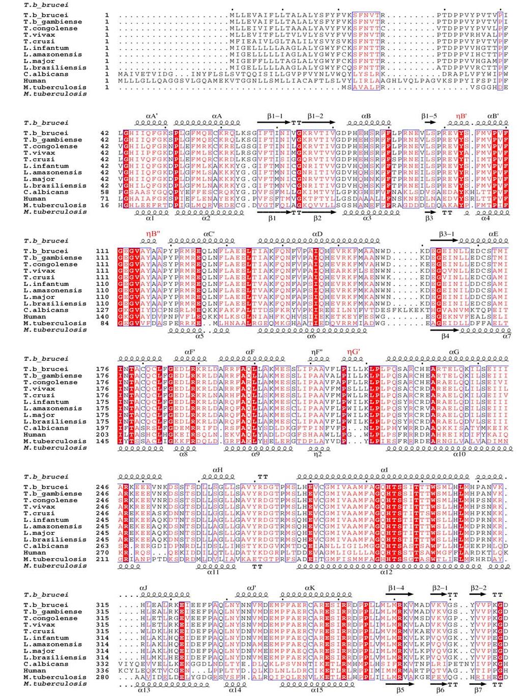
, Candida albicans (24% identity to Tbb4DM), human (27% identity) and Mycobacterium tuberculosis (27%")
5 Supplemental Figure S4. Sequence alignment of 4DMs from Trypanosomatidae (75% average amino acid sequence identity), Candida albicans (24% identity to Tbb4DM), human (27% identity) and Mycobacterium tuberculosis (27% identity). Secondary structure elements of 4DM from Tbb and Mt are shown at the top and the bottom, respectively. The P450-fold nomenclature is provided for Tbb4DM; differences in the secondary structure elements are indicated in red.
 and four")
6 Supplemental Figure S5. Superimposition of four molecules of ligand-free (white) and four molecules of VI-bound (red) Tbb4DM (distal view).

7 G F I G H C K B D L K J J Supplemental Figure S6. View of the superposed ligand-free and VI-bound Tbb4DM structures from the proximal (opposite to Fig.2b) side. Some shift toward the heme in the VI-bound structure can be seen for helices C, D, H, J and the C-terminal part of helix G. Elongated secondary structure elements (helices C, H and G in all four molecules) are marked in red near the places of their extensions. The tendency to form additional helix-like turns has been also observed for azole-bound drug-metabolizing CYP2B4 [SU and 2V0M] and for substrate-bound CYP46A [2q9f] and might indicate some additional molecule surface stabilization.


8 a Y6 Y A29 F24 L V46 T295 I45 F48 b VI C422 Heme Supplemental Figure S7. 2Fo-Fc electron density map for VI in the active site of Tbb4DM contoured at σ (a) and 2.5 σ (b). The bond distances (Å) are marked.
9 a Substrate conversion (%) C. albicans 4DM 5min 60min Human 4DM b VI/4DM (molar ratio) Absorbance Kd=0.37 µm Wavelength, nm Supplemental Figure S8. a. Inhibition of C. albicans and human 4DM activity by VI. Enzyme concentration 0.5 µm, substrate (lanosterol) concentration 50 µm, SD<0%. The other details of the reaction conditions are as described in (9). b. Binding of VI to human 4DM, difference absorbance spectra. The human 4DM concentration.7 µm; mm VI solution in DMS, titration range µm, titration step 0.5 µm
 and clinical antifungal drug ketoconazole (white bars) on 4DMs from two pathogens, Tbb and Candida albicans, and from human.")
10 a b c 00 B Y H V46 I/E 2 3 H T295 A29 Tbb C.alb Human 4 H I Supplemental Figure S9. VI amide group fragment as the most likely cause for its selectivity towards 4DM from pathogenic microbes. a. Comparison of inhibitory effects of VI (black bars) and clinical antifungal drug ketoconazole (white bars) on 4DMs from two pathogens, Tbb and Candida albicans, and from human. I/E 2 represents the inhibitor/enzyme ratio that causes a two-fold decrease in the initial rate of reaction, log scale. b. Chemical structures of ketoconazole (), VI (2) and two VI-scaffold derivatives: the β-phenyl azole SDZ (3) with the inhibitory potency and antiparasitic effect in Trypanosomatidae comparable to VI and the α-phenyl azole SDZ (4), a competitive, short-term inhibitor that binds with the same apparent Kd (<00 nm) but can be easily replaced in the enzyme active site by substrate (). c. VI-induced hydrogen bonding network between helices B and I. V46 and its mutation to isoleucine (grey), corresponding to I488 in human 4DM, are shown. Heme is presented as spheres.
11 Supplemental Table S. Data collection and refinement statistics Ligand-free Inhibitor-bound Data collection ative Iron SAD (peak) ative Wavelength, Å Space group P P P Cell dimensions a, b, c, Å 59.8,79.9, , 79.9, , 79., 6.0 α, β, γ, 74.2, 8.6, , 8., , 79., 68.6 umber of molecules per asymmetric unit esolution, Å ( )* ( ) (.9-.87) merge (0.470) (0.536) (0.603) I/σ(I) 8.5 (3.) 44.5 (3.4) 28 (.8) Completeness (%) 97.8 (96.7) 96.8 (95.2) (9.7) edundancy 4.0 (3.8) 7.8 (7.7) 3.9 (3.5) efinement esolution, Å umber of reflections 42,726 44,527 work/ / free 0.95/ /0.238 Average B-factors protein heme/inhibitor 22 23/32 water ms deviations Bond lengths, Å Bond angles, Cα positions between 4 molecules, Å *Values in parenthesis are for highest-resolution shell.
Table S1. Overview of used PDZK1 constructs and their binding affinities to peptides. Related to figure 1.
 Table S1. Overview of used PDZK1 constructs and their binding affinities to peptides. Related to figure 1. PDZK1 constru cts Amino acids MW [kda] KD [μm] PEPT2-CT- FITC KD [μm] NHE3-CT- FITC KD [μm] PDZK1-CT-
SUPPLEMENTARY INFORMATION
 SUPPLEMENTARY INFORMATION doi:10.1038/nature11524 Supplementary discussion Functional analysis of the sugar porter family (SP) signature motifs. As seen in Fig. 5c, single point mutation of the conserved
SUPPLEMENTARY INFORMATION
 doi:10.1038/nature11054 Supplementary Fig. 1 Sequence alignment of Na v Rh with NaChBac, Na v Ab, and eukaryotic Na v and Ca v homologs. Secondary structural elements of Na v Rh are indicated above the
SUPPLEMENTARY INFORMATION
 Supplementary Results DNA binding property of the SRA domain was examined by an electrophoresis mobility shift assay (EMSA) using synthesized 12-bp oligonucleotide duplexes containing unmodified, hemi-methylated,
SUPPLEMENTARY INFORMATION
 Table of Contents Page Supplementary Table 1. Diffraction data collection statistics 2 Supplementary Table 2. Crystallographic refinement statistics 3 Supplementary Fig. 1. casic1mfc packing in the R3
Supporting Information
 Supporting Information Ottmann et al. 10.1073/pnas.0907587106 Fig. S1. Primary structure alignment of SBT3 with C5 peptidase from Streptococcus pyogenes. The Matchmaker tool in UCSF Chimera (http:// www.cgl.ucsf.edu/chimera)
SUPPLEMENTARY INFORMATION
 Supplementary materials Figure S1 Fusion protein of Sulfolobus solfataricus SRP54 and a signal peptide. a, Expression vector for the fusion protein. The signal peptide of yeast dipeptidyl aminopeptidase
2015 AP Biology Unit 2 PRETEST- Introduction to the Cell and Biochemistry
 Name: Class: _ Date: _ 2015 AP Biology Unit 2 PRETEST- Introduction to the Cell and Biochemistry Multiple Choice Identify the choice that best completes the statement or answers the question. 1) In what
SUPPLEMENTARY INFORMATION
 Supplementary Table 1: Amplitudes of three current levels. Level 0 (pa) Level 1 (pa) Level 2 (pa) TrkA- TrkH WT 200 K 0.01 ± 0.01 9.5 ± 0.01 18.7 ± 0.03 200 Na * 0.001 ± 0.01 3.9 ± 0.01 12.5 ± 0.03 200
Supplementary Figure 1. Biochemical and sequence alignment analyses the
 Supplementary Figure 1. Biochemical and sequence alignment analyses the interaction of OPTN and TBK1. (a) Analytical gel filtration chromatography analysis of the interaction between TBK1 CTD and OPTN(1-119).
Nature Structural & Molecular Biology: doi: /nsmb Supplementary Figure 1
 Supplementary Figure 1 Crystallization. a, Crystallization constructs of the ET B receptor are shown, with all of the modifications to the human wild-type the ET B receptor indicated. Residues interacting
SUPPLEMENTARY INFORMATION
 Dph2 SeMet (iron-free) # Dph2 (iron-free) Dph2-[4Fe-4S] Data collection Space group P2 1 2 1 2 1 P2 1 2 1 2 1 P2 1 2 1 2 1 Cell dimensions a, b, c (Å) 58.26, 82.08, 160.42 58.74, 81.87, 160.01 55.70, 80.53,
SUPPLEMENTARY INFORMATION
 doi:10.1038/nature11085 Supplementary Tables: Supplementary Table 1. Summary of crystallographic and structure refinement data Structure BRIL-NOP receptor Data collection Number of crystals 23 Space group
Sensitive NMR Approach for Determining the Binding Mode of Tightly Binding Ligand Molecules to Protein Targets
 Supporting information Sensitive NMR Approach for Determining the Binding Mode of Tightly Binding Ligand Molecules to Protein Targets Wan-Na Chen, Christoph Nitsche, Kala Bharath Pilla, Bim Graham, Thomas
Biochemistry 3100 Sample Problems Binding proteins, Kinetics & Catalysis
 (1) Draw an approximate denaturation curve for a typical blood protein (eg myoglobin) as a function of ph. (2) Myoglobin is a simple, single subunit binding protein that has an oxygen storage function
SI Text S1 Solution Scattering Data Collection and Analysis. SI references
 SI Text S1 Solution Scattering Data Collection and Analysis. The X-ray photon energy was set to 8 kev. The PILATUS hybrid pixel array detector (RIGAKU) was positioned at a distance of 606 mm from the sample.
Supplemental Information for: Characterizing the Membrane-Bound State of Cytochrome P450 3A4: Structure, Depth of Insertion and Orientation
 Supplemental Information for: Characterizing the Membrane-Bound State of Cytochrome P450 3A4: Structure, Depth of Insertion and Orientation Javier L. Baylon, Ivan L. Lenov, Stephen G. Sligar and Emad Tajkhorshid
Biomolecules: lecture 10
 Biomolecules: lecture 10 - understanding in detail how protein 3D structures form - realize that protein molecules are not static wire models but instead dynamic, where in principle every atom moves (yet
Supplementary Figures
 1 Supplementary Figures Supplementary Figure 1 Type I FGFR1 inhibitors (a) Chemical structures of a pyrazolylaminopyrimidine inhibitor (henceforth referred to as PAPI; PDB-code of the FGFR1-PAPI complex:
SUPPLEMENTARY INFORMATION
 Supplementary Table 1: Data collection, phasing and refinement statistics ChbC/Ta 6 Br 12 Native ChbC Data collection Space group P4 3 2 1 2 P4 3 2 1 2 Cell dimensions a, c (Å) 132.75, 453.57 132.81, 452.95
Supplementary Figure 1. Aligned sequences of yeast IDH1 (top) and IDH2 (bottom) with isocitrate
 and IDH2 (bottom) with isocitrate") SUPPLEMENTARY FIGURE LEGENDS Supplementary Figure 1. Aligned sequences of yeast IDH1 (top) and IDH2 (bottom) with isocitrate dehydrogenase from Escherichia coli [ICD, pdb 1PB1, Mesecar, A. D., and Koshland,
SUPPLEMENTARY FIGURE LEGENDS Supplementary Figure 1. Aligned sequences of yeast IDH1 (top) and IDH2 (bottom) with isocitrate dehydrogenase from Escherichia coli [ICD, pdb 1PB1, Mesecar, A. D., and Koshland,
SUPPLEMENTARY INFORMATION
 Fig. 1 Influences of crystal lattice contacts on Pol η structures. a. The dominant lattice contact between two hpol η molecules (silver and gold) in the type 1 crystals. b. A close-up view of the hydrophobic
SUPPLEMENTARY INFORMATION
 Supplementary Table S1 Kinetic Analyses of the AMSH-LP mutants AMSH-LP K M (μm) k cat x 10-3 (s -1 ) WT 71.8 ± 6.3 860 ± 65.4 T353A 76.8 ± 11.7 46.3 ± 3.7 F355A 58.9 ± 10.4 5.33 ± 0.30 proximal S358A 75.1
1. What is an ångstrom unit, and why is it used to describe molecular structures?
 1. What is an ångstrom unit, and why is it used to describe molecular structures? The ångstrom unit is a unit of distance suitable for measuring atomic scale objects. 1 ångstrom (Å) = 1 10-10 m. The diameter
SUPPLEMENTARY INFORMATION
 SUPPLEMENTARY INFORMATION doi:10.1038/nature11744 Supplementary Table 1. Crystallographic data collection and refinement statistics. Wild-type Se-Met-BcsA-B SmCl 3 -soaked EMTS-soaked Data collection Space
Chemistry 1506: Allied Health Chemistry 2. Section 10: Enzymes. Biochemical Catalysts. Outline
 Chemistry 1506 Dr. Hunter s Class Section 10 Notes - Page 1/14 Chemistry 1506: Allied Health Chemistry 2 Section 10: Enzymes Biochemical Catalysts. Outline SECTION 10.1 INTRODUCTION...2 SECTION SECTION
Protein Structure. W. M. Grogan, Ph.D. OBJECTIVES
 Protein Structure W. M. Grogan, Ph.D. OBJECTIVES 1. Describe the structure and characteristic properties of typical proteins. 2. List and describe the four levels of structure found in proteins. 3. Relate
Supplemental Information. Molecular Basis of Spectral Diversity. in Near-Infrared Phytochrome-Based. Fluorescent Proteins
 Chemistry & Biology, Volume 22 Supplemental Information Molecular Basis of Spectral Diversity in Near-Infrared Phytochrome-Based Fluorescent Proteins Daria M. Shcherbakova, Mikhail Baloban, Sergei Pletnev,
The structure of a nucleolytic ribozyme that employs a catalytic metal ion. Yijin Liu, Timothy J. Wilson and David M.J. Lilley
 SUPPLEMENTARY INFORMATION The structure of a nucleolytic ribozyme that employs a catalytic metal ion Yijin Liu, Timothy J. Wilson and David M.J. Lilley Cancer Research UK Nucleic Acid Structure Research
Supplementary Information for
 Supplementary Information for Structural basis for the inhibition of Mycobacterium tuberculosis L,D-transpeptidase by meropenem, a drug effective against extensively drug-resistant strains Hyoun Sook Kim
The structure of vanadium nitrogenase reveals an unusual bridging ligand
 SUPPLEMENTARY INFORMATION The structure of vanadium nitrogenase reveals an unusual bridging ligand Daniel Sippel and Oliver Einsle Lehrstuhl Biochemie, Institut für Biochemie, Albert-Ludwigs-Universität
Supporting Information
 Electronic Supplementary Material (ESI) for Physical Chemistry Chemical Physics. This journal is the Owner Societies 2016 Supporting Information Lipid molecules can induce an opening of membrane-facing
Enzymes are macromolecules (proteins) that act as a catalyst
 that act as a catalyst") Chapter 8.4 Enzymes Enzymes speed up metabolic reactions by lowering energy barriers Even though a reaction is spontaneous (exergonic) it may be incredibly slow Enzymes cause hydrolysis to occur at a faster
Chapter 8.4 Enzymes Enzymes speed up metabolic reactions by lowering energy barriers Even though a reaction is spontaneous (exergonic) it may be incredibly slow Enzymes cause hydrolysis to occur at a faster
Diphthamide biosynthesis requires a radical iron-sulfur enzyme. Pennsylvania State University, University Park, Pennsylvania 16802, USA
 Diphthamide biosynthesis requires a radical iron-sulfur enzyme Yang Zhang, 1,4 Xuling Zhu, 1,4 Andrew T. Torelli, 1 Michael Lee, 2 Boris Dzikovski, 1 Rachel Koralewski, 1 Eileen Wang, 1 Jack Freed, 1 Carsten
2054, Chap. 8, page 1
 2054, Chap. 8, page 1 I. Metabolism: Energetics, Enzymes, and Regulation (Chapter 8) A. Energetics and work 1. overview a. energy = ability to do work (1) chemical, transport, mechanical (2) ultimate source
SUPPLEMENTARY INFORMATION
 SUPPLEMENTARY INFORMATION Structure of human carbamoyl phosphate synthetase: deciphering the on/off switch of human ureagenesis Sergio de Cima, Luis M. Polo, Carmen Díez-Fernández, Ana I. Martínez, Javier
Chapter 8: An Introduction to Metabolism
 AP Biology Reading Guide Name Chapter 8: An Introduction to Metabolism Concept 8.1 An organism s metabolism transforms matter and energy, subject to the laws of thermodynamics 1. Define metabolism. 2.
Biological Chemistry and Metabolic Pathways
 Biological Chemistry and Metabolic Pathways 1. Reaction a. Thermodynamics b. Kinetics 2. Enzyme a. Structure and Function b. Regulation of Activity c. Kinetics d. Inhibition 3. Metabolic Pathways a. REDOX
SUPPLEMENTARY INFORMATION
 doi:10.1038/nature10955 Supplementary Figures Supplementary Figure 1. Electron-density maps and crystallographic dimer structures of the motor domain. (a f) Stereo views of the final electron-density maps
Scattering Lecture. February 24, 2014
 Scattering Lecture February 24, 2014 Structure Determination by Scattering Waves of radiation scattered by different objects interfere to give rise to an observable pattern! The wavelength needs to close
Nitrogenase MoFe protein from Clostridium pasteurianum at 1.08 Å resolution: comparison with the Azotobacter vinelandii MoFe protein
 Acta Cryst. (2015). D71, 274-282, doi:10.1107/s1399004714025243 Supporting information Volume 71 (2015) Supporting information for article: Nitrogenase MoFe protein from Clostridium pasteurianum at 1.08
SUPPLEMENTARY INFORMATION. Pistol Ribozyme Adopts a Pseudoknot Fold. Facilitating Site-specific In-line Cleavage
 UPPLEMENTAY INFMATIN Pistol ibozyme Adopts a Pseudoknot Fold Facilitating ite-specific In-line Cleavage Aiming en 1,2,4, Nikola Vušurović 3,4, Jennifer Gebetsberger 3, Pu Gao 2, Michael Juen 3, Christoph
SUPPLEMENTARY FIGURES
 SUPPLEMENTARY FIGURES Supplementary Figure 1 Protein sequence alignment of Vibrionaceae with either a 40-residue insertion or a 44-residue insertion. Identical residues are indicated by red background.
Detailed description of overall and active site architecture of PPDC- 3dThDP, PPDC-2HE3dThDP, PPDC-3dThDP-PPA and PPDC- 3dThDP-POVA
 Online Supplemental Results Detailed description of overall and active site architecture of PPDC- 3dThDP, PPDC-2HE3dThDP, PPDC-3dThDP-PPA and PPDC- 3dThDP-POVA Structure solution and overall architecture
Fluorine in Peptide and Protein Engineering
 Fluorine in Peptide and Protein Engineering Rita Fernandes Porto, February 11 th 2016 Supervisor: Prof. Dr. Beate Koksch 1 Fluorine a unique element for molecule design The most abundant halogen in earth
SUPPLEMENTARY INFORMATION
 doi:10.1038/nature12045 Supplementary Table 1 Data collection and refinement statistics. Native Pt-SAD X-ray source SSRF BL17U SPring-8 BL41XU Wavelength (Å) 0.97947 1.07171 Space group P2 1 2 1 2 1 P2
Nature Structural & Molecular Biology: doi: /nsmb Supplementary Figure 1
 Supplementary Figure 1 Identification of the ScDcp2 minimal region interacting with both ScDcp1 and the ScEdc3 LSm domain. Pull-down experiment of untagged ScEdc3 LSm with various ScDcp1-Dcp2-His 6 fragments.
SUPPLEMENTARY INFORMATION. doi: /nature07461
 Figure S1 Electrophysiology. a ph-activation of. Two-electrode voltage clamp recordings of Xenopus oocytes expressing in comparison to waterinjected oocytes. Currents were recorded at 40 mv. The ph of
Protein Dynamics. The space-filling structures of myoglobin and hemoglobin show that there are no pathways for O 2 to reach the heme iron.
 Protein Dynamics The space-filling structures of myoglobin and hemoglobin show that there are no pathways for O 2 to reach the heme iron. Below is myoglobin hydrated with 350 water molecules. Only a small
SUPPLEMENTARY INFORMATION
 www.nature.com/nature 1 Figure S1 Sequence alignment. a Structure based alignment of the plgic of E. chrysanthemi (ELIC), the acetylcholine binding protein from the snail Lymnea stagnalis (AchBP, PDB code
1) NMR is a method of chemical analysis. (Who uses NMR in this way?) 2) NMR is used as a method for medical imaging. (called MRI )
 NMR is a method of chemical analysis. (Who uses NMR in this way?) 2) NMR is used as a method for medical imaging. (called MRI )") Uses of NMR: 1) NMR is a method of chemical analysis. (Who uses NMR in this way?) 2) NMR is used as a method for medical imaging. (called MRI ) 3) NMR is used as a method for determining of protein, DNA,
Uses of NMR: 1) NMR is a method of chemical analysis. (Who uses NMR in this way?) 2) NMR is used as a method for medical imaging. (called MRI ) 3) NMR is used as a method for determining of protein, DNA,
Pymol Practial Guide
 Pymol Practial Guide Pymol is a powerful visualizor very convenient to work with protein molecules. Its interface may seem complex at first, but you will see that with a little practice is simple and powerful
SUPPLEMENTARY INFORMATION
 Data collection Supplementary Table 1 Statistics of data collection, phasing and refinement Native Se-MAD Space group P2 1 2 1 2 1 P2 1 2 1 2 1 Cell dimensions a, b, c (Å) 50.4, 94.2, 115.4 49.8, 94.2,
Bacterial protease uses distinct thermodynamic signatures for substrate recognition
 Bacterial protease uses distinct thermodynamic signatures for substrate recognition Gustavo Arruda Bezerra, Yuko Ohara-Nemoto, Irina Cornaciu, Sofiya Fedosyuk, Guillaume Hoffmann, Adam Round, José A. Márquez,
Chapter 25 Organic and Biological Chemistry
 Chapter 25 Organic and Biological Chemistry Organic Chemistry The chemistry of carbon compounds. Carbon has the ability to form long chains. Without this property, large biomolecules such as proteins,
NB-DNJ/GCase-pH 7.4 NB-DNJ+/GCase-pH 7.4 NB-DNJ+/GCase-pH 4.5
 SUPPLEMENTARY TABLES Suppl. Table 1. Protonation states at ph 7.4 and 4.5. Protonation states of titratable residues in GCase at ph 7.4 and 4.5. Histidine: HID, H at δ-nitrogen; HIE, H at ε-nitrogen; HIP,
Chapter 8: An Introduction to Metabolism
 Chapter 8: An Introduction to Metabolism Key Concepts 8.1 An organism s metabolism transforms matter and energy, subject to the laws of thermodynamics 8.2 The free-energy change of a reaction tells us
The structure of a nucleolytic ribozyme that employs a catalytic metal ion Liu, Yijin; Wilson, Timothy; Lilley, David
 University of Dundee The structure of a nucleolytic ribozyme that employs a catalytic metal ion Liu, Yijin; Wilson, Timothy; Lilley, David Published in: Nature Chemical Biology DOI: 10.1038/nchembio.2333
Supplementary Information. Structural basis for precursor protein-directed ribosomal peptide macrocyclization
 Supplementary Information Structural basis for precursor protein-directed ribosomal peptide macrocyclization Kunhua Li 1,3, Heather L. Condurso 1,3, Gengnan Li 1, Yousong Ding 2 and Steven D. Bruner 1*
Table S1. Primers used for the constructions of recombinant GAL1 and λ5 mutants. GAL1-E74A ccgagcagcgggcggctgtctttcc ggaaagacagccgcccgctgctcgg
 SUPPLEMENTAL DATA Table S1. Primers used for the constructions of recombinant GAL1 and λ5 mutants Sense primer (5 to 3 ) Anti-sense primer (5 to 3 ) GAL1 mutants GAL1-E74A ccgagcagcgggcggctgtctttcc ggaaagacagccgcccgctgctcgg
Table 1. Crystallographic data collection, phasing and refinement statistics. Native Hg soaked Mn soaked 1 Mn soaked 2
 Table 1. Crystallographic data collection, phasing and refinement statistics Native Hg soaked Mn soaked 1 Mn soaked 2 Data collection Space group P2 1 2 1 2 1 P2 1 2 1 2 1 P2 1 2 1 2 1 P2 1 2 1 2 1 Cell
Membrane Proteins: 1. Integral proteins: 2. Peripheral proteins: 3. Amphitropic proteins:
 Membrane Proteins: 1. Integral proteins: proteins that insert into/span the membrane bilayer; or covalently linked to membrane lipids. (Interact with the hydrophobic part of the membrane) 2. Peripheral
Dr. Yonca Yuzugullu PERG (Protein Engineering Research Group)
") Dr. Yonca Yuzugullu PERG (Protein Engineering Research Group) BSc, 1997 Ankara University, Turkey PhD, 2010 Middle East Technical University, Turkey Lecturer in Department of Biology Kocaeli University,
Dr. Yonca Yuzugullu PERG (Protein Engineering Research Group) BSc, 1997 Ankara University, Turkey PhD, 2010 Middle East Technical University, Turkey Lecturer in Department of Biology Kocaeli University,
Structural characterization of NiV N 0 P in solution and in crystal.
 Supplementary Figure 1 Structural characterization of NiV N 0 P in solution and in crystal. (a) SAXS analysis of the N 32-383 0 -P 50 complex. The Guinier plot for complex concentrations of 0.55, 1.1,
Examples of Protein Modeling. Protein Modeling. Primary Structure. Protein Structure Description. Protein Sequence Sources. Importing Sequences to MOE
 Examples of Protein Modeling Protein Modeling Visualization Examination of an experimental structure to gain insight about a research question Dynamics To examine the dynamics of protein structures To
Viewing and Analyzing Proteins, Ligands and their Complexes 2
 2 Viewing and Analyzing Proteins, Ligands and their Complexes 2 Overview Viewing the accessible surface Analyzing the properties of proteins containing thousands of atoms is best accomplished by representing
RNA Polymerase I Contains a TFIIF-Related DNA-Binding Subcomplex
 Molecular Cell, Volume 39 Supplemental Information RNA Polymerase I Contains a TFIIFRelated DNABinding Subcomplex Sebastian R. Geiger, Kristina Lorenzen, Amelie Schreieck, Patrizia Hanecker, Dirk Kostrewa,
Chemistry in Biology. Section 1. Atoms, Elements, and Compounds
 Section 1 Atoms, Elements, and Compounds Atoms! Chemistry is the study of matter.! Atoms are the building blocks of matter.! Neutrons and protons are located at the center of the atom.! Protons are positively
You are advised to spend an equal amount of time on each question.
 UNIVERSITY OF EAST ANGLIA School of Chemistry Main Series UG Examination 2015-16 BIOPHYSICAL CHEMISTRY CHE-5601Y Time allowed: 2 hours Answer THREE questions. You are advised to spend an equal amount of
SUPPLEMENTARY INFORMATION
 doi:10.1038/nature10458 Active Site Remodeling in the Bifunctional Fructose-1,6- bisphosphate aldolase/phosphatase Juan Du, Rafael F. Say, Wei Lü, Georg Fuchs & Oliver Einsle SUPPLEMENTARY FIGURES Figure
THE CRYSTAL STRUCTURE OF THE SGT1-SKP1 COMPLEX: THE LINK BETWEEN
 THE CRYSTAL STRUCTURE OF THE SGT1-SKP1 COMPLEX: THE LINK BETWEEN HSP90 AND BOTH SCF E3 UBIQUITIN LIGASES AND KINETOCHORES Oliver Willhoft, Richard Kerr, Dipali Patel, Wenjuan Zhang, Caezar Al-Jassar, Tina
CHEM 3653 Exam # 1 (03/07/13)
") 1. Using phylogeny all living organisms can be divided into the following domains: A. Bacteria, Eukarya, and Vertebrate B. Archaea and Eukarya C. Bacteria, Eukarya, and Archaea D. Eukarya and Bacteria
1. Using phylogeny all living organisms can be divided into the following domains: A. Bacteria, Eukarya, and Vertebrate B. Archaea and Eukarya C. Bacteria, Eukarya, and Archaea D. Eukarya and Bacteria
Supplementary information for:
 SUPPLEMETARY IFRMATI Supplementary information for: Structure of a β 1 -adrenergic G protein-coupled receptor Tony Warne, Maria J. Serrano-Vega, Jillian G. Baker#, Rouslan Moukhametzianov, Patricia C.
Supporting Information
 Supporting Information Decoding Allosteric Networks in Biocatalysts: Rational Approach to Therapies and Biotechnologies Johannes T. Cramer 1,2, Jana I. Führing 1, Petra Baruch 2, Christian Brütting 3,
Affinity labels for studying enzyme active sites. Irreversible Enzyme Inhibition. Inhibition of serine protease with DFP
 Irreversible Enzyme Inhibition Irreversible inhibitors form stable covalent bonds with the enzyme (e.g. alkylation or acylation of an active site side chain) There are many naturally-occurring and synthetic
Multiple choice: 10 questions, worth 2 points each. Circle all of the correct answers. Some questions many have more than one correct answer.
 Biochemistry I Fall 2014 Exam 1 Dr. Stone Name Page 1 of 6 Some constants and equations that may be useful: K w = [H+][OH-]= 1 x 10 14 K a for H 2 CO 3 = 2.7 x 10⁻⁴ K eq for the formation of carbonic acid
Supplementary Materials for
 www.sciencesignaling.org/cgi/content/full/5/243/ra68/dc1 Supplementary Materials for Superbinder SH2 Domains Act as Antagonists of Cell Signaling Tomonori Kaneko, Haiming Huang, Xuan Cao, Xing Li, Chengjun
Chapter 8: An Introduction to Metabolism
 Chapter 8: An Introduction to Metabolism Name Period Concept 8.1 An organism s metabolism transforms matter and energy, subject to the laws of thermodynamics 1. Define metabolism. 2. There are two types
BIOCHEMISTRY/MOLECULAR BIOLOGY
 Enzymes Activation Energy Chemical reactions require an initial input of energy activation energy large biomolecules are stable must absorb energy to break bonds cellulose energy CO 2 + H 2 O + heat Activation
Supplementary Figure 1 Crystal packing of ClR and electron density maps. Crystal packing of type A crystal (a) and type B crystal (b).
 and type B crystal (b).") Supplementary Figure 1 Crystal packing of ClR and electron density maps. Crystal packing of type A crystal (a) and type B crystal (b). Crystal contacts at B-C loop are magnified and stereo view of A-weighted
Supplementary Figure 1 Crystal packing of ClR and electron density maps. Crystal packing of type A crystal (a) and type B crystal (b). Crystal contacts at B-C loop are magnified and stereo view of A-weighted
SUPPLEMENTARY INFORMATION
 doi:1.138/nature1737 Supplementary Table 1 variant Description FSEC - 2B12 a FSEC - 6A1 a K d (leucine) c Leucine uptake e K (wild-type like) K (Y18F) K (TS) K (TSY) K288A mutant, lipid facing side chain
Interpreting and evaluating biological NMR in the literature. Worksheet 1
 Interpreting and evaluating biological NMR in the literature Worksheet 1 1D NMR spectra Application of RF pulses of specified lengths and frequencies can make certain nuclei detectable We can selectively
Supplementary Material
 upplementary Material Molecular docking and ligand specificity in fragmentbased inhibitor discovery Chen & hoichet 26 27 (a) 2 1 2 3 4 5 6 7 8 9 10 11 12 15 16 13 14 17 18 19 (b) (c) igure 1 Inhibitors
CHAPTER 29 HW: AMINO ACIDS + PROTEINS
 CAPTER 29 W: AMI ACIDS + PRTEIS For all problems, consult the table of 20 Amino Acids provided in lecture if an amino acid structure is needed; these will be given on exams. Use natural amino acids (L)
Supplementary figure 1 Application of tmfret in LeuT. (a) To assess the feasibility of using tmfret for distance-dependent measurements in LeuT, a
 To assess the feasibility of using tmfret for distance-dependent measurements in LeuT, a") Supplementary figure 1 Application of tmfret in LeuT. (a) To assess the feasibility of using tmfret for distance-dependent measurements in LeuT, a series of tmfret-pairs comprised of single cysteine mutants
Supplementary figure 1 Application of tmfret in LeuT. (a) To assess the feasibility of using tmfret for distance-dependent measurements in LeuT, a series of tmfret-pairs comprised of single cysteine mutants
Ch. 3 Metabolism and Enzymes
 Ch. 3 Metabolism and Enzymes Originally prepared by Kim B. Foglia. Revised and adapted by Nhan A. Pham Flow of energy through life Life is built on chemical reactions that enable energy to flow through
Structural basis of PROTAC cooperative recognition for selective protein degradation
 SUPPLEMENTARY INFORMATION Structural basis of PROTAC cooperative recognition for selective protein degradation Morgan S. Gadd 1, Andrea Testa 1, Xavier Lucas 1, Kwok-Ho Chan, Wenzhang Chen, Douglas J.
Structure and evolution of the spliceosomal peptidyl-prolyl cistrans isomerase Cwc27
 Acta Cryst. (2014). D70, doi:10.1107/s1399004714021695 Supporting information Volume 70 (2014) Supporting information for article: Structure and evolution of the spliceosomal peptidyl-prolyl cistrans isomerase
SUPPLEMENTARY INFORMATION
 Parallel Allostery by camp and PDE Coordinates Activation and Termination Phases in camp Signaling Srinath Krishnamurthy, 1 Nikhil Kumar Tulsian, 1 Arun Chandramohan, 1 and Ganesh S. Anand 1, * 1 Department
Objective: Students will be able identify peptide bonds in proteins and describe the overall reaction between amino acids that create peptide bonds.
 Scott Seiple AP Biology Lesson Plan Lesson: Primary and Secondary Structure of Proteins Purpose:. To understand how amino acids can react to form peptides through peptide bonds.. Students will be able
Final Chem 4511/6501 Spring 2011 May 5, 2011 b Name
 Key 1) [10 points] In RNA, G commonly forms a wobble pair with U. a) Draw a G-U wobble base pair, include riboses and 5 phosphates. b) Label the major groove and the minor groove. c) Label the atoms of
The Basics of General, Organic, and Biological Chemistry
 The Basics of General, Organic, and Biological Chemistry By Ball, Hill and Scott Download PDF at https://open.umn.edu/opentextbooks/bookdetail.aspx?bookid=40 Page 5 Chapter 1 Chemistry, Matter, and Measurement
Principles of Physical Biochemistry
 Principles of Physical Biochemistry Kensal E. van Hold e W. Curtis Johnso n P. Shing Ho Preface x i PART 1 MACROMOLECULAR STRUCTURE AND DYNAMICS 1 1 Biological Macromolecules 2 1.1 General Principles
Overview. The peptide bond. Page 1
 Overview Secondary structure: the conformation of the peptide backbone The peptide bond, steric implications Steric hindrance and sterically allowed conformations. Ramachandran diagrams Side chain conformations
Infrared Spectroscopy: How to use the 5 zone approach to identify functional groups
 Infrared Spectroscopy: How to use the 5 zone approach to identify functional groups Definition: Infrared Spectroscopy is the study of the Infrared Spectrum. An Infrared Spectrum is the plot of photon energy
Three-dimensional structure of a viral genome-delivery portal vertex
 Three-dimensional structure of a viral genome-delivery portal vertex Adam S. Olia 1, Peter E. Prevelige Jr. 2, John E. Johnson 3 and Gino Cingolani 4 1 Department of Biological Sciences, Purdue University,
BSc and MSc Degree Examinations
 Examination Candidate Number: Desk Number: BSc and MSc Degree Examinations 2018-9 Department : BIOLOGY Title of Exam: Molecular Biology and Biochemistry Part I Time Allowed: 1 hour and 30 minutes Marking
A primer on pharmacology pharmacodynamics
 A primer on pharmacology pharmacodynamics Drug binding & effect Universidade do Algarve Faro 2017 by Ferdi Engels, Ph.D. 1 Pharmacodynamics Relation with pharmacokinetics? dosage plasma concentration site
The living world has a hierarchy of organizational levels - from molecules to ecosystems
 The living world has a hierarchy of organizational levels - from molecules to ecosystems In order to understand the whole, biologists study the parts (reductionism) With each level, new properties EMERGE
Structural insights into Aspergillus fumigatus lectin specificity - AFL binding sites are functionally non-equivalent
 Acta Cryst. (2015). D71, doi:10.1107/s1399004714026595 Supporting information Volume 71 (2015) Supporting information for article: Structural insights into Aspergillus fumigatus lectin specificity - AFL