Structure of a bacterial multi-drug ABC transporter
|
|
|
- Roy Briggs
- 5 years ago
- Views:
Transcription
1 1 Structure of a bacterial multi-drug ABC transporter Roger J. P. Dawson and Kaspar P. Locher Institute of Molecular Biology and Biophysics, ETH Zurich, 8093 Zurich, Switzerland Supplementary Information Methods Expression and purification of S. aureus Sav1866. The Sav1866 gene was amplified from Staphyloccus aureus genomic DNA (ATCC no D-S) by polymerase chain reaction and inserted via NdeI/BamHI restriction sites into a modified pet19b (Novagen) expression vector, attaching a N-terminal decahistidine affinity tag to the protein. The resulting plasmid was verified by DNA sequencing. The protein was overexpressed in E.coli BL21-codon plus (DE3)-RIPL (Stratagene) grown in an 10 liter fermentor at 37 C in terrific broth medium supplemented with 1 % (w/v) glucose. Expression was induced by the addition of 0.4 mm isopropyl-β-dthiogalactopyranoside (IPTG) at an optical cell density (A 600 ) of for 1.5 hours. All subsequent procedures were performed at 4 C unless specified differently. Cells were harvested by centrifugation, resuspended in 50 mm Tris ph 8.2, 500 mm NaCl and disrupted using a M-110L microfluidizer (Microfluidics) at psi external pressure. Cell membranes were produced by ultracentrifugation at 100,000xg. Membrane proteins were solubilized in 100 mm sodium phosphate ph 8.0, 200 mm NaCl, 15 % (w/v) glycerol, 20 mm imidazole, 0.1 % (w/v) n-dodecyl-β-dmaltopyranoside (DDM, Anatrace) and 1 % (w/v) polyoxyethylene-8-dodecylether (anapoe-c 12 E 8, Anatrace) for 1.5 hours, while all subsequent buffers only contained C 12 E 8 as detergent. The solubilized membrane proteins were loaded onto a NiNTA superflow affinity column (Qiagen), washed with 50 mm imidazole, and Sav1866 was eluted with 200 mm imidazole. The buffer was exchanged to 10 mm Tris ph 8.2, 100 mm NaCl by desalting and the protein was concentrated to 15 mg/ml using an Amicon Ultra-15 concentrator unit (Millipore) with a molecular cutoff of 100 kda.
2 2 ATPase activity assay. ATPase activity of purified S.aureus Sav1866 in C 12 E 8 detergent was measured at room temperature as described earlier for the E. coli vitamin B 12 transporter BtuCD 1. For inhibition, 1 mm freshly boiled sodium ortho-vanadate was added to the solution. Inorganic phosphate was assayed by a modified molybdate protocol 2. Reconstitution in proteoliposomes. Purified Sav1866 protein was reconstituted in proteoliposomes as described earlier for the E. coli vitamin B 12 transporter BtuCD 1. Drug-stimulated ATPase activity was monitored by adding various drugs to the proteoliposomes. Crystallization and structure determination. Sav1866 was crystallized by vapor diffusion in sitting drops at 10 C against a reservoir containing 20 % (w/v) polyethylene glycol 6000, 50 mm Li 3 -citrate, 150 mm K 3 -citrate, 100 mm Na 2 HPO 4 and 3 mm MgCl 2. ADP was added to the concentrated protein to 1 mm, and the protein to reservoir ratio in the sitting drop was 2:1. Box-shaped crystals appeared after 5 days and matured to full size within 2-3 weeks. They belonged to the space group C2 with one full transporter (Sav1866) 2 in the asymmetric unit. The crystals were cryo-protected in 15% (w/v) glycerol before flash-freezing in liquid nitrogen. Diffraction data were collected at the protein crystallography beamline S06 PX at the Swiss Light Sourse (SLS) and processed with Denzo and Scalepack 3. The structure was solved by multiple isomorphous replacement with anomalous scattering using data from xenon (collected at a wavelength of Å), Ta 6 Br 14 (collected at a wavelength of Å), selenomethionine (collected at a wavelength of Å), 2 -iodo-adp (collected at a wavelength of Å), and ethyl mercury phosphate (EMP, collected at a wavelength of Å) derivative crystals (Table S1). Native data was collected at a wavelength of Å. Initial phases were obtained using SOLVE 4 and were used to calculate anomalous cross-fourier maps using programs from the CCP4 suite 5. Additional heavy atom positions were refined using SHARP 6 and the FOM of centric/acentric reflections was 0.32/0.26 overall for phasing from 30 to 3.0 Å resolution. Solvent flattening and non-crystallographic averaging was performed using Solomon 7 and DM 8. This yielded electron density of excellent quality (Fig. S2), and a
3 3 model of the entire protein (residues 1 to 578) was built using O 9. Chain tracing was aided by the known positions of methionines from the selenomethioinine data, and the location of ADP was indicated by the presence of the iodine signal from the 2'-iodo- ADP data (Fig. S3). Refinement was carried out using CNS 10, and except for crystal contact regions in the extracellular loops and a few residues with evidently different density in the two subunit, strict two-fold non-crystallographic symmetry was imposed. Ramachandran analysis revealed 86.0% in the most favorable, 14.0% in the additional allowed, and no residues in the generously allowed or disallowed regions.
4 4 Table S1 Data collection and refinement statistics Native Xenon Ta 3 Br 14 SelenoMet 2 -I-ADP EMP Data Collection Space group C2 C2 C2 C2 C2 C2 Cell dimensions a, b, c (Å) α, β, γ (º) Resolution (Å) R sym or R merge * 8.1 (56.9) 10.9 (45.7) 10.8 (27.8) 8.1 (35.1) 7.9 (45.9) 9.9 (53.3) I/σI 20.9 (2.18) 14.5 (2.00) 13.3 (3.96) 16.7 (2.83) 19.5 (1.99) 15.0 (2.40) Completeness (%) 99.1 (87.9) 98.0 (85.9) 95.2 (73.9) 97.6 (79.2) 98.0 (79.9) 100 (100) Redundancy 7.4 (5.3) 7.1 (5.1) 6.6 (5.5) 6.0 (4.4) 6.2 (5.1) 6.4 (5.9) Refinement Resolution (Å) No. reflections 54627/4176 R work/ R free 0.255/0.272 No. atoms Protein 9170 ADP 54 Na 2 Water 16 B-factors Protein Ligand 83.2 Ion 27.5 Water 30.9 R.m.s deviations Bond lengths (Å) Bond angles (º) 1.4 *Highest resolution shell is shown in parenthesis.
5 5 Table S2 Comparison of TM helix arrangements of Sav1866 and inverted S. typhimurium MsbA. TM helix of inverted S.typhimurium MsbA TM helix of S. aureus TM helix direction Sav same yes 2 4' same no 3 5' same no 4 2 same yes 5 6 opposite yes 6 3 opposite yes TM helix in same subunit
6 6 Supplementary References 1. Borths, E. L., Poolman, B., Hvorup, R. N., Locher, K. P. & Rees, D. C. In vitro functional characterization of BtuCD-F, the Escherichia coli ABC transporter for vitamin B-12 uptake. Biochemistry 44, (2005). 2. Chifflet, S., Torriglia, A., Chiesa, R. & Tolosa, S. A method for the determination of inorganic phosphate in the presence of labile organic phosphate and high concentrations of protein: Application to lens ATPases. Anal. Biochem. 168, 1-4 (1988). 3. Otwinowski, Z. & Minor, W. Processing of X-ray diffraction data collected in oscillation mode. Methods Enzymol. 276, (1997). 4. Terwilliger, T. C. & Berendzen, J. Automated MAD and MIR structure solution. Acta Crystallogr. D55, (1999). 5. CCP4. The CCP4 (Collaborative Computational Project Number 4) suite: programs for protein crystallography. Acta Crystallogr. D50, (1994). 6. de la Fortelle, E. & Bricogne, G. Maximum-likelihood heavy-atom parameter refinement for multiple isomorphous replacement and multiwavelength anomalous diffraction methods. Methods Enzymol. 276, (1997). 7. Abrahams, J. P. & Leslie, A. G. W. Methods used in the structure determination of bovine mitochondrial F-1 ATPase. Acta Crystallogr. D52, (1996). 8. Cowtan, K. D. & Main, P. Phase combination and cross validation in iterated density-modification calculations. Acta Crystallogr. D52, (1996). 9. Jones, T. A., Zou, J. Y., Cowan, S. W. & Kjeldgaard, M. Improved Methods for Building Protein Models in Electron- Density Maps and the Location of Errors in These Models. Acta Crystallogr. A47, (1991). 10. Brunger, A. T. et al. Crystallography & NMR system: A new software suite for macromolecular structure determination. Acta Crystallogr. D54, (1998).
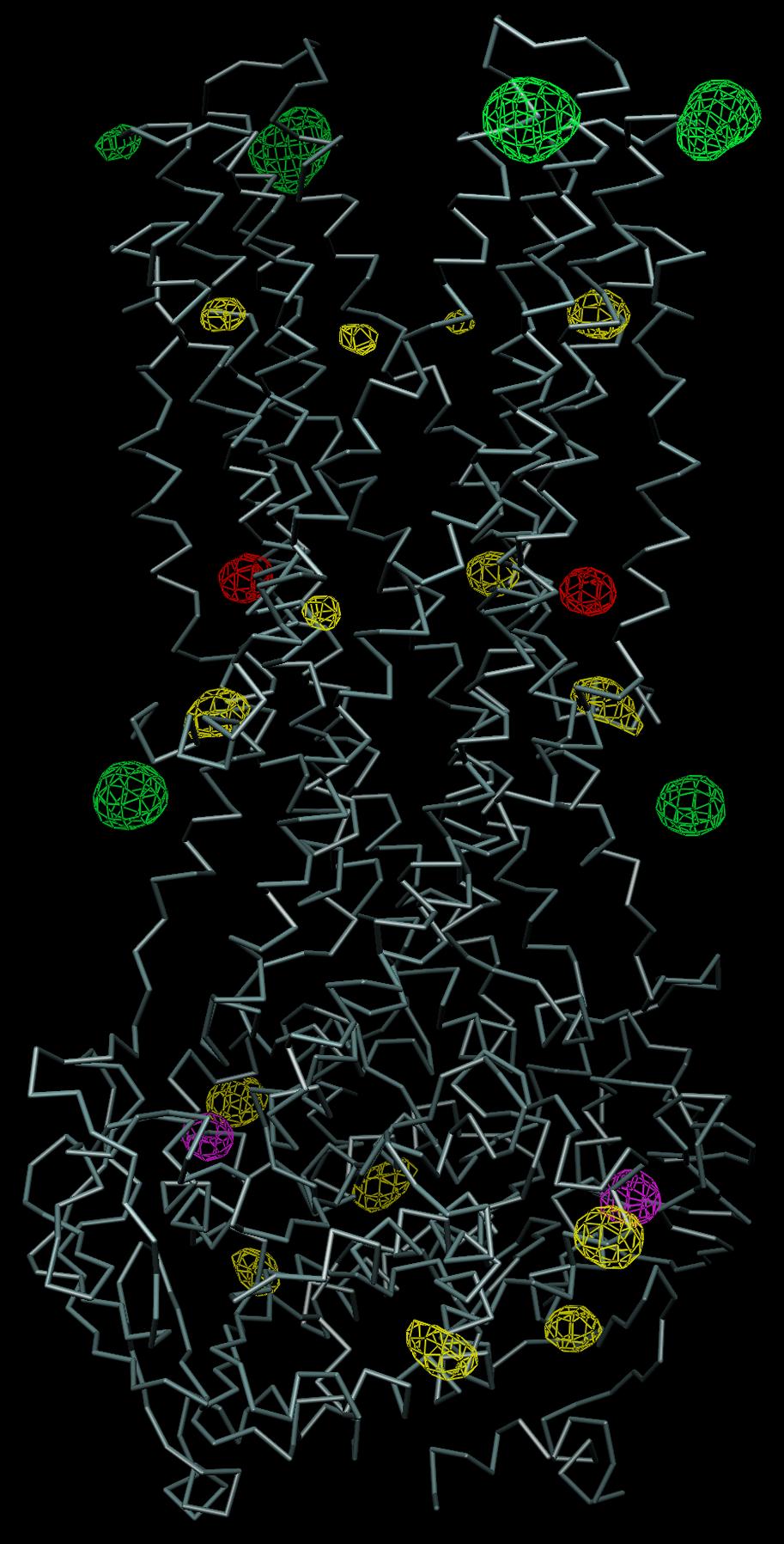
7 7 Supplementary Figure Legends Figure S1 ATPase activity of purified Sav1866. a, Sav1866 protein in C 12 E 8 detergent solution. Inorganic phosphate generated by ATP hydrolysis is shown as a function of time (closed symbols). A control reaction (open symbols) was inhibited by addition of 1 mm ortho-vanadate at 10 minutes. Error bars indicate standard deviations, all reactions were performed in triplicates. b, Similar to a, but using Sav1866 reconstituted in liposomes (closed symbols). A moderate stimulation of the ATPase activity upon addition of 1 mm doxorubicin is evident (open symbols). c, Drugstimulated ATPase activity. Initial ATPase rates were determined in triplicates by linear regression of time points similar to experiments shown in panel b. Closed squares indicate relative ATPase rates upon addition of Hoechst33342, whereas open symbols indicate those with vinblastine. Figure S2 Experimental electron density. Stereo view of the backbone of the Sav1866 transmembrane domains in red, with the experimental electron density map contoured at 1σ (blue mesh). Figure S3 Anomalous electron density maps. The backbone of Sav1866 is in white. Anomalous cross-fourier maps were calculated using experimental phases and the anomalous differences of the following data sets: Selenomethiononine (yellow, contoured at 7σ), Ta 6 Br 14 (green, contoured at 5σ), 2'-iodo-ADP (purple, countoured at 8σ), and xenon (red, contoured at 15σ). The selenomethiononine peaks were helpful in chain tracing. Figure S4 Superposition of Sav1866 and MsbA. a, Stereo views of protein backbones of the transmembrane domains of a single subunit of Salmonella typhimurium MsbA (pdb entry 1Z2R, purple) and Sav1866 (green) after manual superpositions of the entire transporters. No agreement in the helix arrangement is apparent. b, The structure of MsbA was inverted using the coordinate manipulation program pdbset 5, and the resulting molecule was superimposed onto Sav1866. The transmembrane domain of a single monomer of inverted MsbA is shown in purple, while that of subunit A of Sav1866 is shown in green and transmembrane helices TM1 and TM2 of subunit B are shown in yellow. The approximate superposition of TM helices is evident. The differences in helix directionality and subunit assignment are detailed in Table S2.
8 Fig. S1 8

9 9 Fig. S2 Fig. S3

10 10 Fig. S4
SUPPLEMENTARY INFORMATION
 Structure of an ABC transporter-binding protein complex Kaspar Hollenstein, Dominik C. Frei, and Kaspar P. Locher Institute of Molecular Biology and Biophysics, ETH Zurich, 8093 Zurich, Switzerland Supplementary
Supplementary materials. Crystal structure of the carboxyltransferase domain. of acetyl coenzyme A carboxylase. Department of Biological Sciences
 Supplementary materials Crystal structure of the carboxyltransferase domain of acetyl coenzyme A carboxylase Hailong Zhang, Zhiru Yang, 1 Yang Shen, 1 Liang Tong Department of Biological Sciences Columbia
Supporting Information
 Supporting Information Structural Analysis of the Binding of Type I, I 1/2, and II Inhibitors to Eph Tyrosine Kinases Jing Dong, *1 Hongtao Zhao, 1 Ting Zhou, 1 Dimitrios Spiliotopoulos, 1 Chitra Rajendran,
Supplemental Data. Structure of the Rb C-Terminal Domain. Bound to E2F1-DP1: A Mechanism. for Phosphorylation-Induced E2F Release
 Supplemental Data Structure of the Rb C-Terminal Domain Bound to E2F1-DP1: A Mechanism for Phosphorylation-Induced E2F Release Seth M. Rubin, Anne-Laure Gall, Ning Zheng, and Nikola P. Pavletich Section
SUPPLEMENTARY INFORMATION
 Table of Contents Page Supplementary Table 1. Diffraction data collection statistics 2 Supplementary Table 2. Crystallographic refinement statistics 3 Supplementary Fig. 1. casic1mfc packing in the R3
Full-length GlpG sequence was generated by PCR from E. coli genomic DNA. (with two sequence variations, D51E/L52V, from the gene bank entry aac28166),
,") Supplementary Methods Protein expression and purification Full-length GlpG sequence was generated by PCR from E. coli genomic DNA (with two sequence variations, D51E/L52V, from the gene bank entry aac28166),
Supplementary Methods Protein expression and purification Full-length GlpG sequence was generated by PCR from E. coli genomic DNA (with two sequence variations, D51E/L52V, from the gene bank entry aac28166),
SUPPLEMENTARY INFORMATION
 Supplementary Table 1: Data collection, phasing and refinement statistics ChbC/Ta 6 Br 12 Native ChbC Data collection Space group P4 3 2 1 2 P4 3 2 1 2 Cell dimensions a, c (Å) 132.75, 453.57 132.81, 452.95
SUPPLEMENTARY INFORMATION
 SUPPLEMENTARY INFORMATION doi:10.1038/nature11524 Supplementary discussion Functional analysis of the sugar porter family (SP) signature motifs. As seen in Fig. 5c, single point mutation of the conserved
SUPPLEMENTARY INFORMATION
 doi:10.1038/nature12045 Supplementary Table 1 Data collection and refinement statistics. Native Pt-SAD X-ray source SSRF BL17U SPring-8 BL41XU Wavelength (Å) 0.97947 1.07171 Space group P2 1 2 1 2 1 P2
Supporting Information
 Supporting Information Structural Basis of the Antiproliferative Activity of Largazole, a Depsipeptide Inhibitor of the Histone Deacetylases Kathryn E. Cole 1, Daniel P. Dowling 1,2, Matthew A. Boone 3,
type GroEL-GroES complex. Crystals were grown in buffer D (100 mm HEPES, ph 7.5,
 Supplementary Material Supplementary Materials and Methods Structure Determination of SR1-GroES-ADP AlF x SR1-GroES-ADP AlF x was purified as described in Materials and Methods for the wild type GroEL-GroES
Macromolecular X-ray Crystallography
 Protein Structural Models for CHEM 641 Fall 07 Brian Bahnson Department of Chemistry & Biochemistry University of Delaware Macromolecular X-ray Crystallography Purified Protein X-ray Diffraction Data collection
SUPPLEMENTARY INFORMATION
 doi: 10.108/nature0608 a c pmol L-[ H]Leu / mg LeuT pmol L-[ H]Leu / min / mg LeuT 900 50 600 450 00 150 200 150 100 0 0.0 2.5 5.0.5 10.0.5 50 N Cl CMI IMI DMI H C CH N N H C CH N Time (min) 0 0 100 200
Supporting online materials. For Zhou et al., LeuT-desipramine structure suggests. how antidepressants inhibit human neurotransmitter transporters
 Supporting online materials For Zhou et al., LeuT-desipramine structure suggests how antidepressants inhibit human neurotransmitter transporters Materials and methods LeuT overexpression and purification.
SOLVE and RESOLVE: automated structure solution, density modification and model building
 Journal of Synchrotron Radiation ISSN 0909-0495 SOLVE and RESOLVE: automated structure solution, density modification and model building Thomas Terwilliger Copyright International Union of Crystallography
SUPPLEMENTARY INFORMATION
 Supplementary materials Figure S1 Fusion protein of Sulfolobus solfataricus SRP54 and a signal peptide. a, Expression vector for the fusion protein. The signal peptide of yeast dipeptidyl aminopeptidase
Table 1. Crystallographic data collection, phasing and refinement statistics. Native Hg soaked Mn soaked 1 Mn soaked 2
 Table 1. Crystallographic data collection, phasing and refinement statistics Native Hg soaked Mn soaked 1 Mn soaked 2 Data collection Space group P2 1 2 1 2 1 P2 1 2 1 2 1 P2 1 2 1 2 1 P2 1 2 1 2 1 Cell
SUPPLEMENTARY INFORMATION
 Supplementary Table 1: Amplitudes of three current levels. Level 0 (pa) Level 1 (pa) Level 2 (pa) TrkA- TrkH WT 200 K 0.01 ± 0.01 9.5 ± 0.01 18.7 ± 0.03 200 Na * 0.001 ± 0.01 3.9 ± 0.01 12.5 ± 0.03 200
High-resolution crystal structure of ERAP1 with bound phosphinic transition-state analogue inhibitor
 High-resolution crystal structure of ERAP1 with bound phosphinic transition-state analogue inhibitor Petros Giastas 1, Margarete Neu 2, Paul Rowland 2, and Efstratios Stratikos 1 1 National Center for
Overview - Macromolecular Crystallography
 Overview - Macromolecular Crystallography 1. Overexpression and crystallization 2. Crystal characterization and data collection 3. The diffraction experiment 4. Phase problem 1. MIR (Multiple Isomorphous
Marcin Nowotny, Sergei A. Gaidamakov, Rodolfo Ghirlando, Susana M. Cerritelli, Robert J. Crouch, and Wei Yang
 Molecular Cell, Volume 28 Supplemental Data Structure of Human RNase H1 Complexed with an RNA/DNA Hybrid: Insight into HIV Reverse Transcription Marcin Nowotny, Sergei A. Gaidamakov, Rodolfo Ghirlando,
Supporting Information. Synthesis of Aspartame by Thermolysin : An X-ray Structural Study
 Supporting Information Synthesis of Aspartame by Thermolysin : An X-ray Structural Study Gabriel Birrane, Balaji Bhyravbhatla, and Manuel A. Navia METHODS Crystallization. Thermolysin (TLN) from Calbiochem
SUPPLEMENTARY INFORMATION. doi: /nature07461
 Figure S1 Electrophysiology. a ph-activation of. Two-electrode voltage clamp recordings of Xenopus oocytes expressing in comparison to waterinjected oocytes. Currents were recorded at 40 mv. The ph of
University of Groningen
 University of Groningen Molecular basis of transport and regulation in the Na+/betaine symporter BetP Ressl, Susanne; Terwisscha van Scheltinga, Anke C.; Vonrhein, Clemens; Ott, Vera; Ziegler, Christine
SUPPLEMENTARY INFORMATION
 doi:10.1038/nature11085 Supplementary Tables: Supplementary Table 1. Summary of crystallographic and structure refinement data Structure BRIL-NOP receptor Data collection Number of crystals 23 Space group
SUPPLEMENTARY INFORMATION
 doi:1.138/nature1737 Supplementary Table 1 variant Description FSEC - 2B12 a FSEC - 6A1 a K d (leucine) c Leucine uptake e K (wild-type like) K (Y18F) K (TS) K (TSY) K288A mutant, lipid facing side chain
Pathogenic C9ORF72 Antisense Repeat RNA Forms a Double Helix with Tandem C:C Mismatches
 Supporting Information Pathogenic C9ORF72 Antisense Repeat RNA Forms a Double Helix with Tandem C:C Mismatches David W. Dodd, Diana R. Tomchick, David R. Corey, and Keith T. Gagnon METHODS S1 RNA synthesis.
Table S1. Overview of used PDZK1 constructs and their binding affinities to peptides. Related to figure 1.
 Table S1. Overview of used PDZK1 constructs and their binding affinities to peptides. Related to figure 1. PDZK1 constru cts Amino acids MW [kda] KD [μm] PEPT2-CT- FITC KD [μm] NHE3-CT- FITC KD [μm] PDZK1-CT-
SUPPLEMENTARY INFORMATION
 doi:10.1038/nature10955 Supplementary Figures Supplementary Figure 1. Electron-density maps and crystallographic dimer structures of the motor domain. (a f) Stereo views of the final electron-density maps
SUPPLEMENTARY INFORMATION
 doi:10.1038/nature11054 Supplementary Fig. 1 Sequence alignment of Na v Rh with NaChBac, Na v Ab, and eukaryotic Na v and Ca v homologs. Secondary structural elements of Na v Rh are indicated above the
Web-based Auto-Rickshaw for validation of the X-ray experiment at the synchrotron beamline
 Web-based Auto-Rickshaw for validation of the X-ray experiment at the synchrotron beamline Auto-Rickshaw http://www.embl-hamburg.de/auto-rickshaw A platform for automated crystal structure determination
SUPPLEMENTARY INFORMATION
 doi:10.1038/nature10458 Active Site Remodeling in the Bifunctional Fructose-1,6- bisphosphate aldolase/phosphatase Juan Du, Rafael F. Say, Wei Lü, Georg Fuchs & Oliver Einsle SUPPLEMENTARY FIGURES Figure
Structural Mechanism for the Fidelity Modulation of DNA Polymerase λ. 128 Academia Road Sec. 2, Nankang, Taipei, 115, Taiwan
 SUPPORTING INFORMATION Structural Mechanism for the Fidelity Modulation of DNA Polymerase λ Mu-Sen Liu, 1,3 Hsin-Yue Tsai, 1,# Xiao-Xia Liu, 1,# Meng-Chiao Ho, 1,3 Wen-Jin Wu, 1,* and Ming-Daw Tsai 1,2,3,*
research papers 1. Introduction Thomas C. Terwilliger a * and Joel Berendzen b
 Acta Crystallographica Section D Biological Crystallography ISSN 0907-4449 Discrimination of solvent from protein regions in native Fouriers as a means of evaluating heavy-atom solutions in the MIR and
Anomalous dispersion
 Selenomethionine MAD Selenomethionine is the amino acid methionine with the Sulfur replaced by a Selenium. Selenium is a heavy atom that also has the propery of "anomalous scatter" at some wavelengths,
New Delhi Metallo-β-Lactamase: Structural Insights into β- Lactam Recognition and Inhibition
 Supporting Information New Delhi Metallo-β-Lactamase: Structural Insights into β- Lactam Recognition and Inhibition Dustin T. King, Liam J. Worrall, Robert Gruninger, Natalie C.J. Strynadka* AUTHOR ADDRESS:
Electronic Supplementary Information (ESI) for Chem. Commun. Unveiling the three- dimensional structure of the green pigment of nitrite- cured meat
 for Chem. Commun. Unveiling the three- dimensional structure of the green pigment of nitrite- cured meat") Electronic Supplementary Information (ESI) for Chem. Commun. Unveiling the three- dimensional structure of the green pigment of nitrite- cured meat Jun Yi* and George B. Richter- Addo* Department of Chemistry
Electronic Supplementary Information (ESI) for Chem. Commun. Unveiling the three- dimensional structure of the green pigment of nitrite- cured meat Jun Yi* and George B. Richter- Addo* Department of Chemistry
SUPPLEMENTARY INFORMATION
 www.nature.com/nature 1 Figure S1 Sequence alignment. a Structure based alignment of the plgic of E. chrysanthemi (ELIC), the acetylcholine binding protein from the snail Lymnea stagnalis (AchBP, PDB code
Protein crystallography. Garry Taylor
 Protein crystallography Garry Taylor X-ray Crystallography - the Basics Grow crystals Collect X-ray data Determine phases Calculate ρ-map Interpret map Refine coordinates Do the biology. Nitrogen at -180
Nature Structural & Molecular Biology: doi: /nsmb Supplementary Figure 1
 Supplementary Figure 1 Identification of the ScDcp2 minimal region interacting with both ScDcp1 and the ScEdc3 LSm domain. Pull-down experiment of untagged ScEdc3 LSm with various ScDcp1-Dcp2-His 6 fragments.
Crystal lattice Real Space. Reflections Reciprocal Space. I. Solving Phases II. Model Building for CHEM 645. Purified Protein. Build model.
 I. Solving Phases II. Model Building for CHEM 645 Purified Protein Solve Phase Build model and refine Crystal lattice Real Space Reflections Reciprocal Space ρ (x, y, z) pronounced rho F hkl 2 I F (h,
S-SAD and Fe-SAD Phasing using X8 PROTEUM
 S-SAD and Fe-SAD Phasing using X8 PROTEUM Kristina Djinovic Carugo Dept. for Structural and Computational Biology Max F. Perutz Labs Univ. Vienna, Austria Outline Fe-SAD on chlorite dismutase from Candidatus
Plasmid Relevant features Source. W18N_D20N and TrXE-W18N_D20N-anti
 Table S1. E. coli plasmids Plasmid Relevant features Source pdg680 T. reesei XynII AA 2-190 with C-terminal His 6 tag optimized for E. coli expression in pjexpress401 Wan et al. (in press) psbn44d psbn44h
SUPPLEMENTARY INFORMATION
 SUPPLEMENTARY INFORMATION doi:10.1038/nature11744 Supplementary Table 1. Crystallographic data collection and refinement statistics. Wild-type Se-Met-BcsA-B SmCl 3 -soaked EMTS-soaked Data collection Space
IgE binds asymmetrically to its B cell receptor CD23
 Supplementary Information IgE binds asymmetrically to its B cell receptor CD23 Balvinder Dhaliwal 1*, Marie O. Y. Pang 2, Anthony H. Keeble 2,3, Louisa K. James 2,4, Hannah J. Gould 2, James M. McDonnell
SUPPLEMENTARY INFORMATION
 SUPPLEMENTARY INFORMATION doi:10.1038/nature11539 Supplementary Figure 1 Schematic representation of plant (A) and mammalian (B) P 2B -ATPase domain organization. Actuator (A-), nucleotide binding (N-),
X-ray Crystallography. Kalyan Das
 X-ray Crystallography Kalyan Das Electromagnetic Spectrum NMR 10 um - 10 mm 700 to 10 4 nm 400 to 700 nm 10 to 400 nm 10-1 to 10 nm 10-4 to 10-1 nm X-ray radiation was discovered by Roentgen in 1895. X-rays
Structurale, Université Grenoble Alpes, CNRS, CEA, Grenoble, France
 Supplementary Information to Lysine relay mechanism coordinates intermediate transfer in vitamin B6 biosynthesis Matthew J. Rodrigues 1,2, Volker Windeisen 1,3, Yang Zhang 4, Gabriela Guédez 3, Stefan
Phase problem: Determining an initial phase angle α hkl for each recorded reflection. 1 ρ(x,y,z) = F hkl cos 2π (hx+ky+ lz - α hkl ) V h k l
 = F hkl cos 2π (hx+ky+ lz - α hkl ) V h k l") Phase problem: Determining an initial phase angle α hkl for each recorded reflection 1 ρ(x,y,z) = F hkl cos 2π (hx+ky+ lz - α hkl ) V h k l Methods: Heavy atom methods (isomorphous replacement Hg, Pt)
Phase problem: Determining an initial phase angle α hkl for each recorded reflection 1 ρ(x,y,z) = F hkl cos 2π (hx+ky+ lz - α hkl ) V h k l Methods: Heavy atom methods (isomorphous replacement Hg, Pt)
Supplementary Information
 Supplementary Information The direct role of selenocysteine in [NiFeSe] hydrogenase maturation and catalysis Marta C. Marques a, Cristina Tapia b, Oscar Gutiérrez-Sanz b, Ana Raquel Ramos a, Kimberly L.
Chapter 6. The interaction of Src SH2 with the focal adhesion kinase catalytic domain studied by NMR
 The interaction of Src SH2 with the focal adhesion kinase catalytic domain studied by NMR 103 Abstract The interaction of the Src SH2 domain with the catalytic domain of FAK, including the Y397 SH2 domain
SUPPLEMENTARY INFORMATION
 Data collection Supplementary Table 1 Statistics of data collection, phasing and refinement Native Se-MAD Space group P2 1 2 1 2 1 P2 1 2 1 2 1 Cell dimensions a, b, c (Å) 50.4, 94.2, 115.4 49.8, 94.2,
Supporting Information
 Supporting Information Fera et al. 10.1073/pnas.1409954111 SI Methods Compliance. All work related to human subjects complied with protocols approved by the Duke University Health System Institutional
Supplementary Information. Cryo-EM Studies of Drp1 Reveal Cardiolipin Interactions that Activate
 Supplementary Information Cryo-EM Studies of Drp1 Reveal Cardiolipin Interactions that Activate the Helical Oligomer Christopher A. Francy 1, 2, 3, Ryan W. Clinton 1, 2, 3, Chris Fröhlich 4, 5, Colleen
X-ray Crystallography
 2009/11/25 [ 1 ] X-ray Crystallography Andrew Torda, wintersemester 2009 / 2010 X-ray numerically most important more than 4/5 structures Goal a set of x, y, z coordinates different properties to NMR History
Supporting Information
 Supporting Information Horne et al. 10.1073/pnas.0902663106 SI Materials and Methods Peptide Synthesis. Protected 3 -amino acids were purchased from PepTech. Cyclically constrained -residues, Fmoc-ACPC
Determining Protein Structure BIBC 100
 Determining Protein Structure BIBC 100 Determining Protein Structure X-Ray Diffraction Interactions of x-rays with electrons in molecules in a crystal NMR- Nuclear Magnetic Resonance Interactions of magnetic
Supporting Online Material for
 www.sciencemag.org/cgi/content/full/318/5857/1744/dc1 Supporting Online Material for The Structure of a Human p110α/p85α Complex Elucidates the Effects of Oncogenic PI3Kα Mutations Chuan-Hsiang Huang,
The Potassium Ion Channel: Rahmat Muhammad
 The Potassium Ion Channel: 1952-1998 1998 Rahmat Muhammad Ions: Cell volume regulation Electrical impulse formation (e.g. sodium, potassium) Lipid membrane: the dielectric barrier Pro: compartmentalization
Potassium channel gating and structure!
 Reading: Potassium channel gating and structure Hille (3rd ed.) chapts 10, 13, 17 Doyle et al. The Structure of the Potassium Channel: Molecular Basis of K1 Conduction and Selectivity. Science 280:70-77
SI Text S1 Solution Scattering Data Collection and Analysis. SI references
 SI Text S1 Solution Scattering Data Collection and Analysis. The X-ray photon energy was set to 8 kev. The PILATUS hybrid pixel array detector (RIGAKU) was positioned at a distance of 606 mm from the sample.
SUPPLEMENTARY INFORMATION
 Supplementary Figure 1: The HpUreI crystal used for collection of native diffraction data. The crystal belongs to spacegroup P4 2 2 1 2 and has an approximate maximal dimension of 0.25 mm. Supplementary
Nature Structural & Molecular Biology: doi: /nsmb Supplementary Figure 1
 Supplementary Figure 1 Chemical structure of LPS and LPS biogenesis in Gram-negative bacteria. a. Chemical structure of LPS. LPS molecule consists of Lipid A, core oligosaccharide and O-antigen. The polar
for Molecular Biology and Neuroscience and Institute of Medical Microbiology, Rikshospitalet-Radiumhospitalet
 SUPPLEMENTARY INFORMATION TO Structural basis for enzymatic excision of N -methyladenine and N 3 -methylcytosine from DNA Ingar Leiros,5, Marivi P. Nabong 2,3,5, Kristin Grøsvik 3, Jeanette Ringvoll 2,
Supplemental Data SUPPLEMENTAL FIGURES
 Supplemental Data CRYSTAL STRUCTURE OF THE MG.ADP-INHIBITED STATE OF THE YEAST F 1 C 10 ATP SYNTHASE Alain Dautant*, Jean Velours and Marie-France Giraud* From Université Bordeaux 2, CNRS; Institut de
Direct Methods and Many Site Se-Met MAD Problems using BnP. W. Furey
 Direct Methods and Many Site Se-Met MAD Problems using BnP W. Furey Classical Direct Methods Main method for small molecule structure determination Highly automated (almost totally black box ) Solves structures
Supporting Information
 Supporting Information Design and Synthesis of Potent HIV-1 Protease Inhibitors Containing Bicyclic Oxazolidinone Scaffold as the P2-Ligands: Structure-Activity Studies, Biological and X-ray Structural
The structure of vanadium nitrogenase reveals an unusual bridging ligand
 SUPPLEMENTARY INFORMATION The structure of vanadium nitrogenase reveals an unusual bridging ligand Daniel Sippel and Oliver Einsle Lehrstuhl Biochemie, Institut für Biochemie, Albert-Ludwigs-Universität
Nature Structural & Molecular Biology: doi: /nsmb Supplementary Figure 1
 Supplementary Figure 1 Crystallization. a, Crystallization constructs of the ET B receptor are shown, with all of the modifications to the human wild-type the ET B receptor indicated. Residues interacting
Supporting Information
 Supporting Information Wan et al. 10.1073/pnas.1415856111 SI Materials and Methods Proposals for Keto-Enol Tautomerization of Substrate and the DHFR Catalytic Mechanism. As noted in the main text, competing
Supporting Information for. Jesinghaus, Rachael Barry, Zemer Gitai, Justin Kollman and Enoch P. Baldwin
 Supporting Information for Inhibition of E. coli CTP synthetase by NADH and other nicotinamides, and their mutual interactions with CTP and GTP Chris Habrian, Adithi Chandrasekhara, Bita Shahrvini, Brian
Mavis Agbandje-McKenna, Robert McKenna* Department of Biochemistry and Molecular Biology and Department of
 Ultra-High Resolution X-Ray Diffraction from Crystals of the Kinetic Mutant of Human Carbonic Anhydrase II, His 64 Ala, and its Complexes with Proton Acceptor/Donors. David Duda, Chingkuang Tu, David.
- Introduction of x-ray crystallography: what it s used for, how it works, applications in science - Different methods used to generate data - Case
 - Introduction of x-ray crystallography: what it s used for, how it works, applications in science - Different methods used to generate data - Case studies emphasizing the importance of the technique -
Acta Crystallographica Section D
 Supporting information Acta Crystallographica Section D Volume 70 (2014) Supporting information for article: Structural characterization of the virulence factor Nuclease A from Streptococcus agalactiae
Structure and Function of Neisseria gonorrhoeae MtrF Illuminates a Class of Antimetabolite Efflux Pumps
 Cell Reports Supplemental Information Structure and Function of Neisseria gonorrhoeae MtrF Illuminates a Class of Antimetabolite Efflux Pumps Chih-Chia Su, Jani Reddy Bolla, Nitin Kumar, Abhijith Radhakrishnan,
Acta Crystallographica Section F
 Supporting information Acta Crystallographica Section F Volume 70 (2014) Supporting information for article: Chemical conversion of cisplatin and carboplatin with histidine in a model protein crystallised
ml. ph 7.5 ph 6.5 ph 5.5 ph 4.5. β 2 AR-Gs complex + GDP β 2 AR-Gs complex + GTPγS
 a UV28 absorption (mau) 9 8 7 5 3 β 2 AR-Gs complex β 2 AR-Gs complex + GDP β 2 AR-Gs complex + GTPγS β 2 AR-Gs complex dissociated complex excess nucleotides b 9 8 7 5 3 β 2 AR-Gs complex β 2 AR-Gs complex
University of Groningen
 University of Groningen In vitro functional characterization of BtuCD-F, the Escherichia coli ABC transporter for vitamin B-12 uptake Borths, EL; Poolman, Berend; Hvorup, RN; Locher, KP; Rees, DC; Hvorup,
Department of Biochemistry, University of Zürich, Winterthurerstrasse 190, CH-8057 Zürich, Switzerland
 Supporting information Twenty crystal structures of bromodomain and PHD finger containing protein 1 (BRPF1)/ligand complexes reveal conserved binding motifs and rare interactions Jian Zhu and Amedeo Caflisch*
SUPPLEMENTARY INFORMATION
 UPPEER ORO doi:10.1038/nature10753 D D D D P E ntracellular C1 W P P C EC1 D Q R H C D W D R C C2 D E D E C R Q Q W P W W R P P EC2 EC3 P C C P W P W W P C W H R C R E C3 P R R P P P C Extracellular embrane
Cks1 CDK1 CDK1 CDK1 CKS1. are ice- lobe. conserved. conserved
 Cks1 d CKS1 Supplementary Figure 1 The -Cks1 crystal lattice. (a) Schematic of the - Cks1 crystal lattice. -Cks1 crystallizes in a lattice that contains c 4 copies of the t - Cks1 dimer in the crystallographic
MULTIPLE CHOICE. Choose the one alternative that best completes the statement or answers the question. 1)
") MULTIPLE CHOICE. Choose the one alternative that best completes the statement or answers the question. 1) 1) Which of the following statements about the atom A) It has 12 neutrons in its nucleus. B) It
MULTIPLE CHOICE. Choose the one alternative that best completes the statement or answers the question. 1) 1) Which of the following statements about the atom A) It has 12 neutrons in its nucleus. B) It
Production of Recombinant Annexin V from plasmid pet12a-papi
 Tait Research Laboratory Page 1 of 5 Principle Production of Recombinant Annexin V from plasmid pet12a-papi Annexin V is expressed cytoplasmically in BL21(DE3) E. coli (Novagen) with the pet vector system
MULTIPLE CHOICE. Choose the one alternative that best completes the statement or answers the question. Figure 2.1
 Exam Name MULTIPLE CHOICE. Choose the one alternative that best completes the statement or answers the question. Figure 2.1 1) Which compound in Figure 2.1 is an ester? 1) A) a b c d e Answer: D 2) A scientist
Supporting Information. UV-induced ligand exchange in MHC class I protein crystals
 Supporting Information for the article entitled UV-induced ligand exchange in MHC class I protein crystals by Patrick H.N. Celie 1, Mireille Toebes 2, Boris Rodenko 3, Huib Ovaa 3, Anastassis Perrakis
SUPPLEMENTARY FIGURES
 SUPPLEMENTARY FIGURES Supplementary Figure 1 Protein sequence alignment of Vibrionaceae with either a 40-residue insertion or a 44-residue insertion. Identical residues are indicated by red background.
SHELXC/D/E. Andrea Thorn
 SHELXC/D/E Andrea Thorn What is experimental phasing? Experimental phasing is what you do if MR doesn t work. What is experimental phasing? Experimental phasing methods depend on intensity differences.
Supporting Information
 Supporting Information Self-Assembly of Glutathione S-transferases into Nanowires Wei Zhang, a Quan Luo,* a Lu Miao, a Yushi Bai, a Zeyuan Dong, a Jiayun Xu, a and Junqiu Liu* a a State Key Laboratory
Scattering by two Electrons
 Scattering by two Electrons p = -r k in k in p r e 2 q k in /λ θ θ k out /λ S q = r k out p + q = r (k out - k in ) e 1 Phase difference of wave 2 with respect to wave 1: 2π λ (k out - k in ) r= 2π S r
Cloning, preliminary characterization, and crystallization of nucleoside hydrolases from Caenorhabditis elegans and Campylobacter jejuni.
 Cloning, preliminary characterization, and crystallization of nucleoside hydrolases from Caenorhabditis elegans and Campylobacter jejuni. Wim Versées, * Els Van Holsbeke, Stefan De Vos, Klaas Decanniere,
Transporters and Membrane Motors Nov 15, 2007
 BtuB OM vitamin B12 transporter F O F 1 ATP synthase Human multiple drug resistance transporter P-glycoprotein Transporters and Membrane Motors Nov 15, 2007 Transport and membrane motors Concentrations
Measuring S using an analytical ultracentrifuge. Moving boundary
 Measuring S using an analytical ultracentrifuge Moving boundary [C] t = 0 t 1 t 2 0 top r bottom 1 dr b r b (t) r b ω 2 = S ln = ω 2 S (t-t dt r b (t o ) o ) r b = boundary position velocity = dr b dt
SUPPLEMENTARY INFORMATION
 Supplementary Results DNA binding property of the SRA domain was examined by an electrophoresis mobility shift assay (EMSA) using synthesized 12-bp oligonucleotide duplexes containing unmodified, hemi-methylated,
Introduction to single crystal X-ray analysis
 Technical articles Introduction to single crystal X-ray analysis IX. Protein structure analysis and small molecule structure analysis Akihito Yamano* 1. Introduction The previous series have discussed
MULTIPLE CHOICE. Choose the one alternative that best completes the statement or answers the question. C is FALSE?
 Exam Name MULTIPLE CHOICE. Choose the one alternative that best completes the statement or answers the question. 1) Which of the following statements about the atom 12 6 C is FALSE? 1) A) It has 12 neutrons
Supplementary Figure 1. Biochemical and sequence alignment analyses the
 Supplementary Figure 1. Biochemical and sequence alignment analyses the interaction of OPTN and TBK1. (a) Analytical gel filtration chromatography analysis of the interaction between TBK1 CTD and OPTN(1-119).
Supplemental Information
 Supplemental Information Combinatorial Readout of Unmodified H3R2 and Acetylated H3K14 by the Tandem PHD Finger of MOZ Reveals a Regulatory Mechanism for HOXA9 Transcription Yu Qiu 1, Lei Liu 1, Chen Zhao
Structural Basis for Methyl Transfer by a Radical SAM Enzyme
 www.sciencemag.org/cgi/content/full/science.1205358/dc1 Supporting Online Material for Structural Basis for Methyl Transfer by a Radical SAM Enzyme Amie K. Boal, Tyler L. Grove, Monica I. McLaughlin, Neela
Experimental phasing in Crank2
 Experimental phasing in Crank2 Pavol Skubak and Navraj Pannu Biophysical Structural Chemistry, Leiden University, The Netherlands http://www.bfsc.leidenuniv.nl/software/crank/ X-ray structure solution
Microbiology: An Introduction, 12e (Tortora) Chapter 2 Chemical Principles. 2.1 Multiple Choice Questions
 Chapter 2 Chemical Principles. 2.1 Multiple Choice Questions") Microbiology An Introduction 12th Edition Tortora TEST BANK Full download at: https://testbankreal.com/download/microbiology-an-introduction-12thedition-tortora-test-bank/ Microbiology An Introduction
Microbiology An Introduction 12th Edition Tortora TEST BANK Full download at: https://testbankreal.com/download/microbiology-an-introduction-12thedition-tortora-test-bank/ Microbiology An Introduction