Department of Biochemistry, University of Zürich, Winterthurerstrasse 190, CH-8057 Zürich, Switzerland
|
|
|
- Ilene Pitts
- 6 years ago
- Views:
Transcription
1 Supporting information Twenty crystal structures of bromodomain and PHD finger containing protein 1 (BRPF1)/ligand complexes reveal conserved binding motifs and rare interactions Jian Zhu and Amedeo Caflisch* Department of Biochemistry, University of Zürich, Winterthurerstrasse 190, CH-8057 Zürich, Switzerland * caflisch@bioc.uzh.ch Table of contents Fig S1. Top ranked binding mode of fragments 1, 2, 3 and Fig S2. False positive hits from fragment docking Fig S3. Plot of similarity matrix of compounds 1 to BromoScan assay... 4 Fig S4. Dose-response curves for compounds obtained using the competition binding assay at DiscoveRx with BRPF1b Fig S5. IC50 value of compound 20 determined by AlphaScreen binding assay Table S1. X-ray data collection and refinement statistics for complex structures with 1, 2, 3 and Table S2. X-ray data collection and refinement statistics for complex structures with 5, 6, 7 and Table S3. X-ray data collection and refinement statistics for complex structures with 9, 10, 11 and Table S4. X-ray data collection and refinement statistics for complex structures with 13, 14, 15 and Table S5. X-ray data collection and refinement statistics for complex structures with 18, 19, and Fig S6. Close view of binding mode of compounds 1, 2, 3, 4, 5 and Fig S7. Close view of binding mode of compounds 7, 8, 9, 10, 11 and Fig S8. Close view of binding mode of compounds 13, 14, 15, 16, 17, 18, 19 and Fig S9. Chemical structures of inhibitors for structural comparison Fig S10. Binding mode comparison of the dihydroquinolin ligands bound to bromodomain proteins Fig S11. Binding mode comparison of the 9H-purine ligands bound to bromodomain proteins


2 Fig S1. Comparison of docked pose obtained by the fragment-docking program SEED (carbon atoms in magenta) and binding mode in the crystal structure (carbon atoms in yellow) of fragments 1, 2, 3 and 8. Heteroatoms are colored as follows: N (blue), O (red), S (yellow), and Br (maroon). The six conserved water molecules in the Kac binding pocket are labeled W1 to W6. 1
3 Fig S2. False positive hits from fragment docking by SEED. These fragments were among the 13 fragments purchased from 30 top ranked ones, but binding is not observed by X-ray crystallography (soaking into BRPF1 apo crystals). 2
 was")

 of the circles indicate similarity.")

4 Fig S3. Similarity matrix of compounds 1 to 16. The similarity (Tanimoto coefficient) was calculated based on the RDKit fingerprint which is implemented in the RDKit 1 toolkit. The size and darkness (vertical legend on the right) of the circles indicate similarity. The hierarchical complete-link algorithm with the R programming language generates six clusters (orange boxes). 3
5 BromoScan assay 2 BromoScan assays on BRPF1 for the 14 compounds were performed at DiscoveRx. T7 phage strains displaying bromodomains were grown in parallel in 24-well blocks in an E. coli host derived from the BL21 strain. E. coli were grown to log-phase and infected with T7 phage from a frozen stock (multiplicity of infection= 0.4) and incubated with shaking at 32 C until lysis ( minutes). The lysates were centrifuged (5,000 x g) and filtered (0.2 μm) to remove cell debris. Streptavidin-coated magnetic beads were treated with biotinylated small molecule or acetylated peptide ligands for 30 minutes at room temperature to generate affinity resins for bromodomain assays. The liganded beads were blocked with excess biotin and washed with blocking buffer (SeaBlock (Pierce), 1 % BSA, 0.05 % Tween 20, 1 mm DTT) to remove unbound ligand and to reduce non-specific phage binding. Binding reactions were assembled by combining bromodomains, liganded affinity beads, and test compounds in 1x binding buffer (17% SeaBlock, 0.33x PBS, 0.04% Tween 20, 0.02% BSA, 0.004% Sodium azide, 7.4 mm DTT). Test compounds were prepared as 1000X stocks in 100% DMSO and subsequently diluted 1:10 in monoethylene glycol (MEG) to create stocks at 100X the screening concentration (resulting stock solution is 10% DMSO/90% MEG). The compounds were then diluted directly into the assays such that the final concentration of DMSO and MEG were 0.1% and 0.9%, respectively. All reactions were performed in polystyrene 96-well plates in a final volume of ml. The assay plates were incubated at room temperature with shaking for 1 hour and the affinity beads were washed with wash buffer (1x PBS, 0.05% Tween 20). The beads were then re-suspended in elution buffer (1x PBS, 0.05% Tween 20, 2 μm nonbiotinylated affinity ligand) and incubated at room temperature with shaking for 30 minutes. The bromodomain concentration in the eluates was measured by qpcr. Binding constants (Kds) were calculated with a standard dose-response curve using the Hill equation: Signal Background Response = Background + 1+(K / Dose ) Curves were fitted using a non-linear least square fit with the Levenberg-Marquardt algorithm. 4
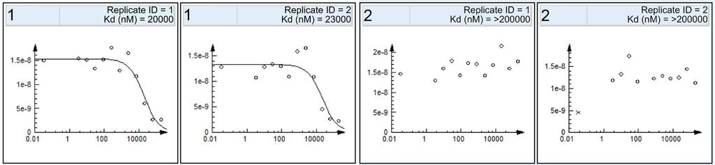
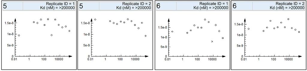
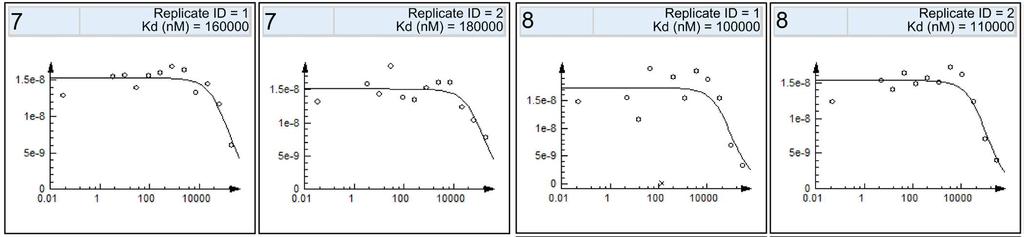
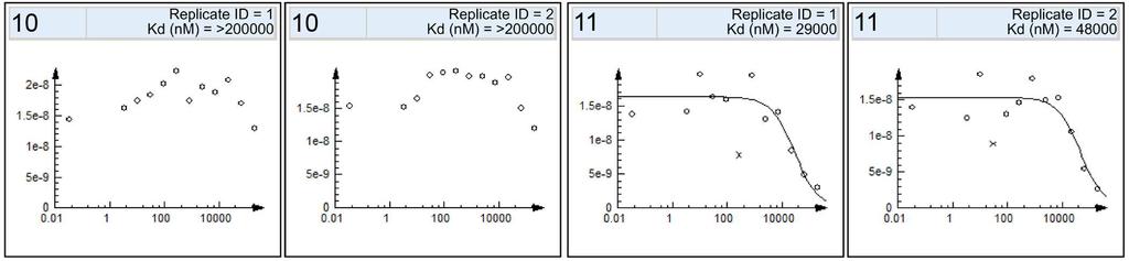
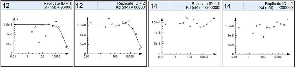
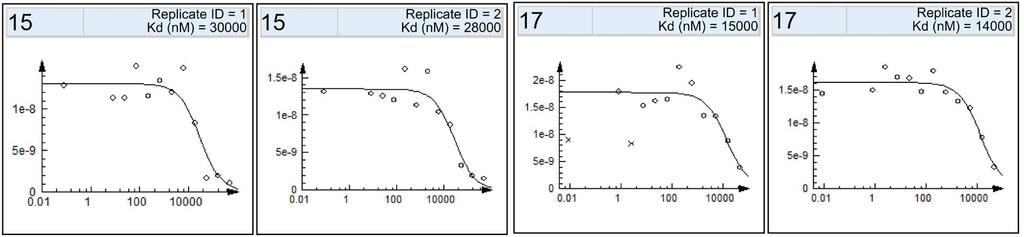
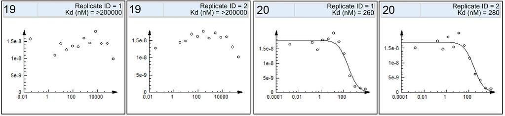
6 Fig S4. Dose-response curves in duplicates for the 16 compounds tested for binding to the BRPF1 bromodomain in the competition binding assay at DiscoveRx. 5
7 Fig S5. IC50 value of compound 20 determined by the AlphaScreen binding assay 3 at Reaction Biology. 6
8 Table S1. X-ray data collection and refinement statistics for complex structures of the BRPF1 bromodomain and compounds 1, 2, 3 and 4. Data Collection PDB ID 5EQ1 5EWD 5E3G 5E3D ligand space group P3221 P3221 P3221 P3221 Cell dimensions a, b, c (Å) 60.38, 60.38, , 60.67, , 60.86, , 60.73, α, β, γ ( ) 90.00, 90.00, , 90.00, , 90.00, , 90.00, resolution (Å) unique observations* 19761(2835) 18839(2686) 16662(2374) 14799(2108) completeness* 99.9 (100.0) 100.0(100.0) 100.0(100.0) 99.7(99.1) redundancy* 9.4 (8.9) 9.6(9.6) 9.7(10.0) 9.5(9.6) Rmerge* (0.338) 0.029(0.359) 0.040(0.442) 0.031(0.405) I/σI* 25.9 (6.1) 34.7(5.7) 25.6(4.9) 30.3(5.1) Refinement Rwork/Rfree* 0.189(0.229)/0.194(0.292) 0.199(0.233)/0.225(0.252) 0.196(0.208)/0.205(0.268) 0.192(0.241)/0.222(0.319) lengths (Å) angles ( ) no. of non-hydrogen atom / average B-factor (Å 2 ) protein 946/ / / /48.64 ligand 13/ / / /59.34 water 80/ / / /48.63 residues in protein chain Ramanchandran Favored allowed disallowed * Highest resolution shell is shown in parentheses. 7
9 Table S2. X-ray data collection and refinement statistics for complex structures of the BRPF1 bromodomain and compounds 5, 6, 7 and 8. Data Collection PDB ID 5C87 5EM3 5EWH 5C85 ligand space group P3221 P3221 P3221 P3221 Cell dimensions a, b, c (Å) 60.56, 60.56, , 60.14, , 60.44, , 60.72, α, β, γ ( ) 90.00, 90.00, , 90.00, , 90.00, , 90.00, resolution (Å) unique observations* 20025(2861) 26409(3814) 16966(2453) 14925(2129) completeness* 100.0(100.0) 99.7(99.7) 100.0(100.0) 100.0(100.0) redundancy* 9.1(6.0) 9.2(8.8) 9.5(9.9) 9.7(9.5) Rmerge* 0.048(0.341) 0.049(0.236) 0.073(0.358) 0.031(0.326) I/σI* 26.6(4.8) 25.4(8.4) 16.3(5.3) 37.1(6.5) Refinement Rwork/Rfree* 0.181(0.221)/0.198(0.26) 0.173(0.187)/0.196(0.224) 0.190(0.212)/0.223(0.288) 0.207(0.241)/0.225(0.269) lengths (Å) angles ( ) no. of non-hydrogen atom / average B-factor (Å 2 ) protein 947/ / / /50.59 ligand 11/ / / /55.99 water 124/ / / /48.55 residues in protein chain Ramanchandran (extra serine residue 625 at the N terminal ) Favored allowed disallowed * Highest resolution shell is shown in parentheses. 8
10 Table S3. X-ray data collection and refinement statistics for complex structures of the BRPF1 bromodomain and compounds 9, 10, 11 and 12. Data Collection PDB ID 5DYC 5DY7 5EPS 5EPR ligand space group P3221 P3221 P3221 P3221 Cell dimensions a, b, c (Å) 60.81, 60.81, , 60.67, , 60.63, , 60.92, α, β, γ ( ) 90.00, 90.00, , 90.00, , 90.00, , 90.00, resolution (Å) unique observations* 16556(2353) 15153(2094) 23061(3316) 16718(2400) completeness* 99.5(99.3) 99.2(95.5) 100.0(99.8) 100.0(100.0) redundancy* 9.7(10.0) 9.5(9.3) 9.5(9.3) 9.5(9.9) Rmerge* 0.039(0.408) 0.060(0.440) 0.042(0.285) 0.035(0.366) I/σI* 28.2(5.4) 18.7(4.5) 25.4(6.6) 28.6(5.6) Refinement Rwork/Rfree* 0.198(0.237)/0.235(0.289) 0.179(0.243)/0.204(0.306) 0.188(0.202)/0.198(0.250) 0.198(0.264)/0.220(0.285) lengths (Å) angles ( ) no. of non-hydrogen atom / average B-factor protein 951/ / / /44.14 ligand 12/ / / /59.44 water 82/ / / /45.87 residues in protein chain Ramanchandran(%) Favored allowed disallowed * Highest resolution shell is shown in parentheses. 9
11 Table S4. X-ray data collection and refinement statistics for complex structures of the BRPF1 bromodomain and compounds 13, 14, 15 and 16. Data Collection PDB ID 5EWC 5DYA 5ETB 5ETD ligand space group P3221 P3221 P3221 P3221 Cell dimensions a, b, c (Å) 60.93, 60.93, , 61.16, , 60.37, , 60.32, α, β, γ ( ) 90.00, 90.00, , 90.00, , 90.00, , 90.00, resolution (Å) unique observations* 14043(1999) 16557(2370) 31032(4300) 26520(3791) completeness* 100.0(99.9) 99.8(100.0) 99.3(95.6) 99.9(99.5) redundancy* 9.7(9.7) 9.5(9.8) 8.7(4.9) 9.2(8.4) Rmerge* 0.049(0.406) 0.027(0.417) 0.038(0.158) 0.044(0.334) I/σI* 24.1(5.1) 37.1(5.2) 31.1(7.8) 22.9(5.3) Refinement Rwork/Rfree* 0.194(0.252)/0.211(0.312) 0.186(0.214)/0.213(0.264) 0.174(0.204)/0.186(0.235) 0.176(0.215)/0.186(0.219) lengths (Å) angles ( ) no. of non-hydrogen atom / average B-factor protein 942/ / / /25.24 ligand 17/ / / /27.97 water 91/ / / /34.71 residues in protein chain Ramanchandran Favored allowed disallowed * Highest resolution shell is shown in parentheses. 10
12 Table S5. X-ray data collection and refinement statistics for complex structures of the BRPF1 bromodomain and compounds 18, 19, and 20. Data Collection PDB ID 5EV9 5EVA 5C7N ligand space group P3221 P3221 P3221 Cell dimensions a, b, c (Å) 60.36, 60.36, , 60.36, , 60.50, α, β, γ ( ) 90.00, 90.00, , 90.00, , 90.00, resolution (Å) unique observations* 24170(3476) 24128(3476) 13890(1986) completeness* 100.0(99.9) 99.9(100.0) 100.0(100.0) redundancy* 9.4(9.3) 9.3(9.0) 9.7(9.8) Rmerge* 0.032(0.285) 0.041(0.292) 0.044(0.420) I/σI* 33.0(6.9) 27.6(7.0) 27.5(5.4) Refinement Rwork/Rfree* 0.188(0.242)/0.221(0.257) 0.183(0.222)/0.197(0.235) 0.180(0.229)/0.220(0.289) angles ( ) lengths (Å) no. of non-hydrogen atom / average B-factor protein 939/ / /45.50 ligand 19/ / /62.80 water 130/ / /48.89 residues in protein chain Ramanchandran Favored allowed disallowed * Highest resolution shell is shown in parentheses. 11


.")


13 Fig S6. Close view of binding mode of compounds 1, 2, 3, 4, 5 and 6. Conserved water molecules in the binding pocket are labeled W1 to W6 (pink spheres). 2Fo-Fc electron density maps contoured at 1 σ for ligands are shown by a mesh. Two alternative conformations of fragment 6 are shown in yellow and cyan. 12


14 Fig S7. Same as Figure S7 for compounds 7, 8, 9, 10, 11 and 12. For fragments 7 and 12, 2Fo-Fc electron density maps are contoured at 0.8 σ. 13


15 Fig S8. Same as Figure S7 for compounds 13, 14, 15, 16, 17, 18, 19 and 20. For ligands 19 and 20, 2Fo-Fc electron density maps are contoured at 0.8 σ. 14
16 Fig S9. Chemical structures of inhibitors for structural comparison. (a)n-[1,3-dimethyl-2-oxo-6-(piperidin-1-yl)-2,3-dihydro-1h-benzimidazol-5-yl]-2-methoxybenzamide, (b) 6-(5-bromo-2-methoxyphenyl)-9H-purin-2-amine, (c)(3r)-n-[3-(3,4-dihydroquinolin-1(2h)-yl)propyl]-3-methyl-2-oxo-1,2,3,4-tetrahydroquinoxaline-5- carboxamide, and (d) (3R)-N-[3-(7-methoxy-3,4-dihydroquinolin-1(2H)-yl)propyl]-3-methyl-2-oxo- 1,2,3,4-tetrahydroquinoxaline-5-carboxamide. 15

 shows that the binding modes are")
 In BRPF1, the bromine substituent of fragment 9, the")
17 Fig S10. Comparison of the binding modes of dihydroquinoline ligands in BRPF1 (top) and CREBBP (bottom). The structural alignment (middle) shows that the binding modes are different. (Top) In BRPF1, the bromine substituent of fragment 9, the trifluromethyl of 10, and the carboxylate of 14 occupy the same position, while fragment 12 (5EPR) is devoid of substituent and its orientation is slightly shifted. 16
 with the previously reported 2-amine-9H-purine ligands of the BRD9 (4XY8, bottom) and BRD4 (4XY9, bottom)")
. 2. Quinn, E.; Wodicka, L.; Ciceri, P.; Pallares, G.; Pickle, E.; Torrey, A.; Floyd, M.")
18 Fig S11. Comparison of the binding modes of mercaptopurine fragments 3 and 4 in BRPF1 (top) with the previously reported 2-amine-9H-purine ligands of the BRD9 (4XY8, bottom) and BRD4 (4XY9, bottom) bromodomains. The structural alignment (middle) shows that the binding modes are different which is consistent with the fact that these ligands share only the purine scaffold. Reference 1. RDKit: cheminformatics and machine learning software (accessed January 3, 2016). 2. Quinn, E.; Wodicka, L.; Ciceri, P.; Pallares, G.; Pickle, E.; Torrey, A.; Floyd, M.; Hunt, J.; Treiber, D. Abstract 4238: BROMOscan - a high throughput, quantitative ligand binding platform identifies best-in-class bromodomain inhibitors from a screen of mature compounds targeting other protein classes Cancer Res. 2013, 73, Philpott, M.; Yang, J.; Tumber, T.; Fedorov, O.; Uttarkar, S.; Filippakopoulos, P.; Picaud, S.; Keates, T.; Felletar, I.; Ciulli, A.; Knapp, S.; Heightman, T. D., Bromodomain-peptide displacement assays for interactome mapping and inhibitor discovery. Mol. BioSyst. 2011, 7,
Supporting Information
 Supporting Information Binding motifs in the CBP bromodomain: an analysis of 20 crystal structures of complexes with small molecules Jian Zhu, 1 Jing Dong, 1 Laurent Batiste, 1 Andrea Unzue, 2 Aymeric
Supporting Information
 Supporting Information Virtual Screen to NMR (VS2NMR): Discovery of fragment hits for the CBP bromodomain Dimitrios Spiliotopoulos, *,a Jian Zhu, a Eike-Christian Wamhoff, b,c Nicholas Deerain, a Jean-Rémy
Nature Structural & Molecular Biology doi: /nsmb Supplementary Figure 1. CRBN binding assay with thalidomide enantiomers.
 Supplementary Figure 1 CRBN binding assay with thalidomide enantiomers. (a) Competitive elution assay using thalidomide-immobilized beads coupled with racemic thalidomide. Beads were washed three times
FRAGMENT SCREENING IN LEAD DISCOVERY BY WEAK AFFINITY CHROMATOGRAPHY (WAC )
") FRAGMENT SCREENING IN LEAD DISCOVERY BY WEAK AFFINITY CHROMATOGRAPHY (WAC ) SARomics Biostructures AB & Red Glead Discovery AB Medicon Village, Lund, Sweden Fragment-based lead discovery The basic idea:
FRAGMENT SCREENING IN LEAD DISCOVERY BY WEAK AFFINITY CHROMATOGRAPHY (WAC ) SARomics Biostructures AB & Red Glead Discovery AB Medicon Village, Lund, Sweden Fragment-based lead discovery The basic idea:
Supporting Information
 Supporting Information Discovery and optimization of a selective ligand for the Switch/Sucrose Non-Fermenting-related bromodomains of Polybromo protein-1 by the use of virtual screening and hydration analysis.
SUPPLEMENTARY INFORMATION
 SUPPLEMENTARY INFORMATION doi:10.1038/nature11524 Supplementary discussion Functional analysis of the sugar porter family (SP) signature motifs. As seen in Fig. 5c, single point mutation of the conserved
Supporting Information
 Protein-Observed Fluorine NMR is a Complementary Ligand Discovery Method to 1 H CPMG Ligand- Observed NMR. Andrew K. Urick, 1,2 Luis Pablo Calle, 3 Juan F. Espinosa, 3 Haitao Hu, 2 * William C. K. Pomerantz
SUPPLEMENTARY INFORMATION
 doi:10.1038/nature11085 Supplementary Tables: Supplementary Table 1. Summary of crystallographic and structure refinement data Structure BRIL-NOP receptor Data collection Number of crystals 23 Space group
SUPPLEMENTARY INFORMATION
 doi:10.1038/nature11054 Supplementary Fig. 1 Sequence alignment of Na v Rh with NaChBac, Na v Ab, and eukaryotic Na v and Ca v homologs. Secondary structural elements of Na v Rh are indicated above the
Supporting Information
 Supporting Information Structural Analysis of the Binding of Type I, I 1/2, and II Inhibitors to Eph Tyrosine Kinases Jing Dong, *1 Hongtao Zhao, 1 Ting Zhou, 1 Dimitrios Spiliotopoulos, 1 Chitra Rajendran,
Table of contents 1. Thermal denaturation assay with BRD4(BD2) incubated with the covalent inhibitors vs. their. non-covalent controls.
 incubated with the covalent inhibitors vs. their. non-covalent controls.") Supporting Information DESIGN AND CHARACTERIZATION OF NOVEL COVALENT BROMODOMAIN AND EXTRA-TERMINAL DOMAIN (BET) INHIBITORS TARGETING A METHIONINE Olesya A. Kharenko a *, Reena G. Patel a, S. David Brown
Supporting Information DESIGN AND CHARACTERIZATION OF NOVEL COVALENT BROMODOMAIN AND EXTRA-TERMINAL DOMAIN (BET) INHIBITORS TARGETING A METHIONINE Olesya A. Kharenko a *, Reena G. Patel a, S. David Brown
Supporting Information
 S-1 Supporting Information Flaviviral protease inhibitors identied by fragment-based library docking into a structure generated by molecular dynamics Dariusz Ekonomiuk a, Xun-Cheng Su b, Kiyoshi Ozawa
Table 1. Crystallographic data collection, phasing and refinement statistics. Native Hg soaked Mn soaked 1 Mn soaked 2
 Table 1. Crystallographic data collection, phasing and refinement statistics Native Hg soaked Mn soaked 1 Mn soaked 2 Data collection Space group P2 1 2 1 2 1 P2 1 2 1 2 1 P2 1 2 1 2 1 P2 1 2 1 2 1 Cell
SUPPLEMENTARY INFORMATION
 Table of Contents Page Supplementary Table 1. Diffraction data collection statistics 2 Supplementary Table 2. Crystallographic refinement statistics 3 Supplementary Fig. 1. casic1mfc packing in the R3
Supplementary Figure 1 Crystal contacts in COP apo structure (PDB code 3S0R)
") Supplementary Figure 1 Crystal contacts in COP apo structure (PDB code 3S0R) Shown in cyan and green are two adjacent tetramers from the crystallographic lattice of COP, forming the only unique inter-tetramer
Supplementary Figure 1 Crystal contacts in COP apo structure (PDB code 3S0R) Shown in cyan and green are two adjacent tetramers from the crystallographic lattice of COP, forming the only unique inter-tetramer
SUPPLEMENTARY INFORMATION
 Supplementary Table 1: Amplitudes of three current levels. Level 0 (pa) Level 1 (pa) Level 2 (pa) TrkA- TrkH WT 200 K 0.01 ± 0.01 9.5 ± 0.01 18.7 ± 0.03 200 Na * 0.001 ± 0.01 3.9 ± 0.01 12.5 ± 0.03 200
Supplementary Materials for
 www.sciencesignaling.org/cgi/content/full/5/243/ra68/dc1 Supplementary Materials for Superbinder SH2 Domains Act as Antagonists of Cell Signaling Tomonori Kaneko, Haiming Huang, Xuan Cao, Xing Li, Chengjun
Plasmid Relevant features Source. W18N_D20N and TrXE-W18N_D20N-anti
 Table S1. E. coli plasmids Plasmid Relevant features Source pdg680 T. reesei XynII AA 2-190 with C-terminal His 6 tag optimized for E. coli expression in pjexpress401 Wan et al. (in press) psbn44d psbn44h
type GroEL-GroES complex. Crystals were grown in buffer D (100 mm HEPES, ph 7.5,
 Supplementary Material Supplementary Materials and Methods Structure Determination of SR1-GroES-ADP AlF x SR1-GroES-ADP AlF x was purified as described in Materials and Methods for the wild type GroEL-GroES
Acta Crystallographica Section F
 Supporting information Acta Crystallographica Section F Volume 70 (2014) Supporting information for article: Chemical conversion of cisplatin and carboplatin with histidine in a model protein crystallised
SUPPLEMENTARY INFORMATION
 Supplementary Results DNA binding property of the SRA domain was examined by an electrophoresis mobility shift assay (EMSA) using synthesized 12-bp oligonucleotide duplexes containing unmodified, hemi-methylated,
Joana Pereira Lamzin Group EMBL Hamburg, Germany. Small molecules How to identify and build them (with ARP/wARP)
") Joana Pereira Lamzin Group EMBL Hamburg, Germany Small molecules How to identify and build them (with ARP/wARP) The task at hand To find ligand density and build it! Fitting a ligand We have: electron
Joana Pereira Lamzin Group EMBL Hamburg, Germany Small molecules How to identify and build them (with ARP/wARP) The task at hand To find ligand density and build it! Fitting a ligand We have: electron
SUPPLEMENTARY INFORMATION
 SUPPLMTARY IFORMATIO a doi:10.108/nature10402 b 100 nm 100 nm c SAXS Model d ulers assigned to reference- Back-projected free class averages class averages Refinement against single particles Reconstructed
Discovery of Decamidine as a New and Potent PRMT1 Inhibitor
 Electronic Supplementary Material (ESI) for MedChemComm. This journal is The Royal Society of Chemistry 2017 Discovery of Decamidine as a New and Potent PRMT1 Inhibitor Jing Zhang, a Kun Qian, a Chunli
Table S1. Overview of used PDZK1 constructs and their binding affinities to peptides. Related to figure 1.
 Table S1. Overview of used PDZK1 constructs and their binding affinities to peptides. Related to figure 1. PDZK1 constru cts Amino acids MW [kda] KD [μm] PEPT2-CT- FITC KD [μm] NHE3-CT- FITC KD [μm] PDZK1-CT-
Supplementary materials. Crystal structure of the carboxyltransferase domain. of acetyl coenzyme A carboxylase. Department of Biological Sciences
 Supplementary materials Crystal structure of the carboxyltransferase domain of acetyl coenzyme A carboxylase Hailong Zhang, Zhiru Yang, 1 Yang Shen, 1 Liang Tong Department of Biological Sciences Columbia
Supplementary Material
 upplementary Material Molecular docking and ligand specificity in fragmentbased inhibitor discovery Chen & hoichet 26 27 (a) 2 1 2 3 4 5 6 7 8 9 10 11 12 15 16 13 14 17 18 19 (b) (c) igure 1 Inhibitors
Dr. Sander B. Nabuurs. Computational Drug Discovery group Center for Molecular and Biomolecular Informatics Radboud University Medical Centre
 Dr. Sander B. Nabuurs Computational Drug Discovery group Center for Molecular and Biomolecular Informatics Radboud University Medical Centre The road to new drugs. How to find new hits? High Throughput
Nature Structural & Molecular Biology: doi: /nsmb Supplementary Figure 1
 Supplementary Figure 1 Identification of the ScDcp2 minimal region interacting with both ScDcp1 and the ScEdc3 LSm domain. Pull-down experiment of untagged ScEdc3 LSm with various ScDcp1-Dcp2-His 6 fragments.
Spherotech, Inc Irma Lee Circle, Unit 101, Lake Forest, Illinois PARTICLE COATING PROCEDURES
 SPHERO TM Technical Note STN-1 Rev C. 041106 Introduction Currently, there are several methods of attaching biological ligands to polystyrene particles. These methods include adsorption to plain polystyrene
Supporting Information
 Supporting Information Decoding Allosteric Networks in Biocatalysts: Rational Approach to Therapies and Biotechnologies Johannes T. Cramer 1,2, Jana I. Führing 1, Petra Baruch 2, Christian Brütting 3,
Supplemental Information. Molecular Basis of Spectral Diversity. in Near-Infrared Phytochrome-Based. Fluorescent Proteins
 Chemistry & Biology, Volume 22 Supplemental Information Molecular Basis of Spectral Diversity in Near-Infrared Phytochrome-Based Fluorescent Proteins Daria M. Shcherbakova, Mikhail Baloban, Sergei Pletnev,
Application Note Antibody-SOMAmer Sandwich Assay
 Application Note Antibody-SOMAmer Sandwich Assay Introduction SOMAmer reagents (Slow Off-rate Modified Aptamers) are DNA-based high affinity (average Kd < 1 nm) protein binding reagents with proprietary
High Sensitivity Polyethylene Glycol (PEG) ELISA Kit
 ELISA Kit") High Sensitivity Polyethylene Glycol (PEG) ELISA Kit Cat. No.:DEIA6158 Pkg.Size:96T Intended use High Sensitivity Polyethylene Glycol (PEG) ELISA Kit is High Sensitivity ELISA for the Measurement of PEG
High Sensitivity Polyethylene Glycol (PEG) ELISA Kit Cat. No.:DEIA6158 Pkg.Size:96T Intended use High Sensitivity Polyethylene Glycol (PEG) ELISA Kit is High Sensitivity ELISA for the Measurement of PEG
The copper active site in CBM33 polysaccharide oxygenases
 Supporting Information for: The copper active site in CBM33 polysaccharide oxygenases Glyn R. Hemsworth, Edward J. Taylor, Robbert Q. Kim, Rebecca C. Gregory, Sally J. Lewis, Johan P. Turkenburg, Alison
Computational chemical biology to address non-traditional drug targets. John Karanicolas
 Computational chemical biology to address non-traditional drug targets John Karanicolas Our computational toolbox Structure-based approaches Ligand-based approaches Detailed MD simulations 2D fingerprints
Supplementary Methods
 Supplementary Methods MMPBSA Free energy calculation Molecular Mechanics/Poisson Boltzmann Surface Area (MM/PBSA) has been widely used to calculate binding free energy for protein-ligand systems (1-7).
SUPPLEMENTARY FIGURES
 SUPPLEMENTARY FIGURES Supplementary Figure 1 Protein sequence alignment of Vibrionaceae with either a 40-residue insertion or a 44-residue insertion. Identical residues are indicated by red background.
Nature Structural & Molecular Biology: doi: /nsmb Supplementary Figure 1
 Supplementary Figure 1 Crystallization. a, Crystallization constructs of the ET B receptor are shown, with all of the modifications to the human wild-type the ET B receptor indicated. Residues interacting
IgE binds asymmetrically to its B cell receptor CD23
 Supplementary Information IgE binds asymmetrically to its B cell receptor CD23 Balvinder Dhaliwal 1*, Marie O. Y. Pang 2, Anthony H. Keeble 2,3, Louisa K. James 2,4, Hannah J. Gould 2, James M. McDonnell
Supporting Information for. Jesinghaus, Rachael Barry, Zemer Gitai, Justin Kollman and Enoch P. Baldwin
 Supporting Information for Inhibition of E. coli CTP synthetase by NADH and other nicotinamides, and their mutual interactions with CTP and GTP Chris Habrian, Adithi Chandrasekhara, Bita Shahrvini, Brian
SUPPLEMENTARY FIGURES. Figure S1
 SUPPLEMENTARY FIGURES Figure S1 The substrate for DH domain (2R,3R,4R,6R,7S,8S,9R)-3,7,9-trihydroxy-5-oxo-2,4,6,8 tetramethylundecanoate) was docked as two separate fragments shown in magenta and blue
Supporting Information
 Discovery of kinase inhibitors by high-throughput docking and scoring based on a transferable linear interaction energy model Supporting Information Peter Kolb, Danzhi Huang, Fabian Dey and Amedeo Caflisch
SUPPLEMENTARY INFORMATION
 SUPPLEMENTARY INFORMATION doi:10.1038/nature11744 Supplementary Table 1. Crystallographic data collection and refinement statistics. Wild-type Se-Met-BcsA-B SmCl 3 -soaked EMTS-soaked Data collection Space
RayBio Rat TNF-alpha ELISA Kit (For Lysates)
") RayBio Rat TNF-alpha ELISA Kit (For Lysates) Catalog #: ELR-TNFa-CL User Manual Last revised April 15, 2016 Caution: Extraordinarily useful information enclosed ISO 13485 Certified 3607 Parkway Lane, Suite
RayBio Rat TNF-alpha ELISA Kit (For Lysates) Catalog #: ELR-TNFa-CL User Manual Last revised April 15, 2016 Caution: Extraordinarily useful information enclosed ISO 13485 Certified 3607 Parkway Lane, Suite
KIM-1 ELISA. For the quantitative determination of Kidney Injury Molecule in various biological samples.
 KIM-1 ELISA For the quantitative determination of Kidney Injury Molecule in various biological samples. For Research Use Only. Not For Use In Diagnostic Procedures. Catalog Number: 41-KIMHU-E01 Size: 96
Three-dimensional structure of a viral genome-delivery portal vertex
 Three-dimensional structure of a viral genome-delivery portal vertex Adam S. Olia 1, Peter E. Prevelige Jr. 2, John E. Johnson 3 and Gino Cingolani 4 1 Department of Biological Sciences, Purdue University,
Stabilizing the CH2 domain of an Antibody by Engineering in an Enhanced Aromatic Sequon
 Stabilizing the CH2 domain of an Antibody by Engineering in an Enhanced Aromatic Sequon Wentao Chen,, Leopold Kong, Stephen Connelly, Julia M. Dendle,, Yu Liu,, Ian A. Wilson,#, Evan T. Powers, *, Jeffery
CH 3 CH 2 OH +H 2 O CHO. 2e + 2H + + O 2 H 2 O +HCOOH
 2 4 H CH 3 2e + 2H + + 2 H 2 2 H CH 2 H 2e + 2H + + 2 H 2 2 H +H 2 CH 2e + 2H + + 2 H 2 2 H +HCH Supplemental Figure S. The three-step 4DM reaction, each step requires two reducing equivalents from ADPH
Polyethylene Glycol (PEG), High Sensitive ELISA
, High Sensitive ELISA") K-ASSAY Polyethylene Glycol (PEG), High Sensitive ELISA For the high sensitive quantitative determination of PEG and PEGylated proteins in serum or plasma Cat. No. KT-657 For Research Use Only. 1 K-ASSAY
K-ASSAY Polyethylene Glycol (PEG), High Sensitive ELISA For the high sensitive quantitative determination of PEG and PEGylated proteins in serum or plasma Cat. No. KT-657 For Research Use Only. 1 K-ASSAY
Supplementary Figure 1. Biochemical and sequence alignment analyses the
 Supplementary Figure 1. Biochemical and sequence alignment analyses the interaction of OPTN and TBK1. (a) Analytical gel filtration chromatography analysis of the interaction between TBK1 CTD and OPTN(1-119).
SI Text S1 Solution Scattering Data Collection and Analysis. SI references
 SI Text S1 Solution Scattering Data Collection and Analysis. The X-ray photon energy was set to 8 kev. The PILATUS hybrid pixel array detector (RIGAKU) was positioned at a distance of 606 mm from the sample.
ICM-Chemist-Pro How-To Guide. Version 3.6-1h Last Updated 12/29/2009
 ICM-Chemist-Pro How-To Guide Version 3.6-1h Last Updated 12/29/2009 ICM-Chemist-Pro ICM 3D LIGAND EDITOR: SETUP 1. Read in a ligand molecule or PDB file. How to setup the ligand in the ICM 3D Ligand Editor.
Bovine FSH(Follicle Stimulating Hormone) ELISA Kit
 ELISA Kit") Bovine FSH(Follicle Stimulating Hormone) ELISA Kit Catalogue No.: EB0141 Size: 48T/96T Reactivity: Bovine Detection Range: 1.563-100mIU/ml Sensitivity:
Bovine FSH(Follicle Stimulating Hormone) ELISA Kit Catalogue No.: EB0141 Size: 48T/96T Reactivity: Bovine Detection Range: 1.563-100mIU/ml Sensitivity:
Supplementary figure 1 Application of tmfret in LeuT. (a) To assess the feasibility of using tmfret for distance-dependent measurements in LeuT, a
 To assess the feasibility of using tmfret for distance-dependent measurements in LeuT, a") Supplementary figure 1 Application of tmfret in LeuT. (a) To assess the feasibility of using tmfret for distance-dependent measurements in LeuT, a series of tmfret-pairs comprised of single cysteine mutants
Supplementary figure 1 Application of tmfret in LeuT. (a) To assess the feasibility of using tmfret for distance-dependent measurements in LeuT, a series of tmfret-pairs comprised of single cysteine mutants
Supporting Information. Synthesis of Aspartame by Thermolysin : An X-ray Structural Study
 Supporting Information Synthesis of Aspartame by Thermolysin : An X-ray Structural Study Gabriel Birrane, Balaji Bhyravbhatla, and Manuel A. Navia METHODS Crystallization. Thermolysin (TLN) from Calbiochem
mrna Isolation Kit for Blood/Bone Marrow For isolation mrna from blood or bone marrow lysates Cat. No
 For isolation mrna from blood or bone marrow lysates Cat. No. 1 934 333 Principle Starting material Application Time required Results Key advantages The purification of mrna requires two steps: 1. Cells
Supplementary Information. The protease GtgE from Salmonella exclusively targets. inactive Rab GTPases
 Supplementary Information The protease GtgE from Salmonella exclusively targets inactive Rab GTPases Table of Contents Supplementary Figures... 2 Supplementary Figure 1... 2 Supplementary Figure 2... 3
Supplementary Figures
 1 Supplementary Figures Supplementary Figure 1 Type I FGFR1 inhibitors (a) Chemical structures of a pyrazolylaminopyrimidine inhibitor (henceforth referred to as PAPI; PDB-code of the FGFR1-PAPI complex:
NMR study of complexes between low molecular mass inhibitors and the West Nile virus NS2B-NS3 protease
 University of Wollongong Research Online Faculty of Science - Papers (Archive) Faculty of Science, Medicine and Health 2009 NMR study of complexes between low molecular mass inhibitors and the West Nile
Non-Interfering Protein Assay Kit Cat. No
 Visit our interactive pathways at /pathways User Protocol 488250 Rev. 5 January 2006 RFH Page 1 of 1 Non-Interfering Protein Assay Kit Cat. No. 488250 Note that this user protocol is not lot-specific and
SUPPLEMENTARY INFORMATION
 doi:10.1038/nature12045 Supplementary Table 1 Data collection and refinement statistics. Native Pt-SAD X-ray source SSRF BL17U SPring-8 BL41XU Wavelength (Å) 0.97947 1.07171 Space group P2 1 2 1 2 1 P2
SUPPLEMENTARY INFORMATION
 doi:10.1038/nature10458 Active Site Remodeling in the Bifunctional Fructose-1,6- bisphosphate aldolase/phosphatase Juan Du, Rafael F. Say, Wei Lü, Georg Fuchs & Oliver Einsle SUPPLEMENTARY FIGURES Figure
LanthaScreen Eu Kinase Binding Assay Validation Packet. Optimization of a LanthaScreen Eu Kinase Binding Assay for AURKB
 Page 1 of 18 LanthaScreen Eu Kinase Binding Assay for AURKB Overview This protocol describes how to perform a LanthaScreen Eu Kinase Binding Assay designed to detect and characterize kinase inhibitors.
Development of Pharmacophore Model for Indeno[1,2-b]indoles as Human Protein Kinase CK2 Inhibitors and Database Mining
![Development of Pharmacophore Model for Indeno[1,2-b]indoles as Human Protein Kinase CK2 Inhibitors and Database Mining](/thumbs/91/107031261.jpg "Development of Pharmacophore Model for Indeno[1,2-b]indoles as Human Protein Kinase CK2 Inhibitors and Database Mining") Development of Pharmacophore Model for Indeno[1,2-b]indoles as Human Protein Kinase CK2 Inhibitors and Database Mining Samer Haidar 1, Zouhair Bouaziz 2, Christelle Marminon 2, Tiomo Laitinen 3, Anti Poso
Development of Pharmacophore Model for Indeno[1,2-b]indoles as Human Protein Kinase CK2 Inhibitors and Database Mining Samer Haidar 1, Zouhair Bouaziz 2, Christelle Marminon 2, Tiomo Laitinen 3, Anti Poso
DOCKING TUTORIAL. A. The docking Workflow
 2 nd Strasbourg Summer School on Chemoinformatics VVF Obernai, France, 20-24 June 2010 E. Kellenberger DOCKING TUTORIAL A. The docking Workflow 1. Ligand preparation It consists in the standardization
SUPPLEMENTARY INFORMATION
 Supplementary materials Figure S1 Fusion protein of Sulfolobus solfataricus SRP54 and a signal peptide. a, Expression vector for the fusion protein. The signal peptide of yeast dipeptidyl aminopeptidase
Progress of Compound Library Design Using In-silico Approach for Collaborative Drug Discovery
 21 th /June/2018@CUGM Progress of Compound Library Design Using In-silico Approach for Collaborative Drug Discovery Kaz Ikeda, Ph.D. Keio University Self Introduction Keio University, Tokyo, Japan (Established
SUPPORTING INFORMATION
 SUPPORTING INFORMATION Discovery of a potent class I PRMT fragment inhibitor Renato Ferreira de Freitas 1,$, Mohammad S. Eram 1,$, Magdalena M. Szewczyk 1, Holger Steuber 2, David Smil 1, Hong Wu 1, Fengling
MM-GBSA for Calculating Binding Affinity A rank-ordering study for the lead optimization of Fxa and COX-2 inhibitors
 MM-GBSA for Calculating Binding Affinity A rank-ordering study for the lead optimization of Fxa and COX-2 inhibitors Thomas Steinbrecher Senior Application Scientist Typical Docking Workflow Databases
Supplementary Figure 1
 A R R RA-selective pocket Cl Adenine pocket and hinge-binding moiety Cl ulfonamide series PLX7 PLX Br BR BR TV PLX RI TQ D RI9 C B PLX7 M ulfonamide concentration Monomer Dimer RA-elective Pocket Unoccupied
Nitrogenase MoFe protein from Clostridium pasteurianum at 1.08 Å resolution: comparison with the Azotobacter vinelandii MoFe protein
 Acta Cryst. (2015). D71, 274-282, doi:10.1107/s1399004714025243 Supporting information Volume 71 (2015) Supporting information for article: Nitrogenase MoFe protein from Clostridium pasteurianum at 1.08
Implementation of novel tools to facilitate fragment-based drug discovery by NMR:
 Implementation of novel tools to facilitate fragment-based drug discovery by NMR: Automated analysis of large sets of ligand-observed NMR binding data and 19 F methods Andreas Lingel Global Discovery Chemistry
Supplementary Information
 Supplementary Information An engineered protein antagonist of K-Ras/B-Raf interaction Monique J. Kauke, 1,2 Michael W. Traxlmayr 1,2, Jillian A. Parker 3, Jonathan D. Kiefer 4, Ryan Knihtila 3, John McGee
SUPPLEMENTARY INFORMATION
 Data collection Supplementary Table 1 Statistics of data collection, phasing and refinement Native Se-MAD Space group P2 1 2 1 2 1 P2 1 2 1 2 1 Cell dimensions a, b, c (Å) 50.4, 94.2, 115.4 49.8, 94.2,
NOVABEADS FOOD 1 DNA KIT
 NOVABEADS FOOD 1 DNA KIT NOVABEADS FOOD DNA KIT is the new generation tool in molecular biology techniques and allows DNA isolations from highly processed food products. The method is based on the use
Selective total encapsulation of the sulfate anion by neutral nano-jars
 Supporting Information for Selective total encapsulation of the sulfate anion by neutral nano-jars Isurika R. Fernando, Stuart A. Surmann, Alexander A. Urech, Alexander M. Poulsen and Gellert Mezei* Department
SUPPLEMENTARY INFORMATION
 www.nature.com/nature 1 Figure S1 Sequence alignment. a Structure based alignment of the plgic of E. chrysanthemi (ELIC), the acetylcholine binding protein from the snail Lymnea stagnalis (AchBP, PDB code
Retrieving hits through in silico screening and expert assessment M. N. Drwal a,b and R. Griffith a
 Retrieving hits through in silico screening and expert assessment M.. Drwal a,b and R. Griffith a a: School of Medical Sciences/Pharmacology, USW, Sydney, Australia b: Charité Berlin, Germany Abstract:
Expanded View Figures
 The EMBO Journal Structure of a Dm peptide bound to the OT module Tobias Raisch et al Expanded View Figures A Hs Dm 262 297 685 8 HEAT HEAT MIF4G 9BD 1SHD 761 91 193 169 1152 1317 16 1376 1467 HEAT HEAT
Bis sulfone Reagents. Figure 1.
 Bis sulfone Reagents An intact IgG molecule has four accessible inter chain disulfide bonds that can be reduced to form eight free cysteine thiols, which can serve as sites for conjugation. The reaction
Supporting Information
 Supporting Information Ottmann et al. 10.1073/pnas.0907587106 Fig. S1. Primary structure alignment of SBT3 with C5 peptidase from Streptococcus pyogenes. The Matchmaker tool in UCSF Chimera (http:// www.cgl.ucsf.edu/chimera)
Chapter 6. The interaction of Src SH2 with the focal adhesion kinase catalytic domain studied by NMR
 The interaction of Src SH2 with the focal adhesion kinase catalytic domain studied by NMR 103 Abstract The interaction of the Src SH2 domain with the catalytic domain of FAK, including the Y397 SH2 domain
Supporting Information How does Darunavir prevent HIV-1 protease dimerization?
 Supporting Information How does Darunavir prevent HIV- protease dimerization? Danzhi Huang and Amedeo Caflisch a Department of Biochemistry University of Zürich, Winterthurerstrasse 9 CH-7 Zürich, Switzerland
SUPPLEMENTARY INFORMATION
 Supplementary Table S1 Kinetic Analyses of the AMSH-LP mutants AMSH-LP K M (μm) k cat x 10-3 (s -1 ) WT 71.8 ± 6.3 860 ± 65.4 T353A 76.8 ± 11.7 46.3 ± 3.7 F355A 58.9 ± 10.4 5.33 ± 0.30 proximal S358A 75.1
Structure, mechanism and ensemble formation of the Alkylhydroperoxide Reductase subunits. AhpC and AhpF from Escherichia coli
 Structure, mechanism and ensemble formation of the Alkylhydroperoxide Reductase subunits AhpC and AhpF from Escherichia coli Phat Vinh Dip 1,#, Neelagandan Kamariah 2,#, Malathy Sony Subramanian Manimekalai
Sensitive NMR Approach for Determining the Binding Mode of Tightly Binding Ligand Molecules to Protein Targets
 Supporting information Sensitive NMR Approach for Determining the Binding Mode of Tightly Binding Ligand Molecules to Protein Targets Wan-Na Chen, Christoph Nitsche, Kala Bharath Pilla, Bim Graham, Thomas
Data sheet. UV-Tracer TM Biotin-Maleimide. For Labeling of Thiol-groups with UV-detectable Biotin CLK-B Description
 Cat. No. CLK-B105-10 CLK-B105-100 Amount 10 mg 100 mg For in vitro use only! Quality guaranteed for 12 months Store at -20 C 1. Description UV-Tracer TM Biotin Maleimide for biotinylation reactions of
Detection of Protein Binding Sites II
 Detection of Protein Binding Sites II Goal: Given a protein structure, predict where a ligand might bind Thomas Funkhouser Princeton University CS597A, Fall 2007 1hld Geometric, chemical, evolutionary
This immunoassay kit allows for the in vitro quantitative determination of Aflatoxin M1 concentrations in milk, milk power.
 Aflatoxin M1 (AFM1) ELISA Kit This immunoassay kit allows for the in vitro quantitative determination of Aflatoxin M1 concentrations in milk, milk power. This package insert must be read in its entirety
Rare double spin canting antiferromagnetic behaviours in a. [Co 24 ] cluster
![Rare double spin canting antiferromagnetic behaviours in a. [Co 24 ] cluster](/thumbs/76/73822148.jpg "Rare double spin canting antiferromagnetic behaviours in a. [Co 24 ] cluster") Electronic Supplementary Material (ESI) for ChemComm. This journal is The Royal Society of Chemistry 2016 Rare double spin canting antiferromagnetic behaviours in a [Co 24 ] cluster Guang-Ming Liang, Qing-Ling
Electronic Supplementary Material (ESI) for ChemComm. This journal is The Royal Society of Chemistry 2016 Rare double spin canting antiferromagnetic behaviours in a [Co 24 ] cluster Guang-Ming Liang, Qing-Ling
Assay procedure for. PeliKine compact TM ELISA kit (288 tests) Research Use Only. Sanquin Reagents
 Research Use Only. Sanquin Reagents") Assay procedure for PeliKine compact TM ELISA kit (288 tests) Research Use Only Sanquin Reagents Plesmanlaan 125 1066 CX Amsterdam The Netherlands reagents@sanquin.nl www.sanquinreagents.com For The Netherlands
Assay procedure for PeliKine compact TM ELISA kit (288 tests) Research Use Only Sanquin Reagents Plesmanlaan 125 1066 CX Amsterdam The Netherlands reagents@sanquin.nl www.sanquinreagents.com For The Netherlands
Mouse Myeloperoxidase ELISA
 Mouse Myeloperoxidase ELISA For the quantitative determination of MPO in mouse serum and plasma. For Research Use Only. Not For Use In Diagnostic Procedures. Catalog Number: 41-MPOMS-E01 Size: 96 wells
Supplemental Information
 Supplemental Information Combinatorial Readout of Unmodified H3R2 and Acetylated H3K14 by the Tandem PHD Finger of MOZ Reveals a Regulatory Mechanism for HOXA9 Transcription Yu Qiu 1, Lei Liu 1, Chen Zhao
Supplementary Figure 1. Proposed mechanism for AusE, PrhA, and these mutants. (a) 5 is desaturated
 5 is desaturated") S1 Supplementary Figure 1. Proposed mechanism for AusE, PrhA, and these mutants. (a) 5 is desaturated to form 6 through hydrogen atom abstraction at C-2 followed by the second hydrogen atom abstraction
S1 Supplementary Figure 1. Proposed mechanism for AusE, PrhA, and these mutants. (a) 5 is desaturated to form 6 through hydrogen atom abstraction at C-2 followed by the second hydrogen atom abstraction
MM-PBSA Validation Study. Trent E. Balius Department of Applied Mathematics and Statistics AMS
 MM-PBSA Validation Study Trent. Balius Department of Applied Mathematics and Statistics AMS 535 11-26-2008 Overview MM-PBSA Introduction MD ensembles one snap-shots relaxed structures nrichment Computational
Supporting information
 Supporting information Fluorescent derivatives of AC-42 to probe bitopic orthosteric/allosteric binding mechanisms on muscarinic M1 receptors Sandrine B. Daval, Céline Valant, Dominique Bonnet, Esther
Targeting Low-Druggability Bromodomains: Fragment Based Screening and Inhibitor Design against the BAZ2B Bromodomain
 pubs.acs.org/jmc Terms of Use CC-BY Targeting Low-Druggability Bromodomains: Fragment Based Screening and Inhibitor Design against the BAZ2B Bromodomain Fleur M. Ferguson, Oleg Fedorov, Apirat Chaikuad,
SPHERO TM Magnetic Particles
 SPHER TM Particles SPHER TM Microparticles provide high quality and reproducible results for your application Allow for rapid and reliable binding between the target and magnetic particle Consists of a