The Fic protein Doc uses an inverted substrate to phosphorylate and. inactivate EF-Tu
|
|
|
- Valentine Mathews
- 5 years ago
- Views:
Transcription
1 The Fic protein Doc uses an inverted substrate to phosphorylate and inactivate EF-Tu Daniel Castro-Roa 1, Abel Garcia-Pino 2,3 *, Steven De Gieter 2,3, Nico A.J. van Nuland 2,3, Remy Loris 2,3, Nikolay Zenkin 1 *. 1 Centre for Bacterial Cell Biology, Institute for Cell and Molecular Biosciences, Newcastle University, Baddiley-Clark Building, Richardson Road, Newcastle upon Tyne, NE2 4AX, UK; 2 Structural Biology Brussels, Department of Biotechnology (DBIT), Vrije Universiteit Brussel, Pleinlaan 2, B-1050 Brussels, Belgium; 3 Molecular Recognition Unit, Department of Structural Biology, VIB, Pleinlaan 2, B-1050 Brussels, Belgium D.C-R and A.G-P contributed equally to this work and should be considered co-first authors. *Correspondence to: Nikolay Zenkin, PhD Centre for Bacterial Cell Biology Institute for Cell and Molecular Biosciences Newcastle University Baddiley-Clark Building Richardson Road Newcastle upon Tyne NE2 4AX, UK Phone: +44(0) FAX: +44(0) n.zenkin@ncl.ac.uk Abel Garcia-Pino, PhD Structural Biology Brussels Department of Biotechnology Vrije Universiteit Brussel Building E, Pleinlaan 2 Brussels B-1050, Belgium Phone: +32 (0) FAX: +32 (0) agarciap@vub.ac.be 1


2 Supplementary Results. Supplementary Figure 1. Images of full gels, TLCs and TLEs produced in this work. Note that some gels were cut at the bottom before phosphorimaging to reduce the signal of radiolabeled NTPs migrating at the bottom of the gel. 2
3 Supplementary Figure 2. Kinetics of EF-Tu phosphorylation in the presence of ATP or GTP. Data are mean of three independent experiments and error bars are standard deviations. Data were fitted into a single-exponential equation and normalized to the predicted maximum, which was taken as 100. ± sign represents standard error of the fit. 3
4 4
5 Supplementary Figure 3. Interplay between EF-Tu, Doc and nucleotides: representative ITC titrations. Titration of EF-Tu into Doc in 1mM GDP (a), EF-Tu (free state) into Doc (b), and EF-Tu into Doc in 1 mm of GMPPNP (c). (d) EF-Tu binding to Doc monitored by the changes in intensity ratio (I/I o ) of the 1 H/ 15 N HSQC spectrum of Doc. Residues S27, R38, R64, L77 as function of EF-Tu concentration were used as probe. (e) AMPPNP binding to Doc followed by chemical shift perturbations (Δδ) as function of AMPPNP concentration of the 1 H/ 15 N HSQC spectrum of Doc. Residues Y20, F68, N78 were used as probe. Titration of non-hydrolysable nucleotides into the pre-formed Doc:EF-Tu:GDP complex AMPPNP (f), GMPPNP (g), and UMPPNP (h). Titration of Doc mutants with EF-Tu in 1 mm GDP, Doc N78W (i), Doc H66Y (j), Doc R64G (k), and Doc with the EF-Tu T382V mutant (l). Titration of AMPPNP into the Doc N78W :EF-Tu:GDP complex (m). Titration of Doc with EF-Tu in the NMR conditions (n). Titrations in the presence of Phd (the antitoxin domain of Phd) and 1 mm GDP, EF-Tu into Doc (o) and AMPPNP into the preformed Doc:EF-Tu complex (p). See Supplementary Table 1 and Online Methods for further details. 5
6 6
7 Supplementary Figure 4. LC-MS/MS analysis of peptides from EF-Tu and EF-Tu treated with Doc and ATP. The analysis of the LC-MS/MS spectra (the EF-Tu spectra in (a) and the spectra of the Doc-treated EF-Tu in (b)) shows that the peptide consisting of the region 374 FAIREGGRTVGAGVVAK 390 has a mass of Da (m/z ratio ) in the non-treated EF-Tu, and a mass of Da (m/z ratio ) in the Doc-treated EF-Tu. The difference in mass between both peptides equals Da, which is almost identical to the average increase in mass expected from the introduction of a phosphate group ( Da). Bottom part of each panel is magnification of the upper part. Other clusters of peaks are other peptides. Peaks in clusters are natural isotopes of the same peptide. 7
8 Supplementary Figure 5. Characterization of the EF-Tu and Doc mutants by CD spectroscopy. (a) The Figure shows that the EF-Tu T382V mutant has a nearly identical far UV CD spectrum as the wild type protein (Figure inset, EF-Tu T382V in red and EF-Tu in blue) and both proteins unfold approximately at the same temperature (EF-Tu T382V at 52.6 C and EF- Tu at 53.2 C), which suggests that this surface mutation has a negligible effect on the overall structure and stability of the protein. (b) The R64G (in blue) and H66Y (in red) surface mutations do not affect the overall secondary structure of Doc (in black) as monitored by far UV CD. All CD measurements were done on a Jasco 715 spectropolarimeter, in Tris-HCl ph 7.4, 40 mm NH 4 Cl, 10 mm MgCl 2, 1 mm TCEP. 8
9 Supplementary Figure 6. Dephosphorylation of EF-Tu by Doc in the presence of GDP. The scheme of the experiment is shown above the radiogram (see also Fig. 3). EF-Tu 32 P- phosphorylated by Doc for 30 min to ensure full usage of γ[ 32 P]-ATP was then incubated with or without 5 µm Phd and/or 1 mm GDP for 2 hours and products analyzed by TLC. For GDP mobility standard α[ 32 P]-GTP was used in the reaction of EF-Tu phosphorylation, which resulted in formation of α[ 32 P]-GDP. Nonradioactive standards, visualized under UV254 are marked with radioactive spots before phosphorimaging. Not all EF-Tu can be dephosphorylated even after prolonged incubation due to either aggregation or to competition from phosphorylation. The identity of the of EF-Tu spot at the start of chromatogram is verified by addition of Ni 2+ -NTA-agarose beads that sequester the His-tagged EF-Tu before spotting on TLC plate 9


10 10
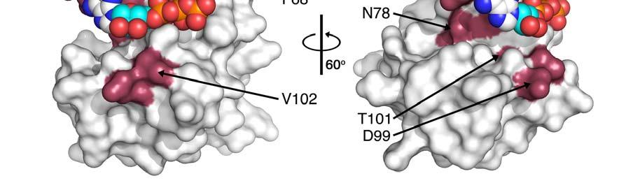
11 Supplementary Figure 7. Assignment of Doc and NMR chemical shift perturbations. (a) 1 H- 15 N HSQC spectrum of Doc and cross peak assignment (b) Chemical shift perturbations observed in the 1 H- 15 N HSQC spectrum of Doc upon addition of 0 μm, 34.0 μm, 58.0 μm μm, μm of EF-Tu. (c) Chemical shift perturbations observed in the 1 H- 15 N HSQC spectrum of Doc upon addition of 0 mm, 1.4 mm, 2.7 mm 9.0 mm, 15.0 mm, 25.8 mm and 40 mm of AMPPNP. (d) Mapping on the surface of Doc of the observed chemical shifts perturbations (in red) used for the docking of AMPPNP on Doc. Residues R19, Y20, G22, L23, G25, F68, R74, N78, D99, T101 and V102 are shown in red (see Figure 5 and Supplementary Table 3 for further details). 11
 and the theoretical curves (in black) derived from the models, for Doc (a), EF-Tu:GDP (b) and Doc:EF-Tu:GDP (c).")
 P(r) functions obtained from the scattering curves using GNOM 21 for Doc (in black), EF-Tu:GDP (in blue) and Doc:EF-Tu:GDP (in red).")
12 Supplementary Figure 8. Determination of experimental SAXS parameters. Guinier analysis of the experimental SAXS curves (in red) and the theoretical curves (in black) derived from the models, for Doc (a), EF-Tu:GDP (b) and Doc:EF-Tu:GDP (c). In every case the curves corresponding to the experimental data are displayed up by one logarithmic unit for clarity. (d) P(r) functions obtained from the scattering curves using GNOM 21 for Doc (in black), EF-Tu:GDP (in blue) and Doc:EF-Tu:GDP (in red). (e) Stereo view of Doc:EF- Tu:GDP representative solutions that fit to the experimental data with χ 2 between 0.9 and 1.1. In the Figure Doc is represented as ribbons and EF-Tu as a blue surface. The solutions superimpose with a core r.m.s.d below 1.5 Å over 510 Cα atoms. Plots of r.m.s.d. versus χ 2 12
13 (f) and χ 2 versus model number (g). Selected solutions were clustered into three groups (blue, green and orange circles). Blue lines demark the χ 2 range of the final solutions. 13


14 Supplementary Figure 9. Chemical shift based model of Doc bound to ATP. The ATP bound to Doc in the complex is shown as purple sticks. The orientation of the nucleotide in the active site is antiparallel to that observed in FIC-like proteins (shown in green, based on the structure of NmFic in complex with AMPPNP, pdbid 3S6A 1 ), presenting the γ-phosphate moiety toward H66 and the site where EF-Tu binds. Doc is colored in light grey and active site residues H66, K73 and R74 are shown as black lines. In typical Fic domains K73 is replaced by a glycine, which removes the steric hindrance and allows nucleotide binding, and constitutes a major difference in the active site motif between both subfamilies. 14

.")
15 Supplementary Figure 10. Phd binding site overlaps the NTP binding site on Doc. When bound to Doc, the C-terminal domain of Phd (in yellow, based on the coordinates of the Doc:Phd complex, pdbid 3K33 24 ) occupies the NTP site (represented by the bound ATP molecule in purple). Note that the site where the NTP binds in Fic-like domains (in green) remains free in the Doc-Phd complex. 15
16 Supplementary Table 1. Interplay between Doc, EF-Tu and nucleotides. The binding affinities were determined from fitting a single interaction model to the experimental data from ITC and NMR titrations. Data represent mean values ± s.d. See Supplementary Figure 3 for representative titrations. Experiment Technique Kd Number of experiments EF-Tu titrated into Doc ITC 8 ± 4 μm 3 EF-Tu titrated into Doc in phosphate ITC 6 ± 1 μm 3 EF-Tu titrated into Doc in phosphate NMR 16.3 μm 1 EF-Tu titrated into Doc in 1mM GDP ITC 1.7 ± 0.7 μm 3 EF-Tu titrated into Doc in 1mM GMPPNP ITC 50 ± 7 μm 3 EF-Tu titrated into Doc H66Y in 1mM GDP ITC 4 ± 2 μm 3 EF-Tu T382V titrated into Doc in 1mM GDP ITC 10 ± 7 μm 3 EF-Tu titrated into Doc R64G in 1mM GDP ITC no binding 2 EF-Tu titrated into Doc in 1mM GDP in Phd ITC no binding 2 EF-Tu titrated into Doc N78W in 1mM GDP ITC 3 ± 1 μm 3 AMPPNP titrated into Doc NMR 7.2 mm 1 AMPPNP titrated into (preformed Doc:EF-Tu:GDP) ITC 0.26 ± 0.05 μm 3 GMPPNP titrated into (preformed Doc:EF-Tu:GDP) ITC 4.4 ± 0.4 μm 3 UMPPNP titrated into (preformed Doc:EF-Tu:GDP) ITC no binding 2 AMPPNP titrated into (preformed Doc N78W :EF-Tu:GDP) ITC 45 ± 1 μm 3 AMPPNP titrated into Doc:EF-Tu:GDP and Phd ITC no binding 2 Supplementary Table 2. SAXS parameters. Theoretical and experimental molecular weights of Doc, EF-Tu, and the Doc:EF-Tu as obtained from the SAXS curves. Using an R SAS cutoff of and Chi-values of 1.5 or lower, model-data agreements can be reliably identified (Rambo & Tainer, Nature 2013) Specie Experimental Molecular Weight SAXS (kda) Experimental Molecular Weight MALS (kda) Theoretical Molecular Weight (kda) Rg (Å) (exps/model) Dmax(Å) χ 2 R SAS 16
17 Doc / EF-Tu / Doc:EF-Tu:GDP / Additional SAXS parameters: Specie Vc (model ) Vc (exp) V SAS Rg (model) Rg (exp) Io (model) Io (exp) Doc EF-Tu Doc:EF-Tu:GDP Supplementary Table 3. Chemical shift perturbations used for docking. Residues with chemical shift perturbations above 2σ selected for the docking experiments. Residue Experiment S27 R64 H66 R19 Y20 G22 L23 G25 F68 R74 N78 D99 Docking of EF-Tu to Doc Docking of EF-Tu to Doc Docking of EF-Tu to Doc 17
18 T101 V102 18
Chapter 6. The interaction of Src SH2 with the focal adhesion kinase catalytic domain studied by NMR
 The interaction of Src SH2 with the focal adhesion kinase catalytic domain studied by NMR 103 Abstract The interaction of the Src SH2 domain with the catalytic domain of FAK, including the Y397 SH2 domain
Nature Structural & Molecular Biology: doi: /nsmb Supplementary Figure 1
 Supplementary Figure 1 Identification of the ScDcp2 minimal region interacting with both ScDcp1 and the ScEdc3 LSm domain. Pull-down experiment of untagged ScEdc3 LSm with various ScDcp1-Dcp2-His 6 fragments.
Table S1. Overview of used PDZK1 constructs and their binding affinities to peptides. Related to figure 1.
 Table S1. Overview of used PDZK1 constructs and their binding affinities to peptides. Related to figure 1. PDZK1 constru cts Amino acids MW [kda] KD [μm] PEPT2-CT- FITC KD [μm] NHE3-CT- FITC KD [μm] PDZK1-CT-
Supplementary Information. Overlap between folding and functional energy landscapes for. adenylate kinase conformational change
 Supplementary Information Overlap between folding and functional energy landscapes for adenylate kinase conformational change by Ulrika Olsson & Magnus Wolf-Watz Contents: 1. Supplementary Note 2. Supplementary
Supplemental Information. Structural and Mechanistic Paradigm. of Leptin Receptor Activation Revealed
 Structure, Volume 22 Supplemental Information Structural and Mechanistic Paradigm of Leptin Receptor Activation Revealed by Complexes with Wild-Type and Antagonist Leptins Kedar Moharana, Lennart Zabeau,
Supplemental data for
 Supplemental data for A Real-Time Guanine Nucleotide Exchange Assay using NMR: Activation of RhoA by PDZ- RhoGEF. Geneviève M.C. Gasmi-Seabrook 1,3, Christopher B. Marshall 1,3, Melissa Cheung 1,3, Bryan
Supplementary figure 1 Application of tmfret in LeuT. (a) To assess the feasibility of using tmfret for distance-dependent measurements in LeuT, a
 To assess the feasibility of using tmfret for distance-dependent measurements in LeuT, a") Supplementary figure 1 Application of tmfret in LeuT. (a) To assess the feasibility of using tmfret for distance-dependent measurements in LeuT, a series of tmfret-pairs comprised of single cysteine mutants
Supplementary figure 1 Application of tmfret in LeuT. (a) To assess the feasibility of using tmfret for distance-dependent measurements in LeuT, a series of tmfret-pairs comprised of single cysteine mutants
Structural characterization of NiV N 0 P in solution and in crystal.
 Supplementary Figure 1 Structural characterization of NiV N 0 P in solution and in crystal. (a) SAXS analysis of the N 32-383 0 -P 50 complex. The Guinier plot for complex concentrations of 0.55, 1.1,
Supplementary Information. The protease GtgE from Salmonella exclusively targets. inactive Rab GTPases
 Supplementary Information The protease GtgE from Salmonella exclusively targets inactive Rab GTPases Table of Contents Supplementary Figures... 2 Supplementary Figure 1... 2 Supplementary Figure 2... 3
Nature Structural and Molecular Biology: doi: /nsmb Supplementary Figure 1. Definition and assessment of ciap1 constructs.
 Supplementary Figure 1 Definition and assessment of ciap1 constructs. (a) ciap1 constructs used in this study are shown as primary structure schematics with domains colored as in the main text. Mutations
Supplementary Figure 1. Biochemical and sequence alignment analyses the
 Supplementary Figure 1. Biochemical and sequence alignment analyses the interaction of OPTN and TBK1. (a) Analytical gel filtration chromatography analysis of the interaction between TBK1 CTD and OPTN(1-119).
Supplementary Information
 Supplementary Information Structural analysis of leader peptide binding enables leaderfree cyanobactin processing Jesko Koehnke 1,2, Greg Mann 1,2, Andrew F Bent 1,2, Hannes Ludewig 1, Sally Shirran 1,
Nature Structural & Molecular Biology: doi: /nsmb Supplementary Figure 1
 Supplementary Figure 1 Crystallization. a, Crystallization constructs of the ET B receptor are shown, with all of the modifications to the human wild-type the ET B receptor indicated. Residues interacting
Supplementary Materials for
 advances.sciencemag.org/cgi/content/full/1/9/e1500511/dc1 Supplementary Materials for Contractility parameters of human -cardiac myosin with the hypertrophic cardiomyopathy mutation R403Q show loss of
SOCS3 binds specific receptor JAK complexes to control cytokine signaling by direct kinase inhibition SUPPLEMENTARY INFORMATION
 SOCS3 binds specific receptor JAK complexes to control cytokine signaling by direct kinase inhibition Nadia J. Kershaw 1,2, James M. Murphy 1,2, Nicholas P.D. Liau 1,2, Leila N. Varghese 1,2, Artem Laktyushin
SUPPLEMENTARY INFORMATION
 SUPPLEMENTARY INFORMATION doi:10.1038/nature11539 Supplementary Figure 1 Schematic representation of plant (A) and mammalian (B) P 2B -ATPase domain organization. Actuator (A-), nucleotide binding (N-),
Supplementary Figure 1 Crystal contacts in COP apo structure (PDB code 3S0R)
") Supplementary Figure 1 Crystal contacts in COP apo structure (PDB code 3S0R) Shown in cyan and green are two adjacent tetramers from the crystallographic lattice of COP, forming the only unique inter-tetramer
Supplementary Figure 1 Crystal contacts in COP apo structure (PDB code 3S0R) Shown in cyan and green are two adjacent tetramers from the crystallographic lattice of COP, forming the only unique inter-tetramer
The Aβ40 and Aβ42 peptides self-assemble into separate homomolecular fibrils in binary mixtures but cross-react during primary nucleation
 Electronic Supplementary Material (ESI) for Chemical Science. This journal is The Royal Society of Chemistry 2015 The Aβ40 and Aβ42 peptides self-assemble into separate homomolecular fibrils in binary
Sensitive NMR Approach for Determining the Binding Mode of Tightly Binding Ligand Molecules to Protein Targets
 Supporting information Sensitive NMR Approach for Determining the Binding Mode of Tightly Binding Ligand Molecules to Protein Targets Wan-Na Chen, Christoph Nitsche, Kala Bharath Pilla, Bim Graham, Thomas
Supporting Information
 Supporting Information Micelle-Triggered b-hairpin to a-helix Transition in a 14-Residue Peptide from a Choline-Binding Repeat of the Pneumococcal Autolysin LytA HØctor Zamora-Carreras, [a] Beatriz Maestro,
Supplementary figure 1. Comparison of unbound ogm-csf and ogm-csf as captured in the GIF:GM-CSF complex. Alignment of two copies of unbound ovine
 Supplementary figure 1. Comparison of unbound and as captured in the GIF:GM-CSF complex. Alignment of two copies of unbound ovine GM-CSF (slate) with bound GM-CSF in the GIF:GM-CSF complex (GIF: green,
Supplementary Figures
 1 Supplementary Figures Supplementary Figure 1 Type I FGFR1 inhibitors (a) Chemical structures of a pyrazolylaminopyrimidine inhibitor (henceforth referred to as PAPI; PDB-code of the FGFR1-PAPI complex:
SUPPLEMENTARY INFORMATION
 doi:10.1038/nature10955 Supplementary Figures Supplementary Figure 1. Electron-density maps and crystallographic dimer structures of the motor domain. (a f) Stereo views of the final electron-density maps
Serine-7 but not serine-5 phosphorylation primes RNA polymerase II CTD for P-TEFb recognition
 Supplementary Information to Serine-7 but not serine-5 phosphorylation primes RNA polymerase II CTD for P-TEFb recognition Nadine Czudnochowski 1,2, *, Christian A. Bösken 1, * & Matthias Geyer 1 1 Max-Planck-Institut
Supplementary Information. Structural basis for precursor protein-directed ribosomal peptide macrocyclization
 Supplementary Information Structural basis for precursor protein-directed ribosomal peptide macrocyclization Kunhua Li 1,3, Heather L. Condurso 1,3, Gengnan Li 1, Yousong Ding 2 and Steven D. Bruner 1*
Supplementary Information for
 Electronic Supplementary Material (ESI) for Analyst. This journal is The Royal Society of Chemistry 2015 Supplementary Information for The use of Ion Mobility Mass Spectrometry to assist Protein Design:
Supplementary Information for. Direct nitration and azidation of aliphatic carbons by an iron-dependent halogenase
 Supplementary Information for Direct nitration and azidation of aliphatic carbons by an iron-dependent halogenase Megan L Matthews, Wei-chen Chang, Andrew P Layne, Linde A Miles, Carsten Krebs, J Martin
THE CRYSTAL STRUCTURE OF THE SGT1-SKP1 COMPLEX: THE LINK BETWEEN
 THE CRYSTAL STRUCTURE OF THE SGT1-SKP1 COMPLEX: THE LINK BETWEEN HSP90 AND BOTH SCF E3 UBIQUITIN LIGASES AND KINETOCHORES Oliver Willhoft, Richard Kerr, Dipali Patel, Wenjuan Zhang, Caezar Al-Jassar, Tina
Supplemental Information
 Supplemental Information Combinatorial Readout of Unmodified H3R2 and Acetylated H3K14 by the Tandem PHD Finger of MOZ Reveals a Regulatory Mechanism for HOXA9 Transcription Yu Qiu 1, Lei Liu 1, Chen Zhao
SUPPLEMENTARY INFORMATION
 Supplementary materials Figure S1 Fusion protein of Sulfolobus solfataricus SRP54 and a signal peptide. a, Expression vector for the fusion protein. The signal peptide of yeast dipeptidyl aminopeptidase
Analysis of nucleotide binding to p97 reveals the properties of a tandem AAA hexameric ATPase
 SUPPLEMENTARY INFORMATION Analysis of nucleotide binding to p97 reveals the properties of a tandem AAA hexameric ATPase Louise C Briggs, Geoff S Baldwin, Non Miyata, Hisao Kondo, Xiaodong Zhang, Paul S
Purification, SDS-PAGE and cryo-em characterization of the MCM hexamer and Cdt1 MCM heptamer samples.
 Supplementary Figure 1 Purification, SDS-PAGE and cryo-em characterization of the MCM hexamer and Cdt1 MCM heptamer samples. (a-b) SDS-PAGE analysis of the hexamer and heptamer samples. The eluted hexamer
SUPPLEMENTARY INFORMATION
 SUPPLEMENTARY INFORMATION doi:10.1038/nature11524 Supplementary discussion Functional analysis of the sugar porter family (SP) signature motifs. As seen in Fig. 5c, single point mutation of the conserved
Impact of the crystallization condition on importin-β conformation
 Supporting information Volume 72 (2016) Supporting information for article: Impact of the crystallization condition on importin-β conformation Marcel J. Tauchert, Clément Hémonnot, Piotr Neumann, Sarah
Supplemental Information. Molecular Basis of Spectral Diversity. in Near-Infrared Phytochrome-Based. Fluorescent Proteins
 Chemistry & Biology, Volume 22 Supplemental Information Molecular Basis of Spectral Diversity in Near-Infrared Phytochrome-Based Fluorescent Proteins Daria M. Shcherbakova, Mikhail Baloban, Sergei Pletnev,
Supplemental Information. The Mitochondrial Fission Receptor MiD51. Requires ADP as a Cofactor
 Structure, Volume 22 Supplemental Information The Mitochondrial Fission Receptor MiD51 Requires ADP as a Cofactor Oliver C. Losón, Raymond Liu, Michael E. Rome, Shuxia Meng, Jens T. Kaiser, Shu-ou Shan,
National de la Recherche Scientifique and Université Paris Descartes, Paris, France.
 FAST-RESPONSE CALMODULIN-BASED FLUORESCENT INDICATORS REVEAL RAPID INTRACELLULAR CALCIUM DYNAMICS Nordine Helassa a, Xiao-hua Zhang b, Ianina Conte a,c, John Scaringi b, Elric Esposito d, Jonathan Bradley
Supporting Information
 Supporting Information Arai et al. 10.1073/pnas.15179911 SI Text Protein Expression and Purification. Myb3 (mouse, residues 84 315) was expressed in Escherichia coli as a fusion with the B1 domain of protein
Supplementary Materials: Localization and Spectroscopic Analysis of the Cu(I) Binding Site in Wheat Metallothionein Ec-1
 Binding Site in Wheat Metallothionein Ec-1") S1 of S8 Supplementary Materials: Localization and Spectroscopic Analysis of the Cu(I) Binding Site in Wheat Metallothionein Ec-1 Katsiaryna Tarasava, Jens Loebus and Eva Freisinger Figure S1. Deconvoluted
S1 of S8 Supplementary Materials: Localization and Spectroscopic Analysis of the Cu(I) Binding Site in Wheat Metallothionein Ec-1 Katsiaryna Tarasava, Jens Loebus and Eva Freisinger Figure S1. Deconvoluted
SUPPLEMENTARY INFORMATION
 Supplementary Table 1: Amplitudes of three current levels. Level 0 (pa) Level 1 (pa) Level 2 (pa) TrkA- TrkH WT 200 K 0.01 ± 0.01 9.5 ± 0.01 18.7 ± 0.03 200 Na * 0.001 ± 0.01 3.9 ± 0.01 12.5 ± 0.03 200
Supplementary Information. The Solution Structural Ensembles of RNA Kink-turn Motifs and Their Protein Complexes
 Supplementary Information The Solution Structural Ensembles of RNA Kink-turn Motifs and Their Protein Complexes Xuesong Shi, a Lin Huang, b David M. J. Lilley, b Pehr B. Harbury a,c and Daniel Herschlag
Interpreting and evaluating biological NMR in the literature. Worksheet 1
 Interpreting and evaluating biological NMR in the literature Worksheet 1 1D NMR spectra Application of RF pulses of specified lengths and frequencies can make certain nuclei detectable We can selectively
Structural basis for catalytically restrictive dynamics of a high-energy enzyme state
 Supplementary Material Structural basis for catalytically restrictive dynamics of a high-energy enzyme state Michael Kovermann, Jörgen Ådén, Christin Grundström, A. Elisabeth Sauer-Eriksson, Uwe H. Sauer
Supporting information for
 Supporting information for Rewiring multi-domain protein switches: transforming a fluorescent Zn 2+ -sensor into a light-responsive Zn 2+ binding protein Stijn J.A. Aper and Maarten Merkx Laboratory of
SUPPLEMENTARY INFORMATION
 Supplementary Table S1 Kinetic Analyses of the AMSH-LP mutants AMSH-LP K M (μm) k cat x 10-3 (s -1 ) WT 71.8 ± 6.3 860 ± 65.4 T353A 76.8 ± 11.7 46.3 ± 3.7 F355A 58.9 ± 10.4 5.33 ± 0.30 proximal S358A 75.1
Supplementary Information to
 Supplementary Information to Wiesner et al.: A change in conformational dynamics underlies the activation of Eph receptor tyrosine kinases Supplementary Material and Methods Cloning and Mutagenesis Site-directed
Bacterial protease uses distinct thermodynamic signatures for substrate recognition
 Bacterial protease uses distinct thermodynamic signatures for substrate recognition Gustavo Arruda Bezerra, Yuko Ohara-Nemoto, Irina Cornaciu, Sofiya Fedosyuk, Guillaume Hoffmann, Adam Round, José A. Márquez,
SUPPLEMENTARY INFORMATION
 Table of Contents Page Supplementary Table 1. Diffraction data collection statistics 2 Supplementary Table 2. Crystallographic refinement statistics 3 Supplementary Fig. 1. casic1mfc packing in the R3
for Molecular Biology and Neuroscience and Institute of Medical Microbiology, Rikshospitalet-Radiumhospitalet
 SUPPLEMENTARY INFORMATION TO Structural basis for enzymatic excision of N -methyladenine and N 3 -methylcytosine from DNA Ingar Leiros,5, Marivi P. Nabong 2,3,5, Kristin Grøsvik 3, Jeanette Ringvoll 2,
Table S1. Primers used for the constructions of recombinant GAL1 and λ5 mutants. GAL1-E74A ccgagcagcgggcggctgtctttcc ggaaagacagccgcccgctgctcgg
 SUPPLEMENTAL DATA Table S1. Primers used for the constructions of recombinant GAL1 and λ5 mutants Sense primer (5 to 3 ) Anti-sense primer (5 to 3 ) GAL1 mutants GAL1-E74A ccgagcagcgggcggctgtctttcc ggaaagacagccgcccgctgctcgg
Supplementary material
 Supplementary material Phosphorylation of the mitochondrial autophagy receptor Nix enhances its interaction with LC3 proteins Vladimir V. Rogov 1,*, Hironori Suzuki 2,3,*, Mija Marinković 4, Verena Lang
pyridoxal phosphate synthase
 Supplementary Information 13 C-NMR snapshots of the complex reaction coordinate of pyridoxal phosphate synthase Jeremiah W. Hanes, Ivan Keresztes, and Tadhg P. Begley * Department of Chemistry and Chemical
ml. ph 7.5 ph 6.5 ph 5.5 ph 4.5. β 2 AR-Gs complex + GDP β 2 AR-Gs complex + GTPγS
 a UV28 absorption (mau) 9 8 7 5 3 β 2 AR-Gs complex β 2 AR-Gs complex + GDP β 2 AR-Gs complex + GTPγS β 2 AR-Gs complex dissociated complex excess nucleotides b 9 8 7 5 3 β 2 AR-Gs complex β 2 AR-Gs complex
Nature Structural & Molecular Biology: doi: /nsmb.3194
 Supplementary Figure 1 Mass spectrometry and solution NMR data for -syn samples used in this study. (a) Matrix-assisted laser-desorption and ionization time-of-flight (MALDI-TOF) mass spectrum of uniformly-
SUPPLEMENTARY INFORMATION
 Figure S1. Secondary structure of CAP (in the camp 2 -bound state) 10. α-helices are shown as cylinders and β- strands as arrows. Labeling of secondary structure is indicated. CDB, DBD and the hinge are
SUPPLEMENTARY ONLINE DATA
 SUPPLEMENTARY ONLINE DATA Secreted Isoform of Human Lynx1 (SLURP-2): Spatial Structure and Pharmacology of Interaction with Different Types of Acetylcholine Receptors E.N. Lyukmanova 1,2,*, M.A. Shulepko
Structural basis of PROTAC cooperative recognition for selective protein degradation
 SUPPLEMENTARY INFORMATION Structural basis of PROTAC cooperative recognition for selective protein degradation Morgan S. Gadd 1, Andrea Testa 1, Xavier Lucas 1, Kwok-Ho Chan, Wenzhang Chen, Douglas J.
Supplementary Information
 Supplementary Information An engineered protein antagonist of K-Ras/B-Raf interaction Monique J. Kauke, 1,2 Michael W. Traxlmayr 1,2, Jillian A. Parker 3, Jonathan D. Kiefer 4, Ryan Knihtila 3, John McGee
Protein Dynamics. The space-filling structures of myoglobin and hemoglobin show that there are no pathways for O 2 to reach the heme iron.
 Protein Dynamics The space-filling structures of myoglobin and hemoglobin show that there are no pathways for O 2 to reach the heme iron. Below is myoglobin hydrated with 350 water molecules. Only a small
NMR in Medicine and Biology
 NMR in Medicine and Biology http://en.wikipedia.org/wiki/nmr_spectroscopy MRI- Magnetic Resonance Imaging (water) In-vivo spectroscopy (metabolites) Solid-state t NMR (large structures) t Solution NMR
In Situ Gelation-Induced Death of Cancer Cells Based on Proteinosomes
 Supporting information for In Situ Gelation-Induced Death of Cancer Cells Based on Proteinosomes Yuting Zhou, Jianmin Song, Lei Wang*, Xuting Xue, Xiaoman Liu, Hui Xie*, and Xin Huang* MIIT Key Laboratory
SUPPLEMENTARY INFORMATION
 SUPPLEMENTARY INFORMATION Structure of human carbamoyl phosphate synthetase: deciphering the on/off switch of human ureagenesis Sergio de Cima, Luis M. Polo, Carmen Díez-Fernández, Ana I. Martínez, Javier
SUPPLEMENTARY FIGURES
 SUPPLEMENTARY FIGURES Supplementary Figure 1 Protein sequence alignment of Vibrionaceae with either a 40-residue insertion or a 44-residue insertion. Identical residues are indicated by red background.
Dr. Yonca Yuzugullu PERG (Protein Engineering Research Group)
") Dr. Yonca Yuzugullu PERG (Protein Engineering Research Group) BSc, 1997 Ankara University, Turkey PhD, 2010 Middle East Technical University, Turkey Lecturer in Department of Biology Kocaeli University,
Dr. Yonca Yuzugullu PERG (Protein Engineering Research Group) BSc, 1997 Ankara University, Turkey PhD, 2010 Middle East Technical University, Turkey Lecturer in Department of Biology Kocaeli University,
Tridip Sheet, Raja Banerjee*
 Electronic Supplementary Material (ESI) for RSC Advances. This journal is The Royal Society of Chemistry 2016 Supplementary information The C NN motif: an intrinsic lover of sulfate and phosphate ions
Protein Structure Determination using NMR Spectroscopy. Cesar Trinidad
 Protein Structure Determination using NMR Spectroscopy Cesar Trinidad Introduction Protein NMR Involves the analysis and calculation of data collected from multiple NMR techniques Utilizes Nuclear Magnetic
Supporting Information. Labeled Ligand Displacement: Extending NMR-based Screening of Protein Targets
 Supporting Information Labeled Ligand Displacement: Extending NMR-based Screening of Protein Targets Steven L. Swann, Danying Song, Chaohong Sun, Philip J. Hajduk, and Andrew M. Petros Global Pharmaceutical
17. Biomolecular Interaction
 17. Biomolecular Interaction Methods for characterizing biomolecular interactions Sequence-specific DNA binding ligands Molecular mechanisms of drug action and drug resistance In silico compound design
Supplementary Information. Synthesis and biological activity of a CXCR4-targeting bis(cyclam) lipid
 lipid") Electronic Supplementary Material (ESI) for Organic & Biomolecular Chemistry. This journal is The Royal Society of Chemistry 2018 Supplementary Information Synthesis and biological activity of a CXCR4-targeting
Electronic Supplementary Material (ESI) for Organic & Biomolecular Chemistry. This journal is The Royal Society of Chemistry 2018 Supplementary Information Synthesis and biological activity of a CXCR4-targeting
SUPPLEMENTARY INFORMATION
 Data collection Supplementary Table 1 Statistics of data collection, phasing and refinement Native Se-MAD Space group P2 1 2 1 2 1 P2 1 2 1 2 1 Cell dimensions a, b, c (Å) 50.4, 94.2, 115.4 49.8, 94.2,
Figure S1. Interaction of PcTS with αsyn. (a) 1 H- 15 N HSQC NMR spectra of 100 µm αsyn in the absence (0:1, black) and increasing equivalent
 1 H- 15 N HSQC NMR spectra of 100 µm αsyn in the absence (0:1, black) and increasing equivalent") Figure S1. Interaction of PcTS with αsyn. (a) 1 H- 15 N HSQC NMR spectra of 100 µm αsyn in the absence (0:1, black) and increasing equivalent concentrations of PcTS (100 µm, blue; 500 µm, green; 1.5 mm,
Figure S1. Interaction of PcTS with αsyn. (a) 1 H- 15 N HSQC NMR spectra of 100 µm αsyn in the absence (0:1, black) and increasing equivalent concentrations of PcTS (100 µm, blue; 500 µm, green; 1.5 mm,
Introduction to" Protein Structure
 Introduction to" Protein Structure Function, evolution & experimental methods Thomas Blicher, Center for Biological Sequence Analysis Learning Objectives Outline the basic levels of protein structure.
Supporting Information
 Supporting Information Copyright Wiley-VCH Verlag GmbH & Co. KGaA, 69451 Weinheim, 2014 An Ensemble of Rapidly Interconverting Orientations in Electrostatic Protein Peptide Complexes Characterized by NMR
Cryo-EM data collection, refinement and validation statistics
 1 Table S1 Cryo-EM data collection, refinement and validation statistics Data collection and processing CPSF-160 WDR33 (EMDB-7114) (PDB 6BM0) CPSF-160 WDR33 (EMDB-7113) (PDB 6BLY) CPSF-160 WDR33 CPSF-30
Supporting Information for. Jesinghaus, Rachael Barry, Zemer Gitai, Justin Kollman and Enoch P. Baldwin
 Supporting Information for Inhibition of E. coli CTP synthetase by NADH and other nicotinamides, and their mutual interactions with CTP and GTP Chris Habrian, Adithi Chandrasekhara, Bita Shahrvini, Brian
Supplementary Materials for
 www.sciencesignaling.org/cgi/content/full/5/243/ra68/dc1 Supplementary Materials for Superbinder SH2 Domains Act as Antagonists of Cell Signaling Tomonori Kaneko, Haiming Huang, Xuan Cao, Xing Li, Chengjun
Supplemental Data SUPPLEMENTAL FIGURES
 Supplemental Data CRYSTAL STRUCTURE OF THE MG.ADP-INHIBITED STATE OF THE YEAST F 1 C 10 ATP SYNTHASE Alain Dautant*, Jean Velours and Marie-France Giraud* From Université Bordeaux 2, CNRS; Institut de
Nature Structural and Molecular Biology: doi: /nsmb.2938
 Supplementary Figure 1 Characterization of designed leucine-rich-repeat proteins. (a) Water-mediate hydrogen-bond network is frequently visible in the convex region of LRR crystal structures. Examples
SUPPLEMENTARY INFORMATION
 SUPPLEMENTARY INFORMATION doi:10.1038/nature11744 Supplementary Table 1. Crystallographic data collection and refinement statistics. Wild-type Se-Met-BcsA-B SmCl 3 -soaked EMTS-soaked Data collection Space
Nature Structural & Molecular Biology: doi: /nsmb Supplementary Figure 1
 Supplementary Figure 1 Resonance assignment and NMR spectra for hairpin and duplex A 6 constructs. (a) 2D HSQC spectra of hairpin construct (hp-a 6 -RNA) with labeled assignments. (b) 2D HSQC or SOFAST-HMQC
Supplementary Materials for
 advances.sciencemag.org/cgi/content/full/3/4/e1600663/dc1 Supplementary Materials for A dynamic hydrophobic core orchestrates allostery in protein kinases Jonggul Kim, Lalima G. Ahuja, Fa-An Chao, Youlin
SUPPLEMENTARY FIGURES. Figure S1
 SUPPLEMENTARY FIGURES Figure S1 The substrate for DH domain (2R,3R,4R,6R,7S,8S,9R)-3,7,9-trihydroxy-5-oxo-2,4,6,8 tetramethylundecanoate) was docked as two separate fragments shown in magenta and blue
Supplementary materials. Crystal structure of the carboxyltransferase domain. of acetyl coenzyme A carboxylase. Department of Biological Sciences
 Supplementary materials Crystal structure of the carboxyltransferase domain of acetyl coenzyme A carboxylase Hailong Zhang, Zhiru Yang, 1 Yang Shen, 1 Liang Tong Department of Biological Sciences Columbia
Supporting Information
 Supporting Information Allosteric-activation of GDP-bound Ras isoforms by bisphenol derivative plasticisers Miriam Schöpel 1, Oleksandr Shkura 1, Jana Seidel 1, Klaus Kock 1, Xueyin Zhong 1, Stefanie Löffek
Simulative and experimental characterization of a ph-dependent
 Simulative and experimental characterization of a ph-dependent clamp-like DNA triple-helix nanoswitch Federico Iacovelli, # Andrea Idili, # Alessandro Benincasa, Davide Mariottini, Alessio Ottaviani, Mattia
SUPPLEMENTARY INFORMATION
 Parallel Allostery by camp and PDE Coordinates Activation and Termination Phases in camp Signaling Srinath Krishnamurthy, 1 Nikhil Kumar Tulsian, 1 Arun Chandramohan, 1 and Ganesh S. Anand 1, * 1 Department
Sample preparation and characterization around SAXS
 Sample preparation and characterization around SAXS Experimental verification and validation? Rob Meijers EMBL Hamburg Garbage in? The right stuff Molecular weight Oligomerization state Monodispersity
Acta Crystallographica Section D
 Supporting information Acta Crystallographica Section D Volume 70 (2014) Supporting information for article: Structural characterization of the virulence factor Nuclease A from Streptococcus agalactiae
Structure of the α-helix
 Structure of the α-helix Structure of the β Sheet Protein Dynamics Basics of Quenching HDX Hydrogen exchange of amide protons is catalyzed by H 2 O, OH -, and H 3 O +, but it s most dominated by base
According to the manufacture s direction (Pierce), RNA and DNA
, RNA and DNA") Supplementary method Electrophoretic Mobility-shift assay (EMSA) According to the manufacture s direction (Pierce), RNA and DNA oligonuleotides were firstly labeled by biotin. TAVb (1pM) was incubated
Supplementary method Electrophoretic Mobility-shift assay (EMSA) According to the manufacture s direction (Pierce), RNA and DNA oligonuleotides were firstly labeled by biotin. TAVb (1pM) was incubated
Enhancing hydrogen production of microalgae by redirecting electrons from photosystem I to hydrogenase
 Electronic Supplementary Material (ESI) for Energy & Environmental Science. This journal is The Royal Society of Chemistry 2014 Supplementary information for Enhancing hydrogen production of microalgae
Deconvoluting the responses of polymer-scaffolded dynamic combinatorial libraries to biomacromolecular templates
 Supporting Information Deconvoluting the responses of polymer-scaffolded dynamic combinatorial libraries to biomacromolecular templates Antonio J. Ruiz-Sanchez, a Patrick L. Higgs, a Daniel T. Peters,
Supplementary Figure 1: Power dependence of hot-electrons reduction of 4-NTP to 4-ATP. a) SERS spectra of the hot-electron reduction reaction using
 SERS spectra of the hot-electron reduction reaction using") Supplementary Figure 1: Power dependence of hot-electrons reduction of 4-NTP to 4-ATP. a) SERS spectra of the hot-electron reduction reaction using 633 nm laser excitation at different powers and b) the
Supplementary Figure 1: Power dependence of hot-electrons reduction of 4-NTP to 4-ATP. a) SERS spectra of the hot-electron reduction reaction using 633 nm laser excitation at different powers and b) the
Supporting Information
 Supporting Information Figure S1. 2D ( 1 H- 1 H) COSY90 NMR (300 MHz, 3:1 TFA:TFA-d) spectrum of oligo(l-glu-co- 25%L-Cys) synthesized using from 7:3 L-Glu-(Et) 2 :L-Cys-Et, 0.5 M total substrate concentration,
Supporting Protocol This protocol describes the construction and the force-field parameters of the non-standard residue for the Ag + -site using CNS
 Supporting Protocol This protocol describes the construction and the force-field parameters of the non-standard residue for the Ag + -site using CNS CNS input file generatemetal.inp: remarks file generate/generatemetal.inp
SUPPLEMENTARY INFORMATION
 SUPPLMTARY IFORMATIO a doi:10.108/nature10402 b 100 nm 100 nm c SAXS Model d ulers assigned to reference- Back-projected free class averages class averages Refinement against single particles Reconstructed
Targeting protein-protein interactions: A hot topic in drug discovery
 Michal Kamenicky; Maria Bräuer; Katrin Volk; Kamil Ödner; Christian Klein; Norbert Müller Targeting protein-protein interactions: A hot topic in drug discovery 104 Biomedizin Innovativ patientinnenfokussierte,
SUPPLEMENTARY INFORMATION
 doi:10.1038/nature12045 Supplementary Table 1 Data collection and refinement statistics. Native Pt-SAD X-ray source SSRF BL17U SPring-8 BL41XU Wavelength (Å) 0.97947 1.07171 Space group P2 1 2 1 2 1 P2
James B. Munro, Roger B. Altman, Nathan O Connor, and Scott C. Blanchard
 Molecular Cell, Volume 25 Supplemental Data Identification of Two Distinct Hybrid State Intermediates on the Ribosome James B. Munro, Roger B. Altman, Nathan O Connor, and Scott C. Blanchard Wild-type
type GroEL-GroES complex. Crystals were grown in buffer D (100 mm HEPES, ph 7.5,
 Supplementary Material Supplementary Materials and Methods Structure Determination of SR1-GroES-ADP AlF x SR1-GroES-ADP AlF x was purified as described in Materials and Methods for the wild type GroEL-GroES