Structural basis of PROTAC cooperative recognition for selective protein degradation
|
|
|
- Allan Davidson
- 6 years ago
- Views:
Transcription
1 SUPPLEMENTARY INFORMATION Structural basis of PROTAC cooperative recognition for selective protein degradation Morgan S. Gadd 1, Andrea Testa 1, Xavier Lucas 1, Kwok-Ho Chan, Wenzhang Chen, Douglas J. Lamont, Michael Zengerle, Alessio Ciulli* Division of Biological Chemistry and Drug Discovery, School of Life Sciences, University of Dundee, Dow Street, Dundee, DD1 5EH, Scotland, UK. *To whom correspondence should be addressed: 1 These authors contributed equally to this work Supplementary Figures 1-12 Supplementary Tables 1-3 1

2 Supplementary Results Supplementary Figure 1. Chemical structures of the PROTAC compounds used in this work. 2
 are shown as ribbons coloured according to average B factor per residue. Complex 1 is comprised of chains A D and Complex 2 is comprised of chains E H.")
3 Supplementary Figure 2. Analysis of the asymmetric unit of the Brd4 BD2 :MZ1:VCB crystal structure. a, Two complexes are present in the asymmetric unit. The eight protein chains (A H) are shown as ribbons coloured according to average B factor per residue. Complex 1 is comprised of chains A D and Complex 2 is comprised of chains E H. b, Superposition of the two Brd4 BD2 :MZ1:VCB ternary complexes in the asymmetric unit. The complexes were superposed via the backbone atoms of the VHL substrate-binding domain (residues ). c, Analysis of crystal contact imposing on the induced Brd4 BD2 :VHL interface in Complex 2. Arg177 from VHL in Complex 1 (chain D) interacts with Glu383 from Brd4 BD2 in Complex 2 (chain E), resulting in two observable conformations of the side chain and displacing VHL residue Arg107 in Complex 2 (chain H) from the interface. Due to this and lower average B factors we used the Complex 1 (chains A, B, C and D) for all subsequent analyses. d, e, F o F c omit maps (green meshes) of MZ1 prior to ligand modelling in Complexes 1 (d) and 2 (e) contoured at 3.0σ with a carve radius of 2.2 Å and 2F o F c maps (blue meshes) covering residues of Brd4 BD2 and VHL within 4 Å of MZ1 contoured at 1.5σ with a carve radius of 1.8 Å. 3

4 Supplementary Figure 3. Analysis and comparison of ligand binding at the Brd4 BD2 :MZ1:VHL interface. a, The Brd4 BD2 :MZ1:VHL interface forms a bowl shape. Overall view of MZ1 and the induced interacting proteins. Brd4 BD2 and VHL are shown as surfaces coloured according to electrostatic potential and the underlying ribbon illustrating secondary structure. MZ1 sits on the 4
5 hydrophobic base (white surface) of the bowl and is surrounded by the binding sites of Brd4 BD2 and VHL and two complementary electrostatic arms forming the induced protein-protein interactions (Fig. 1c,d). b, Structure of HIF-1α (dark purple) with VHL (PDB entry 1LM8; teal) reveals contacts made by HIF-1α residues Asp569 and Asp571 with VHL residues Arg108 and Arg107, respectively. c, Ternary structure of Brd4 BD2 (green), MZ1 (yellow carbons) and VHL reveals similar contacts made by Brd4 BD2 residues Asp381 and Glu383 with the same VHL residues. d, Structure of diacetylated histone 4 tail (H4K5acK8ac; orange sticks) bound to Brd4 BD1 (PDB entry 3UVW; not shown) superposed with Brd4 BD2 from the ternary structure (green surface). e. Ternary structure of Brd4 BD2, MZ1 and VHL reveals that VHL does not make contacts with Brd4 BD2 that are similar to those observed made by the bound histone tail. The PEG linker of MZ1 traces a similar line to the peptide backbone of histone 4 between residues Gly4 and Gly7. f, Superposition of JQ1 (purple carbons) bound in Brd2 BD2 (PDB entry 3ONI; not shown) with MZ1 (yellow carbons) bound in Brd4 BD2 (green surface). g, Superposition of VH032 (orange carbons) bound in VHL (PDB entry 4W9H; teal surface) with MZ1 (yellow carbons). 5
 bound to Brd4 BD2 (green) from Complex 1 (a, chain A) and Complex 2 (b, chain E) and VHL (teal).")
6 Supplementary Figure 4. Comparison of protein-ligand interactions involving PEG linkers of MZ1 and MT1. a,b, Structure of MZ1 (yellow sticks) bound to Brd4 BD2 (green) from Complex 1 (a, chain A) and Complex 2 (b, chain E) and VHL (teal). A hydrogen bond (black dashes) between His437 of Brd4 BD2 from Complex 1 and the proximal ether oxygen to JQ1 in MZ1 is shown. c,d, Structure of MT1 (PDB entry 5JWM; yellow sticks) bound to Brd4 BD2 (green) from chain A (c) and chain B (d). Hydrogen bonds (black dashes) between His437 and the ether oxygens of MT1 are shown. The complementary Brd4 BD2 molecule in the MT1-induced dimer is shown in grey in each case. 6
, buried surface area (BSA) of the PPI and root-mean-square deviation (RMSD) for the 100 ns MD simulation of the complex starting from the crystal structure.")
7 Supplementary Figure 5. Computational study of the intermolecular interactions in the Brd4 BD2 :MZ1:VHL complex. a, Radius of gyration (R g ), buried surface area (BSA) of the PPI and root-mean-square deviation (RMSD) for the 100 ns MD simulation of the complex starting from the crystal structure. b, Brd4 BD2 and VHL maintain an extensive interface throughout the MD simulation. Residues of Brd4 BD2 are plotted according to the proportion of the time during the 100 ns of the simulation they spend making an intermolecular contact with VHL. Residues selected for sidedirected mutagenesis are colored distinctly. c, Persistence of individual protein protein interaction contacts in the Brd4 BD2 :MZ1:VHL complex along the MD simulation. For the sake of clarity, value shown is the mean averaged over 5 ns. d, Superposition of MD simulations (light green and blue) at ns with Brd4 BD2 :MZ1:VHL crystal structure (dark green and teal) through Cα atoms of VHL. e, f, Per-residue intermolecular interaction energies derived from MD simulations for Brd4 BD2 (e) or VHL (f) residues contacting MZ1 (yellow) and VHL (cyan) or Brd4 BD2 (green), respectively. Energies are the mean (± 1 s.d.) of energy values calculated as described in Online Methods from one MD run of 100 ns and collected every 10 ps. 7
8 ITC titrations for binary complex MZ1:protein Data: A1intoMZP2_NDH Chi^2/DoF = 8.544E4 N 1.28 ±0540 Sites K 1.47E7 ±2.09E6 M -1 H E4 ±102.0 cal/mol S cal/mol/deg Data: A1intoMZP2_NDH Chi^2/DoF = 4.015E4 N 1.00 ±0235 Sites K 3.81E7 ±5.06E6 M -1 H E4 ±75.96 cal/mol 0.5 S cal/mol/deg Data: A1intoMZP2_NDH Chi^2/DoF = 2.812E4 N ±0221 Sites K 3.28E7 ±3.65E6 M -1 H E4 ±66.26 cal/mol S cal/mol/deg Brd2 BD1 Brd3 BD1 Brd4 BD Data: omzp22ver2_ndh Chi^2/DoF = 6.864E4 N 1.09 ±0594 Sites K 1.76E7 ±3.31E6 M -1 H E4 ±99.29 cal/mol S cal/mol/deg Data: A2intoMZP2_NDH -1 Chi^2/DoF = 4.474E N 1.03 ±0220 Sites K 9.53E7 ±2.12E7 M H E4 ±75.08 cal/mol S cal/mol/deg Brd2 BD2 Brd3 BD2 Brd4 BD Data: A2intoMZP2_NDH Chi^2/DoF = 2.054E4 N 1.02 ±0211 Sites K 6.29E7 ±1.00E7 M -1 H E4 ±51.71 cal/mol S 9 cal/mol/deg Data: A2andbuffe_NDH Chi^2/DoF = 1.955E4 N ±0413 Sites K 1.49E7 ±1.87E6 M -1 H ±62.47 cal/mol S 6.04 cal/mol/deg Brd2 BD1 (KEA) Brd4 BD2 (QVK) VCB 8
9 ITC titrations for ternary complex VCB:MZ1:BD Data: A21andMZP2_NDH Chi^2/DoF = 1.659E5 N 1.24 ±115 Sites K 3.18E7 ±1.38E7 M -1 H ±138.4 cal/mol S 9.00 cal/mol/deg Data: A32andMZP2_NDH Chi^2/DoF = 1.523E4 N 1.03 ±0286 Sites K 2.29E8 ±1.10E8 M -1 H ±42.96 cal/mol S 17.4 cal/mol/deg MZ1: Brd2 BD1 MZ1: Brd3 BD1 MZ1: Brd4 BD Data: dmzp22ver2_ndh Chi^2/DoF = 2.700E4 N 1.09 ±0310 Sites K 4.07E7 ±5.65E6 M -1 H E4 ±59.57 cal/mol S cal/mol/deg Data: A32andMZP2_NDH Chi^2/DoF = 1.523E4 N 1.03 ±0286 Sites K 2.29E8 ±1.10E8 M -1 H ±42.96 cal/mol S 17.4 cal/mol/deg Data: A42andMZP2_NDH Chi^2/DoF = 6780 N ±0133 Sites K 2.28E8 ±5.21E7 M -1 H ±30.26 cal/mol S 8.20 cal/mol/deg MZ1: Brd2 BD2 MZ1: Brd3 BD2 MZ1: Brd4 BD2 MZ1: Brd2 BD1 (KEA) MZ1: Brd4 BD2 (QVK) Supplementary Figure 6. Representative ITC titrations to form binary and ternary complexes. Binary complexes: MZ1 (20 µm in the cell), and BET BD (200 µm in the syringe). For VCB titration, MZ1 (16.8 µm in the cell), and VCB (168 µm) in the syringe. Ternary complexes: MZ1:BD (16.8 µm) in the cell, and VCB (168 µm) in the syringe. All titrations were performed at 25 C. 9
10 Supplementary Figure 7. Analysis of binary and ternary complex formation by AlphaLISA. a, AlphaLISA intensity values titrating Brd4 BD2 against VCB with MZ1 (green) or cismz1 (black circles). b, AlphaLISA intensity values titrating each BET-BD against VCB with MZ1. c, e, g, AlphaLISA intensity values titrating VCB against BET-BDs with MZ2 (c), MZ3 (e), and MZ4 (g). d, f, h, AlphaLISA intensity values titrating each BET-BD against VCB with MZ2 (d), MZ3 (f) and MZ4 (h). i, AlphaLISA intensity values titrating wild-type and mutant BET-BDs against VCB with MZ2. AlphaLISA intensities are the mean (± 1 s.d.) of intensity values from four technical replicates. 10
11 ITC titrations for binary complex AT1:protein Brd2 BD1 Brd3 BD1 Brd4 BD1 Brd2 BD2 Brd3 BD2 Brd4 BD2 Brd2 BD1 (KEA) Brd4 BD2 (QVK) VCB 11
12 ITC titrations for ternary complex VCB:AT1:BD AT1:Brd2 BD1 AT1:Brd3 BD1 AT1:Brd4 BD1 AT1:Brd2 BD2 AT1:Brd3 BD2 AT1:Brd4 BD2 AT1:Brd2 BD1 (KEA) AT1:Brd4 BD2 (QVK) Supplementary Figure 8. Representative ITC titrations to form binary and ternary complexes. Binary complexes: AT1 (20 µm in the cell), and BET BD (200 µm in the syringe). For VCB titration, AT1 (16.8 µm in the cell), VCB (168 µm) in the syringe. Ternary complexes: AT1:BD (16.8 µm) in the cell, VCB (168 µm) in the syringe. All titrations were performed at 25 C. 12
 (a), AT2 (d), AT3 (g), AT4 (j), AT5 (m) and AT6 (p) in HeLa cells treated with indicated concentrations of corresponding compound.")
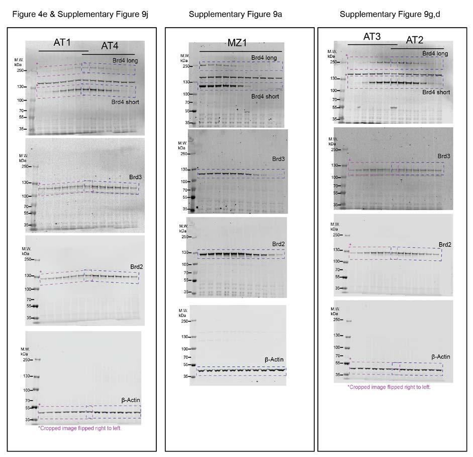
13 Supplementary Figure 9. Analysis of ternary complex recruitment and cellular BET protein degradation by AT1-6. a, d, g, j, m, p, Selective intracellular degradation of Brd4 by MZ1 (Ref. 7 ) (a), AT2 (d), AT3 (g), AT4 (j), AT5 (m) and AT6 (p) in HeLa cells treated with indicated concentrations of corresponding compound. Protein levels are shown from one representative of three biological replicates, visualised by immunoblot (left) and quantified relative to DMSO control (right). b, e, h, k, n, AlphaLISA intensity values titrating VCB against BET-BDs with AT2 (b), AT3 (e), AT4 (h), AT5 (k) and AT6 (n). c, f, i, l, o, AlphaLISA intensity values titrating each BET-BD against VCB with, AT2 (c), AT3 (f), AT4 (i), AT5 (l) and AT6 (o). Western blot intensity values were quantified as described in Online Methods. AlphaLISA intensities are the mean (± 1 s.d.) of intensity values from four technical replicates. Full gels are shown in Supplementary Figure

14 Supplementary Figure 10. Cycloheximide (CHX) chase assay to compare BET protein turnover in cells. HeLa cell protein synthesis was blocked with 100 μg/ml CHX for the given period of time from zero to 24 hours. After the treatment, 30 μg protein extracts from cells were analysed by Western blot (left) probing for BET proteins, cmyc or β-actin to examine depletion rate of these proteins in cells and quantified relative to DMSO control (right). The CHX treatment was effective as levels of cmyc, which is known to have short half-life, were depleted within two hours. All BET proteins were depleted in cells at slower rates compared to cmyc with no distinct differences between Brd2, Brd3 or Brd4. Such observation does not match with the preferential depletion of Brd4 induced by MZ1 and other PROTAC molecules, suggesting that such preference is not due to differences in native protein synthesis or turnover rate. Intensity values were quantified as described in Online Methods and are calculated from one biological replicate. Full gels are shown in Supplementary Figure
15 15

16 Supplementary Figure 11. Full uncut gel images of western blots data. 16

17 Supplementary Figure 12. Model of CRL2 VHL MZ1 Brd4 BD2, in vitro ubiquitination and identification of target lysines. a, Model of CRL2 VHL bound to MZ1 Brd4 BD2. Solvent-exposed lysines of Brd4 BD2 are highlighted as spheres and labelled. b, In vitro ubiquitination of Brd4 BD2 by CRL2 VHL in the presence of increasing concentrations of MZ1. Western blot probing for His 6 purification tag was used to visualize target protein levels. c,d, Ubiquitination sites identified in vitro 17
18 by mass spectrometry, highlighted in green in the sequence alignment. Residue K346 is located at the N-terminal tail of Brd4 BD2 (as indicated in a) and is not visible in our co-crystal structure. Supplementary Data Set 1: Proteomic analysis of relative protein abundance in HeLa cells. Results are graphically represented in Figure 4f,g. See file Supplementary Data Set 1.xls 18
19 Supplementary Table 1. Data collection and refinement statistics (molecular replacement) Brd4 BD2 :MZ1:pVHL-ElonginC-ElonginB (5T35) Data collection Space group P3 2 Cell dimensions a, b, c (Å) 102.3, 102.3, α, β, γ ( ) 90, 90, 120 Resolution (Å) ( )* R merge 7.9 (66.0) I/σ(I) 14.7 (2.1) CC 1/ (57.1) Completeness (%) 99.7 (99.3) Redundancy 4.2 (3.7) Refinement Resolution (Å) No. reflections (3322) R work 20.6 (32.3) R free 23.1 (37.4) No. atoms Protein 7191 Ligand 138 Water 86 B factors Protein 63.7 Ligand 42.5 Water 39.7 R.m.s. deviations Bond lengths (Å) 07 Bond angles ( ) 1.14 * Highest-resolution shell is shown in parentheses. 19
20 Supplementary Table 2. Breakdown of buried surface areas in the ternary complexes ligase:ligand:target. Buried surface areas in ternary complexes (Å 2 ) VHL: MZ1: Brd4 BD2 a CRBN: lenalidomide: CK1α CRBN: CC-885: GSPT1 c Ligase:target PPIs Ligase:ligand Target:ligand Total Buried surface areas were calculated with PISA (see ref. 52 ). The more extensive protein:ligand buried surface areas for our PROTAC ternary structure are consistent with the larger bifunctional nature of the compound MZ1 compared to the phthalimide-based ligands. These are in part compensated by the larger PPI contact areas in the CRBN examples, resulting in all cases in extensive total buried surface areas of > 1800 Å 2, as expected for a productive protein-protein interaction. a crystal structure from this study. b crystal structure from ref. 2 (PDB entry 5FQD). c crystal structure from ref. 3 (PDB code 5HXB). 20
21 Supplementary Table 3. Thermodynamic parameters of formation of binary and ternary complexes between AT1, VCB and BET bromodomains measured by isothermal titration calorimetry. Protein in Species in cell K syringe d (nm) G (kcal mol -1 ) H (kcal mol -1 T S (kcal ) mol -1 ) N α pk d Brd2 BD1 111 ± ± ± ± ± 04 Brd2 BD2 94 ± ± ± ± ± 3 Brd3 BD1 35 ± ± ± ± ± 2 Brd3 BD2 39 ± ± ± ± ± 06 Brd4 BD1 75 ± ± ± ± ± 3 AT1 Brd4 BD2 44 ± ± ± ± ± 2 Brd2 BD1 K 35 ± ± 6 15 ± 1 5 ± ± 0.2 EA Brd4 BD2 Q 38.8 ± ± ± ± ± 1 VK VCB a AT1 a 335 ± ± ± ± ± 4 AT1:Brd2 BD1 280 ± ± ± ± ± ± 0.2 AT1:Brd2 BD2 78 ± ± ± ± ± ± 5 AT1:Brd3 BD1 207 ± ± ± ± ± ± 4 VCB AT1:Brd3 BD2 79 ± ± ± ± ± ± 0.1 AT1:Brd4 BD1 390 ± ± ± ± ± ± 0.2 AT1:Brd4 BD2 46 ± ± ± ± ± ± 7 AT1:Brd2 BD1 KEA 52 ± 18 1 ± ± ± ± ± 0.2 AT1:Brd4 BD2 QVK 160 ± ± ± ± ± ± 0.1 All ITC titrations were performed at 25 C. Values reported are the mean ± s.e.m. from two independent measurements, except for VCB titration into AT1 (line a ) for which values reported are the mean ± s.e.m. from seven independent measurements. 21
SUPPLEMENTARY INFORMATION
 Supplementary materials Figure S1 Fusion protein of Sulfolobus solfataricus SRP54 and a signal peptide. a, Expression vector for the fusion protein. The signal peptide of yeast dipeptidyl aminopeptidase
Supplementary Figure 1. Biochemical and sequence alignment analyses the
 Supplementary Figure 1. Biochemical and sequence alignment analyses the interaction of OPTN and TBK1. (a) Analytical gel filtration chromatography analysis of the interaction between TBK1 CTD and OPTN(1-119).
SUPPLEMENTARY INFORMATION. doi: /nature07461
 Figure S1 Electrophysiology. a ph-activation of. Two-electrode voltage clamp recordings of Xenopus oocytes expressing in comparison to waterinjected oocytes. Currents were recorded at 40 mv. The ph of
SUPPLEMENTARY INFORMATION
 Table of Contents Page Supplementary Table 1. Diffraction data collection statistics 2 Supplementary Table 2. Crystallographic refinement statistics 3 Supplementary Fig. 1. casic1mfc packing in the R3
SUPPLEMENTARY INFORMATION
 doi:10.1038/nature11054 Supplementary Fig. 1 Sequence alignment of Na v Rh with NaChBac, Na v Ab, and eukaryotic Na v and Ca v homologs. Secondary structural elements of Na v Rh are indicated above the
Supplementary Figures
 1 Supplementary Figures Supplementary Figure 1 Type I FGFR1 inhibitors (a) Chemical structures of a pyrazolylaminopyrimidine inhibitor (henceforth referred to as PAPI; PDB-code of the FGFR1-PAPI complex:
SUPPLEMENTARY INFORMATION
 doi:10.1038/nature11085 Supplementary Tables: Supplementary Table 1. Summary of crystallographic and structure refinement data Structure BRIL-NOP receptor Data collection Number of crystals 23 Space group
University of Dundee Structural basis of PROTAC cooperative recognition for selective protein degradation General rights Take down policy
 University of Dundee tructural basis of PRTAC cooperative recognition for selective protein degradation Gadd, Morgan; Testa, Andrea; Lucas, Xavier; Chan, Kwok Ho; Chen, Wenzhang; Lamont, Douglas; Zengerle,
SUPPLEMENTARY INFORMATION
 Supplementary Results DNA binding property of the SRA domain was examined by an electrophoresis mobility shift assay (EMSA) using synthesized 12-bp oligonucleotide duplexes containing unmodified, hemi-methylated,
SUPPLEMENTARY INFORMATION
 SUPPLEMENTARY INFORMATION doi:10.1038/nature11524 Supplementary discussion Functional analysis of the sugar porter family (SP) signature motifs. As seen in Fig. 5c, single point mutation of the conserved
Table 1. Crystallographic data collection, phasing and refinement statistics. Native Hg soaked Mn soaked 1 Mn soaked 2
 Table 1. Crystallographic data collection, phasing and refinement statistics Native Hg soaked Mn soaked 1 Mn soaked 2 Data collection Space group P2 1 2 1 2 1 P2 1 2 1 2 1 P2 1 2 1 2 1 P2 1 2 1 2 1 Cell
SUPPLEMENTARY INFORMATION
 Fig. 1 Influences of crystal lattice contacts on Pol η structures. a. The dominant lattice contact between two hpol η molecules (silver and gold) in the type 1 crystals. b. A close-up view of the hydrophobic
SUPPLEMENTARY INFORMATION
 Supplementary Table 1: Amplitudes of three current levels. Level 0 (pa) Level 1 (pa) Level 2 (pa) TrkA- TrkH WT 200 K 0.01 ± 0.01 9.5 ± 0.01 18.7 ± 0.03 200 Na * 0.001 ± 0.01 3.9 ± 0.01 12.5 ± 0.03 200
Table S1. Overview of used PDZK1 constructs and their binding affinities to peptides. Related to figure 1.
 Table S1. Overview of used PDZK1 constructs and their binding affinities to peptides. Related to figure 1. PDZK1 constru cts Amino acids MW [kda] KD [μm] PEPT2-CT- FITC KD [μm] NHE3-CT- FITC KD [μm] PDZK1-CT-
THE CRYSTAL STRUCTURE OF THE SGT1-SKP1 COMPLEX: THE LINK BETWEEN
 THE CRYSTAL STRUCTURE OF THE SGT1-SKP1 COMPLEX: THE LINK BETWEEN HSP90 AND BOTH SCF E3 UBIQUITIN LIGASES AND KINETOCHORES Oliver Willhoft, Richard Kerr, Dipali Patel, Wenjuan Zhang, Caezar Al-Jassar, Tina
Structural basis for catalytically restrictive dynamics of a high-energy enzyme state
 Supplementary Material Structural basis for catalytically restrictive dynamics of a high-energy enzyme state Michael Kovermann, Jörgen Ådén, Christin Grundström, A. Elisabeth Sauer-Eriksson, Uwe H. Sauer
SUPPLEMENTARY INFORMATION
 www.nature.com/nature 1 Figure S1 Sequence alignment. a Structure based alignment of the plgic of E. chrysanthemi (ELIC), the acetylcholine binding protein from the snail Lymnea stagnalis (AchBP, PDB code
Nitrogenase MoFe protein from Clostridium pasteurianum at 1.08 Å resolution: comparison with the Azotobacter vinelandii MoFe protein
 Acta Cryst. (2015). D71, 274-282, doi:10.1107/s1399004714025243 Supporting information Volume 71 (2015) Supporting information for article: Nitrogenase MoFe protein from Clostridium pasteurianum at 1.08
Supplementary Information. Structural basis for precursor protein-directed ribosomal peptide macrocyclization
 Supplementary Information Structural basis for precursor protein-directed ribosomal peptide macrocyclization Kunhua Li 1,3, Heather L. Condurso 1,3, Gengnan Li 1, Yousong Ding 2 and Steven D. Bruner 1*
Supplementary Materials for
 www.sciencesignaling.org/cgi/content/full/5/243/ra68/dc1 Supplementary Materials for Superbinder SH2 Domains Act as Antagonists of Cell Signaling Tomonori Kaneko, Haiming Huang, Xuan Cao, Xing Li, Chengjun
Supplemental Information
 Supplemental Information Combinatorial Readout of Unmodified H3R2 and Acetylated H3K14 by the Tandem PHD Finger of MOZ Reveals a Regulatory Mechanism for HOXA9 Transcription Yu Qiu 1, Lei Liu 1, Chen Zhao
SUPPLEMENTARY INFORMATION
 doi:1.138/nature1737 Supplementary Table 1 variant Description FSEC - 2B12 a FSEC - 6A1 a K d (leucine) c Leucine uptake e K (wild-type like) K (Y18F) K (TS) K (TSY) K288A mutant, lipid facing side chain
SUPPLEMENTARY INFORMATION
 Supplementary Table S1 Kinetic Analyses of the AMSH-LP mutants AMSH-LP K M (μm) k cat x 10-3 (s -1 ) WT 71.8 ± 6.3 860 ± 65.4 T353A 76.8 ± 11.7 46.3 ± 3.7 F355A 58.9 ± 10.4 5.33 ± 0.30 proximal S358A 75.1
SI Text S1 Solution Scattering Data Collection and Analysis. SI references
 SI Text S1 Solution Scattering Data Collection and Analysis. The X-ray photon energy was set to 8 kev. The PILATUS hybrid pixel array detector (RIGAKU) was positioned at a distance of 606 mm from the sample.
SUPPLEMENTARY INFORMATION
 SUPPLEMENTARY INFORMATION doi:10.1038/nature12242 C. thermophilum 666 RPAVLDNVYIRPALE-GKRVPGKVEIHQNGIRYQSPLSTTQRVDVLFSNIRHLFFQPCQN S. pombe 659 RPAHINDVYVRPAID-GKRLPGFIEIHQNGIRYQSPLRSDSHIDLLFSNMKHLFFQPCEG
Supplementary Materials for
 advances.sciencemag.org/cgi/content/full/3/4/e1600663/dc1 Supplementary Materials for A dynamic hydrophobic core orchestrates allostery in protein kinases Jonggul Kim, Lalima G. Ahuja, Fa-An Chao, Youlin
SUPPLEMENTARY INFORMATION
 Supplementary Table 1: Data collection, phasing and refinement statistics ChbC/Ta 6 Br 12 Native ChbC Data collection Space group P4 3 2 1 2 P4 3 2 1 2 Cell dimensions a, c (Å) 132.75, 453.57 132.81, 452.95
SUPPLEMENTARY FIGURES
 SUPPLEMENTARY FIGURES Supplementary Figure 1 Protein sequence alignment of Vibrionaceae with either a 40-residue insertion or a 44-residue insertion. Identical residues are indicated by red background.
Supplementary Information
 Supplementary Information The direct role of selenocysteine in [NiFeSe] hydrogenase maturation and catalysis Marta C. Marques a, Cristina Tapia b, Oscar Gutiérrez-Sanz b, Ana Raquel Ramos a, Kimberly L.
IgE binds asymmetrically to its B cell receptor CD23
 Supplementary Information IgE binds asymmetrically to its B cell receptor CD23 Balvinder Dhaliwal 1*, Marie O. Y. Pang 2, Anthony H. Keeble 2,3, Louisa K. James 2,4, Hannah J. Gould 2, James M. McDonnell
SUPPLEMENTARY INFORMATION
 SUPPLEMENTARY INFORMATION doi:10.1038/nature11539 Supplementary Figure 1 Schematic representation of plant (A) and mammalian (B) P 2B -ATPase domain organization. Actuator (A-), nucleotide binding (N-),
Structural characterization of NiV N 0 P in solution and in crystal.
 Supplementary Figure 1 Structural characterization of NiV N 0 P in solution and in crystal. (a) SAXS analysis of the N 32-383 0 -P 50 complex. The Guinier plot for complex concentrations of 0.55, 1.1,
Structure and evolution of the spliceosomal peptidyl-prolyl cistrans isomerase Cwc27
 Acta Cryst. (2014). D70, doi:10.1107/s1399004714021695 Supporting information Volume 70 (2014) Supporting information for article: Structure and evolution of the spliceosomal peptidyl-prolyl cistrans isomerase
CH 3 CH 2 OH +H 2 O CHO. 2e + 2H + + O 2 H 2 O +HCOOH
 2 4 H CH 3 2e + 2H + + 2 H 2 2 H CH 2 H 2e + 2H + + 2 H 2 2 H +H 2 CH 2e + 2H + + 2 H 2 2 H +HCH Supplemental Figure S. The three-step 4DM reaction, each step requires two reducing equivalents from ADPH
Experimental and Computational Mutagenesis to Investigate the. Positioning of a General Base within an Enzyme Active Site
 Experimental and Computational Mutagenesis to Investigate the Positioning of a General Base within an Enzyme Active Site Jason P. Schwans, Philip Hanoian, Benjamin J. Lengerich, Fanny Sunden, Ana Gonzalez
Nature Structural & Molecular Biology: doi: /nsmb Supplementary Figure 1
 Supplementary Figure 1 Crystallization. a, Crystallization constructs of the ET B receptor are shown, with all of the modifications to the human wild-type the ET B receptor indicated. Residues interacting
Cks1 CDK1 CDK1 CDK1 CKS1. are ice- lobe. conserved. conserved
 Cks1 d CKS1 Supplementary Figure 1 The -Cks1 crystal lattice. (a) Schematic of the - Cks1 crystal lattice. -Cks1 crystallizes in a lattice that contains c 4 copies of the t - Cks1 dimer in the crystallographic
Structure, mechanism and ensemble formation of the Alkylhydroperoxide Reductase subunits. AhpC and AhpF from Escherichia coli
 Structure, mechanism and ensemble formation of the Alkylhydroperoxide Reductase subunits AhpC and AhpF from Escherichia coli Phat Vinh Dip 1,#, Neelagandan Kamariah 2,#, Malathy Sony Subramanian Manimekalai
Nature Structural & Molecular Biology doi: /nsmb Supplementary Figure 1. CRBN binding assay with thalidomide enantiomers.
 Supplementary Figure 1 CRBN binding assay with thalidomide enantiomers. (a) Competitive elution assay using thalidomide-immobilized beads coupled with racemic thalidomide. Beads were washed three times
FW 1 CDR 1 FW 2 CDR 2
 Supplementary Figure 1 Supplementary Figure 1: Interface of the E9:Fas structure. The two interfaces formed by V H and V L of E9 with Fas are shown in stereo. The Fas receptor is represented as a surface
Rational Design of Thermodynamic and Kinetic Binding Profiles by. Optimizing Surface Water Networks Coating Protein Bound Ligands
 SUPPORTING INFORMATION Rational Design of Thermodynamic and Kinetic Binding Profiles by Optimizing Surface Water Networks Coating Protein Bound Ligands Stefan G. Krimmer,, Jonathan Cramer,, Michael Betz,
Supplementary Information. The protease GtgE from Salmonella exclusively targets. inactive Rab GTPases
 Supplementary Information The protease GtgE from Salmonella exclusively targets inactive Rab GTPases Table of Contents Supplementary Figures... 2 Supplementary Figure 1... 2 Supplementary Figure 2... 3
Structure and RNA-binding properties. of the Not1 Not2 Not5 module of the yeast Ccr4 Not complex
 Structure and RNA-binding properties of the Not1 Not2 Not5 module of the yeast Ccr4 Not complex Varun Bhaskar 1, Vladimir Roudko 2,3, Jerome Basquin 1, Kundan Sharma 4, Henning Urlaub 4, Bertrand Seraphin
SUPPLEMENTARY INFORMATION
 SUPPLEMENTARY INFORMATION Structure of human carbamoyl phosphate synthetase: deciphering the on/off switch of human ureagenesis Sergio de Cima, Luis M. Polo, Carmen Díez-Fernández, Ana I. Martínez, Javier
SUPPLEMENTARY INFORMATION
 doi:10.1038/nature12045 Supplementary Table 1 Data collection and refinement statistics. Native Pt-SAD X-ray source SSRF BL17U SPring-8 BL41XU Wavelength (Å) 0.97947 1.07171 Space group P2 1 2 1 2 1 P2
Nature Structural & Molecular Biology: doi: /nsmb Supplementary Figure 1
 Supplementary Figure 1 Identification of the ScDcp2 minimal region interacting with both ScDcp1 and the ScEdc3 LSm domain. Pull-down experiment of untagged ScEdc3 LSm with various ScDcp1-Dcp2-His 6 fragments.
SUPPLEMENTARY INFORMATION
 doi:10.1038/nature10458 Active Site Remodeling in the Bifunctional Fructose-1,6- bisphosphate aldolase/phosphatase Juan Du, Rafael F. Say, Wei Lü, Georg Fuchs & Oliver Einsle SUPPLEMENTARY FIGURES Figure
The structure of a nucleolytic ribozyme that employs a catalytic metal ion. Yijin Liu, Timothy J. Wilson and David M.J. Lilley
 SUPPLEMENTARY INFORMATION The structure of a nucleolytic ribozyme that employs a catalytic metal ion Yijin Liu, Timothy J. Wilson and David M.J. Lilley Cancer Research UK Nucleic Acid Structure Research
Supplemental Information. Molecular Basis of Spectral Diversity. in Near-Infrared Phytochrome-Based. Fluorescent Proteins
 Chemistry & Biology, Volume 22 Supplemental Information Molecular Basis of Spectral Diversity in Near-Infrared Phytochrome-Based Fluorescent Proteins Daria M. Shcherbakova, Mikhail Baloban, Sergei Pletnev,
Supporting Information
 Supporting Information Ottmann et al. 10.1073/pnas.0907587106 Fig. S1. Primary structure alignment of SBT3 with C5 peptidase from Streptococcus pyogenes. The Matchmaker tool in UCSF Chimera (http:// www.cgl.ucsf.edu/chimera)
Sensitive NMR Approach for Determining the Binding Mode of Tightly Binding Ligand Molecules to Protein Targets
 Supporting information Sensitive NMR Approach for Determining the Binding Mode of Tightly Binding Ligand Molecules to Protein Targets Wan-Na Chen, Christoph Nitsche, Kala Bharath Pilla, Bim Graham, Thomas
SUPPLEMENTARY INFORMATION
 doi:10.1038/nature10955 Supplementary Figures Supplementary Figure 1. Electron-density maps and crystallographic dimer structures of the motor domain. (a f) Stereo views of the final electron-density maps
RNA Polymerase I Contains a TFIIF-Related DNA-Binding Subcomplex
 Molecular Cell, Volume 39 Supplemental Information RNA Polymerase I Contains a TFIIFRelated DNABinding Subcomplex Sebastian R. Geiger, Kristina Lorenzen, Amelie Schreieck, Patrizia Hanecker, Dirk Kostrewa,
Supplementary Information
 Supplementary Information Structural analysis of leader peptide binding enables leaderfree cyanobactin processing Jesko Koehnke 1,2, Greg Mann 1,2, Andrew F Bent 1,2, Hannes Ludewig 1, Sally Shirran 1,
Supplementary figure 1 Application of tmfret in LeuT. (a) To assess the feasibility of using tmfret for distance-dependent measurements in LeuT, a
 To assess the feasibility of using tmfret for distance-dependent measurements in LeuT, a") Supplementary figure 1 Application of tmfret in LeuT. (a) To assess the feasibility of using tmfret for distance-dependent measurements in LeuT, a series of tmfret-pairs comprised of single cysteine mutants
Supplementary figure 1 Application of tmfret in LeuT. (a) To assess the feasibility of using tmfret for distance-dependent measurements in LeuT, a series of tmfret-pairs comprised of single cysteine mutants
Supplementary Information
 1 Supplementary Information Figure S1 The V=0.5 Harker section of an anomalous difference Patterson map calculated using diffraction data from the NNQQNY crystal at 1.3 Å resolution. The position of the
Supplementary Information
 Supplementary Information Resveratrol Serves as a Protein-Substrate Interaction Stabilizer in Human SIRT1 Activation Xuben Hou,, David Rooklin, Hao Fang *,,, Yingkai Zhang Department of Medicinal Chemistry
Computational engineering of cellulase Cel9A-68 functional motions through mutations in its linker region. WT 1TF4 (crystal) -90 ERRAT PROVE VERIFY3D
 -90 ERRAT PROVE VERIFY3D") Electronic Supplementary Material (ESI) for Physical Chemistry Chemical Physics. This journal is the Owner Societies 218 Supplementary Material: Computational engineering of cellulase Cel9-68 functional
Electronic Supplementary Material (ESI) for Physical Chemistry Chemical Physics. This journal is the Owner Societies 218 Supplementary Material: Computational engineering of cellulase Cel9-68 functional
T H E J O U R N A L O F G E N E R A L P H Y S I O L O G Y. jgp
 S u p p l e m e n ta l m at e r i a l jgp Lee et al., http://www.jgp.org/cgi/content/full/jgp.201411219/dc1 T H E J O U R N A L O F G E N E R A L P H Y S I O L O G Y S u p p l e m e n ta l D I S C U S
Structural insights into Aspergillus fumigatus lectin specificity - AFL binding sites are functionally non-equivalent
 Acta Cryst. (2015). D71, doi:10.1107/s1399004714026595 Supporting information Volume 71 (2015) Supporting information for article: Structural insights into Aspergillus fumigatus lectin specificity - AFL
SUPPLEMENTARY INFORMATION
 SUPPLEMENTARY INFORMATION doi:10.1038/nature11744 Supplementary Table 1. Crystallographic data collection and refinement statistics. Wild-type Se-Met-BcsA-B SmCl 3 -soaked EMTS-soaked Data collection Space
Supporting Information
 Supporting Information Discovery and optimization of a selective ligand for the Switch/Sucrose Non-Fermenting-related bromodomains of Polybromo protein-1 by the use of virtual screening and hydration analysis.
NMR study of complexes between low molecular mass inhibitors and the West Nile virus NS2B-NS3 protease
 University of Wollongong Research Online Faculty of Science - Papers (Archive) Faculty of Science, Medicine and Health 2009 NMR study of complexes between low molecular mass inhibitors and the West Nile
Structure and Function of Neisseria gonorrhoeae MtrF Illuminates a Class of Antimetabolite Efflux Pumps
 Cell Reports Supplemental Information Structure and Function of Neisseria gonorrhoeae MtrF Illuminates a Class of Antimetabolite Efflux Pumps Chih-Chia Su, Jani Reddy Bolla, Nitin Kumar, Abhijith Radhakrishnan,
Supplementary Figure 1 Crystal contacts in COP apo structure (PDB code 3S0R)
") Supplementary Figure 1 Crystal contacts in COP apo structure (PDB code 3S0R) Shown in cyan and green are two adjacent tetramers from the crystallographic lattice of COP, forming the only unique inter-tetramer
Supplementary Figure 1 Crystal contacts in COP apo structure (PDB code 3S0R) Shown in cyan and green are two adjacent tetramers from the crystallographic lattice of COP, forming the only unique inter-tetramer
Nature Structural and Molecular Biology: doi: /nsmb.2938
 Supplementary Figure 1 Characterization of designed leucine-rich-repeat proteins. (a) Water-mediate hydrogen-bond network is frequently visible in the convex region of LRR crystal structures. Examples
Viewing and Analyzing Proteins, Ligands and their Complexes 2
 2 Viewing and Analyzing Proteins, Ligands and their Complexes 2 Overview Viewing the accessible surface Analyzing the properties of proteins containing thousands of atoms is best accomplished by representing
Supplemental Data SUPPLEMENTAL FIGURES
 Supplemental Data CRYSTAL STRUCTURE OF THE MG.ADP-INHIBITED STATE OF THE YEAST F 1 C 10 ATP SYNTHASE Alain Dautant*, Jean Velours and Marie-France Giraud* From Université Bordeaux 2, CNRS; Institut de
Supporting Information. UV-induced ligand exchange in MHC class I protein crystals
 Supporting Information for the article entitled UV-induced ligand exchange in MHC class I protein crystals by Patrick H.N. Celie 1, Mireille Toebes 2, Boris Rodenko 3, Huib Ovaa 3, Anastassis Perrakis
Supplementary figure 1. Comparison of unbound ogm-csf and ogm-csf as captured in the GIF:GM-CSF complex. Alignment of two copies of unbound ovine
 Supplementary figure 1. Comparison of unbound and as captured in the GIF:GM-CSF complex. Alignment of two copies of unbound ovine GM-CSF (slate) with bound GM-CSF in the GIF:GM-CSF complex (GIF: green,
Structurale, Université Grenoble Alpes, CNRS, CEA, Grenoble, France
 Supplementary Information to Lysine relay mechanism coordinates intermediate transfer in vitamin B6 biosynthesis Matthew J. Rodrigues 1,2, Volker Windeisen 1,3, Yang Zhang 4, Gabriela Guédez 3, Stefan
Supplemental Information. The Mitochondrial Fission Receptor MiD51. Requires ADP as a Cofactor
 Structure, Volume 22 Supplemental Information The Mitochondrial Fission Receptor MiD51 Requires ADP as a Cofactor Oliver C. Losón, Raymond Liu, Michael E. Rome, Shuxia Meng, Jens T. Kaiser, Shu-ou Shan,
The Fic protein Doc uses an inverted substrate to phosphorylate and. inactivate EF-Tu
 The Fic protein Doc uses an inverted substrate to phosphorylate and inactivate EF-Tu Daniel Castro-Roa 1, Abel Garcia-Pino 2,3 *, Steven De Gieter 2,3, Nico A.J. van Nuland 2,3, Remy Loris 2,3, Nikolay
Bacterial protease uses distinct thermodynamic signatures for substrate recognition
 Bacterial protease uses distinct thermodynamic signatures for substrate recognition Gustavo Arruda Bezerra, Yuko Ohara-Nemoto, Irina Cornaciu, Sofiya Fedosyuk, Guillaume Hoffmann, Adam Round, José A. Márquez,
NB-DNJ/GCase-pH 7.4 NB-DNJ+/GCase-pH 7.4 NB-DNJ+/GCase-pH 4.5
 SUPPLEMENTARY TABLES Suppl. Table 1. Protonation states at ph 7.4 and 4.5. Protonation states of titratable residues in GCase at ph 7.4 and 4.5. Histidine: HID, H at δ-nitrogen; HIE, H at ε-nitrogen; HIP,
Acta Cryst. (2017). D73, doi: /s
. D73, doi: /s") Acta Cryst. (2017). D73, doi:10.1107/s2059798317010932 Supporting information Volume 73 (2017) Supporting information for article: Designing better diffracting crystals of biotin carboxyl carrier protein
Acta Cryst. (2017). D73, doi:10.1107/s2059798317010932 Supporting information Volume 73 (2017) Supporting information for article: Designing better diffracting crystals of biotin carboxyl carrier protein
SUPPLEMENTARY INFORMATION
 Data collection Supplementary Table 1 Statistics of data collection, phasing and refinement Native Se-MAD Space group P2 1 2 1 2 1 P2 1 2 1 2 1 Cell dimensions a, b, c (Å) 50.4, 94.2, 115.4 49.8, 94.2,
Supplementary information
 Supplementary information The structural basis of modularity in ECF-type ABC transporters Guus B. Erkens 1,2, Ronnie P-A. Berntsson 1,2, Faizah Fulyani 1,2, Maria Majsnerowska 1,2, Andreja Vujičić-Žagar
Plasmid Relevant features Source. W18N_D20N and TrXE-W18N_D20N-anti
 Table S1. E. coli plasmids Plasmid Relevant features Source pdg680 T. reesei XynII AA 2-190 with C-terminal His 6 tag optimized for E. coli expression in pjexpress401 Wan et al. (in press) psbn44d psbn44h
LS1a Fall 2014 Problem Set #2 Due Monday 10/6 at 6 pm in the drop boxes on the Science Center 2 nd Floor
 LS1a Fall 2014 Problem Set #2 Due Monday 10/6 at 6 pm in the drop boxes on the Science Center 2 nd Floor Note: Adequate space is given for each answer. Questions that require a brief explanation should
Identifying Interaction Hot Spots with SuperStar
 Identifying Interaction Hot Spots with SuperStar Version 1.0 November 2017 Table of Contents Identifying Interaction Hot Spots with SuperStar... 2 Case Study... 3 Introduction... 3 Generate SuperStar Maps
Supplementary Figure 1. Aligned sequences of yeast IDH1 (top) and IDH2 (bottom) with isocitrate
 and IDH2 (bottom) with isocitrate") SUPPLEMENTARY FIGURE LEGENDS Supplementary Figure 1. Aligned sequences of yeast IDH1 (top) and IDH2 (bottom) with isocitrate dehydrogenase from Escherichia coli [ICD, pdb 1PB1, Mesecar, A. D., and Koshland,
SUPPLEMENTARY FIGURE LEGENDS Supplementary Figure 1. Aligned sequences of yeast IDH1 (top) and IDH2 (bottom) with isocitrate dehydrogenase from Escherichia coli [ICD, pdb 1PB1, Mesecar, A. D., and Koshland,
Crystal Structure of Fibroblast Growth Factor 9 (FGF9) Reveals Regions. Implicated in Dimerization and Autoinhibition
 Reveals Regions. Implicated in Dimerization and Autoinhibition") JBC Papers in Press. Published on November 1, 2000 as Manuscript M006502200 Crystal Structure of Fibroblast Growth Factor 9 (FGF9) Reveals Regions Implicated in Dimerization and Autoinhibition 1 Copyright
JBC Papers in Press. Published on November 1, 2000 as Manuscript M006502200 Crystal Structure of Fibroblast Growth Factor 9 (FGF9) Reveals Regions Implicated in Dimerization and Autoinhibition 1 Copyright
Structural and mechanistic insight into the substrate. binding from the conformational dynamics in apo. and substrate-bound DapE enzyme
 Electronic Supplementary Material (ESI) for Physical Chemistry Chemical Physics. This journal is the Owner Societies 215 Structural and mechanistic insight into the substrate binding from the conformational
Supplementary Information
 Supplementary Information An engineered protein antagonist of K-Ras/B-Raf interaction Monique J. Kauke, 1,2 Michael W. Traxlmayr 1,2, Jillian A. Parker 3, Jonathan D. Kiefer 4, Ryan Knihtila 3, John McGee
Nature Structural and Molecular Biology: doi: /nsmb.2783
 Supplementary Figure 1: Crystallized chimera construct (mhv1cc). (a) Sequence alignment between mhv1cc and other VSDs. These sequences (mhv1cc, Kv1.2 Kv2.1; shaker family voltage gated potassium channel
SUPPLEMENTARY INFORMATION
 Dph2 SeMet (iron-free) # Dph2 (iron-free) Dph2-[4Fe-4S] Data collection Space group P2 1 2 1 2 1 P2 1 2 1 2 1 P2 1 2 1 2 1 Cell dimensions a, b, c (Å) 58.26, 82.08, 160.42 58.74, 81.87, 160.01 55.70, 80.53,
Isothermal Titration Calorimetry in Drug Discovery. Geoff Holdgate Structure & Biophysics, Discovery Sciences, AstraZeneca October 2017
 Isothermal Titration Calorimetry in Drug Discovery Geoff Holdgate Structure & Biophysics, Discovery Sciences, AstraZeneca October 217 Introduction Introduction to ITC Strengths / weaknesses & what is required
Supporting Information
 Supporting Information Micelle-Triggered b-hairpin to a-helix Transition in a 14-Residue Peptide from a Choline-Binding Repeat of the Pneumococcal Autolysin LytA HØctor Zamora-Carreras, [a] Beatriz Maestro,
SUPPLEMENTARY INFORMATION
 Supplementary Figure 1: The HpUreI crystal used for collection of native diffraction data. The crystal belongs to spacegroup P4 2 2 1 2 and has an approximate maximal dimension of 0.25 mm. Supplementary
Stabilizing the CH2 domain of an Antibody by Engineering in an Enhanced Aromatic Sequon
 Stabilizing the CH2 domain of an Antibody by Engineering in an Enhanced Aromatic Sequon Wentao Chen,, Leopold Kong, Stephen Connelly, Julia M. Dendle,, Yu Liu,, Ian A. Wilson,#, Evan T. Powers, *, Jeffery
The structure of the deubiquitinase USP15 reveals a misaligned catalytic triad and an open ubiquitin-binding channel
 SUPPORTING INFORMATION The structure of the deubiquitinase USP15 reveals a misaligned catalytic triad and an open ubiquitin-binding channel Stephanie J. Ward, Hayley E. Gratton, Peni Indrayudha #, Camille
Serine-7 but not serine-5 phosphorylation primes RNA polymerase II CTD for P-TEFb recognition
 Supplementary Information to Serine-7 but not serine-5 phosphorylation primes RNA polymerase II CTD for P-TEFb recognition Nadine Czudnochowski 1,2, *, Christian A. Bösken 1, * & Matthias Geyer 1 1 Max-Planck-Institut
Microcalorimetry for the Life Sciences
 Microcalorimetry for the Life Sciences Why Microcalorimetry? Microcalorimetry is universal detector Heat is generated or absorbed in every chemical process In-solution No molecular weight limitations Label-free
PDBe TUTORIAL. PDBePISA (Protein Interfaces, Surfaces and Assemblies)
") PDBe TUTORIAL PDBePISA (Protein Interfaces, Surfaces and Assemblies) http://pdbe.org/pisa/ This tutorial introduces the PDBePISA (PISA for short) service, which is a webbased interactive tool offered by
PDBe TUTORIAL PDBePISA (Protein Interfaces, Surfaces and Assemblies) http://pdbe.org/pisa/ This tutorial introduces the PDBePISA (PISA for short) service, which is a webbased interactive tool offered by
Full wwpdb X-ray Structure Validation Report i
 Full wwpdb X-ray Structure Validation Report i Jan 14, 2019 11:10 AM EST PDB ID : 6GYW Title : Crystal structure of DacA from Staphylococcus aureus Authors : Tosi, T.; Freemont, P.S.; Grundling, A. Deposited
17. Biomolecular Interaction
 17. Biomolecular Interaction Methods for characterizing biomolecular interactions Sequence-specific DNA binding ligands Molecular mechanisms of drug action and drug resistance In silico compound design
Structure of the α-helix
 Structure of the α-helix Structure of the β Sheet Protein Dynamics Basics of Quenching HDX Hydrogen exchange of amide protons is catalyzed by H 2 O, OH -, and H 3 O +, but it s most dominated by base
Expanded View Figures
 The EMBO Journal Structure of a Dm peptide bound to the OT module Tobias Raisch et al Expanded View Figures A Hs Dm 262 297 685 8 HEAT HEAT MIF4G 9BD 1SHD 761 91 193 169 1152 1317 16 1376 1467 HEAT HEAT
Detailed description of overall and active site architecture of PPDC- 3dThDP, PPDC-2HE3dThDP, PPDC-3dThDP-PPA and PPDC- 3dThDP-POVA
 Online Supplemental Results Detailed description of overall and active site architecture of PPDC- 3dThDP, PPDC-2HE3dThDP, PPDC-3dThDP-PPA and PPDC- 3dThDP-POVA Structure solution and overall architecture