Nature Structural & Molecular Biology: doi: /nsmb.3194
|
|
|
- Quentin Park
- 5 years ago
- Views:
Transcription
1 Supplementary Figure 1 Mass spectrometry and solution NMR data for -syn samples used in this study. (a) Matrix-assisted laser-desorption and ionization time-of-flight (MALDI-TOF) mass spectrum of uniformly- 13 C, 15 N- enriched -syn monomer (blue) and natural abundance monomer (red). The molecular mass of natural abundance -syn monomer was calculated to be kda, in agreement with the experimental mass spectrum. The mass of kda for the U- 13 C, 15 N-labeled sample corresponded to a ~98% incorporation of 13 C and 15 N during protein expression. (b) 15 N- 1 H HSQC spectrum of monomeric -syn prior to fibrillization. The chemical shifts agreed well with the published assignments (Eliezer, D., Kutluay, E., Bussell, R. & Browne, G. Conformational properties of α-synuclein in its free and lipid-associated states. J. Mol. Biol. 307, (2001).)
2
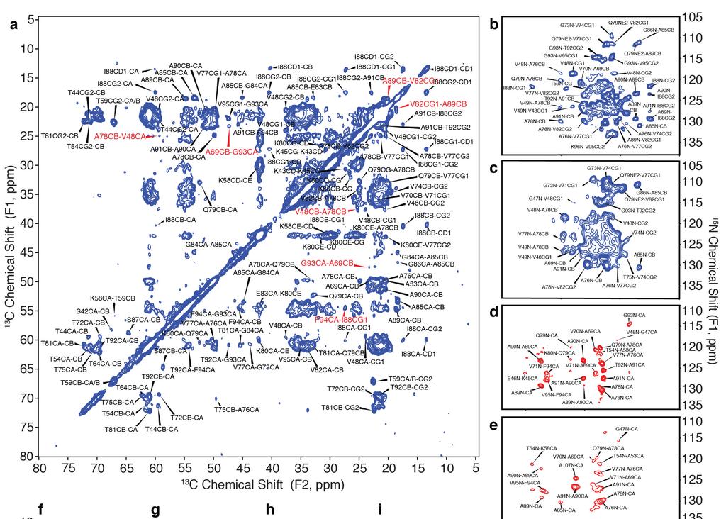
3 Supplementary Figure 2 SSNMR spectroscopy data for -syn fibrils, exhibiting cross-peaks indicative of long-range restraints and intermolecular registry of the fibrils. (a) 2D 13 C- 13 C SSNMR { 1 H}- 13 C-{ 1 H- 1 H}- 13 C spectrum (Lange, A., Luca, S. & Baldus, M. Structural constraints from proton-mediated rare-spin correlation spectroscopy in rotating solids. J. Am. Chem. Soc. 124, (2002).) of dilute -syn fibrils (sample D from Table 1) showing unambiguous, long-range intramolecular correlations. Data were acquired at 750 MHz 1 H frequency, 12.5 khz MAS and 400 s 1 H- 1 H mixing time, with 12 days signal averaging. Red labels correspond to unambiguous long-range distances, including those from A78 to V48, from A69 to G93, from F94 to I88, and from V82 to A89. These intramolecular long-range restraints were inconsistent with structural models that exhibit a domain swap. (b-e) 2D 15 N, 13 C SSNMR transferred echo double resonance (TEDOR, Jaroniec, C.P., Filip, C. & Griffin, R.G. 3D TEDOR NMR experiments for the simultaneous measurement of multiple carbon-nitrogen distances in uniformly 13 C, 15 N-labeled solids. J. Am. Chem. Soc. 124, (2002).) data showing intermolecular interactions. The data were collected for four samples: (b) 1,3-13 C glycerol, 15 N-labeled fibrils (sample B), signal averaged for 28 hr; (c) 50:50 [1,3-13 C]glycerol, natural abundance (n.a.) nitrogen: n.a. carbon, 15 N labeled (sample E), signal averaged for 81 hr; (d) [2-13 C]glycerol, 15 N labeled (sample C), signal averaged for 113 hr; and (e) 50: C glycerol, n.a. nitrogen: n.a. carbon, 15 N labeled (sample F), signal averaged for 124 hr. Data was collected with mixing times of (c, e) 14.4 ms at 500 MHz 1 H frequency, 11.1 khz MAS, and (b, d) 16.0 ms at 600 MHz 1 H frequency, 10.0 khz MAS. Cross-peaks present in both parts b and c, or d and e, demonstrate a parallel, in-register arrangement (Debelouchina, G.T., Platt, G.W., Bayro, M.J., Radford, S.E. & Griffin, R.G. Intermolecular alignment in 2 -microglobulin amyloid fibrils. J. Am. Chem. Soc. 132, (2010).) (f k) Spectra of sample B collected at 750 MHz 1 H frequency, 12.5 khz MAS (f-i) 2D 13 C- 13 C spectra using dipolar-assisted rotational resonance (DARR, Takegoshi, K., Nakamura, S. & Terao, T. 13 C- 1 H dipolar-assisted rotational resonance in magic-angle spinning NMR. Chem. Phys. Lett. 344, (2001).) mixing times of (f) 50 ms, signal averaged for 10.9 hr, (g) 100 ms, signal averaged for 5.7 hr, (h) 200 ms, signal averaged for 12.0 hr, and (i) 300 ms, signal averaged for 12.7 hr. Several long-range correlations (red) were observed between the F94 aromatic ring resonances (CD and CE) and I88, A90 and A91. (j-k) 2D planes from a 3D 15 N- 13 CO- 13 CX correlation spectrum signal averaged for 24 hr. Long-range correlations observed at this relatively short mixing time indicate internuclear distances <5 Å.

 Side view showing the -sheet packing between each monomer as well as the disordered tails.")
 Backbone traces for neighboring monomers drawn in blue, yellow, orange and red.")
 Side and top views of the I88-A91-F94 pocket perpendicular to the fibril axis, exhibiting short")
4 Supplementary Figure 3 Full-length structure of the -syn fibril, illustrating important features in expanded regions. (a) View along the fibril axis showing the highly ordered core and the disordered tails. (b) Side view showing the -sheet packing between each monomer as well as the disordered tails. (c-f) Core interactions illustrating NMR distance restraints with dotted lines. (g-l) Backbone traces for neighboring monomers drawn in blue, yellow, orange and red. (g, h) Side and top views of the salt bridge from E46 to K80 of the neighboring monomer. (i, j) Side and top views of the I88-A91-F94 pocket perpendicular to the fibril axis, exhibiting short intermolecular distances. (k, l) Side and top view of the Q79 side chain exhibiting a glutamine ladder, with intermolecular hydrogen bonds involving Nε2 and Oε1 moieties.
5 Supplementary Figure 4 SSNMR internal validation of structural restraints and dihedral angles for the -syn fibril structure. (a) The backbone trace is shown in grey, and black lines represent unambiguous distance restraints. Blue lines correspond to the shortest observed distance among the possible ambiguous assignments. The resulting structure is the lowest energy conformer consistent with all of the data. (b-c) Ramachandran probability maps of accepted dihedral angle regions (Lovell, S.C. et al. Structure validation by C geometry:, and C deviation. Proteins 50, (2003).) and plotted in Chimera (Pettersen, E.F. et al. UCSF Chimera a visualization system for exploratory research and analysis. J. Comput. Chem. 25, (2004).) for residues The only residue not in the accepted range is E57, which is part of an unstructured loop and lacks NMR restraints in the simulated annealing calculations. All glycine residues are within the accepted Ramachandran space.
6 Supplementary Figure 5 Cross-validation of the SSNMR structure of -syn fibrils with electron microscopy and X-ray fiber diffraction. (a) Chemical shift comparison of the fibril sample for solid-state NMR before and after washing with 4-(2-hydroxyethyl)-1- piperazineethanesulfonic acid (HEPES) buffer and sonication for electron microscopy. (b) Expansions of overlaid 2D 13 C- 13 C spectra of the U- 13 C, 15 N -syn fibril as prepared for solid-state NMR (red) and fibrils washed with HEPES buffer as the samples for electron microscopy (blue). Spectra were acquired at 600 MHz 1 H frequency, 13.3 khz magic-angle spinning (MAS) and 50 ms dipolar-assisted rotational resonance (DARR, Takegoshi, K., Nakamura, S. & Terao, T. 13 C- 1 H dipolar-assisted rotational resonance in magic-angle spinning NMR. Chem. Phys. Lett. 344, (2001).). (c) Equatorial intensity plot of the calculated and experimental fiber diffraction pattern of the -syn fibril. The solid line shows the experimental fiber diffraction pattern (equatorial intensity projected from Figure 4c) compared to the simulated pattern (dotted line) from the atomic coordinates of the structure presented in Figure 3. The simulated pattern recapitulates the main features of the experimental pattern including the fine features near 0.12 Å -1. The correlation coefficient between the two patterns is 0.77, in good agreement with previous amyloid fibril fiber diffraction comparisons.
7 Supplementary Figure 6 Comparison of the C chemical-shift perturbations for three early-onset Parkinson's disease mutant forms of -syn, relative to the wild-type chemical shifts. (a c) Chemical shift perturbation plots of the absolute deviation of the C shifts for A30P, E46K, and A53T previously published (Lemkau, L.R. et al. Mutant protein A30P α-synuclein adopts wild-type fibril structure, despite slower fibrillation kinetics. J. Biol. Chem. 287, (2012); Lemkau, L.R. et al. Site-specific perturbations of α-synuclein fibril structure by the Parkinson's disease associated mutations A53T and E46K. PLoS One 8, e49750 (2013).) relative to the wild-type chemical shifts used in this work.
8 Supplementary Table 1. Solid-state nuclear magnetic resonance experiments Sample Experiment Magnet MAS Rate (khz) Mixing (ms) Exp Time (hr) A 2D 13 C- 13 C with DARR mixing 17.6 T (WB) A 2D 15 N-{ 13 CO}- 13 CX with DARR mixing 17.6 T (WB) A 2D 15 N-{ 13 CA}- 13 CX with DARR mixing 17.6 T (WB) A 2D 13 CA-{ 15 N}-{ 13 CO}- 13 CX with DARR mixing 17.6 T (WB) A 3D 13 CA-{ 15 N}- 13 CO 17.6 T (WB) N/A 4 A 3D 15 N- 13 CA- 13 CX with DARR mixing 17.6 T (WB) A 3D 15 N- 13 CO- 13 CX with DARR mixing 17.6 T (WB) B 3D 15 N- 13 CO- 13 CX with DARR mixing 11.7 T (WB) B 2D 13 C- 13 C with DARR mixing 11.7 T (WB) B 2D 13 C- 13 C with DARR mixing 11.7 T (WB) B 2D 15 N- 13 C with TEDOR mixing 14.1 T (WB) B 2D 15 N- 13 C with TEDOR mixing 14.1 T (WB) , 3.2, 4.8, 6.4, 9.6, 12.8, , 3.2, 4.8, 6.4, 8.0, 9.6, 12.8, , 13, 13, 9, 13, 13, 13 7, 14, 21, 35, 21, 21, 28, 28 B 2D 13 C- 13 C with DARR mixing 14.1 T (WB) B 3D 15 N- 13 CO- 13 CX with DARR mixing 14.1 T (WB) B 2D 13 C- 13 C with DARR mixing 14.1 T (WB) B 2D 13 C- 13 C with DARR mixing 14.1 T (WB) B 2D 13 C- 13 C with DARR mixing 17.6 T (NB) , 100, 200, 300, 400, , 100, 200, 300, 400, , 11, 12, 19, 33, 42 11, 6, 12, 13, 13, 14 B 3D 15 N- 13 CO- 13 CX with DARR mixing 17.6 T (NB) B 3D 15 N- 13 CO- 13 CX with DARR mixing 17.6 T (NB) B 3D 15 N- 13 CO- 13 CX with DARR mixing 17.6 T (NB) B 3D 15 N- 13 CO- 13 CX with DARR mixing 17.6 T (NB) C 3D 15 N- 13 CA- 13 CX with DARR mixing 11.7 T (WB) C 2D 13 C- 13 C with DARR mixing 14.1 T (WB) C 2D 15 N- 13 C with TEDOR mixing 14.1 T (WB) , 200, 300, 400, , 3.2, 4.8, 6.4, 9.6, 12.8, , 18, 18, 30, 48 17, 22, 30, 22, 30, 113 C 3D 15 N- 13 CA- 13 CX with DARR mixing 17.6 T (WB) C 3D 15 N- 13 CO- 13 CX with DARR mixing 17.6 T (WB) C 3D 13 CA- 15 N- 13 CO 17.6 T (WB) N/A 19 C 2D 13 C- 13 C with DARR mixing 17.6 T (WB) C 2D 13 C- 13 C with DARR mixing 17.6 T (WB) C 3D 13 C- 13 C- 13 C with DARR mixing 17.6 T (WB) , C 3D 15 N- 13 CA- 13 CX with DARR mixing 17.6 T (WB) C 3D 15 N- 13 CO- 13 CX with DARR mixing 17.6 T (WB) D 2D 13 C- 13 C with 1H-1H mixing 17.6 T (WB) D 2D 13 C- 13 C with DARR mixing 17.6 T (WB) E 2D 15 N- 13 C with TEDOR mixing 11.7 T (WB) F 2D 15 N- 13 C with TEDOR mixing 11.7 T (WB) Total Time 3049
SUPPLEMENTARY INFORMATION
 DOI: 10.1038/NCHEM.1299 Protein fold determined by paramagnetic magic-angle spinning solid-state NMR spectroscopy Ishita Sengupta 1, Philippe S. Nadaud 1, Jonathan J. Helmus 1, Charles D. Schwieters 2
Bayro, 2011, JACS Intermolecular structure determination of amyloid fibrils with magic-angle spinning and dynamic nuclear polarization NMR
 Bayro, 2011, JACS Intermolecular structure determination of amyloid fibrils with magic-angle spinning and dynamic nuclear polarization NMR Presented by: Daniel Droste 17.10.2013 Seminar Magnetische Resonanz
Dipole Recoupling at High Spinning Frequencies. and. High Magnetic Fields
 Dipole Recoupling at High Spinning Frequencies and High Magnetic Fields Stowe, Vermont January 2010 Francis Bitter Magnet Laboratory and Department of Chemistry Massachusetts Institute of Technology Outline
Electronic Supplemental Information
 Electronic Supplementary Material (ESI) for ChemComm. This journal is The Royal Society of Chemistry 2018 1 Electronic Supplemental Information Coexisting Order and Disorder Within a Common 40-Residue
SUPPLEMENTARY MATERIAL FOR
 SUPPLEMENTARY MATERIAL FOR THE LIPID-BINDING DOMAIN OF WILD TYPE AND MUTANT ALPHA- SYNUCLEIN: COMPACTNESS AND INTERCONVERSION BETWEEN THE BROKEN- AND EXTENDED-HELIX FORMS. Elka R. Georgieva 1, Trudy F.
NIH Public Access Author Manuscript Angew Chem Int Ed Engl. Author manuscript; available in PMC 2010 November 17.
 NIH Public Access Author Manuscript Published in final edited form as: Angew Chem Int Ed Engl. 2009 ; 48(31): 5708 5710. doi:10.1002/anie.200901520. Long-Range Aliphatic Correlations in Protein MAS NMR
Introduction to" Protein Structure
 Introduction to" Protein Structure Function, evolution & experimental methods Thomas Blicher, Center for Biological Sequence Analysis Learning Objectives Outline the basic levels of protein structure.
Chapter 6. The interaction of Src SH2 with the focal adhesion kinase catalytic domain studied by NMR
 The interaction of Src SH2 with the focal adhesion kinase catalytic domain studied by NMR 103 Abstract The interaction of the Src SH2 domain with the catalytic domain of FAK, including the Y397 SH2 domain
SUPPLEMENTARY INFORMATION
 5 N 4 8 20 22 24 2 28 4 8 20 22 24 2 28 a b 0 9 8 7 H c (kda) 95 0 57 4 28 2 5.5 Precipitate before NMR expt. Supernatant before NMR expt. Precipitate after hrs NMR expt. Supernatant after hrs NMR expt.
Supplementary Information
 1 Supplementary Information Figure S1 The V=0.5 Harker section of an anomalous difference Patterson map calculated using diffraction data from the NNQQNY crystal at 1.3 Å resolution. The position of the
Supplementary Materials for
 advances.sciencemag.org/cgi/content/full/4/1/eaau413/dc1 Supplementary Materials for Structure and dynamics conspire in the evolution of affinity between intrinsically disordered proteins Per Jemth*, Elin
NMR in Medicine and Biology
 NMR in Medicine and Biology http://en.wikipedia.org/wiki/nmr_spectroscopy MRI- Magnetic Resonance Imaging (water) In-vivo spectroscopy (metabolites) Solid-state t NMR (large structures) t Solution NMR
I690/B680 Structural Bioinformatics Spring Protein Structure Determination by NMR Spectroscopy
 I690/B680 Structural Bioinformatics Spring 2006 Protein Structure Determination by NMR Spectroscopy Suggested Reading (1) Van Holde, Johnson, Ho. Principles of Physical Biochemistry, 2 nd Ed., Prentice
Formed by a TTR Mutant. Supporting Information
 Possible Existence of α-sheets in the Amyloid Fibrils Formed by a TTR 105-115 Mutant Supporting Information Mary Rose Hilaire, 1, Bei Ding, 1,2, Debopreeti Mukherjee, 1, Jianxin Chen, 1,2 and Feng Gai
Atomic structure and handedness of the building block of a biological assembly
 Supporting Information: Atomic structure and handedness of the building block of a biological assembly Antoine Loquet, Birgit Habenstein, Veniamin Chevelkov, Suresh Kumar Vasa, Karin Giller, Stefan Becker,
Supplementary figure 1. Comparison of unbound ogm-csf and ogm-csf as captured in the GIF:GM-CSF complex. Alignment of two copies of unbound ovine
 Supplementary figure 1. Comparison of unbound and as captured in the GIF:GM-CSF complex. Alignment of two copies of unbound ovine GM-CSF (slate) with bound GM-CSF in the GIF:GM-CSF complex (GIF: green,
NIH Public Access Author Manuscript Biochemistry. Author manuscript; available in PMC 2006 April 13.
 NIH Public Access Author Manuscript Published in final edited form as: Biochemistry. 2006 January 17; 45(2): 498 512. Experimental constraints on quaternary structure in Alzheimer's β-amyloid fibrils Aneta
Table S1. Primers used for the constructions of recombinant GAL1 and λ5 mutants. GAL1-E74A ccgagcagcgggcggctgtctttcc ggaaagacagccgcccgctgctcgg
 SUPPLEMENTAL DATA Table S1. Primers used for the constructions of recombinant GAL1 and λ5 mutants Sense primer (5 to 3 ) Anti-sense primer (5 to 3 ) GAL1 mutants GAL1-E74A ccgagcagcgggcggctgtctttcc ggaaagacagccgcccgctgctcgg
NMR in Structural Biology
 NMR in Structural Biology Exercise session 2 1. a. List 3 NMR observables that report on structure. b. Also indicate whether the information they give is short/medium or long-range, or perhaps all three?
DNP enhanced frequency-selective TEDOR experiments in bacteriorhodopsin
 DNP enhanced frequency-selective TEDOR experiments in bacteriorhodopsin Journal of Magnetic Resonance 202 (2010) 9-13 Bajaj S. V., Mak-Jurkauskus, A. L., Belenky, M., Herzfeld, J. and Griffin, R. MR Seminar
Christopher Pavlik Bioanalytical Chemistry March 2, 2011
 Nuclear Magnetic Resonance of Proteins Christopher Pavlik Bioanalytical Chemistry March 2, 2011 Nuclear Magnetic Resonance NMR Application of a magnetic field causes absorption of EM energy that induces
Timescales of Protein Dynamics
 Timescales of Protein Dynamics From Henzler-Wildman and Kern, Nature 2007 Dynamics from NMR Show spies Amide Nitrogen Spies Report On Conformational Dynamics Amide Hydrogen Transverse Relaxation Ensemble
Protein dynamics from NMR Relaxation data
 Protein dynamics from NMR Relaxation data Clubb 3/15/17 (S f2 ) ( e ) Nitrogen-15 relaxation ZZ-exchange R 1 = 1/T 1 Longitudinal relaxation (decay back to z-axis) R 2 = 1/T 2 Spin-spin relaxation (dephasing
THE TANGO ALGORITHM: SECONDARY STRUCTURE PROPENSITIES, STATISTICAL MECHANICS APPROXIMATION
 THE TANGO ALGORITHM: SECONDARY STRUCTURE PROPENSITIES, STATISTICAL MECHANICS APPROXIMATION AND CALIBRATION Calculation of turn and beta intrinsic propensities. A statistical analysis of a protein structure
Protein structure determination by solid-state NMR
 Protein structure determination by solid-state NMR Birgit Habenstein Supervised by Anja Böckmann Solid state NMR proteinaceous targets Structural studies and structure determination at atomic level Membrane
Sensitive NMR Approach for Determining the Binding Mode of Tightly Binding Ligand Molecules to Protein Targets
 Supporting information Sensitive NMR Approach for Determining the Binding Mode of Tightly Binding Ligand Molecules to Protein Targets Wan-Na Chen, Christoph Nitsche, Kala Bharath Pilla, Bim Graham, Thomas
Timescales of Protein Dynamics
 Timescales of Protein Dynamics From Henzler-Wildman and Kern, Nature 2007 Summary of 1D Experiment time domain data Fourier Transform (FT) frequency domain data or Transverse Relaxation Ensemble of Nuclear
Protein NMR. Bin Huang
 Protein NMR Bin Huang Introduction NMR and X-ray crystallography are the only two techniques for obtain three-dimentional structure information of protein in atomic level. NMR is the only technique for
NMR study of complexes between low molecular mass inhibitors and the West Nile virus NS2B-NS3 protease
 University of Wollongong Research Online Faculty of Science - Papers (Archive) Faculty of Science, Medicine and Health 2009 NMR study of complexes between low molecular mass inhibitors and the West Nile
X 1 H rotational-echo double-resonance NMR for torsion angle determination of peptides
 Chemical Physics Letters 380 (2003) 742 748 www.elsevier.com/locate/cplett X 1 H rotational-echo double-resonance NMR for torsion angle determination of peptides Neeraj Sinha, Mei Hong * Department of
Supporting information for. Towards automatic protein backbone assignment using proton-detected 4D solid-state NMR data
 Supporting information for Towards automatic protein backbone assignment using proton-detected 4D solid-state NMR data Shengqi Xiang 1, Veniamin Chevelkov 1,2, Stefan Becker 1, Adam Lange 1,2,3 * 1 Max
Supplementary Figure 1.
 a b c d e f g 1 Supplementary Figure 1. Identification of unfolded regions in the Chz1-H2A.Z-H2B complex and structure and dynamics of Chz.core-sH2B_H2A.Z. (a) 1 H- 15 N HSQC spectrum of Chz1. All backbone
Center for Sustainable Environmental Technologies, Iowa State University
 NMR Characterization of Biochars By Catherine Brewer Center for Sustainable Environmental Technologies, Iowa State University Introduction Nuclear magnetic resonance spectroscopy (NMR) uses a very strong
Macromolecular X-ray Crystallography
 Protein Structural Models for CHEM 641 Fall 07 Brian Bahnson Department of Chemistry & Biochemistry University of Delaware Macromolecular X-ray Crystallography Purified Protein X-ray Diffraction Data collection
Acta Crystallographica Section D
 Supporting information Acta Crystallographica Section D Volume 70 (2014) Supporting information for article: Structural basis of the heterodimerization of the MST and RASSF SARAH domains in the Hippo signalling
Solid-State NMR Structural Studies of Proteins Using Paramagnetic Probes
 Solid-State NMR Structural Studies of Proteins Using Paramagnetic Probes Christopher Jaroniec Department of Chemistry & Biochemistry The Ohio State University Protein Structure by MAS Solid-State NMR D
Sequential Assignment Strategies in Proteins
 Sequential Assignment Strategies in Proteins NMR assignments in order to determine a structure by traditional, NOE-based 1 H- 1 H distance-based methods, the chemical shifts of the individual 1 H nuclei
Introduction solution NMR
 2 NMR journey Introduction solution NMR Alexandre Bonvin Bijvoet Center for Biomolecular Research with thanks to Dr. Klaartje Houben EMBO Global Exchange course, IHEP, Beijing April 28 - May 5, 20 3 Topics
Chemical Shift Anisotropy & Multidimensional Recoupling for Uniformly Labeled Proteins
 Chemical Shift Anisotropy & Multidimensional Recoupling for Uniformly Labeled Proteins Chad M. Rienstra University of Illinois at Urbana-Champaign Winter School on Biomolecular Solid State NMR Jan. 20-25,
D = kt/6 a. d /2D. For sucrose/h 2O, we have D = 0.521X10
 600 400 200 0-200 -400 M 0 M 0 D D = kt/6a 2 d /2D a d a For sucrose/h 2O, we have D = 0.521X10⁻⁵ cm 2-1 s, then -13 d = 1 nm -11 d = 10 nm NMR frequency ~ 10 8 Hz All spin interactions become isotropic
Table S1. Overview of used PDZK1 constructs and their binding affinities to peptides. Related to figure 1.
 Table S1. Overview of used PDZK1 constructs and their binding affinities to peptides. Related to figure 1. PDZK1 constru cts Amino acids MW [kda] KD [μm] PEPT2-CT- FITC KD [μm] NHE3-CT- FITC KD [μm] PDZK1-CT-
Protein Structure Determination using NMR Spectroscopy. Cesar Trinidad
 Protein Structure Determination using NMR Spectroscopy Cesar Trinidad Introduction Protein NMR Involves the analysis and calculation of data collected from multiple NMR techniques Utilizes Nuclear Magnetic
Interpreting and evaluating biological NMR in the literature. Worksheet 1
 Interpreting and evaluating biological NMR in the literature Worksheet 1 1D NMR spectra Application of RF pulses of specified lengths and frequencies can make certain nuclei detectable We can selectively
(Supplementary Information)
") (Supplementary Information) Peptidomimetic-based Multi-Domain Targeting Offers Critical Evaluation of Aβ Structure and Toxic Function Sunil Kumar 1*, Anja Henning-Knechtel 2, Mazin Magzoub 2, and Andrew
(Supplementary Information) Peptidomimetic-based Multi-Domain Targeting Offers Critical Evaluation of Aβ Structure and Toxic Function Sunil Kumar 1*, Anja Henning-Knechtel 2, Mazin Magzoub 2, and Andrew
Nature Structural and Molecular Biology: doi: /nsmb Supplementary Figure 1. Definition and assessment of ciap1 constructs.
 Supplementary Figure 1 Definition and assessment of ciap1 constructs. (a) ciap1 constructs used in this study are shown as primary structure schematics with domains colored as in the main text. Mutations
SUPPORTING INFORMATION. for. Investigations of dynamic amyloid-like structures of the Wnt signalling pathway by solid-state NMR
 Electronic Supplementary Material (ESI) for ChemComm. This journal is The Royal Society of Chemistry 2018 SUPPORTING INFORMATION for Investigations of dynamic amyloid-like structures of the Wnt signalling
Structure, mechanism and ensemble formation of the Alkylhydroperoxide Reductase subunits. AhpC and AhpF from Escherichia coli
 Structure, mechanism and ensemble formation of the Alkylhydroperoxide Reductase subunits AhpC and AhpF from Escherichia coli Phat Vinh Dip 1,#, Neelagandan Kamariah 2,#, Malathy Sony Subramanian Manimekalai
Structural characterization of NiV N 0 P in solution and in crystal.
 Supplementary Figure 1 Structural characterization of NiV N 0 P in solution and in crystal. (a) SAXS analysis of the N 32-383 0 -P 50 complex. The Guinier plot for complex concentrations of 0.55, 1.1,
Automated Assignment of Backbone NMR Data using Artificial Intelligence
 Automated Assignment of Backbone NMR Data using Artificial Intelligence John Emmons στ, Steven Johnson τ, Timothy Urness*, and Adina Kilpatrick* Department of Computer Science and Mathematics Department
Cross Polarization 53 53
 Cross Polarization 53 Why don t we normally detect protons in the solid-state BPTI Strong couplings between protons ( >20kHz) Homogeneous interaction Not readily averaged at moderate spinning speeds Rhodopsin
Introduction to Comparative Protein Modeling. Chapter 4 Part I
 Introduction to Comparative Protein Modeling Chapter 4 Part I 1 Information on Proteins Each modeling study depends on the quality of the known experimental data. Basis of the model Search in the literature
T H E J O U R N A L O F G E N E R A L P H Y S I O L O G Y. jgp
 S u p p l e m e n ta l m at e r i a l jgp Lee et al., http://www.jgp.org/cgi/content/full/jgp.201411219/dc1 T H E J O U R N A L O F G E N E R A L P H Y S I O L O G Y S u p p l e m e n ta l D I S C U S
High Frequency sonoatrp of 2-Hydroxyethyl Acrylate in an Aqueous Medium
 Electronic Supplementary Material (ESI) for Polymer Chemistry. This journal is The Royal Society of Chemistry 2018 High Frequency sonoatrp of 2-Hydroxyethyl Acrylate in an Aqueous Medium Joe Collins, Thomas
SUPPLEMENTARY INFORMATION
 Figure S1. Secondary structure of CAP (in the camp 2 -bound state) 10. α-helices are shown as cylinders and β- strands as arrows. Labeling of secondary structure is indicated. CDB, DBD and the hinge are
NMR, X-ray Diffraction, Protein Structure, and RasMol
 NMR, X-ray Diffraction, Protein Structure, and RasMol Introduction So far we have been mostly concerned with the proteins themselves. The techniques (NMR or X-ray diffraction) used to determine a structure
An engineered scorpion toxin analogue with improved Kv1.3 selectivity displays reduced conformational flexibility
 An engineered scorpion toxin analogue with improved Kv1.3 selectivity displays reduced conformational flexibility Adam Bartok a,#, Krisztina Fehér b,c,#,, Andrea Bodor d, Kinga Rákosi e, Gábor K. Tóth
A Combined Optical and EPR Spectroscopy Study: Azobenzene-Based Biradicals as Reversible Molecular Photoswitches
 Electronic Supplementary Material (ESI) for Physical Chemistry Chemical Physics. This journal is the Owner Societies 2017 A Combined Optical and EPR Spectroscopy Study: Azobenzene-Based Biradicals as Reversible
Supporting Information. Copyright Wiley-VCH Verlag GmbH & Co. KGaA, Weinheim, 2009
 Supporting Information Copyright Wiley-VCH Verlag GmbH & Co. KGaA, 69451 Weinheim, 2009 Helical Hairpin Structure of a potent Antimicrobial Peptide MSI-594 in Lipopolysaccharide Micelles by NMR Anirban
Details of Protein Structure
 Details of Protein Structure Function, evolution & experimental methods Thomas Blicher, Center for Biological Sequence Analysis Anne Mølgaard, Kemisk Institut, Københavns Universitet Learning Objectives
Principles of NMR Protein Spectroscopy. 2) Assignment of chemical shifts in a protein ( 1 H, 13 C, 15 N) 3) Three dimensional structure determination
 Assignment of chemical shifts in a protein ( 1 H, 13 C, 15 N) 3) Three dimensional structure determination") 1) Protein preparation (>50 aa) 2) Assignment of chemical shifts in a protein ( 1 H, 13 C, 15 N) 3) Three dimensional structure determination Protein Expression overexpression in E. coli - BL21(DE3) 1
1) Protein preparation (>50 aa) 2) Assignment of chemical shifts in a protein ( 1 H, 13 C, 15 N) 3) Three dimensional structure determination Protein Expression overexpression in E. coli - BL21(DE3) 1
CHAPTER 6 CRYSTAL STRUCTURE OF A DEHYDROACETIC ACID SUBSTITUTED SCHIFF BASE DERIVATIVE
 139 CHAPTER 6 CRYSTAL STRUCTURE OF A DEHYDROACETIC ACID SUBSTITUTED SCHIFF BASE DERIVATIVE 6.1 INTRODUCTION This chapter describes the crystal and molecular structure of a dehydroacetic acid substituted
Molecular Modeling lecture 2
 Molecular Modeling 2018 -- lecture 2 Topics 1. Secondary structure 3. Sequence similarity and homology 2. Secondary structure prediction 4. Where do protein structures come from? X-ray crystallography
LS1a Fall 2014 Problem Set #2 Due Monday 10/6 at 6 pm in the drop boxes on the Science Center 2 nd Floor
 LS1a Fall 2014 Problem Set #2 Due Monday 10/6 at 6 pm in the drop boxes on the Science Center 2 nd Floor Note: Adequate space is given for each answer. Questions that require a brief explanation should
BCMB / CHEM 8190 Biomolecular NMR GRADUATE COURSE OFFERING IN NUCLEAR MAGNETIC RESONANCE
 BCMB / CHEM 8190 Biomolecular NMR GRADUATE COURSE OFFERING IN NUCLEAR MAGNETIC RESONANCE "Biomolecular Nuclear Magnetic Resonance" is a course intended for all graduate students with an interest in applications
SUPPLEMENTARY INFORMATION
 doi:10.1038/nature10955 Supplementary Figures Supplementary Figure 1. Electron-density maps and crystallographic dimer structures of the motor domain. (a f) Stereo views of the final electron-density maps
Protein Structure Analysis and Verification. Course S Basics for Biosystems of the Cell exercise work. Maija Nevala, BIO, 67485U 16.1.
 Protein Structure Analysis and Verification Course S-114.2500 Basics for Biosystems of the Cell exercise work Maija Nevala, BIO, 67485U 16.1.2008 1. Preface When faced with an unknown protein, scientists
SUPPLEMENTARY INFORMATION
 In the format provided by the authors and unedited. SUPPLEMENTARY INFORMATION DOI: 10.1038/NCHEM.2720 1 2 3 Tuning underwater adhesion with cation-π interactions Matthew A. Gebbie, Wei Wei, Alex M. Schrader,
Supporting Protocol This protocol describes the construction and the force-field parameters of the non-standard residue for the Ag + -site using CNS
 Supporting Protocol This protocol describes the construction and the force-field parameters of the non-standard residue for the Ag + -site using CNS CNS input file generatemetal.inp: remarks file generate/generatemetal.inp
NMR Assay of Purity and Folding
 NMR Assay of Purity and Folding Don t Need Resonance Assignments or Labeling 1D requires only 10-50 µm protein concentration 2D Provides A More Detailed Assay 15 N- 1 H HSQC 1 H COSY 13 C HSQC also! Analyze
Coupling of Functional Hydrogen Bonds in Pyridoxal-5 -phosphate- Enzyme Model Systems Observed by Solid State NMR
 Supporting Information for Coupling of Functional ydrogen Bonds in Pyridoxal-5 -phosphate- Enzyme Model Systems bserved by Solid State NMR Shasad Sharif, David Schagen, Michael D. Toney, and ans-einrich
Supplementary Figure S1. Urea-mediated buffering mechanism of H. pylori. Gastric urea is funneled to a cytoplasmic urease that is presumably attached
 Supplementary Figure S1. Urea-mediated buffering mechanism of H. pylori. Gastric urea is funneled to a cytoplasmic urease that is presumably attached to HpUreI. Urea hydrolysis products 2NH 3 and 1CO 2
Supporting Information
 Supporting Information Micelle-Triggered b-hairpin to a-helix Transition in a 14-Residue Peptide from a Choline-Binding Repeat of the Pneumococcal Autolysin LytA HØctor Zamora-Carreras, [a] Beatriz Maestro,
Supporting information for Template-directed proton conduction pathway in a coordination framework
 Electronic Supplementary Material (ESI) for Journal of Materials Chemistry A. This journal is The Royal Society of Chemistry 2014 Supporting information for Template-directed proton conduction pathway
Quantification of Dynamics in the Solid-State
 Bernd Reif Quantification of Dynamics in the Solid-State Technische Universität München Helmholtz-Zentrum München Biomolecular Solid-State NMR Winter School Stowe, VT January 0-5, 206 Motivation. Solid
NUCLEAR MAGNETIC RESONANCE AND INTRODUCTION TO MASS SPECTROMETRY
 NUCLEAR MAGNETIC RESONANCE AND INTRODUCTION TO MASS SPECTROMETRY A STUDENT SHOULD BE ABLE TO: 1. Identify and explain the processes involved in proton ( 1 H) and carbon-13 ( 13 C) nuclear magnetic resonance
17. Biomolecular Interaction
 17. Biomolecular Interaction Methods for characterizing biomolecular interactions Sequence-specific DNA binding ligands Molecular mechanisms of drug action and drug resistance In silico compound design
HOMOLOGY MODELING. The sequence alignment and template structure are then used to produce a structural model of the target.
 HOMOLOGY MODELING Homology modeling, also known as comparative modeling of protein refers to constructing an atomic-resolution model of the "target" protein from its amino acid sequence and an experimental
Name: BCMB/CHEM 8190, BIOMOLECULAR NMR FINAL EXAM-5/5/10
 Name: BCMB/CHEM 8190, BIOMOLECULAR NMR FINAL EXAM-5/5/10 Instructions: This is an open book, limited time, exam. You may use notes you have from class and any text book you find useful. You may also use
Examples of Protein Modeling. Protein Modeling. Primary Structure. Protein Structure Description. Protein Sequence Sources. Importing Sequences to MOE
 Examples of Protein Modeling Protein Modeling Visualization Examination of an experimental structure to gain insight about a research question Dynamics To examine the dynamics of protein structures To
1) NMR is a method of chemical analysis. (Who uses NMR in this way?) 2) NMR is used as a method for medical imaging. (called MRI )
 NMR is a method of chemical analysis. (Who uses NMR in this way?) 2) NMR is used as a method for medical imaging. (called MRI )") Uses of NMR: 1) NMR is a method of chemical analysis. (Who uses NMR in this way?) 2) NMR is used as a method for medical imaging. (called MRI ) 3) NMR is used as a method for determining of protein, DNA,
Uses of NMR: 1) NMR is a method of chemical analysis. (Who uses NMR in this way?) 2) NMR is used as a method for medical imaging. (called MRI ) 3) NMR is used as a method for determining of protein, DNA,
Module 13: Chemical Shift and Its Measurement
 Subject Chemistry Paper No and Title Module No and Title Module Tag Paper 12: Organic Spectroscopy CHE_P12_M13_e-Text TABLE OF CONTENTS 1. Learning Outcomes 2. Introduction 3. Shielding and deshielding
H B. θ = 90 o. Lecture notes Part 4: Spin-Spin Coupling. θ θ
 Lecture notes Part 4: Spin-Spin Coupling F. olger Försterling October 4, 2011 So far, spins were regarded spins isolated from each other. owever, the magnetic moment of nuclear spins also have effect on
BMB/Bi/Ch 173 Winter 2018
 BMB/Bi/Ch 173 Winter 2018 Homework Set 8.1 (100 Points) Assigned 2-27-18, due 3-6-18 by 10:30 a.m. TA: Rachael Kuintzle. Office hours: SFL 220, Friday 3/2 4:00-5:00pm and SFL 229, Monday 3/5 4:00-5:30pm.
Probing Hydrogen Bonding by Solid-State NMR. Steven P. Brown
 Probing ydrogen Bonding by Solid-State M Steven P. Brown Solution-State M: Isotropic Interactions Fast isotropic tumbling of the molecules averages to zero all anisotropic broadening Chemical Shift Differentiation
e 2m e c I, (7.1) = g e β B I(I +1), (7.2) = erg/gauss. (7.3)
 = g e β B I(I +1), (7.2) = erg/gauss. (7.3)") Chemistry 126 Molecular Spectra & Molecular Structure Week # 7 Electron Spin Resonance Spectroscopy, Supplement Like the hydrogen nucleus, an unpaired electron in a sample has a spin of I=1/2. The magnetic
Chemistry 126 Molecular Spectra & Molecular Structure Week # 7 Electron Spin Resonance Spectroscopy, Supplement Like the hydrogen nucleus, an unpaired electron in a sample has a spin of I=1/2. The magnetic
Supporting Information
 Supporting Information C-terminal truncated -synuclein fibrils contain strongly twisted β-sheets Aditya Iyer, Steven J. Roeters ǂ, Vladimir Kogan Ί, Sander Woutersen *ǂ, Mireille M.A.E Claessens * and
Using NMR to study Macromolecular Interactions. John Gross, BP204A UCSF. Nov 27, 2017
 Using NMR to study Macromolecular Interactions John Gross, BP204A UCSF Nov 27, 2017 Outline Review of basic NMR experiment Multidimensional NMR Monitoring ligand binding Structure Determination Review:
Quantitative Solid-State NMR Study on Ligand Surface Interaction in
 Supporting Information: Quantitative Solid-State NMR Study on Ligand Surface Interaction in Cysteine-capped CdSe Magic-Sized Clusters Takuya Kurihara, Yasuto Noda,* and K. Takegoshi Division of Chemistry,
Oneshot45 and T 1 -DOSY: the gun and the knife of mixture analysis
 Oneshot45 and T 1 -DOSY: the gun and the knife of mixture analysis Adolfo Botana, Juan A. Aguilar, Mathias Nilsson, Gareth A. Morris School of Chemistry, University of Manchester, Oxford Road, Manchester
NMR Resonance Assignment Assisted by Mass Spectrometry
 NMR Resonance Assignment Assisted by Mass Spectrometry This lecture talked about a NMR resonance assignment assisted by mass spectrometry [1, 2]. 1 Motivation Nuclear magnetic resonance (NMR) provides
Module 20: Applications of PMR in Structural Elucidation of Simple and Complex Compounds and 2-D NMR spectroscopy
 Subject Chemistry Paper No and Title Module No and Title Module Tag Paper 12: Organic Spectroscopy Module 20: Applications of PMR in Structural Elucidation of Simple and Complex Compounds and 2-D NMR spectroscopy
Supplementary Figures
 Supplementary Figures Supplementary Figure 1. The asymmetric unit in para-iodio-phenylalanine crystal. The 50% probability ellipsoid representation was prepared using the Mercury Software. Colors are as
Basics of protein structure
 Today: 1. Projects a. Requirements: i. Critical review of one paper ii. At least one computational result b. Noon, Dec. 3 rd written report and oral presentation are due; submit via email to bphys101@fas.harvard.edu
Supporting Information Elucidating Lithium-Ion and Proton Dynamics in Anti- Perovskite Solid Electrolytes
 Electronic Supplementary Material (ESI) for Energy & Environmental Science. This journal is The Royal Society of Chemistry 2018 Supporting Information Elucidating Lithium-Ion and Proton Dynamics in Anti-
Basic principles of multidimensional NMR in solution
 Basic principles of multidimensional NMR in solution 19.03.2008 The program 2/93 General aspects Basic principles Parameters in NMR spectroscopy Multidimensional NMR-spectroscopy Protein structures NMR-spectra
Solving the three-dimensional solution structures of larger
 Accurate and rapid docking of protein protein complexes on the basis of intermolecular nuclear Overhauser enhancement data and dipolar couplings by rigid body minimization G. Marius Clore* Laboratory of
Structural Biology of Parkinson s disease: structural and functional insights into the interaction of toxic oligomeric α synuclein with membranes
 International Society for Neurochemistry CAEN grant Category 1B 2012 2013 Research report Dr María Soledad Celej Structural Biology of Parkinson s disease: structural and functional insights into the interaction
Useful background reading
 Overview of lecture * General comment on peptide bond * Discussion of backbone dihedral angles * Discussion of Ramachandran plots * Description of helix types. * Description of structures * NMR patterns
CHAPTER 29 HW: AMINO ACIDS + PROTEINS
 CAPTER 29 W: AMI ACIDS + PRTEIS For all problems, consult the table of 20 Amino Acids provided in lecture if an amino acid structure is needed; these will be given on exams. Use natural amino acids (L)
Nitrogenase MoFe protein from Clostridium pasteurianum at 1.08 Å resolution: comparison with the Azotobacter vinelandii MoFe protein
 Acta Cryst. (2015). D71, 274-282, doi:10.1107/s1399004714025243 Supporting information Volume 71 (2015) Supporting information for article: Nitrogenase MoFe protein from Clostridium pasteurianum at 1.08