Supporting Information
|
|
|
- Calvin Kelly
- 6 years ago
- Views:
Transcription
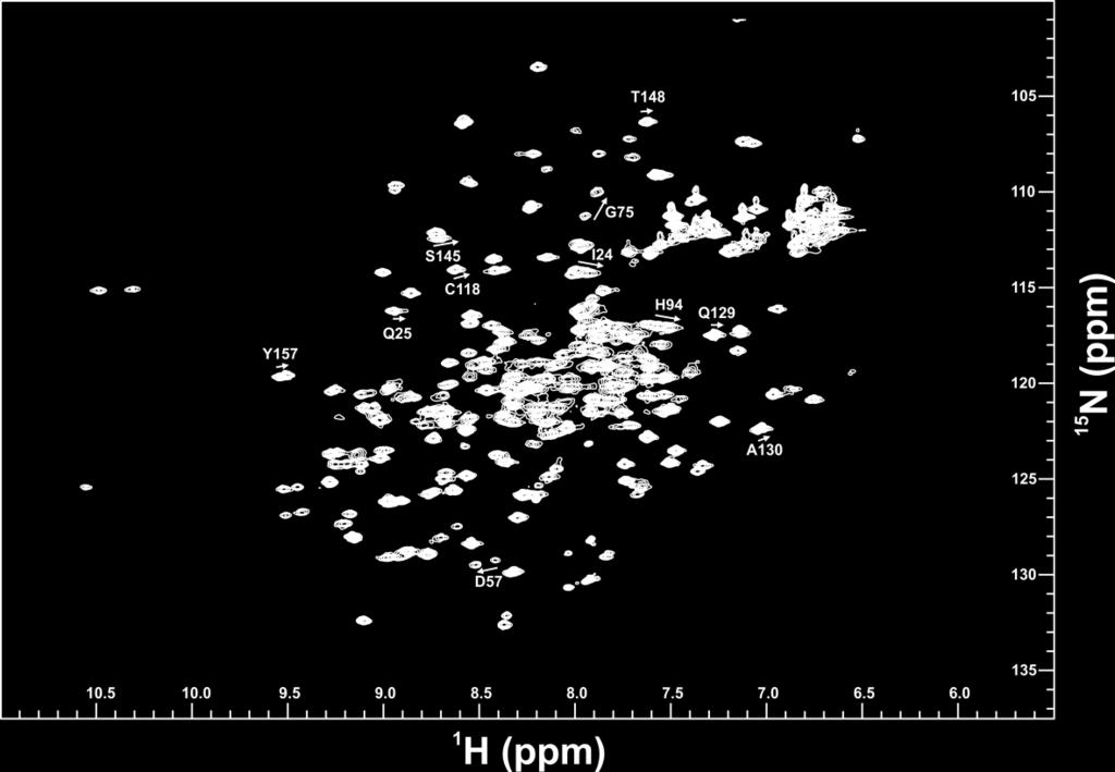
1 Supporting Information Allosteric-activation of GDP-bound Ras isoforms by bisphenol derivative plasticisers Miriam Schöpel 1, Oleksandr Shkura 1, Jana Seidel 1, Klaus Kock 1, Xueyin Zhong 1, Stefanie Löffek 2, Iris Helfrich 2, Hagen S. Bachmann 3, Jürgen Scherkenbeck 4, Christian Herrmann 1, and Raphael Stoll 1, * 1 Faculty of Chemistry and Biochemistry, Ruhr University of Bochum, Universitätsstr. 150, D Bochum, Germany 2 Skin Cancer Unit of the Dermatology Department, West German Cancer Center, University Hospital Essen, University Duisburg-Essen and the German Cancer Consortium (DKTK), D Essen, Germany 3 Institute of Pharmacogenetics, Universitätsklinikum Essen, Hufelandstr. 55, D Essen, Germany 4 Faculty of Mathematics and Natural Sciences, University of Wuppertal, Gaußstr. 20, D Wuppertal * Correspondence: raphael.stoll@ruhr-uni-bochum.de; Tel.:
2 Abbreviations 1D, one-dimensional; 2D, two-dimensional; BPs, Bisphenols; BPA, Bisphenol A; BPS, Bisphenol S; EDC, endocrine disrupting chemical; GAP, GTPase activating protein; GDI, guanine nucleotide dissociation inhibition; GDP, guanosine diphosphate; GTP, guanosine triphosphate; GEF, guanine nucleotide exchange factor; HSQC, heteronuclear single quantum coherence; KD dissociation constant; NMR, nuclear magnetic resonance; RMSD, root mean square deviation; SD, standard deviation; Sos, son of sevenless, BP, bisphenols; BPA, BPAF, BPB, BPE, BPF, BPNH2. Table S1 Bisphenols tested in this study, with varying bridging moieties at the central sp 3 -hybridised carbon atom. AFX was used to characterise the binding of one phenolic ring to K-Ras4B. ND stands for not determined. Structural formula Bisphenol CAS Systematic name (IUPAC) Comment A ,2-Bis(4-hydroxyphenyl) propane K D =0.6 ± 0.2 mm AF ,2-Bis(4-hydroxyphenyl) hexafluoropropane K D =0.4 ± 0.1 mm AP ,1-Bis(4-hydroxyphenyl)-1- phenyl-ethane insoluble 2
3 B ,2-Bis(4- hydroxyphenyl)butane K D =3.6 ± 0.7 mm BP Bis-(4-hydroxyphenyl) diphenylmethane insoluble C Bis(4-hydroxyphenyl)-2,2- dichlorethylene denaturation of protein E ,1-Bis(4- hydroxyphenyl)ethane K D =7 ± 0.7 mm F Bis(4- hydroxyphenyl)methane K D =14 ± 2 mm FL ,9-Bis(4- hydroxyphenyl)fluorene insoluble M ,3-Bis(2-(4-hydroxyphenyl)- 2-propyl)benzene denaturation of protein P ,4-Bis(2-(4-hydroxyphenyl)- 2-propyl)benzene denaturation of protein 3
4 S Bis(4-hydroxyphenyl)sulfone K D =6 ± 0.7 mm Z ,1-Bis(4-hydroxyphenyl)- cyclohexane denaturation of protein NH ,1-Bis(4-hydroxyphenyl)- cyclohexane K D =0.4 ± 0.1 mm 4
 were chosen in order to calculate the K D value that yielded 347 ± 5 µm.")
5 Supporting Figures Fig. S1 Supporting Figure 1. On the left hand side, the chemical shift differences (in ppm.) are plotted against the ligand concentration (in mm). For the binding pocket, six representative residues (L6, V9, Y40, L56, T74 and G75) were chosen in order to calculate the K D value that yielded 347 ± 5 µm. In the right hand panel, a HADDOCK-based model is shown, in which the amino acids with a weighted chemical shift above once the standard deviation (SD) are annotated in orange and those with a shift with a value twice the SD in red. The BPAF and GDP molecules are represented in green and blue, respectively. 5
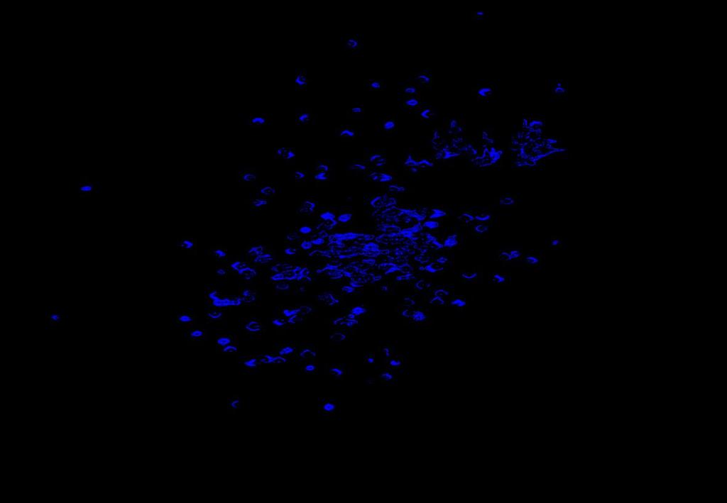
6 Fig. S2 Supporting Figure 2. Overall view of the NMR chemical shift perturbation at 600 MHz and 298 K observed in 2D 1 H- 15 N HSQC spectra of H-Ras, K-Ras4B, N- Ras, and Rap-1A bound to GDP upon titration with Bisphenol AF, ranging from the black reference to a ratio of 1:25, shown in magenta. Rap-1A did not show significant binding at all. 6


7 Fig. S3 Supporting Figure 3. NMR-based competitive titration of 15 N-enriched GDPbound K-Ras4B with 14 N SOS cat and BPAF at 600 MHz and at 298 K. Different 2D 1 H- 15 N HSQC spectra of this titration are superimposed. The reference spectrum of the K-Ras4B GDP protein is shown in black. The 2D 1 H- 15 N HSQC spectrum of K-Ras4B GDP in the presence of twofold molar excess of the GEF -protein SOS cat is depicted in blue. The 2D 1 H- 15 N HSQC spectrum of a sample that contained K- Ras4B GDP, SOS c at, and BPAF in a molar ratio of 1:2:4 is shown in magenta. The recovery of backbone amide proton NMR resonances is indicated by dashed yellow circles. It is important to note that the recovered resonances of residues from the binding pocket exhibit chemical shift perturbations compared to ligand-free 2D 1 H- 15 N HSQC spectra of 15 N-enriched GDP-bound K-Ras4B that match those observed during the titration of GDP-bound K-Ras4B with BPAF only. 7
8 Fig. S4 Supporting Figure 4. 1D 19 F-Spectra at 235 MHz and at 298 K. In black, the reference BPAF in PBS at ph 7.4 is shown. In blue, the chemical shift corresponding to the protein-bound BPAF (at a molar ratio of 1:1) is presented. 8
9 Fig. S5 9
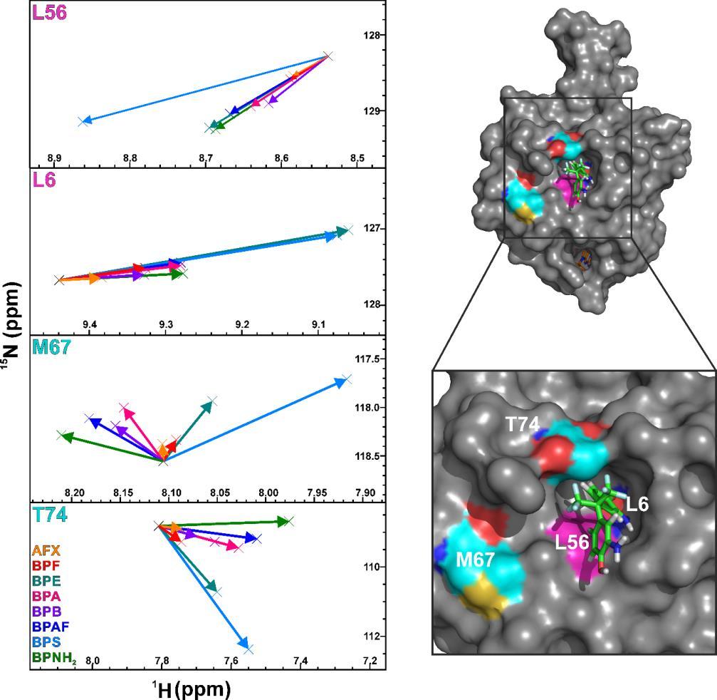
10 Supporting Figure 5 (refer to previous page). In the upper left hand panel, a vectorial shift analysis of L6, L56 (in pink) as representative residues of the binding pocket is shown based on the addition of BPNH 2 derived from the NMR chemical shift perturbation observed in 2D 1 H- 15 N HSQC spectra. The different shifts follow the same pattern, besides of L56 in BPS. In cyan, two representative amino acids from the rim of the binding pocket are shown and the vectorial shift patterns of M67 and T74 are characterised by a high dispersion in the 1 H as well as in the 15 N dimension. In the upper right hand panel, L6, L56, M67, and T74 are highlighted in pink and cyan on the protein surface of GDP-bound K-Ras4B complexed to BPNH 2. The lower part of this figure shows an overlay of 2D 1 H- 15 N HSQC spectra of K-Ras4B bound to GDP in the absence (black) and presence (blue) of Bisphenol AF recorded at 700 MHz and 298 K. Chemical shift perturbations observed are highlighted by arrows. 10
 based on the chemical shift perturbation of Y157, normalised against the shift of the GppNHp-activated Ras")
11 Fig. S6 Supporting Figure 6. Column chart of the activation levels of different bisphenols and AFX (Trifluormethylphenol) based on the chemical shift perturbation of Y157, normalised against the shift of the GppNHp-activated Ras (100%). AFX does not lead to an activation of Ras. In contrast, BPS and BPF cause an activation level of around 20 % each, whereas BPE and BPB induce activation levels of 37.3 % and 32.8 %, respectively. Remarkably, BPA generates an activation level of nearly 50 %, only surpassed by BPAF (57.5 %) and BPNH 2 (64.2 %). 11
12 Fig. S7 Supporting Figure 7. GDI assay carried out in triplicate (black, red, green; upper panel): The GDI (guanine nucleotide dissociation inhibitor) assay is carried out similar to the SOS cat assay: Instead of mantgdp, the non-hydrolysable GTP analogue mantgppnhp is used but no SOS cat is added here [17][18]. Again, the dissociation of the fluorescent nucleotide from Ras in the presence of various BPNH2 concentrations is detected by the decrease of fluorescence and the time dependence yields the k obs values. A plot of the kobs values versus BPNH2 concentration fitted by a binding isotherm yields the K D value of the BPNH 2 complex (please also refer to Figure 4).This assay reveals a KD value of 0.34±0.02 mm for BPNH2. MTT cytotoxicity assay (lower four panels): The IC50 values in Hela cells exposed for 72 hrs to BPs were determined as follows: µm (BPA), µm (BPAF), µm (BPNH 2), µm (BPS). 12
13 Acknowledgements We are grateful to the Deutsche Krebshilfe ( and ), the DFG (SFB 642, INST 213/757-1 FUGG), and the RUB Research School Plus for generous financial support. 13
Supplemental data for
 Supplemental data for A Real-Time Guanine Nucleotide Exchange Assay using NMR: Activation of RhoA by PDZ- RhoGEF. Geneviève M.C. Gasmi-Seabrook 1,3, Christopher B. Marshall 1,3, Melissa Cheung 1,3, Bryan
of the Guanine Nucleotide Exchange Factor FARP2
 Structure, Volume 21 Supplemental Information Structural Basis for Autoinhibition of the Guanine Nucleotide Exchange Factor FARP2 Xiaojing He, Yi-Chun Kuo, Tyler J. Rosche, and Xuewu Zhang Inventory of
Supplementary Information. The protease GtgE from Salmonella exclusively targets. inactive Rab GTPases
 Supplementary Information The protease GtgE from Salmonella exclusively targets inactive Rab GTPases Table of Contents Supplementary Figures... 2 Supplementary Figure 1... 2 Supplementary Figure 2... 3
Acta Crystallographica Section D
 Supporting information Acta Crystallographica Section D Volume 70 (2014) Supporting information for article: Structural basis of the heterodimerization of the MST and RASSF SARAH domains in the Hippo signalling
NMR study of complexes between low molecular mass inhibitors and the West Nile virus NS2B-NS3 protease
 University of Wollongong Research Online Faculty of Science - Papers (Archive) Faculty of Science, Medicine and Health 2009 NMR study of complexes between low molecular mass inhibitors and the West Nile
Molecular Mechanism for Conformational Dynamics of Ras GTP Elucidated from In-Situ Structural Transition in Crystal
 Molecular Mechanism for Conformational Dynamics of Ras GTP Elucidated from In-Situ Structural Transition in Crystal Shigeyuki Matsumoto, Nao Miyano, Seiki Baba, Jingling Liao, Takashi Kawamura, Chiemi
Supplementary Information. Overlap between folding and functional energy landscapes for. adenylate kinase conformational change
 Supplementary Information Overlap between folding and functional energy landscapes for adenylate kinase conformational change by Ulrika Olsson & Magnus Wolf-Watz Contents: 1. Supplementary Note 2. Supplementary
Supporting Information
 Supporting Information Allosteric communication disrupted by small molecule binding to the Imidazole glycerol phosphate synthase protein-protein interface. Ivan Rivalta*,#, George P. Lisi #, Ning-Shiuan
17. Biomolecular Interaction
 17. Biomolecular Interaction Methods for characterizing biomolecular interactions Sequence-specific DNA binding ligands Molecular mechanisms of drug action and drug resistance In silico compound design
Sensitive NMR Approach for Determining the Binding Mode of Tightly Binding Ligand Molecules to Protein Targets
 Supporting information Sensitive NMR Approach for Determining the Binding Mode of Tightly Binding Ligand Molecules to Protein Targets Wan-Na Chen, Christoph Nitsche, Kala Bharath Pilla, Bim Graham, Thomas
Nature Structural & Molecular Biology: doi: /nsmb Supplementary Figure 1
 Supplementary Figure 1 Identification of the ScDcp2 minimal region interacting with both ScDcp1 and the ScEdc3 LSm domain. Pull-down experiment of untagged ScEdc3 LSm with various ScDcp1-Dcp2-His 6 fragments.
Chapter 6. The interaction of Src SH2 with the focal adhesion kinase catalytic domain studied by NMR
 The interaction of Src SH2 with the focal adhesion kinase catalytic domain studied by NMR 103 Abstract The interaction of the Src SH2 domain with the catalytic domain of FAK, including the Y397 SH2 domain
NMR in Medicine and Biology
 NMR in Medicine and Biology http://en.wikipedia.org/wiki/nmr_spectroscopy MRI- Magnetic Resonance Imaging (water) In-vivo spectroscopy (metabolites) Solid-state t NMR (large structures) t Solution NMR
Activation of a receptor. Assembly of the complex
 Activation of a receptor ligand inactive, monomeric active, dimeric When activated by growth factor binding, the growth factor receptor tyrosine kinase phosphorylates the neighboring receptor. Assembly
Table S1. Primers used for the constructions of recombinant GAL1 and λ5 mutants. GAL1-E74A ccgagcagcgggcggctgtctttcc ggaaagacagccgcccgctgctcgg
 SUPPLEMENTAL DATA Table S1. Primers used for the constructions of recombinant GAL1 and λ5 mutants Sense primer (5 to 3 ) Anti-sense primer (5 to 3 ) GAL1 mutants GAL1-E74A ccgagcagcgggcggctgtctttcc ggaaagacagccgcccgctgctcgg
Protein dynamics from NMR Relaxation data
 Protein dynamics from NMR Relaxation data Clubb 3/15/17 (S f2 ) ( e ) Nitrogen-15 relaxation ZZ-exchange R 1 = 1/T 1 Longitudinal relaxation (decay back to z-axis) R 2 = 1/T 2 Spin-spin relaxation (dephasing
The Fic protein Doc uses an inverted substrate to phosphorylate and. inactivate EF-Tu
 The Fic protein Doc uses an inverted substrate to phosphorylate and inactivate EF-Tu Daniel Castro-Roa 1, Abel Garcia-Pino 2,3 *, Steven De Gieter 2,3, Nico A.J. van Nuland 2,3, Remy Loris 2,3, Nikolay
Supporting Protocol This protocol describes the construction and the force-field parameters of the non-standard residue for the Ag + -site using CNS
 Supporting Protocol This protocol describes the construction and the force-field parameters of the non-standard residue for the Ag + -site using CNS CNS input file generatemetal.inp: remarks file generate/generatemetal.inp
Supporting Information
 Supporting Information Reaction Mechanism of Adenylyltransferase DrrA from Legionella pneumophila Elucidated by Time-Resolved Fourier Transform Infrared Spectroscopy Konstantin Gavriljuk, Jonas Schartner,
SUPPLEMENTARY INFORMATION
 Figure S1. Secondary structure of CAP (in the camp 2 -bound state) 10. α-helices are shown as cylinders and β- strands as arrows. Labeling of secondary structure is indicated. CDB, DBD and the hinge are
Slow conformational dynamics of the guanine nucleotide-binding protein Ras complexed with the GTP analogue GTPcS
 Slow conformational dynamics of the guanine nucleotide-binding protein Ras complexed with the GTP analogue GTPcS Michael Spoerner 1, Andrea Nuehs 1, Christian Herrmann 2, Guido Steiner 1 and Hans Robert
Supplementary Materials for
 advances.sciencemag.org/cgi/content/full/3/4/e1600663/dc1 Supplementary Materials for A dynamic hydrophobic core orchestrates allostery in protein kinases Jonggul Kim, Lalima G. Ahuja, Fa-An Chao, Youlin
Supporting Information
 Migration of Parabens, Bisphenols, Benzophenone-Type UV filters, Triclosan, and Triclocarban from Teethers and Its Implications for Infant Exposure Alexandros G. Asimakopoulos a,, Madhavan Elangovan a,,
1) NMR is a method of chemical analysis. (Who uses NMR in this way?) 2) NMR is used as a method for medical imaging. (called MRI )
 NMR is a method of chemical analysis. (Who uses NMR in this way?) 2) NMR is used as a method for medical imaging. (called MRI )") Uses of NMR: 1) NMR is a method of chemical analysis. (Who uses NMR in this way?) 2) NMR is used as a method for medical imaging. (called MRI ) 3) NMR is used as a method for determining of protein, DNA,
Uses of NMR: 1) NMR is a method of chemical analysis. (Who uses NMR in this way?) 2) NMR is used as a method for medical imaging. (called MRI ) 3) NMR is used as a method for determining of protein, DNA,
SUPPLEMENTARY INFORMATION
 5 N 4 8 20 22 24 2 28 4 8 20 22 24 2 28 a b 0 9 8 7 H c (kda) 95 0 57 4 28 2 5.5 Precipitate before NMR expt. Supernatant before NMR expt. Precipitate after hrs NMR expt. Supernatant after hrs NMR expt.
SUPPLEMENTARY INFORMATION
 SUPPLEMENTARY INFORMATION doi:10.1038/nature11524 Supplementary discussion Functional analysis of the sugar porter family (SP) signature motifs. As seen in Fig. 5c, single point mutation of the conserved
Supplementary Materials for
 advances.sciencemag.org/cgi/content/full/4/1/eaau413/dc1 Supplementary Materials for Structure and dynamics conspire in the evolution of affinity between intrinsically disordered proteins Per Jemth*, Elin
Supporting Information
 Supporting Information Micelle-Triggered b-hairpin to a-helix Transition in a 14-Residue Peptide from a Choline-Binding Repeat of the Pneumococcal Autolysin LytA HØctor Zamora-Carreras, [a] Beatriz Maestro,
Biochemistry 530 NMR Theory and Practice
 Biochemistry 530 NMR Theory and Practice Gabriele Varani Department of Biochemistry and Department of Chemistry University of Washington 1D spectra contain structural information.. but is hard to extract:
Nature Structural & Molecular Biology: doi: /nsmb Supplementary Figure 1
 Supplementary Figure 1 Crystallization. a, Crystallization constructs of the ET B receptor are shown, with all of the modifications to the human wild-type the ET B receptor indicated. Residues interacting
1. Sodium-Nucleotide Complexes
 Investigating the Interaction of Metal Ions with Nucleotides and Macromolecular RNA Using Solid- State 23 Na, 25 Mg and 59 Co NMR for Direct Observation of the Metals 1. Sodium-Nucleotide Complexes Metals
TITAN: Two-dimensional lineshape analysis
 TITAN: Two-dimensional lineshape analysis Chris Waudby Christodoulou Group c.waudby@ucl.ac.uk Andres Ramos Lisa Cabrita John Christodoulou Inhibition of fatty acid synthesis for treatment of tularemia
Carbazole Derivatives Binding to c-kit G-quadruplex DNA
 Supplementary Materials Carbazole Derivatives Binding to c-kit G-quadruplex DNA Agata Głuszyńska 1, *, Bernard Juskowiak 1, Martyna Kuta-Siejkowska 2, Marcin Hoffmann 2 and Shozeb Haider 3 1 Laboratory
Chemical Exchange and Ligand Binding
 Chemical Exchange and Ligand Binding NMR time scale Fast exchange for binding constants Slow exchange for tight binding Single vs. multiple binding mode Calcium binding process of calcium binding proteins
You are advised to spend an equal amount of time on each question.
 UNIVERSITY OF EAST ANGLIA School of Chemistry Main Series UG Examination 2015-16 BIOPHYSICAL CHEMISTRY CHE-5601Y Time allowed: 2 hours Answer THREE questions. You are advised to spend an equal amount of
Structural basis for catalytically restrictive dynamics of a high-energy enzyme state
 Supplementary Material Structural basis for catalytically restrictive dynamics of a high-energy enzyme state Michael Kovermann, Jörgen Ådén, Christin Grundström, A. Elisabeth Sauer-Eriksson, Uwe H. Sauer
NMR Resonance Assignment Assisted by Mass Spectrometry
 NMR Resonance Assignment Assisted by Mass Spectrometry This lecture talked about a NMR resonance assignment assisted by mass spectrometry [1, 2]. 1 Motivation Nuclear magnetic resonance (NMR) provides
References on Kinetics and Mechanisms
 References on Kinetics and Mechanisms Excellent reference for all aspects of enzyme kinetics including important elements of Metabolic Control Analysis of relevance to systems analysis of enzyme function
Effects of Chemical Exchange on NMR Spectra
 Effects of Chemical Exchange on NMR Spectra Chemical exchange refers to any process in which a nucleus exchanges between two or more environments in which its NMR parameters (e.g. chemical shift, scalar
Introduction solution NMR
 2 NMR journey Introduction solution NMR Alexandre Bonvin Bijvoet Center for Biomolecular Research with thanks to Dr. Klaartje Houben EMBO Global Exchange course, IHEP, Beijing April 28 - May 5, 20 3 Topics
Structural Basis of Multivalent Binding to Wheat Germ Agglutinin
 Structural Basis of Multivalent Binding to Wheat Germ Agglutinin David Schwefel, Caroline Maierhofer, Johannes G. Beck, Sonja Seeberger, Kay Diederichs, Heiko M. Möller,*, Wolfram Welte,*, and Valentin
Supporting Information
 S-1 Supporting Information Flaviviral protease inhibitors identied by fragment-based library docking into a structure generated by molecular dynamics Dariusz Ekonomiuk a, Xun-Cheng Su b, Kiyoshi Ozawa
Timescales of Protein Dynamics
 Timescales of Protein Dynamics From Henzler-Wildman and Kern, Nature 2007 Dynamics from NMR Show spies Amide Nitrogen Spies Report On Conformational Dynamics Amide Hydrogen Transverse Relaxation Ensemble
Supplementary Figures
 1 Supplementary Figures Supplementary Figure 1 Type I FGFR1 inhibitors (a) Chemical structures of a pyrazolylaminopyrimidine inhibitor (henceforth referred to as PAPI; PDB-code of the FGFR1-PAPI complex:
Analysis of correlated mutations in Ras G-domain
 www.bioinformation.net Volume 13(6) Hypothesis Analysis of correlated mutations in Ras G-domain Ekta Pathak * Bioinformatics Department, MMV, Banaras Hindu University. Ekta Pathak - E-mail: ektavpathak@gmail.com;
to all three phosphate groups P, P Q, P of ATP, Mg +, Ca +, p
 Volume 4 Number 2 February 1977 Nucleic Acids Research A 31 P - NMR study of the interaction of M^2" 1 " ions with nucleoside diphosphates S.Tran-Dinh and J.M.Neumann Departement de Biologie - Centre d'etudes
Table S1. Overview of used PDZK1 constructs and their binding affinities to peptides. Related to figure 1.
 Table S1. Overview of used PDZK1 constructs and their binding affinities to peptides. Related to figure 1. PDZK1 constru cts Amino acids MW [kda] KD [μm] PEPT2-CT- FITC KD [μm] NHE3-CT- FITC KD [μm] PDZK1-CT-
Timescales of Protein Dynamics
 Timescales of Protein Dynamics From Henzler-Wildman and Kern, Nature 2007 Summary of 1D Experiment time domain data Fourier Transform (FT) frequency domain data or Transverse Relaxation Ensemble of Nuclear
SUPPLEMENTARY INFORMATION
 Parallel Allostery by camp and PDE Coordinates Activation and Termination Phases in camp Signaling Srinath Krishnamurthy, 1 Nikhil Kumar Tulsian, 1 Arun Chandramohan, 1 and Ganesh S. Anand 1, * 1 Department
Investigation of Cation Binding and Sensing by new Crown Ether core substituted Naphthalene Diimide systems
 Electronic Supplementary Material (ESI) for New Journal of Chemistry. This journal is The Royal Society of Chemistry and the Centre National de la Recherche Scientifique 218 Supplementary Material Investigation
Enhancing hydrogen production of microalgae by redirecting electrons from photosystem I to hydrogenase
 Electronic Supplementary Material (ESI) for Energy & Environmental Science. This journal is The Royal Society of Chemistry 2014 Supplementary information for Enhancing hydrogen production of microalgae
Supplemental Information
 Supplemental Information Combinatorial Readout of Unmodified H3R2 and Acetylated H3K14 by the Tandem PHD Finger of MOZ Reveals a Regulatory Mechanism for HOXA9 Transcription Yu Qiu 1, Lei Liu 1, Chen Zhao
Mechanistic insight into inhibition of two-component system signaling
 Supporting Information Mechanistic insight into inhibition of two-component system signaling Samson Francis, a Kaelyn E. Wilke, a Douglas E. Brown a and Erin E. Carlson a,b* a Department of Chemistry,
Protein-protein interactions (PPIs) via NMR. Paola Turano
 via NMR. Paola Turano") Protein-protein interactions (PPIs) via NMR Paola Turano turano@cerm.unifi.it The magnetic field at the The chemical shift nucleus (the effective field) is generally less than the applied field by a fraction
Protein-protein interactions (PPIs) via NMR Paola Turano turano@cerm.unifi.it The magnetic field at the The chemical shift nucleus (the effective field) is generally less than the applied field by a fraction
SUPPLEMENTARY ONLINE DATA
 SUPPLEMENTARY ONLINE DATA Secreted Isoform of Human Lynx1 (SLURP-2): Spatial Structure and Pharmacology of Interaction with Different Types of Acetylcholine Receptors E.N. Lyukmanova 1,2,*, M.A. Shulepko
Protein-protein interactions (PPIs) via NMR. Paola Turano
 via NMR. Paola Turano") Protein-protein interactions (PPIs) via NMR Paola Turano turano@cerm.unifi.it The magnetic field at the The chemical shift nucleus (the effective field) is generally less than the applied field by a fraction
Protein-protein interactions (PPIs) via NMR Paola Turano turano@cerm.unifi.it The magnetic field at the The chemical shift nucleus (the effective field) is generally less than the applied field by a fraction
Presenter: She Zhang
 Presenter: She Zhang Introduction Dr. David Baker Introduction Why design proteins de novo? It is not clear how non-covalent interactions favor one specific native structure over many other non-native
Name: BCMB/CHEM 8190, BIOMOLECULAR NMR FINAL EXAM-5/5/10
 Name: BCMB/CHEM 8190, BIOMOLECULAR NMR FINAL EXAM-5/5/10 Instructions: This is an open book, limited time, exam. You may use notes you have from class and any text book you find useful. You may also use
Electronic Supplementary Information (ESI) for New Journal of Chemistry
 for New Journal of Chemistry") Electronic Supplementary Material (ESI) for New Journal of Chemistry. This journal is The Royal Society of Chemistry and the Centre National de la Recherche Scientifique 217 Electronic Supplementary Information
Electronic Supplementary Material (ESI) for New Journal of Chemistry. This journal is The Royal Society of Chemistry and the Centre National de la Recherche Scientifique 217 Electronic Supplementary Information
Supplemental Information. Molecular Basis of Spectral Diversity. in Near-Infrared Phytochrome-Based. Fluorescent Proteins
 Chemistry & Biology, Volume 22 Supplemental Information Molecular Basis of Spectral Diversity in Near-Infrared Phytochrome-Based Fluorescent Proteins Daria M. Shcherbakova, Mikhail Baloban, Sergei Pletnev,
Using NMR to study Macromolecular Interactions. John Gross, BP204A UCSF. Nov 27, 2017
 Using NMR to study Macromolecular Interactions John Gross, BP204A UCSF Nov 27, 2017 Outline Review of basic NMR experiment Multidimensional NMR Monitoring ligand binding Structure Determination Review:
Targeting protein-protein interactions: A hot topic in drug discovery
 Michal Kamenicky; Maria Bräuer; Katrin Volk; Kamil Ödner; Christian Klein; Norbert Müller Targeting protein-protein interactions: A hot topic in drug discovery 104 Biomedizin Innovativ patientinnenfokussierte,
Nature Structural & Molecular Biology: doi: /nsmb Supplementary Figure 1
 Supplementary Figure 1 Resonance assignment and NMR spectra for hairpin and duplex A 6 constructs. (a) 2D HSQC spectra of hairpin construct (hp-a 6 -RNA) with labeled assignments. (b) 2D HSQC or SOFAST-HMQC
I690/B680 Structural Bioinformatics Spring Protein Structure Determination by NMR Spectroscopy
 I690/B680 Structural Bioinformatics Spring 2006 Protein Structure Determination by NMR Spectroscopy Suggested Reading (1) Van Holde, Johnson, Ho. Principles of Physical Biochemistry, 2 nd Ed., Prentice
SUPPLEMENTARY INFORMATION
 Supplementary Results DNA binding property of the SRA domain was examined by an electrophoresis mobility shift assay (EMSA) using synthesized 12-bp oligonucleotide duplexes containing unmodified, hemi-methylated,
NB-DNJ/GCase-pH 7.4 NB-DNJ+/GCase-pH 7.4 NB-DNJ+/GCase-pH 4.5
 SUPPLEMENTARY TABLES Suppl. Table 1. Protonation states at ph 7.4 and 4.5. Protonation states of titratable residues in GCase at ph 7.4 and 4.5. Histidine: HID, H at δ-nitrogen; HIE, H at ε-nitrogen; HIP,
NMR in Structural Biology
 NMR in Structural Biology Exercise session 2 1. a. List 3 NMR observables that report on structure. b. Also indicate whether the information they give is short/medium or long-range, or perhaps all three?
RhoGAP assay kit (Cat. # BK105)
") RhoGAP assay kit (Cat. # BK105) The Ras superfamily of small GTPases (such as Ras, Rho, Rab, Arf and Ran proteins) serve as binary switches cycling between a GDP-bound OFF state and a GTP-bound ON state,
RhoGAP assay kit (Cat. # BK105) The Ras superfamily of small GTPases (such as Ras, Rho, Rab, Arf and Ran proteins) serve as binary switches cycling between a GDP-bound OFF state and a GTP-bound ON state,
A prevalent intraresidue hydrogen bond stabilizes proteins
 Supplementary Information A prevalent intraresidue hydrogen bond stabilizes proteins Robert W. Newberry 1 & Ronald T. Raines 1,2 * 1 Department of Chemistry and 2 Department of Biochemistry, University
Supplementary Figure 1. Biochemical and sequence alignment analyses the
 Supplementary Figure 1. Biochemical and sequence alignment analyses the interaction of OPTN and TBK1. (a) Analytical gel filtration chromatography analysis of the interaction between TBK1 CTD and OPTN(1-119).
Nature Structural & Molecular Biology: doi: /nsmb.3194
 Supplementary Figure 1 Mass spectrometry and solution NMR data for -syn samples used in this study. (a) Matrix-assisted laser-desorption and ionization time-of-flight (MALDI-TOF) mass spectrum of uniformly-
Supporting information for
 Supporting information for Rewiring multi-domain protein switches: transforming a fluorescent Zn 2+ -sensor into a light-responsive Zn 2+ binding protein Stijn J.A. Aper and Maarten Merkx Laboratory of
NMR methods for the determination of protein ligand dissociation constants
 Progress in Nuclear Magnetic Resonance Spectroscopy 51 (2007) 219 242 www.elsevier.com/locate/pnmrs NMR methods for the determination of protein ligand dissociation constants Lee Fielding * Organon BioSciences,
Supporting Information
 Supporting Information Li et al. 10.1073/pnas.1314303110 SI Text Preparation of NMR Samples. Mutagenesis, protein expression, and purification were performed as previously described (1), except that the
Characterization of Reversible Kinase Inhibitors using Microfluidic Mobility-Shift Assays
 Application Note 211 Characterization of Reversible Kinase Inhibitors using Microfluidic Mobility-Shift Assays Introduction Current drug discovery efforts typically focus on developing small molecule inhibitors
LineShapeKin NMR Line Shape Analysis Software for Studies of Protein-Ligand Interaction Kinetics
 LineShapeKin NMR Line Shape Analysis Software for Studies of Protein-Ligand Interaction Kinetics http://lineshapekin.net Spectral intensity Evgenii L. Kovrigin Department of Biochemistry, Medical College
Cryo-EM data collection, refinement and validation statistics
 1 Table S1 Cryo-EM data collection, refinement and validation statistics Data collection and processing CPSF-160 WDR33 (EMDB-7114) (PDB 6BM0) CPSF-160 WDR33 (EMDB-7113) (PDB 6BLY) CPSF-160 WDR33 CPSF-30
Determining Chemical Structures with NMR Spectroscopy the ADEQUATEAD Experiment
 Determining Chemical Structures with NMR Spectroscopy the ADEQUATEAD Experiment Application Note Author Paul A. Keifer, Ph.D. NMR Applications Scientist Agilent Technologies, Inc. Santa Clara, CA USA Abstract
Supporting Information
 Supporting Information Arai et al. 10.1073/pnas.15179911 SI Text Protein Expression and Purification. Myb3 (mouse, residues 84 315) was expressed in Escherichia coli as a fusion with the B1 domain of protein
CELB40060 Membrane Trafficking in Animal Cells. Prof. Jeremy C. Simpson. Lecture 2 COPII and export from the ER
 CELB40060 Membrane Trafficking in Animal Cells Prof. Jeremy C. Simpson Lecture 2 COPII and export from the ER Today s lecture... The COPII coat - localisation and subunits Formation of the COPII coat at
BMB/Bi/Ch 173 Winter 2018
 BMB/Bi/Ch 173 Winter 2018 Homework Set 8.1 (100 Points) Assigned 2-27-18, due 3-6-18 by 10:30 a.m. TA: Rachael Kuintzle. Office hours: SFL 220, Friday 3/2 4:00-5:00pm and SFL 229, Monday 3/5 4:00-5:30pm.
A conserved P-loop anchor limits the structural dynamics that mediate. nucleotide dissociation in EF-Tu.
 Supplemental Material for A conserved P-loop anchor limits the structural dynamics that mediate nucleotide dissociation in EF-Tu. Evan Mercier 1,2, Dylan Girodat 1, and Hans-Joachim Wieden 1 * 1 Alberta
Supplemental Information. The Mitochondrial Fission Receptor MiD51. Requires ADP as a Cofactor
 Structure, Volume 22 Supplemental Information The Mitochondrial Fission Receptor MiD51 Requires ADP as a Cofactor Oliver C. Losón, Raymond Liu, Michael E. Rome, Shuxia Meng, Jens T. Kaiser, Shu-ou Shan,
Supporting Information
 Protein-Observed Fluorine NMR is a Complementary Ligand Discovery Method to 1 H CPMG Ligand- Observed NMR. Andrew K. Urick, 1,2 Luis Pablo Calle, 3 Juan F. Espinosa, 3 Haitao Hu, 2 * William C. K. Pomerantz
Supporting Information. Supramolecular Host-Guest Chemistry-based Folate/Riboflavin Functionalization and. Cancer Cell Labeling of Nanoparticle
 Supporting Information Supramolecular Host-Guest Chemistry-based Folate/Riboflavin Functionalization and Cancer Cell Labeling of Nanoparticle Suman Pal, 1 Chumki Dalal 1 and Nikhil R. Jana 1, * 1 Centre
CD Basis Set of Spectra that is used is that derived from comparing the spectra of globular proteins whose secondary structures are known from X-ray
 CD Basis Set of Spectra that is used is that derived from comparing the spectra of globular proteins whose secondary structures are known from X-ray crystallography An example of the use of CD Modeling
Identifying Interaction Hot Spots with SuperStar
 Identifying Interaction Hot Spots with SuperStar Version 1.0 November 2017 Table of Contents Identifying Interaction Hot Spots with SuperStar... 2 Case Study... 3 Introduction... 3 Generate SuperStar Maps
Efficient Interaction Between Two GTPases. Allows the Chloroplast SRP Pathway to Bypass. the Requirement for an SRP RNA
 9 Chapter 1: Efficient Interaction Between Two GTPases Allows the Chloroplast SRP Pathway to Bypass the Requirement for an SRP RNA A version of this chapter has been published as: Jaru-Ampornpan, P., Chandrasekar,
Supporting Information
 Supporting Information Boehr et al. 10.1073/pnas.0914163107 SI Text Materials and Methods. R 2 relaxation dispersion experiments. 15 NR 2 relaxation dispersion data measured at 1 H Larmor frequencies of
Interpreting and evaluating biological NMR in the literature. Worksheet 1
 Interpreting and evaluating biological NMR in the literature Worksheet 1 1D NMR spectra Application of RF pulses of specified lengths and frequencies can make certain nuclei detectable We can selectively
Serine-7 but not serine-5 phosphorylation primes RNA polymerase II CTD for P-TEFb recognition
 Supplementary Information to Serine-7 but not serine-5 phosphorylation primes RNA polymerase II CTD for P-TEFb recognition Nadine Czudnochowski 1,2, *, Christian A. Bösken 1, * & Matthias Geyer 1 1 Max-Planck-Institut
Longitudinal-relaxation enhanced fast-pulsing techniques: New tools for biomolecular NMR spectroscopy
 Longitudinal-relaxation enhanced fast-pulsing techniques: New tools for biomolecular NMR spectroscopy Bernhard Brutscher Laboratoire de Résonance Magnétique Nucléaire Institut de Biologie Structurale -
Supplementary Figure 1 Crystal packing of ClR and electron density maps. Crystal packing of type A crystal (a) and type B crystal (b).
 and type B crystal (b).") Supplementary Figure 1 Crystal packing of ClR and electron density maps. Crystal packing of type A crystal (a) and type B crystal (b). Crystal contacts at B-C loop are magnified and stereo view of A-weighted
Supplementary Figure 1 Crystal packing of ClR and electron density maps. Crystal packing of type A crystal (a) and type B crystal (b). Crystal contacts at B-C loop are magnified and stereo view of A-weighted
PROTEIN NMR SPECTROSCOPY
 List of Figures List of Tables xvii xxvi 1. NMR SPECTROSCOPY 1 1.1 Introduction to NMR Spectroscopy 2 1.2 One Dimensional NMR Spectroscopy 3 1.2.1 Classical Description of NMR Spectroscopy 3 1.2.2 Nuclear
Electronic Supplementary Information
 Electronic Supplementary Material (ESI) for Journal of Materials Chemistry B. This journal is The Royal Society of Chemistry 2017 Electronic Supplementary Information Rapid detection of intracellular Cys
LS1a Fall 2014 Problem Set #2 Due Monday 10/6 at 6 pm in the drop boxes on the Science Center 2 nd Floor
 LS1a Fall 2014 Problem Set #2 Due Monday 10/6 at 6 pm in the drop boxes on the Science Center 2 nd Floor Note: Adequate space is given for each answer. Questions that require a brief explanation should
Molecular Modeling lecture 2
 Molecular Modeling 2018 -- lecture 2 Topics 1. Secondary structure 3. Sequence similarity and homology 2. Secondary structure prediction 4. Where do protein structures come from? X-ray crystallography
Supporting Information
 Supporting Information Decoding Allosteric Networks in Biocatalysts: Rational Approach to Therapies and Biotechnologies Johannes T. Cramer 1,2, Jana I. Führing 1, Petra Baruch 2, Christian Brütting 3,
Supporting Information
 Supporting Information Copyright Wiley-VCH Verlag GmbH & Co. KGaA, 69451 Weinheim, 2014 An Ensemble of Rapidly Interconverting Orientations in Electrostatic Protein Peptide Complexes Characterized by NMR
Supplementary Information. Synthesis and biological activity of a CXCR4-targeting bis(cyclam) lipid
 lipid") Electronic Supplementary Material (ESI) for Organic & Biomolecular Chemistry. This journal is The Royal Society of Chemistry 2018 Supplementary Information Synthesis and biological activity of a CXCR4-targeting
Electronic Supplementary Material (ESI) for Organic & Biomolecular Chemistry. This journal is The Royal Society of Chemistry 2018 Supplementary Information Synthesis and biological activity of a CXCR4-targeting