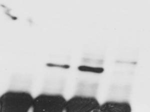
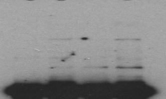
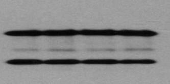
TNFα 18hr. Control. CHX 18hr. TNFα+ CHX 18hr. TNFα: 18 18hr (KDa) PARP. Cleaved. Cleaved. Cleaved. Caspase3. Pellino3 shrna. Control shrna.
|
|
|
- Jasmin Bridges
- 5 years ago
- Views:
Transcription


 for 18h. (a) Cells were photographed (bar μm).")

 or presence of human TNF (40 ng/ml) and/or cycloheximide")

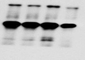
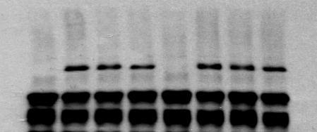
1 Survival ( %) a. TNFα 18hr b. Control sirna Pellino3 sirna TNFα: 18 18hr c. Control shrna Pellino3 shrna Caspase3 Actin Control d. Control shrna Pellino3 shrna *** CHX 18hr 40 TNFα+ CHX 18hr f. Control shrna 0 CHX TNF + CHX 18 hour Pellino3 shrna Pellino3 β-actin Control shrna Pellino3 shrna TNFα min I Bα p-p38 P e. p-jnk JNK TNFα+ CHX: 18 18hr Caspase-3 Caspase-8 Actin 18 Actin Supplementary Figure S1.:Knockdown of Pellino3 in Hela and 1321N1 astrocytoma leads to increased sensitization to TNF-induced apoptosis. (a, b) Hela cells were transfected with control or Pellino3-specific sirna and then treated with human TNF (40 ng/ml) for 18h. (a) Cells were photographed (bar μm). (b) Cell lysates were probed by immunoblotting for expression levels of, cleaved caspase 3 and b-actin. (c, d) 1321N1 astrocytoma cells were infected with lentivirus control or Pellino3-shRNA and treated in the absence (control) or presence of human TNF (40 ng/ml) and/or cycloheximide (CHX) (10 μg/ml). (c) Cells were photographed (bar μm). Immunoblot analysis demonstrating efficacy of Pellino3 protein knockdown is shown. (d) Cells were analyzed for cell viability using MTT assays (mean +/- SEM of 3 independent experiments) ***p<0.001 (two-way ANOVA). (e) Cell lysates were immunblotted for, caspase 3, caspase 8 and b-actin. and caspase 8 are indicated by arrows. (f) Cell lysates blotted for phosphorylated JNK (p-jnk),p38 (p-p38) and total I Bα, JNK and P38


 for 18 h.")
 Cells were stained by TUNEL and Hoechst and subjected to confocal microscopy. Bar μm.")
, CHX (10 μg/ml) and ZVAD ( mm).")
2 Input Input sirnap3 sirnac a. b. TNFα18hr TNFα: 18 18hr Caspase-3 Actin Pellino3 TUNEL Hoechst Merge c. IP: sirnacontrol sirnapellino3 Z-VAD-FMK : TNFα + CHX : hr d. IP: sirnacontrol sirnapellino3 Z-VAD-FMK : TNFα + CHX : hr Rip1 Caspase-8 Nonspecific IgGL Rip1 Caspase-8 Supplementary Figure S2: Knockdown of Pellino3 in HEK293 cells leads to increased sensitization to TNF-induced apoptosis. (a, b) HEK293 cells were transfected with control or Pellino3-specific sirna and treated with TNF ( ng/ml) for 18 h. (a) Cell lysates were immunoblotted for, caspase 3, Pellino3 and b-actin. (b) Cells were stained by TUNEL and Hoechst and subjected to confocal microscopy. Bar μm. (c, d) HEK293 cells were transfected with control or Pellino3-specific sirna and treated with TNF ( ng/ml), CHX (10 μg/ml) and ZVAD ( mm). Cell lysates were immunoprecipitated with an (c) anti-caspase 8 or (d) anti- antibody and blotted for coprecipitated and caspase 8. The expression levels of and caspase 8 in whole cell lysates ( input ) were also assessed.

3 Supplementary Figure S3: Schematic representation of mutant forms of Pellino3 used in reconstitution and overexpression studies.

4 In vitro K63Ub 1 IB: Ub 55 IB: UbK63only: E1: E2: Pellino3smyc: + Pellino3sFmmyc: + Pellino3sEmmyc: + Supplementary Figure S4: Mutation of the RING-like domain but not the FHA domain of Pellino3 abrogates its E3 ubiquitin ligase activity. Purified recombinant forms of myc-tagged Pellino3s, FHA domain-mutated Pellino3s(P3sFHAm) or RING-like domain-mutated Pellino3s(P3sE3m) (0.5 µg) were incubated with recombinant ubiquitin (1µg) (containing a single lysine at residue 63 (K63 Ub)) in the presence of E1 (ng) and the E2 UbcH13 (400 ng) for 2 h at 37 C. The reactions were terminated by the addition of SDS-PAGE sample buffer. Samples were subjected to PAGE and subsequently to Western immunoblotting using anti-ubiquitin and -myc antibodies.
5 a. IP: Flag b. IP: Flag Flag P3 Flag: P3ΔC-term Flag: -: Input Flag P3 Full-length P3 Δ C-term Input P3 Full-length Flag P3 Δ C-term P3 Flag: + + P3ΔC-term Flag: + + -: + + P3s Full-length P3s Δ C-term P3s Full-length P3s Δ C-term c. IP: Flag d. Flag IP: P3s Full-length Flag Input P3 Full-length P3 Δ C-term Input Flag P3 Flag: P3ΔC-term Flag: + Caspase-8 : P3 Full-length Flag P3 Δ C-term P3 Flag: + P3ΔC-term Flag: Caspase-8 : + + P3s Full-length P3s Δ C-term Supplementary Figure S5: The FHA domain of Pellino3 is sufficient to interact with but not caspase8. HEK293 cells were co-transfected with indicated combinations of constructs encoding Flag-tagged Pellino3s or its C-terminal truncation mutant (P3 ΔC-term(1-313aa)) and (a, b) myc-tagged or (c, d) myc-tagged caspase8. Such transfections also included co-expression of the caspase inhibitor CrmA to prevent caspase induced cleavage of and cell apoptosis. Cell lysates were immunoprecipitated (IP) using anti-flag (a, c) or anti-myc (b, d) antibodies followed by immunoblotting of immunopreciptates and cell lysates (input) using indicated antibodies.
6 Annexin V positive(%) Annexin V positive(%) a. Pellino3 +/+ Pellino3 -/ b. Caspase 3 FasL CHX FasL FasL+CHX β-actin c Control Pellino3 +/+ Pellino3 -/- Etoposide d. Etoposide Caspase 3 β-actin Supplementary Figure S6: Loss of Pellino3 has no effect on the pro-apoptotic effects of Fas or genotoxic stress. MEFs were isolated from Pellino3+/+ and Pellino3-/- embryos and treated with (a, b) cycloheximide (CHX) (10 μg/ml) and/or anti-fas antibody (2 µg/ml) for 12h or (c, d) etoposide (0 mm) for 24 hr. (a, c) Cells were analyzed by flow cytometry for percentage of cells staining positive for annexin V staining. Data represent the mean +/- SEM of 3 independent experiments. (b, d) Cell lysates were probed by immunoblotting for expression levels of, cleaved caspase 3 and β-actin.


.")
 and cycloheximide (CHX) (10 μg/ml) (TC) for 10h.")

 Cells were analyzed by flow cytometry for percentage of cells staining")

7 Annexin V positive (%) mp3δcm Retrovirus Vector Retrovirus a. Control TC 10hr b vector mpellino3 Cm ns c. Pellino3 -/- MEF Caspase 3 myc TNFα+ CHX 10hr Retro Retro Vector mp3δcm 10 GFP 30 0 CHX TNF + CHX 1 2 Actin Supplementary Figure S7: The FHA domain of Pellino3 is not sufficient to inhibit the proapoptotic effects of TNF. MEFs, from Pellino3 -/- embryos, were infected with murine stem cell virus (MSCV), lacking (vector retrovirus) or containing the myc-tagged C-terminal truncation mutant of the murine Pellino3 gene (mp3δcm). Infected cells were treated in the absence (control) or presence of murine TNF (40 ng/ml) and cycloheximide (CHX) (10 μg/ml) (TC) for 10h. (a) Cells were photographed using a phase contrast microscope (bar μm) or viewed by fluorescent microscopy to detect infected cells based on expression of MSCV-encoded GFP. (b) Cells were analyzed by flow cytometry for percentage of cells staining positive for annexin V staining. Data represent the mean +/- SEM of 3 independent experiments; n.s: not significant (two-way ANOVA). (c) Cell lysates were probed by immunoblotting for expression levels of, cleaved caspase 3, myc- Pellino3,GFP and b-actin.


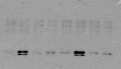
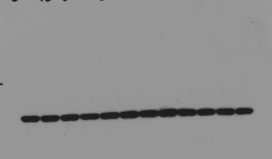
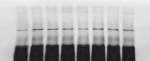
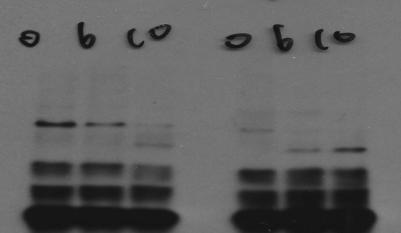
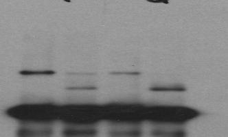
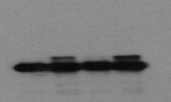
8 Fig. 1d. Control shrna Pellino3 shrna TNFα+ CHX: hr Fig. 1e. IP: Caspase 8 Control shrna Pellino3 shrna Z-VAD-FMK : TNFα + CHX : hr Caspase-3 Caspase-8 18 Pellino3 Caspase 8 Caspase 8 Supplementary Figure S8: Full Blots relating to Figure 1.


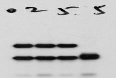
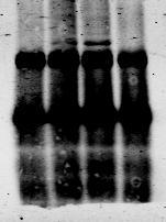
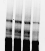
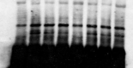
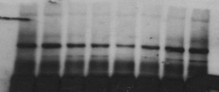
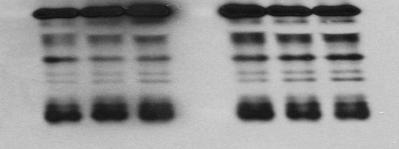
9 P3shRNA+EV P3shRNA+P3s P3shRNA+P3l P3shRNA+P3sF P3shRNA+P3lF P3shRNA+P3sE P3shRNA+P3lE Annexin V positive ( %) Fig. 2b. Fig. 2d. IP: Caspase 8 Pellino3 shrna knockdown TNFα+ CHX: 5 5hr + Z-VAD-FMK : TNFα+ CHX : hr Caspase-3 Caspase-8 40 TNF + CHX 5hr *** ns *** Caspase IP: Caspase 8 Pellino3 shrna knockdown Fig. 2c. + Z-VAD-FMK : TNFα+ CHX : hr Pellino3 Caspase-3 Caspase 8 Supplementary Figure S9: Full Blots relating to Figure 2.


10 Input Input Rip1 input HA input Fig. 3a. HA IP: Fig. 3b. -pulldown Purified P3s: Purified P3l: Purified Rip1: Rip1 Fig. 3c. -pulldown Purified P3smyc: + Purified P3lmyc: + Purified P3sFmmyc: Purified P3lFmmyc: + + HA-Rip1: HA P3smyc: + P3sFHAmmyc: + P3lmyc : + P3lFHAmmyc : + HARip1 : P3l P3l P3s Fig. 3d. Input IP: Flag Procaspase-8myc : Caspase-8DEDmyc : aspase-8caspasemyc + + Pellino3sflag : Fig. 3e. Input IP: Pellino3smyc: + + Pellino3sFHAmmyc: + + Pellino3lmyc : + + Pellino3lFHAmmyc : + + HAcaspase 8: Actin β-actin Procaspase-8 IgG H Caspase-8Caspase Caspase-8DED HA Flag IgG H Pellino3s Fig. 3f. Purified P3smyc: + Purified P3lmyc: + Purified procaspase-8: Caspase 8 -pulldown Fig. 3g. -pulldown Purified P3smyc: + Purified P3lmyc: + Purified Caspase-8Caspase: Caspase 8 30 Pellino3l Pellino3s Supplementary Figure S10: Full Blots relating to Figure 3. Pellino3l Pellino3s


11 Fig. 3h. Flag Pellino3s-flag IgG H Flag- -myc IgG H Pellino3s-myc Pellino3smyc: + + Flag : Pellino3sflag: myc: + + Supplementary Figure S10 (continued): Full Blots relating to Figure 3.


12 Fig. 4a. Z-VAD-FMK : TNFα + CHX : hr IP : Con Fig. 4b. Z-VAD-FMK : TNFα + CHX : hr IP : C8 C8 C8 Con Pellino3 Pellino3 Fig. 4c. TNFα: min IP : TNFR1 TNFR1 TNFR1 TNFR Fig. 4d. IP : Pellino3 Pellino3s EV Pellino3l EV Z-VAD-FMK : TNFα + CHX : hr hr 95 Caspase 8 TRAF2 IgGL TNFR1 Supplementary Figure S11: Full Blots relating to Figure 4.


: Full")

13 Fig. 4e. IP : Fig. 4f. IP : P3sFHAm P3s P3lFHAm P3l Z-VAD-FMK : TNFα + CHX : hr hr P3sE3m EV P3lE3m EV Z-VAD-FMK : TNFα + CHX : hr hr Caspase 8 Caspase 8 Supplementary Figure S11 (continued): Full Blots relating to Figure 4.


14 Fig. 5. IP: Z-VAD-FMK : TNFα+ CHX : hr Caspase 8 52 Flag Pellino3 Pellino3Δc Supplementary Figure S12: Full Blots relating to Figure 5.


15 Fig. 6c. TNF+CHX10hr + + Fig. 6f. Pellino3 -/- MEF TNFα+ CHX 10hr Retro Retro Retro Retro Vector mp3 mp3fm mp3em Caspase 3 Caspase 3 Caspase 3 Caspase 3 β-actin myc Actin GFP 30 Supplementary Figure S13: Full Blots relating to Figure 6.


16 Supplementary Figure S14: Full Blots relating to Figure 7. Fig. 7b. Fig. 7d. TNF + + Smac Control shrna Pellino3 shrna TNF + + Smac Caspase 3 Caspase 3 Pan- ciap Caspase 3 65 Pan-cIAP 65 β-actin β-actin Fig. 7f. Control sirna sirna TNFα+CHX 10hr Z-VAD-FMK Pellino3 +/+ -/- +/+ -/- +/+ -/- +/+ -/- +/+ -/- +/+ -/- Short exposure Long exposure 30 Actin


17 Fig. 8a. TNFα min Fig. 8c. P-IKBα TNFα min IKBα Bcl-x 28 p-p38 38 p-jnk 38 Actin P38 JNK 38 p-erk 40 ERK 40. Actin Supplementary Figure S15: Full Blots relating to Figure 8.


18 Fig. 9c EV TNF CHX + + IκBα SR TNF CHX + + β-actin IκBα SR β-actin Supplementary Figure S16: Full Blots relating to Figure 9c.

19 IP: TNFR1 Supplementary Figure S17: Full Blots relating to Figure 10. IP: Fig. 10a. TNFα: min Fig. 10b. TNFα: min 94 TRAF2 Ub TAK1 65 NEMO TNFR1 Fig. 10c. IP: Fig. 10d. IP: TNFα : min Z-VAD-FMK : TNFα + SMAC : hr Pan-cIAP


20 Fig. 11d. TNFα+ D-gal +/+ -/- Caspase 3 18 Actin Supplementary Figure S18: Full Blots relating to Figure 11d.
SUPPLEMENTARY INFORMATION
 DOI: 10.1038/ncb2362 Figure S1 CYLD and CASPASE 8 genes are co-regulated. Analysis of gene expression across 79 tissues was carried out as described previously [Ref: PMID: 18636086]. Briefly, microarray
Supplementary Figure 1. AnnexinV FITC and Sytox orange staining in wild type, Nlrp3 /, ASC / and casp1/11 / TEC treated with TNF /CHX.
 Supplementary Figure 1. AnnexinV FITC and Sytox orange staining in wild type, Nlrp3 /, ASC / and casp1/11 / TEC treated with TNF /CHX. Phase contrast and widefield fluorescence microscopy (20x magnification).
Supplementary Figure 1.
 Supplementary Figure 1. Characterisation of IHG-1 overexpressing and knockdown cell lines. (A) Total cellular RNA was prepared from HeLa cells stably overexpressing IHG-1 or mts-ihg-1. IHG-1 mrna was quantified
Supplementary Information
 Supplementary Information MAP2/Hoechst Hyp.-AP ph 6.5 Hyp.-SD ph 7.2 Norm.-SD ph 7.2 Supplementary Figure 1. Mitochondrial elongation in cortical neurons by acidosis. Representative images of neuronal
TRADD mediates the tumor necrosis factor-induced apoptosis of L929 cells in the absence of RIP3
 www.nature.com/scientificreports Received: 14 June 2017 Accepted: 9 November 2017 Published: xx xx xxxx OPEN TRADD mediates the tumor necrosis factor-induced apoptosis of L929 cells in the absence of RIP3
FSC-W FSC-H CD4 CD62-L
 Supplementary Fig. 1 a SSC-A FSC-A FSC-W FSC-H SSC-W SSC-H CD4 CD62-L b SSC-A FSC-A FSC-W FSC-A FSC-A 7-AAD FSC-A CD4 IL-9 CD4 c SSC-A FSC-A FSC-W FSC-H SSC-W SSC-H 7-AAD KI67 Annexin-V 7-AAD d I L -5
Apoptosis in Mammalian Cells
 Apoptosis in Mammalian Cells 7.16 2-10-05 Apoptosis is an important factor in many human diseases Cancer malignant cells evade death by suppressing apoptosis (too little apoptosis) Stroke damaged neurons
Role of Mitochondrial Remodeling in Programmed Cell Death in
 Developmental Cell, Vol. 12 Supplementary Data Role of Mitochondrial Remodeling in Programmed Cell Death in Drosophila melanogaster Gaurav Goyal, Brennan Fell, Apurva Sarin, Richard J. Youle, V. Sriram.
SUPPLEMENTAL MATERIAL
 SUPPLEMENTAL MATERIAL Figure S1. Mitochondrial morphology in Fis1-null, Mff-null and Fis1/Mff-null MEF cells. (A) Western blotting of lysates from Fis1-null, Mff-null and Fis1/Mff-null cells. Lysates were
Supplementary Figure 1. Biochemical and sequence alignment analyses the
 Supplementary Figure 1. Biochemical and sequence alignment analyses the interaction of OPTN and TBK1. (a) Analytical gel filtration chromatography analysis of the interaction between TBK1 CTD and OPTN(1-119).
 http://cwp.embo.org/w09-13/index.html Roads to Ruin is s o t p o ap healthy cell autophagic cell death ne cr os is Thanks to Seamus Martin! Evolution of apoptosis signalling cascades Inhibitor Adopted
SUPPLEMENTARY INFORMATION
 doi:10.1038/nature11419 Supplementary Figure 1 Schematic representation of innate immune signaling pathways induced by intracellular Salmonella in cultured macrophages. a, During the infection Salmonella
Supplemental Figures S1 S5
 Beyond reduction of atherosclerosis: PON2 provides apoptosis resistance and stabilizes tumor cells Ines Witte (1), Sebastian Altenhöfer (1), Petra Wilgenbus (1), Julianna Amort (1), Albrecht M. Clement
Figure S1: Extracellular nicotinic acid, but not tryptophan, is sufficient to maintain
 SUPPLEMENTAL INFORMATION Supplemental Figure Legends Figure S1: Extracellular nicotinic acid, but not tryptophan, is sufficient to maintain mitochondrial NAD +. A) Extracellular tryptophan, even at 5 µm,
RESULTS AND DISCUSSION;
 RESULTS AND DISCUSSION; Stress-Induced changes in morphology of 5/9 cells Pro apoptotic agents stimulate 5/xaspase activity UV-B light also stimulated apoptosis and caspase activity in mammalian cells
Enterovirus 71 2B induces cell apoptosis by directly inducing the conformational activation of the pro-apoptotic protein Bax
 JVI Accepted Manuscript Posted Online 24 August 2016 J. Virol. doi:10.1128/jvi.01499-16 Copyright 2016, American Society for Microbiology. All Rights Reserved. 1 2 3 4 5 6 7 8 9 10 11 12 13 14 15 16 17
Death Ligand- Death Receptor. Annexin V. Fas PI MKK7 PIP3. pro-caspase-3. Granzyme B. Caspase Activation. Bid. Bcl-2 Caspase-3 Caspase-12
 International Corporation life. science. discovery. { Apoptosis Apoptosis Kits International Corporation Apoptosis Signal Cascade and Detection Kits Annexin V Kits MEBCYTO Apoptosis Kit Annexin V Apoptosis
4) Please cite Dagda et al J Biol Chem 284: , for any publications or presentations resulting from use or modification of the macro.
 Please cite Dagda et al J Biol Chem 284: , for any publications or presentations resulting from use or modification of the macro.") Supplement Figure S1. Algorithmic quantification of mitochondrial morphology in SH- SY5Y cells treated with known fission/fusion mediators. Parental SH-SY5Y cells were transiently transfected with an empty
Supplement Figure S1. Algorithmic quantification of mitochondrial morphology in SH- SY5Y cells treated with known fission/fusion mediators. Parental SH-SY5Y cells were transiently transfected with an empty
SUPPLEMENTARY INFORMATION
 DOI:.38/ncb97 P ( μm, hours) 1 2 4 P DMSO Figure S1 unningham et al. E 97 65 27 MFN1 GFP-Parkin Opa1 ctin GPDH HEK293 GFP-Parkin 19 115 97 65 27 Mitochondrial Fraction SH-SY5Y GFP-Parkin Mito DMSO Mito
Research Article. Key words: DISC, c-flip, JNK, Mitochondria, Caspase
 Research Article 6459 p38α, but not p38β, inhibits the phosphorylation and presence of c-flip S in DISC to potentiate Fas-mediated caspase-8 activation and type I apoptotic signaling Leon Tourian Jr, Hong
2 The abbreviations used are: FADD, Fas-associated death domain; tbid, truncated
 THE JOURNAL OF BIOLOGICAL CHEMISTRY VOL. 287, NO. 34, pp. 29125 29133, August 17, 2012 2012 by The American Society for Biochemistry and Molecular Biology, Inc. Published in the U.S.A. Death Receptor 6
Direct Interaction between Survivin and Smac/DIABLO Is Essential for the Anti-apoptotic Activity of Survivin during Taxol-induced Apoptosis*
 THE JOURNAL OF BIOLOGICAL CHEMISTRY Vol. 278, No. 25, Issue of June 20, pp. 23130 23140, 2003 2003 by The American Society for Biochemistry and Molecular Biology, Inc. Printed in U.S.A. Direct Interaction
TRAIL-induced apoptosis requires Bax-dependent mitochondrial release of Smac/DIABLO
 TRAIL-induced apoptosis requires Bax-dependent mitochondrial release of Smac/DIABLO Yibin Deng, Yahong Lin, and Xiangwei Wu 1 Huffington Center on Aging and Department of Molecular and Cellular Biology,
Apoptosis: Death Comes for the Cell
 Apoptosis: Death Comes for the Cell Joe W. Ramos joeramos@hawaii.edu From Ingmar Bergman s The Seventh Seal 1 2 Mutations in proteins that regulate cell proliferation, survival and death can contribute
Casper Is a FADD- and Caspase-Related Inducer of Apoptosis
 Immunity, Vol. 6, 751 763, June, 1997, Copyright 1997 by Cell Press Casper Is a FADD- and Caspase-Related Inducer of Apoptosis Hong-Bing Shu, David R. Halpin, the cysteine residue is the structural hallmark
Illegitimate translation causes unexpected gene expression from on-target out-of-frame alleles
 Illegitimate translation causes unexpected gene expression from on-target out-of-frame alleles created by CRISPR-Cas9 Shigeru Makino, Ryutaro Fukumura, Yoichi Gondo* Mutagenesis and Genomics Team, RIKEN
Supplementary Figure 1. SDS-PAGE analysis of GFP oligomer variants with different linkers. Oligomer mixtures were applied to a PAGE gel containing
 Supplementary Figure 1. SDS-PAGE analysis of GFP oligomer variants with different linkers. Oligomer mixtures were applied to a PAGE gel containing 0.1% SDS without boiling. The gel was analyzed by a fluorescent
HOXA5-Induced Apoptosis in Breast Cancer Cells Is Mediated by Caspases 2 and 8
 HOXA5-Induced Apoptosis in Breast Cancer Cells Is Mediated by Caspases 2 and 8 Hexin Chen, Seung Chung and Saraswati Sukumar Mol. Cell. Biol. 2004, 24(2):924. DOI: 10.1128/MCB.24.2.924-935.2004. REFERENCES
Supplemental Materials Molecular Biology of the Cell
 Supplemental Materials Molecular iology of the Cell Figure S1 Krüger et al. Arabidopsis Plasmodium H. sapiens* 1) Xenopus* 1) Drosophila C.elegans S.cerevisae S.pombe L.major T.cruzi T.brucei DCP5 CITH
Supplementary Materials for
 www.sciencesignaling.org/cgi/content/full/6/281/ra51/dc1 Supplementary Materials for Rapgef2 Connects GPCR-Mediated camp Signals to ERK Activation in Neuronal and Endocrine Cells Andrew C. Emery, Maribeth
Supplementary material
 Supplementary material Phosphorylation of the mitochondrial autophagy receptor Nix enhances its interaction with LC3 proteins Vladimir V. Rogov 1,*, Hironori Suzuki 2,3,*, Mija Marinković 4, Verena Lang
Granzyme B Short-Circuits the Need for. Cytotoxic T-Lymphocyte Killing by Directly Cleaving Bid
 REFERENCES CONTENT ALERTS Granzyme B Short-Circuits the Need for Caspase 8 Activity during Granule-Mediated Cytotoxic T-Lymphocyte Killing by Directly Cleaving Bid Michele Barry, Jeffrey A. Heibein, Michael
Nature Medicine: doi: /nm.3776
 C terminal Hsp90 inhibitors restore glucocorticoid sensitivity and relieve a mouse allograft model of Cushing s disease Mathias Riebold, Christian Kozany, Lee Freiburger, Michael Sattler, Michael Buchfelder,
Supplementary Materials
 Electronic Supplementary Material (ESI) for Integrative Biology. This journal is The Royal Society of Chemistry 2015 Predicting genetic interactions from Boolean models of biological networks Supplementary
Isoform B of myosin II heavy chain mediates actomyosin contractility during TNFα-induced apoptosis
 Research Article 1681 Isoform B of myosin II heavy chain mediates actomyosin contractility during TNFα-induced apoptosis Sara Solinet and María Leiza Vitale* Department of Pathology and Cell Biology, Université
The Role of GRASP65 in Golgi Cisternal Stacking and Cell Cycle Progression
 Traffic 2010; 11: 827 842 2010 John Wiley & Sons A/S doi:10.1111/j.1600-0854.2010.01055.x The Role of GRASP65 in Golgi Cisternal Stacking and Cell Cycle Progression Danming Tang, Hebao Yuan and Yanzhuang
Supplementary Figure 1: To test the role of mir-17~92 in orthologous genetic model of ADPKD, we generated Ksp/Cre;Pkd1 F/F (Pkd1-KO) and Ksp/Cre;Pkd1
 and Ksp/Cre;Pkd1") Supplementary Figure 1: To test the role of mir-17~92 in orthologous genetic model of ADPKD, we generated Ksp/Cre;Pkd1 F/F (Pkd1-KO) and Ksp/Cre;Pkd1 F/F ;mir-17~92 F/F (Pkd1-miR-17~92KO) mice. (A) Q-PCR
Supplementary Figure 1: To test the role of mir-17~92 in orthologous genetic model of ADPKD, we generated Ksp/Cre;Pkd1 F/F (Pkd1-KO) and Ksp/Cre;Pkd1 F/F ;mir-17~92 F/F (Pkd1-miR-17~92KO) mice. (A) Q-PCR
Supplementary Figure 1. CoMIC in 293T, HeLa, and HepG2 cells. (a) Mitochondrial morphology in 293T, HeLa and HepG2 cells. Cells were transfected with
 Mitochondrial morphology in 293T, HeLa and HepG2 cells. Cells were transfected with") Supplementary Figure 1. CoMIC in 293T, HeLa, and HepG2 cells. (a) Mitochondrial morphology in 293T, HeLa and HepG2 cells. Cells were transfected with DsRed-mito. Right panels are time-course enlarged images
Supplementary Figure 1. CoMIC in 293T, HeLa, and HepG2 cells. (a) Mitochondrial morphology in 293T, HeLa and HepG2 cells. Cells were transfected with DsRed-mito. Right panels are time-course enlarged images
Supporting Information
 Supporting Information Mullins et al. 10.1073/pnas.0906781106 SI Text Detection of Calcium Binding by 45 Ca 2 Overlay. The 45 CaCl 2 (1 mci, 37 MBq) was obtained from NEN. The general method of 45 Ca 2
Table S1. Sequence of primers used in RT-qPCR
 Table S1. Sequence of primers used in RT-qPCR Primer Name P16Ink4a-F P16Ink4a-R P15Ink4b-F P15Ink4b-R P19Arf-F P19Arf-R P53-F P53-R P21cip1-F P21cip1-R P27kip1-F P27kip1-R P18Ink4c-F P18Ink4c-R P19Ink4d-F
Supplemental Information. The Mitochondrial Fission Receptor MiD51. Requires ADP as a Cofactor
 Structure, Volume 22 Supplemental Information The Mitochondrial Fission Receptor MiD51 Requires ADP as a Cofactor Oliver C. Losón, Raymond Liu, Michael E. Rome, Shuxia Meng, Jens T. Kaiser, Shu-ou Shan,
Supplemental Information
 Supplemental Information Figure S. Regions recognized by the specific antibodies. The amino acid sequence of the major p3-alc species, p3-alc α 35, p3-alc β 37 and p3-alc γ 3, are indicated as red letters.
Roles of the Mammalian Mitochondrial Fission and Fusion Mediators Fis1, Drp1, and Opa1 in Apoptosis
 Molecular Biology of the Cell Vol. 15, 5001 5011, November 2004 Roles of the Mammalian Mitochondrial Fission and Fusion Mediators Fis1, Drp1, and Opa1 in Apoptosis Yang-ja Lee,* Seon-Yong Jeong,* Mariusz
Supplementary Figure 1
 Supplementary Figure 1 Supplementary Figure 1. HSP21 expression in 35S:HSP21 and hsp21 knockdown plants. (a) Since no T- DNA insertion line for HSP21 is available in the publicly available T-DNA collections,
Copyright 2013 The Authors. Deposited on: 16 January 2015
 nn Lefebvre, J. et al. (2013) Caspase-generated fragment of the Met receptor favors apoptosis via the intrinsic pathway independently of its tyrosine kinase activity., 4 (10). e871. ISSN 2041-4889 Copyright
A JNK-Dependent Pathway Is Required for TNF -Induced Apoptosis
 Cell, Vol. 115, 61 70, October 3, 2003, Copyright 2003 by Cell Press A JNK-Dependent Pathway Is Required for TNF -Induced Apoptosis Yibin Deng, 1 Xiaoyang Ren, 1 Lin Yang, Yahong Lin, and Xiangwei Wu*
Molecular Mechanisms of Caspase-Dependent Apoptosis. A Senior Honors Thesis. By Hassan Kamran. The Ohio State University June 2010
 Molecular Mechanisms of Caspase-Dependent Apoptosis A Senior Honors Thesis Presented in Partial Fulfillment of the Requirements for graduation with research distinction in Molecular Genetics in the undergraduate
Thioredoxins (Trx) are cellular redox enzymes that have
 are cellular redox enzymes that have") Thioredoxin-2 Inhibits Mitochondria-Located ASK1-Mediated Apoptosis in a JNK-Independent Manner Rong Zhang, Rafia Al-Lamki, Lanfang Bai, Jeffrey W. Streb, Joseph M. Miano, John Bradley, Wang Min Abstract
Thioredoxin-2 Inhibits Mitochondria-Located ASK1-Mediated Apoptosis in a JNK-Independent Manner Rong Zhang, Rafia Al-Lamki, Lanfang Bai, Jeffrey W. Streb, Joseph M. Miano, John Bradley, Wang Min Abstract
Actinobacteria Relative abundance (%) Co-housed CD300f WT. CD300f KO. Colon length (cm) Day 9. Microscopic inflammation score
 Co-housed CD300f WT. CD300f KO. Colon length (cm) Day 9. Microscopic inflammation score") y groups y individuals 9 Actinobacteria Relative abundance (%) acteroidetes Cyanobacteria Deferribacteres Firmicutes Proteobacteria TM Tenericutes Unclassified CDf CDf Co-housed CDf Co-housed CDf CDf CDf
y groups y individuals 9 Actinobacteria Relative abundance (%) acteroidetes Cyanobacteria Deferribacteres Firmicutes Proteobacteria TM Tenericutes Unclassified CDf CDf Co-housed CDf Co-housed CDf CDf CDf
Mechanisms of Cell Death
 Mechanisms of Cell Death CELL DEATH AND FORMATION OF THE SEMICIRCULAR CANALS Carol M. Troy, MD PhD August 24, 2009 FROM: Fekete et al., Development 124: 2451 (1997) PHENOMENOLOGY OF CELL DEATH I. DEVELOPMENT
Serine-7 but not serine-5 phosphorylation primes RNA polymerase II CTD for P-TEFb recognition
 Supplementary Information to Serine-7 but not serine-5 phosphorylation primes RNA polymerase II CTD for P-TEFb recognition Nadine Czudnochowski 1,2, *, Christian A. Bösken 1, * & Matthias Geyer 1 1 Max-Planck-Institut
SENP3-mediated desumoylation of dynamin-related protein 1 promotes cell death following ischaemia
 The EMBO Journal (2013) 32, 1514 1528 www.embojournal.org SENP3-mediated desumoylation of dynamin-related protein 1 promotes cell death following ischaemia THE EMBO JOURNAL Chun Guo, Keri L Hildick, Jia
The Pirh2 keratin 8/18 interaction modulates the cellular distribution of mitochondria and UV-induced apoptosis
 (2009) 16, 826 837 & 2009 Macmillan Publishers Limited All rights reserved 1350-9047/09 $32.00 www.nature.com/cdd The Pirh2 keratin 8/18 interaction modulates the cellular distribution of mitochondria
Cell Survival Curves (Chap. 3)
") Cell Survival Curves (Chap. 3) Example of pedigress of EMT6 mouse cells Clone The in vitro cell survival curve Cell Survival Assay Dye exclusion assay: membrane integrity MTT assay: respiratory activity
Cell Survival Curves (Chap. 3) Example of pedigress of EMT6 mouse cells Clone The in vitro cell survival curve Cell Survival Assay Dye exclusion assay: membrane integrity MTT assay: respiratory activity
Exploring the Contextual Sensitivity of Factors that Determine Cellto-Cell Variability in Receptor-Mediated Apoptosis
 Exploring the Contextual Sensitivity of Factors that Determine Cellto-Cell Variability in Receptor-Mediated Apoptosis The Harvard community has made this article openly available. Please share how this
Supplementary Information
 Supplementary Information An engineered protein antagonist of K-Ras/B-Raf interaction Monique J. Kauke, 1,2 Michael W. Traxlmayr 1,2, Jillian A. Parker 3, Jonathan D. Kiefer 4, Ryan Knihtila 3, John McGee
HSV-1 ICP27 induces apoptosis by promoting Bax translocation to mitochondria through interacting with
 BMB Reports BMB Rep. 2017; 50(5): 257-262 www.bmbreports.org HSV-1 ICP27 induces apoptosis by promoting Bax translocation to mitochondria through interacting with 14-3-3 Ji Ae Kim #, Jin Chul Kim,#, Jung
Biochimica et Biophysica Acta
 Biochimica et Biophysica Acta 1832 (2013) 453 474 Contents lists available at SciVerse ScienceDirect Biochimica et Biophysica Acta journal homepage: www.elsevier.com/locate/bbadis PSAP induces a unique
Supplementary Figures
 Supplementary Figures Supplementary Figure 1. Ainsliadimer A (1) inhibited I B phosphorylation in spleen extracts. The mice were received intravenous injection of DMSO, 1 (25 mg/kg) or BMS-345541 (25 mg/kg)
eif3k regulates apoptosis in epithelial cells by releasing caspase 3 from keratin-containing inclusions
 2382 Research Article eif3k regulates apoptosis in epithelial cells by releasing caspase 3 from keratin-containing inclusions Yu-Min Lin 1, Yi-Ru Chen 2, Jia-Ren Lin 3, Won-Jing Wang 2, Akihito Inoko 4,
Modeling a Snap-Action, Variable-Delay Switch Controlling Extrinsic Cell Death
 Modeling a Snap-Action, Variable-Delay Switch Controlling Extrinsic Cell Death John G. Albeck 1[, John M. Burke 1,2[, Sabrina L. Spencer 1,3, Douglas A. Lauffenburger 2,3, Peter K. Sorger 1,2* PLoS BIOLOGY
Nature Structural and Molecular Biology: doi: /nsmb Supplementary Figure 1. Definition and assessment of ciap1 constructs.
 Supplementary Figure 1 Definition and assessment of ciap1 constructs. (a) ciap1 constructs used in this study are shown as primary structure schematics with domains colored as in the main text. Mutations
Changjiang Weng, Yuan Li, Dan Xu, Yong Shi, and Hong Tang **
 THE JOURNAL OF BIOLOGICAL CHEMISTRY Vol. 280, No. 11, Issue of March 18, pp. 10491 10500, 2005 2005 by The American Society for Biochemistry and Molecular Biology, Inc. Printed in U.S.A. Specific Cleavage
Mechanism of Dronc activation in Drosophila cells
 Research Article 5035 Mechanism of Dronc activation in Drosophila cells Israel Muro*, Kristin Monser* and Rollie J. Clem Molecular, Cellular, and Developmental Biology Program, Division of Biology, Ackert
Supporting Information
 Supporting Information Cao et al. 10.1073/pnas.1306220110 Gram - bacteria Hemolymph Cytoplasm PGRP-LC TAK1 signalosome Imd dfadd Dredd Dnr1 Ikk signalosome P Relish Nucleus AMP and effector genes Fig.
Photoreceptor Regulation of Constans Protein in Photoperiodic Flowering
 Photoreceptor Regulation of Constans Protein in Photoperiodic Flowering by Valverde et. Al Published in Science 2004 Presented by Boyana Grigorova CBMG 688R Feb. 12, 2007 Circadian Rhythms: The Clock Within
 DOI: 10.1038/ncb2819 Gαi3 / Actin / Acetylated Tubulin Gαi3 / Actin / Acetylated Tubulin a a Gαi3 a Actin Gαi3 WT Gαi3 WT Gαi3 WT b b Gαi3 b Actin Gαi3 KO Gαi3 KO Gαi3 KO # # Figure S1 Loss of protein
This is an open access publisher version of an article that appears in:
 This is an open access publisher version of an article that appears in: JOURNAL OF CELL BIOLOGY The internet address for this paper is: https://publications.icr.ac.uk/5622/ Published text: PS Ribeiro,
Mechanisms. Cell Death. Carol M. Troy, MD PhD August 24, 2009 PHENOMENOLOGY OF CELL DEATH I. DEVELOPMENT 8/20/2009
 Mechanisms of Cell Death Carol M. Troy, MD PhD August 24, 2009 PHENOMENOLOGY OF CELL DEATH I. DEVELOPMENT A. MORPHOGENESIS: SCULPTING/SHAPING STRUCTURES CREATION OF CAVITIES AND TUBES FUSION OF TISSUE
Journal of Cell Science. Research Article
 Research Article 3511 Toxoplasma gondii infection confers resistance against Bim S -induced apoptosis by preventing the activation and mitochondrial targeting of pro-apoptotic Bax Diana Hippe 1, Arnim
Myxoma Virus M11L Blocks Apoptosis through Inhibition of Conformational Activation of Bax at the Mitochondria
 JOURNAL OF VIROLOGY, Feb. 2006, p. 1140 1151 Vol. 80, No. 3 0022-538X/06/$08.00 0 doi:10.1128/jvi.80.3.1140 1151.2006 Copyright 2006, American Society for Microbiology. All Rights Reserved. Myxoma Virus
SUPPLEMENTARY INFORMATION
 DOI: 10.1038/ncb2647 Figure S1 Other Rab GTPases do not co-localize with the ER. a, Cos-7 cells cotransfected with an ER luminal marker (either KDEL-venus or mch-kdel) and mch-tagged human Rab5 (mch-rab5,
MCL-1ES Induces MCL-1L-Dependent BAX- and BAK- Independent Mitochondrial Apoptosis
 MCL-1ES Induces MCL-1L-Dependent BAX- and BAK- Independent Mitochondrial Apoptosis Jae-Hong Kim, Jeehyeon Bae* College of Pharmacy, Chung-Ang University, Seoul, Korea Abstract MCL-1 (myeloid cell leukemia-1),
Apoptosis EXTRA REQUIREMETS
 Apoptosis Introduction (SLIDE 1) Organisms develop from a single cell, and in doing so an anatomy has to be created. This process not only involves the creation of new cells, but also the removal of cells
SUPPLEMENTARY INFORMATION
 GP2 Type I-piliated bacteria FAE M cell M cell pocket idc T cell mdc Generation of antigenspecific T cells Induction of antigen-specific mucosal immune response Supplementary Figure 1 Schematic diagram
Waithe et al Supplementary Figures
 Waithe et al Supplementary Figures Supplementary Figure 1 Expression and properties of WT and W391A mutant YFP- Ca V 2.2. A Immunoblot using Ca V 2.2 Ab for untransfected cells (UT, lane 1), YFP-Ca V 2.2
Cell Death & Trophic Factors II. Steven McLoon Department of Neuroscience University of Minnesota
 Cell Death & Trophic Factors II Steven McLoon Department of Neuroscience University of Minnesota 1 Remember? Neurotrophins are cell survival factors that neurons get from their target cells! There is a
Supplemental Information. Cand1-Mediated Adaptive Exchange Mechanism. Enables Variation in F-Box Protein Expression
 Molecular Cell, Volume 69 upplemental Information -Mediated Adaptive Exchange Mechanism Enables Variation in F-Box Protein Expression Xing Liu, Justin M. Reitsma, Jennifer L. Mamrosh, Yaru Zhang, Ronny
Apoptosis Detection Using the BD Accuri C6 Flow Cytometer
 Apoptosis Detection Using the BD Accuri C6 Flow Cytometer Stacey Roys, Marketing Applications Specialist, BD Biosciences Cyndy Lane, Senior Product Manager, BD Biosciences 23-14519-00 Outline What is apoptosis?
IAPs. Structure of BIR2 Zn finger domain from XIAP
 IAPs The only known endogenous caspase inhibitors are members of a second family of proteins identified by apoptosis research, the IAP (inhibitor of apoptosis proteins) family. IAPs were originally described
Apoptosis: Death comes for the Cell. Apoptosis: Programmed Cell Death. Apoptosis. Necrosis
 poptosis: eath comes for the Cell Mutations in proteins that regulate cell proliferation, survival and death can contribute to oncogenesis Joe W. Ramos jramos@crch.hawaii.edu From Ingmar Bergman s The
Early Mitochondrial Activation and Cytochrome c Up-regulation during Apoptosis* S
 THE JOURNAL OF BIOLOGICAL CHEMISTRY Vol. 277, No. 52, Issue of December 27, pp. 50842 50854, 2002 2002 by The American Society for Biochemistry and Molecular Biology, Inc. Printed in U.S.A. Early Mitochondrial
Technical Bulletin. Caspase 9 Fluorescein (FLICA) Assay. Catalog Number CS0300 Storage Temperature 2-8 C
 Assay. Catalog Number CS0300 Storage Temperature 2-8 C") 1 Caspase 9 Fluorescein (FLICA) Assay Catalog Number CS0300 Storage Temperature 2-8 C Technical Bulletin Product Description Caspases are detected by immunoprecipitation, immunoblotting with caspase specific
1 Caspase 9 Fluorescein (FLICA) Assay Catalog Number CS0300 Storage Temperature 2-8 C Technical Bulletin Product Description Caspases are detected by immunoprecipitation, immunoblotting with caspase specific
Richik N. Ghosh, Linnette Grove, and Oleg Lapets ASSAY and Drug Development Technologies 2004, 2:
 1 3/1/2005 A Quantitative Cell-Based High-Content Screening Assay for the Epidermal Growth Factor Receptor-Specific Activation of Mitogen-Activated Protein Kinase Richik N. Ghosh, Linnette Grove, and Oleg
Enhanced zinc-finger-nuclease activity with improved obligate heterodimeric architectures
 Nature Methods Enhanced zinc-finger-nuclease activity with improved obligate heterodimeric architectures Yannick Doyon, Thuy D Vo, Matthew C Mendel, Shon G Greenberg, Jianbin Wang, Danny F Xia, Jeffrey
The novel conserved mitochondrial inner-membrane protein MTGM regulates mitochondrial morphology and cell proliferation
 2252 Research Article The novel conserved mitochondrial inner-membrane protein MTGM regulates mitochondrial morphology and cell proliferation Jian Zhao 1, *, Tong Liu 1, Shao-Bo Jin 2, Nikolay Tomilin
Caspase-2 is an initiator caspase responsible for pore-forming toxin-mediated apoptosis
 The EMBO Journal (2012) 31, 2615 2628 & 2012 European Molecular Biology Organization All Rights Reserved 0261-4189/12 www.embojournal.org Caspase-2 is an initiator caspase responsible for pore-forming
Apoptosis and Cancer. Carol M. Troy, MD, PhD October 26, 2016
 Apoptosis and Cancer Carol M. Troy, MD, PhD October 26, 2016 PHENOMENOLOGY OF CELL DEATH -1 I. DEVELOPMENT A. MORPHOGENESIS: SCULPTING/SHAPING STRUCTURES FUSION OF TISSUE MASSES (PALATE/NEURAL TUBE) CREATION
Fig. S1. Proliferation and cell cycle exit are affected by the med mutation. (A,B) M-phase nuclei are visualized by a-ph3 labeling in wild-type (A)
 M-phase nuclei are visualized by a-ph3 labeling in wild-type (A)") Fig. S1. Proliferation and cell cycle exit are affected by the med mutation. (A,B) M-phase nuclei are visualized by a-ph3 labeling in wild-type (A) and mutant (B) 4 dpf retinae. The central retina of the
Fig. S1. Proliferation and cell cycle exit are affected by the med mutation. (A,B) M-phase nuclei are visualized by a-ph3 labeling in wild-type (A) and mutant (B) 4 dpf retinae. The central retina of the
Supplementary information
 Supplementary information General Strategy for Direct Cytosolic Protein Delivery via Protein- Nanoparticle Co-Engineering Rubul Mout, Moumita Ray, Tristan Tay, Kanae Sasaki, Gulen Yesilbag Tonga, Vincent
Figure S1, related to Figure 1. Mitochondrial localization of Mfn2 ActA and ER localization of Mfn2 IYFFT and their functional analyses.
 Molecular Cell, Volume 51 Supplemental Information MITOL Regulates Endoplasmic Reticulum-Mitochondria Contacts via Mitofusin2 Ayumu Sugiura, Shun Nagashima, Takeshi Tokuyama, Taku Amo, Yohei Matsuki, Satoshi
DNA DAMAGE-INDUCED APOPTOSIS: INHIBITION BY CALMODULIN ANTAGONIST, FAS RECEPTOR ANTIBODY AND CASPASE INHIBITORS
 DNA DAMAGE-INDUCED APOPTOSIS: INHIBITION BY CALMODULIN ANTAGONIST, FAS RECEPTOR ANTIBODY AND CASPASE INHIBITORS R. Ray, B. J. Benton, M. E. Burke, T. Rockwood, K. R. Bhat, D. R. Anderson, J. P. Petrali,
SUPPLEMENTARY INFORMATION
 DOI: 10.1038/ncb3267 Supplementary Figure 1 A group of genes required for formation or orientation of annular F-actin bundles and aecm ridges: RNAi phenotypes and their validation by standard mutations.
Structural basis of PROTAC cooperative recognition for selective protein degradation
 SUPPLEMENTARY INFORMATION Structural basis of PROTAC cooperative recognition for selective protein degradation Morgan S. Gadd 1, Andrea Testa 1, Xavier Lucas 1, Kwok-Ho Chan, Wenzhang Chen, Douglas J.
Table of Contents. Experiment: Apoptosis Assay using Caspase 3/7 with Hoechst... 2
 Table of Contents Experiment: Apoptosis Assay using Caspase 3/7 with Hoechst... 2 Celigo Setup... 2 Assay Protocol and Plate Setup... 2 Results... 3 Drug-treated MDA-MB-231 and Jurkat cells showed an increase
RayBio CaspGLOW TM Fluorescein Active Caspase-3 Staining Kit
 RayBio CaspGLOW TM Fluorescein Active Caspase-3 Staining Kit User Manual Version 1.0 May 10 th, 2015 RayBio Caspase-3 Fluorometric Assay Kit Protocol (Cat#: 68FLS-Casp3-S) RayBiotech, Inc. We Provide You
Cleavage of BID by Caspase 8 Mediates the Mitochondrial Damage in the Fas Pathway of Apoptosis
 Cell, Vol. 94, 491 501, August 21, 1998, Copyright 1998 by Cell Press Cleavage of BID by Caspase 8 Mediates the Mitochondrial Damage in the Fas Pathway of Apoptosis Honglin Li, Hong Zhu, Chi-jie Xu, and
Kyung-Won LEE, a,b Hyun-Ju JUNG, c Hee-Juhn PARK, c Deog-Gon KIM, d Jin-Yong LEE, d and Kyung-Tae LEE*,a,b
 854 Biol. Pharm. Bull. 28(5) 854 859 (2005) Vol. 28, No. 5 b -D-Xylopyranosyl-(1 3)-b -D-glucuronopyranosyl Echinocystic Acid Isolated from the Roots of Codonopsis lanceolata Induces Caspase-Dependent
Role of Unc51.1 and its binding partners in CNS axon outgrowth
 Role of Unc51.1 and its binding partners in CNS axon outgrowth Toshifumi Tomoda, Jee Hae Kim, Caixin Zhan, and Mary E. Hatten 1 Laboratory of Developmental Neurobiology, The Rockefeller University, New
Full-length GlpG sequence was generated by PCR from E. coli genomic DNA. (with two sequence variations, D51E/L52V, from the gene bank entry aac28166),
,") Supplementary Methods Protein expression and purification Full-length GlpG sequence was generated by PCR from E. coli genomic DNA (with two sequence variations, D51E/L52V, from the gene bank entry aac28166),
Supplementary Methods Protein expression and purification Full-length GlpG sequence was generated by PCR from E. coli genomic DNA (with two sequence variations, D51E/L52V, from the gene bank entry aac28166),