Supplementary Figure 1.
|
|
|
- Austen Bennett
- 5 years ago
- Views:
Transcription
1 Supplementary Figure 1. Characterisation of IHG-1 overexpressing and knockdown cell lines. (A) Total cellular RNA was prepared from HeLa cells stably overexpressing IHG-1 or mts-ihg-1. IHG-1 mrna was quantified by real time PCR. Results were normalised to 18srRNA, and are expressed as relative quantification (RQ) versus cells transduced with an Empty Vector (EV). (B) Whole cell lysates from cells in A were analysed by immunoblotting with antibodies specific for V5. (C) Total cellular RNA was prepared from HeLa cells stably expressing tetracycline-inducible shrnai constructs specific for IHG-1 or with a scrambled construct (Scr) cultured in the presence or absence of doxycycline (dox) for 96 hours. IHG-1 mrna was quantified as in A. (D) Whole cell lysates from cells in C were analysed by immunoblotting with antibodies specific for IHG-1. -actin demonstrates equal protein loading. (E) Whole cell lysates from HeLa cells were incubated with GTP-agarose or unbound agarose beads for 2 hours. Mfn2 bound to agarose beads was assessed by Western blotting.
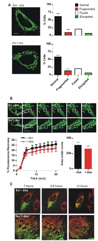
2 Supplementary Figure 2. Doxycycline treatment of cells does not alter mitochondrial morphology or fusion. (A) HeLa cells expressing tetracycline-inducible scrambled shrnai constructs (Scr) were transduced with lentiviral vector expressing mtgfp. Cells were then cultured in the presence or absence of doxycycline (dox) for 96 hours. Representative confocal images of each cell line are shown on the left. Mitochondrial morphology was scored as normal, fragmented, fused or elongated for at least 15 cells for each per experiment. Graphs in the right hand panel present the data as the mean and standard error of the mean for 3 independent experiments (n=45). Black squares= Scr ShRNA cell lines dox; red triangles = Scr ShRNA cell lines + dox. (B) Fluorescence recovery after photobleaching (FRAP) assessments were carried out by live cell imaging of cells in A. Fluorescence recovery was analysed for at least 15 cells for each cell line per experiment. Fluorescence recovery over time is expressed as a percentage of the original fluorescence prior to photobleaching (left panel). For each individual cell assessed the AUC was calculated and is presented in the right hand panel. Results are presented as the mean and standard error of the mean for 3 independent experiments (n=45). Scale bar, 10 µm. (C) Cells from A were transduced with lentiviral vector expressing either mtgfp or mtrfp. GFP and RFP cells for each cell line were co-cultured and fused by incubation with polyethylene glycol (PEG). Mitochondrial fusion, as measured by overlapping RFP and GFP fluorescence, was assessed over 12 hours. Representative confocal images for each cell line are shown. Scale bar, 20 µm.
3
4 Supplementary Figure 3. Doxycycline treatment of cells does not alter respiration or ATP production. (A) HK2 cells expressing tetracycline-inducible scrambled shrnai constructs (Scr) were cultured in the presence or absence of doxycycline (dox) for 96 hours. Oxygen consumption rate (OCR) was then measured. Basal respiration and respiratory capacity are shown for each cell line. (B) Intracellular ATP levels were measured in cells from A. *, p<0.05; **, p<0.01; mean SEM; n = 4 throughout.
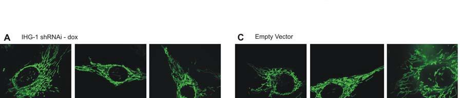
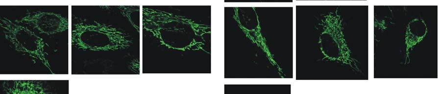
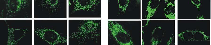
5 Supplementary Figure 4. Altered expression of IHG-1 results in altered mitochondrial morphology. HeLa cells expressing tetracycline-inducible shrnai constructs specific for IHG-1 were transduced with lentiviral vector expressing mtgfp. Cells were then cultured in the absence (A) or presence (B) of doxycycline (dox) for 96 hours. Control HeLa cells (Empty vector) (C) or cells stably overexpressing IHG-1 (D) were transduced with lentiviral vector expressing mtgfp. Ten confocal images of each cell line are shown.


6
 cultured in the presence or absence of doxycycline (dox) for 96 hours were")
7 Supplementary Figure 5. Loss of IHG-1 expression does not increase mitochondrial association with lysosomes. HeLa cells stably expressing tetracycline-inducible shrnai constructs specific for IHG-1 or with a scrambled construct (Scr) cultured in the presence or absence of doxycycline (dox) for 96 hours were stained with MitoTracker Green and LysoTracker Red (both from Invitrogen). Fluorescence was analysed by confocal microscopy. Apoptosis was induced by treating cells with staurosporine.
 Total cellular RNA was prepared from HK2 cells stably expressing tetracycline-inducible shrnai constructs specific for IHG-1 or with a scrambled construct (Scr) cultured in the presence of")
8 Supplementary Figure 6. IHG-1 does not alter expression of components of the mitochondrial fusion and fission machinery. (A) Total cellular RNA was prepared from HK2 cells stably expressing tetracycline-inducible shrnai constructs specific for IHG-1 or with a scrambled construct (Scr) cultured in the presence of doxycycline (dox) for 96 hours. Drp-1, Fis1, Mfn1, Mfn2 and OPA1 mrna were quantified by real time PCR. Results were normalised to 18srRNA, and are expressed as relative quantification (RQ) versus cells transduced with a scrambled construct (Scr). (B) Whole cell lysates from cells in A were analysed by immunoblotting with antibodies specific for Mfn1, Mfn2 and Drp-1. (C) Whole cell lysates from HeLa cells stably overexpressing IHG-1 or control cells (EV) were analysed by immunoblotting with antibodies specific for Mfn1, Mfn2 and V5. Actin antibody binding demonstrates equal protein loading.
9 Supplementary Figure 7. IHG-1 increases mitochondrial fusion. HeLa cells expressing tetracyclineinducible shrnai constructs specific for IHG-1 were transduced with lentiviral vector expressing either mtgfp or mtrfp. Cells were cultured in the (A) absence or (B) presence of doxycycline (dox) for 96 hours. GFP and RFP cells for each cell line were co-cultured and fused by incubation with polyethylene glycol (PEG). Mitochondrial fusion, as measured by overlapping RFP and GFP fluorescence, was assessed over 12 hours. Representative confocal images for each cell line are shown. Scale bar, 20 µm. (C) Control HeLa cells (Empty Vector) and cells stably overexpressing (D) IHG-1 were transduced with lentiviral vector expressing either mtgfp or mtrfp. Mitochondrial fusion was assessed as in A and B over 10 hours. Representative confocal images for each cell line are shown. Scale bar, 20 µm.

10
11 Supplementary Figure 8. IHG-1 protects cells from ROS-induced loss of mitochondrial membrane potential. HeLa cells stably overexpressing IHG-1 or control cells (Empty Vector) were treated with 10 mu/ml glucose oxidase. Mitochondrial membrane potential (MMP) was assessed at 16 hours using JC-1 staining and flow cytometric analysis. *, p<0.05; mean SEM; n = 3.
12 Supplementary Figure 9. Effect of TGF-β1 on the expression of Mfn2, Drp1 and mitochondrial electron transport chain complexes. HK2 cells stably expressing tetracycline-inducible shrnai constructs specific for IHG-1 or with a scrambled construct (Scr) cultured in the presence of doxycycline (dox) for 96 hours were stimulated with TGF-β1 (5ng/ml) for the indicated times. Whole cell lysates were prepared and analysed by immunoblotting with antibodies specific for (A) Mfn2 and Drp1 and (B) complex II (Succinate dehydrogenase [ubiquinone] iron-sulfur subunit [SDHB]) and complex V (ATP synthase alpha-subunit [ATP5A]) of the mitochondrial electron transport chain. Actin antibody binding demonstrates equal protein loading. Alterations in protein expression were analysed by densitometry. Results are shown as the mean +/- SEM for at least 3 independent experiments. Differences in means are not significant.
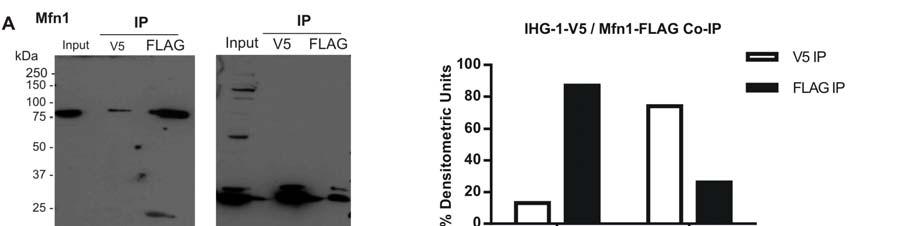
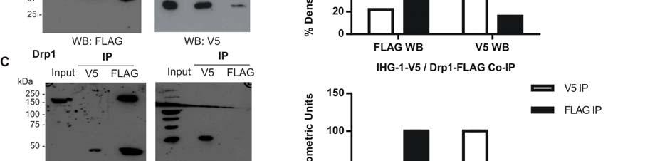
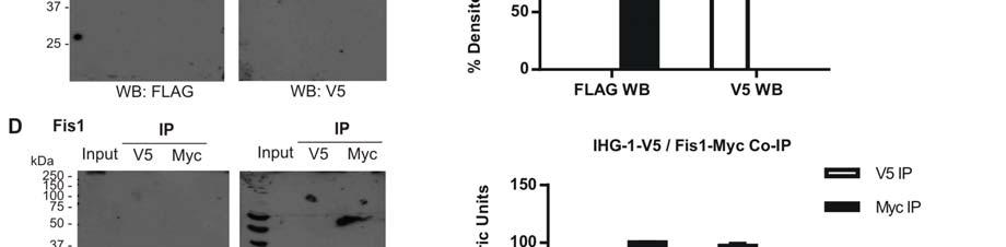
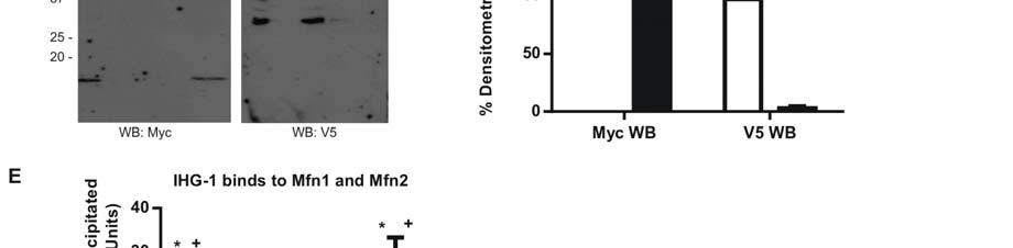
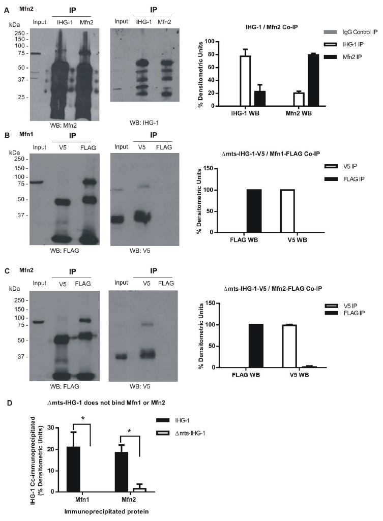
13 Supplementary Figure 10. IHG-1 binds to Mfn1 and Mfn2. HEK293T cells were co-transfected withihg-1-v5 and FLAG-Mitofusin 1 (Mfn1) (A), Mitofusin 2 (Mfn2) (B), Dynamin-related protein 1 (Drp1) (C) or Myc-tagged mitochondrial fission protein 1 (Fis1) (D). Binding of proteins was assessed by immunoprecipitation (IP) and Western blotting. Unprocessed images of original Western blot films are shown for Figure 4 A-D along with densitometric analysis. (E) Comparison of levels of IHG-1 protein co-immunoprecipitated by the indicated proteins. Results are shown as the mean +/- SEM for at least 2 independent experiments. *, p<0.05; **, P<0.01 versus Drp1. +, p<0.05; versus Fis1.


14
15 Supplementary Figure 11. IHG-1 binds to Mfn1 and Mfn2. (A) Endogenous IHG-1 or Mfn2 was immunoprecipitated from HeLa cells. Co-immunoprecipitation of the alternate protein was assessed by Western blotting. Unprocessed images of original Western blot films are shown for Figure 4 A-D along with densitometric analysis. HEK293T cells were co-transfected with mts-ihg-1-v5 and either FLAG-tagged- Mfn1 (B) or Mfn2 (C). Binding of proteins was assessed by immunoprecipitation and Western blotting. Unprocessed images of original Western blot films are shown for Figure 4 A-D along with densitometric analysis. HEK293T cells were co-transfected with mts-ihg-1-v5 and either FLAG-tagged- Mfn1 (B) or Mfn2 (C). Binding of proteins was assessed by immunoprecipitation and Western blotting. Unprocessed images of original Western blot films are shown for Figure 4 A-D along with densitometric analysis. (D) Comparison of levels of IHG-1 and mts-ihg-1 protein co-immunoprecipitated by the Mfn1 and Mfn2. Results are shown as the mean +/- SEM for at least 2 independent experiments..*, p<0.05.
16
Supplementary Information
 Supplementary Information MAP2/Hoechst Hyp.-AP ph 6.5 Hyp.-SD ph 7.2 Norm.-SD ph 7.2 Supplementary Figure 1. Mitochondrial elongation in cortical neurons by acidosis. Representative images of neuronal
SUPPLEMENTAL MATERIAL
 SUPPLEMENTAL MATERIAL Figure S1. Mitochondrial morphology in Fis1-null, Mff-null and Fis1/Mff-null MEF cells. (A) Western blotting of lysates from Fis1-null, Mff-null and Fis1/Mff-null cells. Lysates were
4) Please cite Dagda et al J Biol Chem 284: , for any publications or presentations resulting from use or modification of the macro.
 Please cite Dagda et al J Biol Chem 284: , for any publications or presentations resulting from use or modification of the macro.") Supplement Figure S1. Algorithmic quantification of mitochondrial morphology in SH- SY5Y cells treated with known fission/fusion mediators. Parental SH-SY5Y cells were transiently transfected with an empty
Supplement Figure S1. Algorithmic quantification of mitochondrial morphology in SH- SY5Y cells treated with known fission/fusion mediators. Parental SH-SY5Y cells were transiently transfected with an empty
IHG-1 INCREASES MITOCHONDRIAL FUSION AND BIOENERGETIC FUNCTION
 Page 1 of 46 Diabetes IHG-1 INCREASES MITOCHONDRIAL FUSION AND BIOENERGETIC FUNCTION Fionnuala B. Hickey *, James B. Corcoran *, Brenda Griffin *, Una Bhreathnach *, Heather Mortiboys #, Helen M. Reid,
Supplementary Figure 1. CoMIC in 293T, HeLa, and HepG2 cells. (a) Mitochondrial morphology in 293T, HeLa and HepG2 cells. Cells were transfected with
 Mitochondrial morphology in 293T, HeLa and HepG2 cells. Cells were transfected with") Supplementary Figure 1. CoMIC in 293T, HeLa, and HepG2 cells. (a) Mitochondrial morphology in 293T, HeLa and HepG2 cells. Cells were transfected with DsRed-mito. Right panels are time-course enlarged images
Supplementary Figure 1. CoMIC in 293T, HeLa, and HepG2 cells. (a) Mitochondrial morphology in 293T, HeLa and HepG2 cells. Cells were transfected with DsRed-mito. Right panels are time-course enlarged images
IHG-1 Increases Mitochondrial Fusion and Bioenergetic Function
 4314 Diabetes Volume 63, December 2014 Fionnuala B. Hickey, 1,2,3 James B. Corcoran, 1,2 Brenda Griffin, 1,4 Una Bhreathnach, 1,2 Heather Mortiboys, 5 Helen M. Reid, 4 Darrell Andrews, 1,4 Shane Byrne,
Supplemental material
 Supplemental material THE JOURNAL OF CELL BIOLOGY Mourier et al., http://www.jcb.org/cgi/content/full/jcb.201411100/dc1 Figure S1. Size and mitochondrial content in Mfn1 and Mfn2 knockout hearts. (A) Body
TNFα 18hr. Control. CHX 18hr. TNFα+ CHX 18hr. TNFα: 18 18hr (KDa) PARP. Cleaved. Cleaved. Cleaved. Caspase3. Pellino3 shrna. Control shrna.
 PARP. Cleaved. Cleaved. Cleaved. Caspase3. Pellino3 shrna. Control shrna.") Survival ( %) a. TNFα 18hr b. Control sirna Pellino3 sirna TNFα: 18 18hr c. Control shrna Pellino3 shrna Caspase3 Actin Control d. Control shrna Pellino3 shrna *** 100 80 60 CHX 18hr 40 TNFα+ CHX 18hr
Survival ( %) a. TNFα 18hr b. Control sirna Pellino3 sirna TNFα: 18 18hr c. Control shrna Pellino3 shrna Caspase3 Actin Control d. Control shrna Pellino3 shrna *** 100 80 60 CHX 18hr 40 TNFα+ CHX 18hr
SUPPLEMENTARY INFORMATION
 DOI: 10.1038/ncb2362 Figure S1 CYLD and CASPASE 8 genes are co-regulated. Analysis of gene expression across 79 tissues was carried out as described previously [Ref: PMID: 18636086]. Briefly, microarray
Mitochondrial Dynamics Is a Distinguishing Feature of Skeletal Muscle Fiber Types and Regulates Organellar Compartmentalization
 Cell Metabolism Supplemental Information Mitochondrial Dynamics Is a Distinguishing Feature of Skeletal Muscle Fiber Types and Regulates Organellar Compartmentalization Prashant Mishra, Grigor Varuzhanyan,
Supplemental Information. The Mitochondrial Fission Receptor MiD51. Requires ADP as a Cofactor
 Structure, Volume 22 Supplemental Information The Mitochondrial Fission Receptor MiD51 Requires ADP as a Cofactor Oliver C. Losón, Raymond Liu, Michael E. Rome, Shuxia Meng, Jens T. Kaiser, Shu-ou Shan,
SUPPLEMENTARY INFORMATION
 DOI: 10.1038/ncb2647 Figure S1 Other Rab GTPases do not co-localize with the ER. a, Cos-7 cells cotransfected with an ER luminal marker (either KDEL-venus or mch-kdel) and mch-tagged human Rab5 (mch-rab5,
SUPPLEMENTARY INFORMATION
 DOI:.38/ncb97 P ( μm, hours) 1 2 4 P DMSO Figure S1 unningham et al. E 97 65 27 MFN1 GFP-Parkin Opa1 ctin GPDH HEK293 GFP-Parkin 19 115 97 65 27 Mitochondrial Fraction SH-SY5Y GFP-Parkin Mito DMSO Mito
Figure S1: Extracellular nicotinic acid, but not tryptophan, is sufficient to maintain
 SUPPLEMENTAL INFORMATION Supplemental Figure Legends Figure S1: Extracellular nicotinic acid, but not tryptophan, is sufficient to maintain mitochondrial NAD +. A) Extracellular tryptophan, even at 5 µm,
Supporting Information
 Supporting Information Wang et al. 10.1073/pnas.0804871105 SI Materials and Methods Cell Culture and Transfection. Human neuroblastoma M17 cells were grown as described before (1). Transfection was performed
FSC-W FSC-H CD4 CD62-L
 Supplementary Fig. 1 a SSC-A FSC-A FSC-W FSC-H SSC-W SSC-H CD4 CD62-L b SSC-A FSC-A FSC-W FSC-A FSC-A 7-AAD FSC-A CD4 IL-9 CD4 c SSC-A FSC-A FSC-W FSC-H SSC-W SSC-H 7-AAD KI67 Annexin-V 7-AAD d I L -5
Supplementary Figure 1. Confirmation of REF52-hE2F1p::4NLS-d4Venus reporter cells and characterization of E2F dynamics. (a) Alignment of E2F dynamics
 Alignment of E2F dynamics") Supplementary Figure 1. Confirmation of REF52-hE2F1p::4NLS-d4Venus reporter cells and characterization of E2F dynamics. (a) Alignment of E2F dynamics trajectories to endogenous E2F1 mrna expression. Gray
Supplementary Figure 1. Confirmation of REF52-hE2F1p::4NLS-d4Venus reporter cells and characterization of E2F dynamics. (a) Alignment of E2F dynamics trajectories to endogenous E2F1 mrna expression. Gray
Membrane depolarization activates the mitochondrial protease OMA1 by stimulating self-cleavage
 Scientific Report Membrane depolarization activates the mitochondrial protease OMA1 by stimulating self-cleavage Kuan Zhang, Huihui Li & Zhiyin Song* Abstract Mitochondrial inner membrane fusion depends
Mitofusin 1 and 2 play distinct roles in mitochondrial fusion reactions via GTPase activity
 Research Article 6535 Mitofusin 1 and 2 play distinct roles in mitochondrial fusion reactions via GTPase activity Naotada Ishihara, Yuka Eura and Katsuyoshi Mihara* Department of Molecular Biology, Graduate
Role of Mitochondrial Remodeling in Programmed Cell Death in
 Developmental Cell, Vol. 12 Supplementary Data Role of Mitochondrial Remodeling in Programmed Cell Death in Drosophila melanogaster Gaurav Goyal, Brennan Fell, Apurva Sarin, Richard J. Youle, V. Sriram.
Roles of the Mammalian Mitochondrial Fission and Fusion Mediators Fis1, Drp1, and Opa1 in Apoptosis
 Molecular Biology of the Cell Vol. 15, 5001 5011, November 2004 Roles of the Mammalian Mitochondrial Fission and Fusion Mediators Fis1, Drp1, and Opa1 in Apoptosis Yang-ja Lee,* Seon-Yong Jeong,* Mariusz
Mitochondrie et muscle lisse. Vladimir Veksler
 Mitochondrie et muscle lisse Vladimir Veksler Main mitochonrial functions Energy production ATP consumption rate in smooth and striated muscle smooth muscle striated (cardiac) muscle at rest 30 µmol/min/g
Mitochondrial Structure, Function and Dynamics Are Temporally Controlled by c-myc
 Mitochondrial Structure, Function and Dynamics Are Temporally Controlled by c-myc J. Anthony Graves 1 *, Yudong Wang 2, Sunder Sims-Lucas 3, Edward Cherok 1, Kristi Rothermund 1, Maria F. Branca 4, Jennifer
Supplemental Figures S1 S5
 Beyond reduction of atherosclerosis: PON2 provides apoptosis resistance and stabilizes tumor cells Ines Witte (1), Sebastian Altenhöfer (1), Petra Wilgenbus (1), Julianna Amort (1), Albrecht M. Clement
SUPPLEMENTARY INFORMATION
 DOI: 10.1038/ncb2215 Figure S1 Number of egfp-vps4a bursts versus cellular expression levels. The total number of egfp-vps4a bursts, counted at the end of each movie (frame 2000, after 1h 28 min) are plotted
Mfn2 modulates the UPR and mitochondrial function via repression of PERK
 The EMBO Journal (2013) 32, 2348 2361 www.embojournal.org Mfn2 modulates the UPR and mitochondrial function via repression of PERK THE EMBO JOURNAL Juan Pablo Muñoz 1,2,3, Saška Ivanova 1,2,3, Jana Sánchez-Wandelmer
Supplementary Figure 1. Real time in vivo imaging of SG secretion. (a) SGs from Drosophila third instar larvae that express Sgs3-GFP (green) and
 SGs from Drosophila third instar larvae that express Sgs3-GFP (green) and") Supplementary Figure 1. Real time in vivo imaging of SG secretion. (a) SGs from Drosophila third instar larvae that express Sgs3-GFP (green) and Lifeact-Ruby (red) were imaged in vivo to visualize secretion
Supplementary Figure 1. Real time in vivo imaging of SG secretion. (a) SGs from Drosophila third instar larvae that express Sgs3-GFP (green) and Lifeact-Ruby (red) were imaged in vivo to visualize secretion
T H E J O U R N A L O F C E L L B I O L O G Y
 T H E J O U R N A L O F C E L L B I O L O G Y Supplemental material Eisner et al., http://www.jcb.org/cgi/content/full/jcb.201312066/dc1 Figure S1. Mitochondrial continuity in adult skeletal muscle. (A)
supplementary information
 DOI: 10.1038/ncb2119 Figure S1 Analysis of IRGM localization, GST-IRGMd, GST-IRGMdS47N and GST-IRGMb protein purification, and analysis of their binding to cardiolipin. a. Mitochondria were purified by
Supplementary Figure 1. AnnexinV FITC and Sytox orange staining in wild type, Nlrp3 /, ASC / and casp1/11 / TEC treated with TNF /CHX.
 Supplementary Figure 1. AnnexinV FITC and Sytox orange staining in wild type, Nlrp3 /, ASC / and casp1/11 / TEC treated with TNF /CHX. Phase contrast and widefield fluorescence microscopy (20x magnification).
Supplementary Figure 1: To test the role of mir-17~92 in orthologous genetic model of ADPKD, we generated Ksp/Cre;Pkd1 F/F (Pkd1-KO) and Ksp/Cre;Pkd1
 and Ksp/Cre;Pkd1") Supplementary Figure 1: To test the role of mir-17~92 in orthologous genetic model of ADPKD, we generated Ksp/Cre;Pkd1 F/F (Pkd1-KO) and Ksp/Cre;Pkd1 F/F ;mir-17~92 F/F (Pkd1-miR-17~92KO) mice. (A) Q-PCR
Supplementary Figure 1: To test the role of mir-17~92 in orthologous genetic model of ADPKD, we generated Ksp/Cre;Pkd1 F/F (Pkd1-KO) and Ksp/Cre;Pkd1 F/F ;mir-17~92 F/F (Pkd1-miR-17~92KO) mice. (A) Q-PCR
Supplementary Figure 1
 Supplementary Figure 1 Supplementary Figure 1. HSP21 expression in 35S:HSP21 and hsp21 knockdown plants. (a) Since no T- DNA insertion line for HSP21 is available in the publicly available T-DNA collections,
Waithe et al Supplementary Figures
 Waithe et al Supplementary Figures Supplementary Figure 1 Expression and properties of WT and W391A mutant YFP- Ca V 2.2. A Immunoblot using Ca V 2.2 Ab for untransfected cells (UT, lane 1), YFP-Ca V 2.2
Supplementary Materials for
 www.sciencesignaling.org/cgi/content/full/6/281/ra51/dc1 Supplementary Materials for Rapgef2 Connects GPCR-Mediated camp Signals to ERK Activation in Neuronal and Endocrine Cells Andrew C. Emery, Maribeth
Supplementary information
 Supplementary information General Strategy for Direct Cytosolic Protein Delivery via Protein- Nanoparticle Co-Engineering Rubul Mout, Moumita Ray, Tristan Tay, Kanae Sasaki, Gulen Yesilbag Tonga, Vincent
G-protein β2 subunit interacts with mitofusin 1 to regulate mitochondrial fusion
 Received 1 Jun 1 Aepted 23 Sep 1 Published 19 Oct 1 DOI: 1.138/ncomms199 G-protein β2 subunit interacts with mitofusin 1 to regulate mitochondrial fusion Juan Zhang 1, Weihua Liu 1, Jianchao Liu 2, Weiming
Supplementary Information
 Supplementary Information An engineered protein antagonist of K-Ras/B-Raf interaction Monique J. Kauke, 1,2 Michael W. Traxlmayr 1,2, Jillian A. Parker 3, Jonathan D. Kiefer 4, Ryan Knihtila 3, John McGee
Mitochondrial kiss-and-run : interplay between mitochondrial motility and fusion fission dynamics
 The EMBO Journal (2009) 28, 3074 3089 & 2009 European Molecular Biology Organization All Rights Reserved 0261-4189/09 www.embojournal.org : interplay between mitochondrial motility and fusion fission dynamics
SUPPLEMENTARY INFORMATION
 doi:10.1038/nature11419 Supplementary Figure 1 Schematic representation of innate immune signaling pathways induced by intracellular Salmonella in cultured macrophages. a, During the infection Salmonella
SUPPLEMENTARY INFORMATION
 Cell viability rate 0.8 0.6 0 0.05 0.1 0.2 0.3 0.4 0.5 0.7 1 Exposure duration (s) Supplementary Figure 1. Femtosecond laser could disrupt and turn off GFP without photons at 473 nm and keep cells alive.
targets. clustering show that different complex pathway
 Supplementary Figure 1. CLICR allows clustering and activation of cytoplasmic protein targets. (a, b) Upon light activation, the Cry2 (red) and LRP6c (green) components co-cluster due to the heterodimeric
Supplementary Figure 1. Biochemical and sequence alignment analyses the
 Supplementary Figure 1. Biochemical and sequence alignment analyses the interaction of OPTN and TBK1. (a) Analytical gel filtration chromatography analysis of the interaction between TBK1 CTD and OPTN(1-119).
Table S1. Sequence of primers used in RT-qPCR
 Table S1. Sequence of primers used in RT-qPCR Primer Name P16Ink4a-F P16Ink4a-R P15Ink4b-F P15Ink4b-R P19Arf-F P19Arf-R P53-F P53-R P21cip1-F P21cip1-R P27kip1-F P27kip1-R P18Ink4c-F P18Ink4c-R P19Ink4d-F
SENP3-mediated desumoylation of dynamin-related protein 1 promotes cell death following ischaemia
 The EMBO Journal (2013) 32, 1514 1528 www.embojournal.org SENP3-mediated desumoylation of dynamin-related protein 1 promotes cell death following ischaemia THE EMBO JOURNAL Chun Guo, Keri L Hildick, Jia
Arabidopsis PPR40 connects abiotic stress responses to mitochondrial electron transport
 Ph.D. thesis Arabidopsis PPR40 connects abiotic stress responses to mitochondrial electron transport Zsigmond Laura Supervisor: Dr. Szabados László Arabidopsis Molecular Genetic Group Institute of Plant
Illegitimate translation causes unexpected gene expression from on-target out-of-frame alleles
 Illegitimate translation causes unexpected gene expression from on-target out-of-frame alleles created by CRISPR-Cas9 Shigeru Makino, Ryutaro Fukumura, Yoichi Gondo* Mutagenesis and Genomics Team, RIKEN
Mitochondries, Hypoxie et Cancer
 Mitochondries, Hypoxie et Cancer CNRS - UMR 6543 Institute of Signaling, Developmental Biology and Cancer Research University of Nice-Sophia Antipolis - FRANCE Colon tumor necrosis Blood vessel Colon tumor
The novel conserved mitochondrial inner-membrane protein MTGM regulates mitochondrial morphology and cell proliferation
 2252 Research Article The novel conserved mitochondrial inner-membrane protein MTGM regulates mitochondrial morphology and cell proliferation Jian Zhao 1, *, Tong Liu 1, Shao-Bo Jin 2, Nikolay Tomilin
Selective Targeting of ER Exit Sites Supports Axon Development
 Traffic 2009; 10: 1669 1684 2009 John Wiley & Sons A/S doi:10.1111/j.1600-0854.2009.00974.x Selective Targeting of ER Exit Sites Supports Axon Development Meir Aridor 1 and Kenneth N. Fish 2, 1 Department
Figure 1. Identification of UGT74E2 as an IBA glycosyltransferase. (A) Relative conversion rates of different plant hormones to their glucosylated
 Relative conversion rates of different plant hormones to their glucosylated") Figure 1. Identification of UGT74E2 as an IBA glycosyltransferase. (A) Relative conversion rates of different plant hormones to their glucosylated form by recombinant UGT74E2. The naturally occurring auxin
Figure 1. Identification of UGT74E2 as an IBA glycosyltransferase. (A) Relative conversion rates of different plant hormones to their glucosylated form by recombinant UGT74E2. The naturally occurring auxin
scientific report Separate fusion of outer and inner mitochondrial membranes scientificreport
 scientificreport Separate fusion of outer and inner mitochondrial membranes Florence Malka 1, Olwenn Guillery 1, Carmen Cifuentes-Diaz 2, Emmanuelle Guillou 3, Pascale elenguer 3, nne Lombès 1 & Manuel
Heather Currinn, Benjamin Guscott, Zita Balklava, Alice Rothnie and Thomas Wassmer*
 Online Resources APP controls the formation of PI(3,5)P 2 vesicles through its binding of the PIKfyve complex. Cellular and Molecular Life Sciences Heather Currinn, Benjamin Guscott, Zita Balklava, Alice
The Mitochondrion. Definition Structure, ultrastructure Functions
 The Mitochondrion Definition Structure, ultrastructure Functions Organelle definition Etymology of the name Carl Benda (1903): (mitos) thread; (khondrion) granule. Light microscopy identification First
Biological Process Term Enrichment
 Biological Process Term Enrichment cellular protein localization cellular macromolecule localization intracellular protein transport intracellular transport generation of precursor metabolites and energy
Regulation of mitochondrial fission and apoptosis by the mitochondrial outer membrane protein hfis1
 Research Article 4141 Regulation of mitochondrial fission and apoptosis by the mitochondrial outer membrane protein hfis1 Tianzheng Yu 1, Randall J. Fox 1, Lindsay S. Burwell 2 and Yisang Yoon 1, * 1 Department
The C Terminus of the L-Type Voltage-Gated Calcium Channel Ca V 1.2 Encodes a Transcription Factor
 The C Terminus of the L-Type Voltage-Gated Calcium Channel Ca V 1.2 Encodes a Transcription Factor Natalia Gomez-Ospina, 1 Fuminori Tsuruta, 1,3 Odmara Barreto-Chang, 1,3 Linda Hu, 2 and Ricardo Dolmetsch
MITOCHONDRIAL MARKERS
 1 MITOCHONDRIAL MARKERS www.ptglab.com 2 INTRODUCTION Mitochondria are important cellular organelles that maintain cellular energy balance, contain key regulators of cell death processes, and play a significant
Downloaded from at University of Washington Health Sciences Libraries, on November 12, 2010
 THE JOURNAL OF BIOLOGICAL CHEMISTRY VOL. 285, NO. 41, pp. 31590 31602, October 8, 2010 2010 by The American Society for Biochemistry and Molecular Biology, Inc. Printed in the U.S.A. Rab32 Modulates Apoptosis
Loss of MARCH5 mitochondrial E3 ubiquitin ligase induces cellular senescence through dynamin-related protein 1 and mitofusin 1
 Research Article 619 Loss of MARCH5 mitochondrial E3 ubiquitin ligase induces cellular senescence through dynamin-related protein 1 and mitofusin 1 Yong-Yea Park 1,2, Seungmin Lee 1,2, Mariusz Karbowski
In order to confirm the contribution of diffusion to the FRAP recovery curves of
 Fonseca et al., Supplementary Information FRAP data analysis ) Contribution of Diffusion to the recovery curves In order to confirm the contribution of diffusion to the FRAP recovery curves of PH::GFP
Mitochondrial oxidative stress causes mitochondrial fragmentation via differential modulation of mitochondrial fission fusion proteins
 Mitochondrial oxidative stress causes mitochondrial fragmentation via differential modulation of mitochondrial fission fusion proteins Shengnan Wu, Feifan Zhou, Zhenzhen Zhang and Da Xing MOE Key Laboratory
The Novel Tail-anchored Membrane Protein Mff Controls Mitochondrial and Peroxisomal Fission in Mammalian Cells
 Molecular Biology of the Cell Vol. 19, 2402 2412, June 2008 The Novel Tail-anchored Membrane Protein Mff Controls Mitochondrial and Peroxisomal Fission in Mammalian Cells Shilpa Gandre-Babbe and Alexander
Supporting Information
 Supporting Information Mullins et al. 10.1073/pnas.0906781106 SI Text Detection of Calcium Binding by 45 Ca 2 Overlay. The 45 CaCl 2 (1 mci, 37 MBq) was obtained from NEN. The general method of 45 Ca 2
Nature Medicine: doi: /nm.3776
 C terminal Hsp90 inhibitors restore glucocorticoid sensitivity and relieve a mouse allograft model of Cushing s disease Mathias Riebold, Christian Kozany, Lee Freiburger, Michael Sattler, Michael Buchfelder,
Supplementary material
 Supplementary material Phosphorylation of the mitochondrial autophagy receptor Nix enhances its interaction with LC3 proteins Vladimir V. Rogov 1,*, Hironori Suzuki 2,3,*, Mija Marinković 4, Verena Lang
ΔG o' = ηf ΔΕ o' = (#e ( V mol) ΔΕ acceptor
 ΔΕ acceptor") Reading: Sec. 19.1 Electron-Transfer Reactions in Mitochondria (listed subsections only) 19.1.1 Electrons are Funneled to Universal Electron Acceptors p. 692/709 19.1.2 Electrons Pass through a Series
Reading: Sec. 19.1 Electron-Transfer Reactions in Mitochondria (listed subsections only) 19.1.1 Electrons are Funneled to Universal Electron Acceptors p. 692/709 19.1.2 Electrons Pass through a Series
Supplementary Materials for
 www.sciencesignaling.org/cgi/content/full/6/301/ra98/dc1 Supplementary Materials for Regulation of Epithelial Morphogenesis by the G Protein Coupled Receptor Mist and Its Ligand Fog Alyssa J. Manning,
Computational Biology, Part 24 Clustering and Unmixing of Subcellular Patterns
 Computational Biology, Part 24 Clustering and Unmixing of Subcellular Patterns Robert F. Murphy Copyright 1996, 1999, 2000-2009. All rights reserved. Unsupervised Learning to Identify High-Resolution Protein
Stoichiometric expression of mthsp40 and mthsp70 modulates mitochondrial morphology and cristae structure via Opa1 L cleavage
 MBoC ARTICLE Stoichiometric expression of mthsp40 and mthsp70 modulates mitochondrial morphology and cristae structure via Opa1 L cleavage Byoungchun Lee a, Younghee Ahn b, Sung-Myung Kang a, Youngjin
7.06 Problem Set
 7.06 Problem Set 5 -- 2006 1. In the first half of the course, we encountered many examples of proteins that entered the nucleus in response to the activation of a cell-signaling pathway. One example of
Appoptosin interacts with mitochondrial outer-membrane. fusion proteins and regulates mitochondrial morphology
 2016. Published by The Company of Biologists Ltd. Appoptosin interacts with mitochondrial outer-membrane fusion proteins and regulates mitochondrial morphology Cuilin Zhang 1, Zhun Shi 1, Lingzhi Zhang
Supplemental Data. Perrella et al. (2013). Plant Cell /tpc
. Plant Cell /tpc") Intensity Intensity Intensity Intensity Intensity Intensity 150 50 150 0 10 20 50 C 150 0 10 20 50 D 0 10 20 Distance (μm) 50 20 40 E 50 F 0 10 20 50 0 15 30 Distance (μm) Supplemental Figure 1: Co-localization
Intensity Intensity Intensity Intensity Intensity Intensity 150 50 150 0 10 20 50 C 150 0 10 20 50 D 0 10 20 Distance (μm) 50 20 40 E 50 F 0 10 20 50 0 15 30 Distance (μm) Supplemental Figure 1: Co-localization
The Role of GRASP65 in Golgi Cisternal Stacking and Cell Cycle Progression
 Traffic 2010; 11: 827 842 2010 John Wiley & Sons A/S doi:10.1111/j.1600-0854.2010.01055.x The Role of GRASP65 in Golgi Cisternal Stacking and Cell Cycle Progression Danming Tang, Hebao Yuan and Yanzhuang
Reconstructing Mitochondrial Evolution?? Morphological Diversity. Mitochondrial Diversity??? What is your definition of a mitochondrion??
 Reconstructing Mitochondrial Evolution?? What is your definition of a mitochondrion?? Morphological Diversity Mitochondria as we all know them: Suprarenal gland Liver cell Plasma cell Adrenal cortex Mitochondrial
Complexes of syndapin II with dynamin II promote vesicle formation at the trans-golgi network
 1504 Research Article Complexes of syndapin II with dynamin II promote vesicle formation at the trans-golgi network Michael M. Kessels 1, *, Jiaxin Dong 2, *,, Wibke Leibig 1,, Peter Westermann 2, and
Under the Radar Screen: How Bugs Trick Our Immune Defenses
 Under the Radar Screen: How Bugs Trick Our Immune Defenses Session 2: Phagocytosis Marie-Eve Paquet and Gijsbert Grotenbreg Whitehead Institute for Biomedical Research Salmonella Gram negative bacteria
p32 protein levels are integral to mitochondrial and endoplasmic reticulum morphology, cell metabolism and survival
 Biochem. J. (2013) 453, 381 391 (Printed in Great Britain) doi:10.1042/bj20121829 381 p32 protein levels are integral to mitochondrial and endoplasmic reticulum morphology, cell metabolism and survival
Original Article MiR-106b-mediated Mfn2 suppression is critical for PKM2 induced mitochondrial fusion
 Am J Cancer Res 2016;6(10):2221-2234 www.ajcr.us /ISSN:2156-6976/ajcr0036379 Original Article MiR-106b-mediated Mfn2 suppression is critical for PKM2 induced mitochondrial fusion Haili Wu 1, Zhuoyu Li
The mitochondrial protein MTP18 contributes to mitochondrial fission in mammalian cells
 Research Article 3049 The mitochondrial protein MTP18 contributes to mitochondrial fission in mammalian cells Daniel Tondera*, Frank Czauderna, Katharina Paulick, Rolf Schwarzer, Jörg Kaufmann and Ansgar
OPA1 dependent cristae modulation is essential for cellular adaptation to metabolic demand
 Manuscript EMBO-2014-88349 OPA1 dependent cristae modulation is essential for cellular adaptation to metabolic demand David A. Patten, Jacob Wong, Mireille Khacho, Vincent Soubannier, Ryan J. Mailloux,
13-3. Synthesis-Secretory pathway: Sort lumenal proteins, Secrete proteins, Sort membrane proteins
 13-3. Synthesis-Secretory pathway: Sort lumenal proteins, Secrete proteins, Sort membrane proteins Molecular sorting: specific budding, vesicular transport, fusion 1. Why is this important? A. Form and
Biochemistry, The University of Western Australia, Crawley WA 6009, Australia.
 Manuscript (MUST INCLUDE TITLE PAGE AND ABSTRACT) Click here to download Manuscript (MUST INCLUDE TITLE PAGE AND ABSTRACT): Lopez et al revised2.docx 1 Estrogen-mediated regulation of mitochondrial gene
The Molecular Characterization of the Mitochondrial Calcium Uniporter
 The Molecular Characterization of the Mitochondrial Calcium Uniporter The Harvard community has made this article openly available. Please share how this access benefits you. Your story matters. Citation
Supplementary Materials for
 www.sciencemag.org/cgi/content/full/science.1243417/dc1 Supplementary Materials for Circadian Clock Cycle Drives Mitochondrial Oxidative Metabolism in Mice Clara Bien Peek, Alison H. Affinati, Kathryn
Death-associated Protein 3 Localizes to the Mitochondria and Is Involved in the Process of Mitochondrial Fragmentation during Cell Death*
 THE JOURNAL OF BIOLOGICAL CHEMISTRY Vol. 279, No. 35, Issue of August 27, pp. 36732 36738, 2004 2004 by The American Society for Biochemistry and Molecular Biology, Inc. Printed in U.S.A. Death-associated
Fission and selective fusion govern mitochondrial segregation and elimination by autophagy
 The EMBO Journal (28) 27, 433 446 & 28 European Molecular Biology Organization All Rights Reserved 26-489/8 www.embojournal.org Fission and selective fusion govern mitochondrial segregation and elimination
Utah School of Medicine, Salt Lake City, UT Tel.: ; Fax: ;
 THE JOURNAL OF BIOLOGICAL CHEMISTRY VOL. 281, NO. 25, pp. 17312 17320, June 23, 2006 2006 by The American Society for Biochemistry and Molecular Biology, Inc. Printed in the U.S.A. Dimeric Dnm1-G385D Interacts
PEX11 family members are membrane elongation factors that coordinate peroxisome proliferation and maintenance
 Research Article 3389 PEX11 family members are membrane elongation factors that coordinate peroxisome proliferation and maintenance Johannes Koch 1, Kornelija Pranjic 1, Anja Huber 1, Adolf Ellinger 2,
Characterization of the mitochondrial protein LETM1, which maintains the mitochondrial tubular shapes and interacts with the AAA-ATPase BCS1L
 2588 Research Article Characterization of the mitochondrial protein LETM1, which maintains the mitochondrial tubular shapes and interacts with the AAA-ATPase BCS1L Shoko Tamai 1, Hiroshi Iida 2, Sadaki
Enterovirus 71 2B induces cell apoptosis by directly inducing the conformational activation of the pro-apoptotic protein Bax
 JVI Accepted Manuscript Posted Online 24 August 2016 J. Virol. doi:10.1128/jvi.01499-16 Copyright 2016, American Society for Microbiology. All Rights Reserved. 1 2 3 4 5 6 7 8 9 10 11 12 13 14 15 16 17
Mitochondrial dynamics in ischemia and reperfusion
 Mitochondrial dynamics in ischemia and reperfusion Derek J Hausenloy Reader in Cardiovascular Medicine, British Heart Foundation Senior Clinical Research Fellow, The Hatter Cardiovascular Institute, University
SUPPLEMENTARY INFORMATION
 doi:10.1038/nature10244 a O07391_MYCAV/127-243 NLPC_HAEIN/80-181 SPR_SHIFL/79-183 P74160_SYNY3/112-245 O24914_HELPY/301-437 Q51835_PORGI/68-178 DPP6_BACSH/163-263 YKFC_BACSU/185-292 YDHO_ECOLI/153-263
Nambin Yim 1, Seung-Wook Ryu 1,3, Eun Chun Han 2, Jonghee Yoon 1, Kyungsun Choi 1 *, Chulhee Choi 1,2,3 * Abstract. Introduction
 Mutant Ubiquitin UBB+1 Induces Mitochondrial Fusion by Destabilizing Mitochondrial Fission-Specific Proteins and Confers Resistance to Oxidative Stress-Induced Cell Death in Astrocytic Cells Nambin Yim
Time allowed: 2 hours Answer ALL questions in Section A, ALL PARTS of the question in Section B and ONE question from Section C.
 UNIVERSITY OF EAST ANGLIA School of Biological Sciences Main Series UG Examination 2015-2016 FUNDAMENTALS OF CELL BIOLOGY AND BIOCHEMISTRY BIO-4004B Time allowed: 2 hours Answer ALL questions in Section
Nature Structural & Molecular Biology: doi: /nsmb Supplementary Figure 1
 Supplementary Figure 1 Identification of the ScDcp2 minimal region interacting with both ScDcp1 and the ScEdc3 LSm domain. Pull-down experiment of untagged ScEdc3 LSm with various ScDcp1-Dcp2-His 6 fragments.
 DOI: 10.1038/ncb2819 Gαi3 / Actin / Acetylated Tubulin Gαi3 / Actin / Acetylated Tubulin a a Gαi3 a Actin Gαi3 WT Gαi3 WT Gαi3 WT b b Gαi3 b Actin Gαi3 KO Gαi3 KO Gαi3 KO # # Figure S1 Loss of protein
Regulation of cargo-selective endocytosis by dynamin 2 GTPase-activating protein girdin
 Article Regulation of cargo-selective endocytosis by dynamin 2 GTPase-activating protein girdin Liang Weng 1, Atsushi Enomoto 1,, Hiroshi Miyoshi 2, Kiyofumi Takahashi 3, Naoya Asai 1, Nobuhiro Morone
WD Repeat Domain of Dictyostelium Myosin Heavy Chain Kinase C Functions in both Substrate Targeting and Cellular Localization,
 WD Repeat Domain of Dictyostelium Myosin Heavy Chain Kinase C Functions in both Substrate Targeting and Cellular Localization, By: Atiya Franklin, Linzi Hyatt, Alyssa Chowdhury, and Paul A. Steimle* Franklin,
APC binds the Miro/Milton motor complex to stimulate transport of mitochondria to the plasma membrane
 MBoC ARTICLE APC binds the Miro/Milton motor complex to stimulate transport of mitochondria to the plasma membrane Kate M. Mills, Mariana G. Brocardo, and Beric R. Henderson Centre for Cancer Research,
A single lysine in the N-terminal region of store-operated channels is critical for STIM1-mediated gating
 Published Online: 29 November, 2010 Supp Info: http://doi.org/10.1085/jgp.201010484 Downloaded from jgp.rupress.org on December 25, 2018 A r t i c l e A single lysine in the N-terminal region of store-operated