Figure S1, related to Figure 1. Mitochondrial localization of Mfn2 ActA and ER localization of Mfn2 IYFFT and their functional analyses.
|
|
|
- Donna Hines
- 6 years ago
- Views:
Transcription
1 Molecular Cell, Volume 51 Supplemental Information MITOL Regulates Endoplasmic Reticulum-Mitochondria Contacts via Mitofusin2 Ayumu Sugiura, Shun Nagashima, Takeshi Tokuyama, Taku Amo, Yohei Matsuki, Satoshi Ishido, Yoshihisa Kudo, Heidi M. McBride, Toshifumi Fukuda, Nobuko Matsushita, Ryoko Inatome, and Shigeru Yanagi Inventory of Supplemental Information Figure S1, related to Figure 1. Mitochondrial localization of Mfn2 ActA and ER localization of Mfn2 IYFFT and their functional analyses. Figure S2, related to Figure 2. MITOL is not involved in the degradation of Mfn2. Figure S3, related to Figure 3. Effect of MITOL knockdown on Mfn2 localization and ER morphology. Figure S4, related to Figure 4 and Figure 7. MITOL and ubiquitination of K192 of Mfn2 are critical for intracellular Ca 2+ homeostasis. Figure S5, related to Figure 5. MITOL knockdown inhibited the formation of high-molecular-weight Mfn1 complexes. Figure S6, related to Figure 7. Ubiquitination at K192 of Mfn2 by MITOL is critical for Mfn2 activation. Supplemental Experimental Procedures cdna cloning, expression vectors, and mutagenesis, Antibodies and reagents, shrna construct, shrna-resistant MITOL, shmitol stable cell lines, Morphological analysis by immunofluorescence microscopy, Electron microscopy, Subcellular fractionation, Immunoprecipitation, GST pull-down assay, Ca 2+ imaging, Blue Native PAGE, GTP-binding assay, GTP hydrolysis assay Supplemental References
2
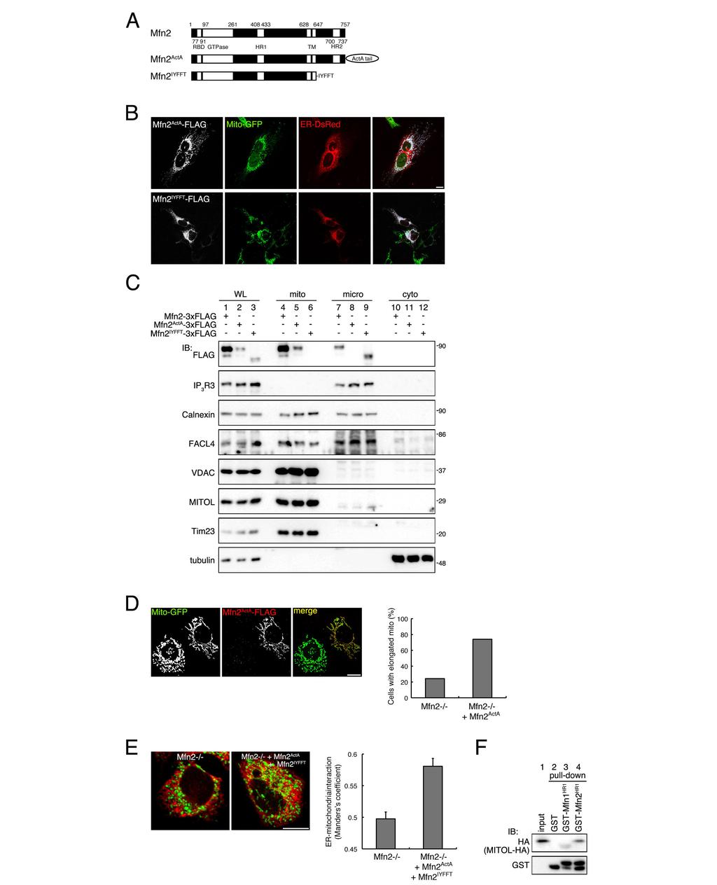
3 Supplemental Figure 1, related to Figure 1. Mitochondrial localization of Mfn2 ActA and ER localization of Mfn2 IYFFT and their functional analyses. (A) Structures of the 2 Mfn2 mutants used in this study. TM, transmembrane domain; RBD, Ras-binding domain; HR, heptad repeat; ActA tail, mitochondria-insertion signal; IYFFT, ER membrane-targeting signal. (B) Mitochondrial localization of Mfn2 ActA and ER localization of Mfn2 IYFFT. Immunofluorescence analysis with Mito-GFP and ER-DsRed was performed on HeLa cells transfected with Mfn2 ActA -FLAG or Mfn2 IYFFT -FLAG. Merged images indicate the mitochondrial localization of Mfn2 ActA and ER localization of Mfn2 IYFFT. (C) Subcellular fractionation demonstrated mitochondrial localization of Mfn2 ActA and ER localization of Mfn2 IYFFT. IB analysis was performed using the indicated Abs in HEK293 cells transfected with wild-type Mfn2, Mfn2 ActA -FLAG, or Mfn2 IYFFT -FLAG. (D) Mitochondrial fusion activity of Mfn2 ActA. Mfn2-/- MEFs were co-transfected with Mfn2 ActA -3 FLAG and Mito-GFP. Mitochondrial morphological changes in Mfn2-/- MEFs co-transfected with Mfn2 ActA -3 FLAG and Mito-GFP were analyzed by confocal microscopy. Percentages of cells with elongated mitochondria obtained from 3 independent experiments (103 Mfn2-/- cells and 73 Mfn2-/- cells expressing Mfn2 ActA ) are shown on the right. Mfn2-/-, FLAG-negative cells. (E) Rescue of MAM formation in Mfn2-/- MEFs expressing Mfn2 ActA and Mfn2 IYFFT. Immunofluorescence analysis was performed on Mfn2-/- MEFs co-transfected with Mfn2 ActA and Mfn2 IYFFT using Mito-GFP and ER-DsRed. Three-dimensional images were reconstructed, and MAM formation as indicated in the merged image was quantified by Manders s coefficient. Error bars indicate ± SE (n = 50). Scale bars represent 10 µm. (F) Relationship between MITOL and Mfn1. The GST-HR1 fragment of Mfn1 does not pull down MITOL. A GST pull-down assay was performed on lysates from HEK293 cells expressing HA-tagged MITOL and GST, GST-Mfn2 HR1 ( aa), or GST-Mfn1 HR1 ( aa), followed by IB analysis with the indicated Abs.
4
5 Supplemental Figure 2, related to Figure 2. MITOL is not involved in the degradation of Mfn2. (A) No obvious change in Mfn2 levels following MITOL knockdown. An IB assay was performed on shgfp- or shmitol-expressing HeLa cells with anti-mfn2 and anti-drp1 Abs, and each protein was quantified. Error bars indicate ± SD. *, p < 0.05 (Student s t-test, n = 3). (B) (C) The rate of Mfn2 degradation was not altered by MITOL knockdown or MITOL overexpression. A cycloheximide (CHX)-chase assay was performed on (B) shgfp- and shmitol-expressing cells or (C) HeLa cells co-transfected with Mfn2-HA and either empty vector (vec) or MITOL. Cells were treated with 10 µg/ml CHX for the indicated times. The level of Mfn2 was monitored by IB with the indicated Abs, and the statistical analysis was performed as described in (A). Error bars indicate ± SD, n = 3. (D) Mfn1 slightly accumulates in shmitol-expressing cells. IB analysis was performed on shgfp- or shmitol-expressing HeLa cells using the indicated Abs. (E) A CHX chase assay showed delayed degradation of Mfn1. Error bars indicate ± SD (n = 3). The reason for Mfn1 accumulation in shmitol-expressing cells is unknown at present. It is possible that increased Mfn1 expression compensates for Mfn2 dysfunction in shmitol-expressing cells. (F) MITOL knockdown induced mitochondrial elongation. Mitochondria of shgfp- and shmitol-expressing HeLa cells were labeled with DsRed-Mito. Mitochondrial morphology was analyzed by confocal microscopy. Graph indicates the mean ± SD of 3 independent experiments (50 cells in each experiment). Scale bar represents 10 µm.

6 Supplemental Figure 3, related to Figure 3. Effect of MITOL knockdown on Mfn2 localization and ER morphology. (A) MITOL knockdown induced Mfn2 mislocalization. An immunofluorescence assay was performed on shgfp- and shmitol-expressing cells transfected with Mfn2-FLAG and DsRed-Mito by staining with anti-flag Ab. Arrowheads indicate punctiform Mfn2 not co-localized to mitochondria. These were quantified by Manders s coefficient. The data in the graph represent the mean ± SE (40 cells in each experiment). Scale bar represents 10 µm.
7 (B) An electron microscopy analysis showed a decrease in the number of mitochondria interacting with ER in MITOL-knockdown cells. Contacted and dissociated positions between the ER and mitochondria are indicated by closed and open arrowheads, respectively. The data in the graph represent the percentages of mitochondria interacting with ER obtained from 70 mitochondria in 7 shgfp or shmitol cells. Error bars indicate ± SD of 3 independent experiments. Scale bar represents 200 nm. (C) MITOL knockdown induced abnormal ER morphology. Morphological analysis using confocal microscopy was performed on shgfp- and shmitol-expressing cells transfected with ER-DsRed. Changes in ER morphology were evaluated from 3 independent experiments (50 cells/experiment). Abnormal ER morphology was defined as disrupted reticular morphology with aggregation or fragmentation. Graph represents the mean ± SD. *, p < 0.05 (chi-square test data from 3 independent experiments). Scale bar represents 10 µm. (D) MITOL localizes to the MAM domain. Each subcellular fraction was isolated from HeLa cells, and IB was performed with the indicated Abs. IP 3 R3 and Calnexin, ER/MAM proteins; FACL4 (long chain fatty acid-coa ligase4), MAM protein; Mfn2, mitochondrial/mam/microsomal protein; VDAC, MAM and mitochondrial outer membrane protein; Tom20, mitochondrial outer membrane protein; α-tubulin, cytosolic protein.
8 Supplemental Figure 4, related to Figure 4 and Figure 7. MITOL and ubiquitination of K192 of Mfn2 are critical for intracellular Ca 2+ homeostasis. shgfp- and shmitol-expressing HeLa cells (A, B), or Mfn2-/- MEFs (C, D) were transfected with the indicated vectors. Mitochondrial (A, C) and cytosolic (B, D) Ca 2+ mobilization was monitored as described in Fig. 4 and Materials and Methods. The data in
 To detect Mfn1 complexes, a blue native (BN)-PAGE retardation assay was performed on MAM fractions derived from shgfp- or shmitol-expressing cells.")
9 the graphs represent the mean ± SE (50 cells). Supplemental Figure 5, related to Figure 5. MITOL knockdown inhibited the formation of high-molecular-weight Mfn1 complexes. (A) To detect Mfn1 complexes, a blue native (BN)-PAGE retardation assay was performed on MAM fractions derived from shgfp- or shmitol-expressing cells. IB after SDS-PAGE (lower panel) is shown as an Mfn1 loading control. (B) MITOL knockdown inhibited the interaction of ER-Mfn2 with Mfn1. An IP-IB assay was performed on shgfp- and shmitol-expressing cells transfected with indicated vectors. The amount of Mfn1 that co-immunoprecipitated with ER-Mfn2 was quantified by densitometry. Relative values are shown in the graph. Error bars indicate ± SD. *, p < 0.05 (Student s t-test, n = 3).
10 Supplemental Figure 6, related to Figure 7. Ubiquitination at K192 of Mfn2 by MITOL is critical for Mfn2 activation. (A) Comparison of the amino acid sequences in the GTPase domains of Mfn1 and Mfn2.
11 The GTPase domain sequences of Mfn1 and Mfn2 were aligned with ClustalW. The arrowhead indicates the lysine residue in Mfn2 that is not conserved in Mfn1. (B) MITOL ubiquitinates the GTPase-defective K109A Mfn2 mutant as well as wild-type Mfn2. Lysates of HeLa cells transfected with the indicated vectors were subjected to an IP-IB assay with the indicated Abs. (C) No changes in the degradation of Mfn2 K109A and K192R mutants. A cycloheximide (CHX)-chase assay was performed on HeLa cells transfected with the indicated vectors. Cells were treated with 10 µg/ml CHX for the indicated times. The level of Mfn2 was monitored over time by IB with an anti-flag Ab. Error bars indicate ± SD, n = 3. (D) (E) Lysine 192 is required for high efficiency binding to and hydrolysis of GTP. Mitochondrial lysates from HEK293 cells transfected with the indicated vectors were used in a GTP-binding assay. Eluted and GTP-bound Mfn2 was analyzed by IB with an anti-ha Ab (D). Immunopurified FLAG-tagged Mfn2 from Mfn2-/- cells was incubated with [ -32 P]GTP at 37 C for 1 h, and analyzed by thin layer chromatography followed by digital autoradiography. (F) (G) The Mfn2 K192R mutant rescue altered mitochondrial morphology, but not ER morphology in Mfn2-/- cells. Mfn2-/- MEFs were cotransfected with empty vector (Vec), FLAG-tagged Mfn2, or Mfn2 K192R and Mito-GFP. Mitochondrial and ER morphology was analyzed by confocal microscopy. The data shown in the graphs indicate the percentage of cells with elongated mitochondria (vec: 87, Mfn2: 62, Mfn2 K192R : 141 cells were observed respectively) or reticular ER (vec: 92, Mfn2: 41, Mfn2 K192R : 95 cells were observed respectively). Graph indicates the mean ± SD of 3 independent experiments (50 cells in each experiment). Scale bars represent 10 µm.
12 Supplemental Experimental Procedures cdna cloning, expression vectors, and mutagenesis To clone cdna encoding human Mfn1 and Mfn2, total RNA isolated from HeLa cells was reverse-transcribed using the Superscript RT kit (Invitrogen) according to the manufacturer s protocol. Full-length cdnas were obtained by PCR and subcloned into pflag CMV14 (Sigma). To generate localization-restricted Mfn2, mitochondrial Mfn2 (Mfn2 ActA ) and ER Mfn2 (Mfn2 IYFFT ) were constructed as previously described (Rojo et al., 2002; Zhu et al., 1996). cdna containing the Mfn2 point mutations K192R or K109A or ubiquitin mutants were generated with the site-directed mutagenesis kit (Stratagene) Antibodies and reagents The anti-mitol rabbit polyclonal Ab was previously described (Yonashiro et al., 2006). Anti-FLAG (M2) and mouse monoclonal anti- -tubulin Abs were purchased from Sigma. The mouse monoclonal anti-c-myc Ab was from Roche. The mouse monoclonal anti-ha Ab was from Babco. The rabbit polyclonal anti-v5 Ab was from MBL. The mouse monoclonal anti-gst Ab was from WAKO. The rabbit polyclonal anti-his Ab was from Cell Signaling Technology. The mouse monoclonal Abs against Drp1, Tom20, Tim23, PDI, and IP 3 R3 were from BD Biosciences. The mouse monoclonal anti-mitofusin2 Ab (XX-1), mouse monoclonal anti-ubiquitin Ab (P4D1), and goat polyclonal anti-calnexin Ab (C-20) were from Santa Cruz. The rabbit polyclonal anti-facl4 Ab was from ABGENT. The rabbit polyclonal anti-vdac Ab was from Calbiochem. The rabbit polyclonal anti-mfn1 Ab was from Abcam. The rabbit polyclonal anti-cytochrome P450 Ab was from Chemicon. The anti-ub K48- and K63-specific Abs were from Millipore. shrna construct For the shrna experiments, 2 complementary oligonucleotides (sense with an added BamHI linker, 5'-gat cgg TTT ACG TCT TGG ATC TTG Ccg aag CAA GAT CCA AGA CGT AAA CCt ttt tt-3'; antisense with an added HindIII linker, 5'-agc taa aaa agg TTT ACG TCT TGG ATC TTG Ctt cgg CAA GAT CCA AGA CGT AAA CC-3') were designed and inserted into the psilencer 3.1-H1 neo vector according to the manufacturer s
13 instructions (Ambion). shrna-resistant MITOL An shrna-resistant MITOL construct was generated by site-directed mutagenesis using following primers: 1st (sense, 5'-AAA ATT GGG TCC AGT TGT GTA CGT GTT GGA TCT TGC AGA T-3'; antisense, 5'-ATC TGC AAG ATC CAA CAC GTA CACA ACTG GAC CCA ATT TT-3'), 2nd (5'-AAA ATT GGG ACC GGT TGT GTA CGT GCT GGA TCT TGC AGA T-3'; antisense, 5'-ATC TGC AAG ATC CAG CAC GTA CAC AAC CGG TCC CAA TTTT-3'). shmitol stable cell lines To establish MITOL-knocked down cell lines, HeLa cells were transfected with shmitol or shgfp as a control. Cells were selected with 1 mg/ml G418 neomycin. Morphological analysis by immunofluorescence microscopy Cells were fixed with 4% paraformaldehyde in phosphate-buffered saline (PBS) for 1 h at room temperature, then washed twice with PBS, permeabilized with 0.2% Triton X-100 in PBS for 5 min, then washed 4 times with PBS, and blocked with 1% bovine serum albumin in PBS, all at room temperature. For double staining, the cells were incubated with appropriate primary Abs for 1 h at room temperature, washed 3 times with PBS, and then incubated with appropriate secondary Abs for 30 min. The samples were washed as described above, mounted using Fluorescent Mounting Medium (Dako), and analyzed using an Olympus IX81 confocal fluorescence microscope. To analyze the interaction between mitochondria and ER, a z-axis image series of cells expressing mitochondrial GFP and ER-DsRed were obtained and stacked. The background signals were removed using the BG subtraction function of ImageJ, and then Manders s colocalization coefficient was calculated. For ER morphological analysis, DsRed-labeled ER was observed in shrna-transfected stable cell lines with a confocal fluorescence microscope. ER morphology (reticular or not,
14 aggregation or fragmentation) was determined. Data are the mean ± SD of 3 independent experiments. Electron microscopy HeLa cells stably expressing shgfp or shmitol were fixed with 2.5% glutaraldehyde in 0.1 M sodium phosphate buffer (ph 7.4). They were postfixed with 2% OsO 4 in the same buffer. After dehydration with a graded series of ethanol, they were substituted by propylene oxide and embedded in Epoxy resin. Silver and gold thin sections were doubly stained with uranyl acetate and lead citrate, and examined with an H-7000 transmission electron microscope (HITACHI, Tokyo, Japan). Subcellular fractionation MAM fractions were isolated from HeLa cells or mouse brain as previously described (Szabadkai et al., 2006; Vance, 1990). HeLa cells or mouse brain were homogenized with homogenization buffer (HB) (5 mm HEPES ph 7.4, 0.5 mm EDTA, 250 mm mannitol) and the crude mitochondrial fraction (pellet after centrifugation at 8,000 g) was subjected to further separation by 30% Percoll gradient centrifugation at 9,500 g for 30 min at 4 C. A low-density band (denoted as the MAM fraction) was resuspended in HB and further purified by removing the pellet after centrifugation at 6,300 g for 10 min. The mitochondrial fraction in the high-density band was resuspended in HB and pelleted by centrifugation (6,300 g for 10 min). The MAM fraction was obtained by centrifugation at 100,000 g for 1 h at 4 C. Microsomes were pelleted from the mitochondrial supernatant by centrifugation at 100,000 g for 1 h. Cytosol was recovered as the supernatant from this centrifugation. SDS-PAGE sample buffer was added to each fraction and analyzed by IB. Immunoprecipitation To analyze the interaction between endogenous MITOL and Mfn2, crude mitochondria from mouse brain cells were solubilized with lysis buffer (0.5% NP-40, 50 mm Tris-HCl ph 7.4, 150 mm NaCl, 5 mm EDTA, 20% sucrose, protease inhibitors). Lysates were centrifuged at 15,000 rpm for 10 min at 4 C, and the supernatant was subjected to IP using
15 an anti-mitol Ab and normal rabbit IgG as a control. For the ubiquitination assay, HeLa cells transfected with vectors as indicated in the figures were lysed with 0.1% SDS RIPA buffer (0.1% SDS, 0.05% DOC, 1% TritonX-100, 10 mm Tris-HCl ph 7.4, 150 mm NaCl, 5 mm EDTA, protease inhibitors), and then incubated with the appropriate Ab. For the in vitro ubiquitination assay, FLAG-tagged Mfn2 and HA-tagged MITOL were purified from HEK293 cells by IP. Immunoprecipitates were incubated with reaction buffer containing 50 mm Tris-HCl, ph 7.4, 2 mm MgCl 2, 4 mm ATP, 100 ng of E1 (Biomol), 400 ng of UbcH5b (Biomol), and 2 g of His-Ub (Biomol) for 2 h at 30 C, and then terminated with 3 SDS sample buffer. GST pull-down assay To determine the specific sites required for MITOL and Mfn2 interaction, GST-fusion deletion mutants were prepared as described previously (Yonashiro et al., 2006). Lysates from mouse brain were incubated with the purified GST-fusion proteins. Precipitates were analyzed by IB. Ca 2+ imaging HeLa cells stably expressing shrnas were seeded on a glass-bottom dish 1 day before measurement. For transfection, HeLa cells or Mfn2-/- MEFs were seeded on a glass-bottom dish 2 days before measurement. After 1 day, cells were transfected with the expression vectors described in supplemental Fig. 5. Cells were washed with BSS buffer (20 mm HEPES, 130 mm NaCl, 5.4 mm KCl, 2 mm CaCl 2, 1 mm MgCl 2, 5.5 mm glucose), stained with 3 µm XRhod 1-AM (Molecular Probes) for 1 h or 3 µm Fura 4-AM (Molecular Probes) for 20 min at RT, washed with BSS, and then incubated in BSS for 30 min. Ca 2+ mobilization induced by treatment with 1 µm histamine (Sigma) for HeLa cells or 200 µm ATP (Sigma) for MEFs was detected by a quantitative fluorescence system using a CCD camera (Orca-ER; Hamamatsu Photonics K.K., Hamamatsu, Shizuoka) through the 40 objective lens of an inverted microscope. The images were processed using software specific for Ca 2+ imaging (Aquacosmos; Hamamatsu Photonics). The time course of the change in [Ca 2+ ]i at a region of interest (ROI) in each cell was calculated as the
16 average brightness of each ROI. For the preparation loaded with fura-4f, the ratio of the fluorescence (>500 nm) induced by alternative excitation at 340 nm and 380 nm (F340/F380) was calculated. In the X rhod-1-stained cells, the ratio of the fluorescence (>580 nm) induced by excitation at 540 nm was calculated using the initial image as a reference. In the present study, F340/F380 data for fura-4f, and F540/F540 data for X rhod-1 were normalized to the time point just before exposure to histamine, which was set to 1.0 (10 cells/experiment, n = 3) Blue Native PAGE Blue-native PAGE (BN-PAGE) was performed according to Schagger (Schagger, 2001). Mitochondria or MAM domains isolated from HeLa cells were solubilized with solubilization buffer (1% digitonin, 20 mm NaCl, 50 mm imidazole ph 7.0, 1 mm EDTA, 10% glycerol, 500 mm 6-aminocaproic acid, protease inhibitors). After centrifugation at 14,000 g for 10 min at 4 C Coomassie Brilliant blue G-250 (0.2% final concentration) was added, and the samples were electrophoresed through 3 12% polyacrylamide gradient gels. The gels were subjected to IB using the Abs described in the figures. GTP-binding assay Mitochondria were isolated from HeLa cells and solubilized in 1% digitonin buffer (50 mm Tris-HCl, 150 mm NaCl, protease inhibitors). After centrifugation at 14,000 g for 10 min at 4 C, the supernatant was incubated with reaction buffer (50 mm Tris-HCl ph 7.4, 150 mm NaCl, 1 mm MgOAc 2, 1 mm DTT, protease inhibitors) containing GTP-agarose beads on ice or at 30 C for 60 min. The bound proteins were eluted with elution buffer (1% digitonin, 50 mm Tris-HCl ph 7.4, 150 mm NaCl, 10 mm GTP, protease inhibitors) for 1 h at 4 C, and 3 SDS-PAGE sample buffer was added to the eluates including GTP-agarose. The amount of Mfn2 bound to GTP was determined by IB with an anti-mfn2 Ab. For the expressed Mfn2, mitochondria from HEK293 cells were lysed in reaction buffer containing 1% Triton X-100 (50 mm Tris-HCl, ph 7.4, 150 mm NaCl, 5 mm MgCl 2, 1 mm DTT, protease inhibitors) and incubated on ice for 20 min. After centrifugation at 20,000 g for 15 min at 4 C, supernatant was incubated with GTP-agarose for 1 h at 37 C.
17 The bound proteins were eluted with elution buffer (1% digitonin, 50 mm Tris-HCl ph 7.4, 150 mm NaCl, 20 mm EDTA, 10 mm GTP, protease inhibitors). GTP hydrolysis assay Endogenous or expressed Mfn2 was purified from mitochondria by IP using an anti-mfn2 or anti-flag Ab. The purified proteins were incubated with 20 L of 20 mm HEPES-KOH buffer (ph 7.4) containing 2 mm MgCl 2, 1 mm dithiothreitol, and 1% Triton X-100 with 1 Ci of [ - 32 P]GTP at 37 C for 1 h. One microliter of the reaction mixture was removed and spotted on a polyethyleneimine-cellulose filter (Merck). The reaction products were resolved by thin layer chromatography in 1 M LiCl 2 and 2 M formic acid. The products were analyzed and quantified using a Bioimage Analyzer BAS2000 (Fuji Film Co.).
18 Supplemental References Rojo, M., Legros, F., Chateau, D., and Lombes, A. (2002). Membrane topology and mitochondrial targeting of mitofusins, ubiquitous mammalian homologs of the transmembrane GTPase Fzo. J Cell Sci 115, Schagger, H. (2001). Blue-native gels to isolate protein complexes from mitochondria. Methods Cell Biol 65, Szabadkai, G., Bianchi, K., Varnai, P., De Stefani, D., Wieckowski, M.R., Cavagna, D., Nagy, A.I., Balla, T., and Rizzuto, R. (2006). Chaperone-mediated coupling of endoplasmic reticulum and mitochondrial Ca2+ channels. J. Cell Biol. 175, Vance, J.E. (1990). Phospholipid synthesis in a membrane fraction associated with mitochondria. J. Biol. Chem. 265, Yonashiro, R., Ishido, S., Kyo, S., Fukuda, T., Goto, E., Matsuki, Y., Ohmura-Hoshino, M., Sada, K., Hotta, H., Yamamura, H., et al. (2006). A novel mitochondrial ubiquitin ligase plays a critical role in mitochondrial dynamics. EMBO J. 25, Zhu, W., Cowie, A., Wasfy, G.W., Penn, L.Z., Leber, B., and Andrews, D.W. (1996). Bcl-2 mutants with restricted subcellular location reveal spatially distinct pathways for apoptosis in different cell types. EMBO J. 15,
SUPPORTING INFORMATION FOR. SEquence-Enabled Reassembly of β-lactamase (SEER-LAC): a Sensitive Method for the Detection of Double-Stranded DNA
: a Sensitive Method for the Detection of Double-Stranded DNA") SUPPORTING INFORMATION FOR SEquence-Enabled Reassembly of β-lactamase (SEER-LAC): a Sensitive Method for the Detection of Double-Stranded DNA Aik T. Ooi, Cliff I. Stains, Indraneel Ghosh *, David J. Segal
SUPPORTING INFORMATION FOR SEquence-Enabled Reassembly of β-lactamase (SEER-LAC): a Sensitive Method for the Detection of Double-Stranded DNA Aik T. Ooi, Cliff I. Stains, Indraneel Ghosh *, David J. Segal
SUPPLEMENTARY DATA - 1 -
 - 1 - SUPPLEMENTARY DATA Construction of B. subtilis rnpb complementation plasmids For complementation, the B. subtilis rnpb wild-type gene (rnpbwt) under control of its native rnpb promoter and terminator
Supplementary Figure 1.
 Supplementary Figure 1. Characterisation of IHG-1 overexpressing and knockdown cell lines. (A) Total cellular RNA was prepared from HeLa cells stably overexpressing IHG-1 or mts-ihg-1. IHG-1 mrna was quantified
Mitofusin 1 and 2 play distinct roles in mitochondrial fusion reactions via GTPase activity
 Research Article 6535 Mitofusin 1 and 2 play distinct roles in mitochondrial fusion reactions via GTPase activity Naotada Ishihara, Yuka Eura and Katsuyoshi Mihara* Department of Molecular Biology, Graduate
Number-controlled spatial arrangement of gold nanoparticles with
 Electronic Supplementary Material (ESI) for RSC Advances. This journal is The Royal Society of Chemistry 2016 Number-controlled spatial arrangement of gold nanoparticles with DNA dendrimers Ping Chen,*
Practical Bioinformatics
 5/2/2017 Dictionaries d i c t i o n a r y = { A : T, T : A, G : C, C : G } d i c t i o n a r y [ G ] d i c t i o n a r y [ N ] = N d i c t i o n a r y. h a s k e y ( C ) Dictionaries g e n e t i c C o
TNFα 18hr. Control. CHX 18hr. TNFα+ CHX 18hr. TNFα: 18 18hr (KDa) PARP. Cleaved. Cleaved. Cleaved. Caspase3. Pellino3 shrna. Control shrna.
 PARP. Cleaved. Cleaved. Cleaved. Caspase3. Pellino3 shrna. Control shrna.") Survival ( %) a. TNFα 18hr b. Control sirna Pellino3 sirna TNFα: 18 18hr c. Control shrna Pellino3 shrna Caspase3 Actin Control d. Control shrna Pellino3 shrna *** 100 80 60 CHX 18hr 40 TNFα+ CHX 18hr
Survival ( %) a. TNFα 18hr b. Control sirna Pellino3 sirna TNFα: 18 18hr c. Control shrna Pellino3 shrna Caspase3 Actin Control d. Control shrna Pellino3 shrna *** 100 80 60 CHX 18hr 40 TNFα+ CHX 18hr
Supplementary Information
 Supplementary Information MAP2/Hoechst Hyp.-AP ph 6.5 Hyp.-SD ph 7.2 Norm.-SD ph 7.2 Supplementary Figure 1. Mitochondrial elongation in cortical neurons by acidosis. Representative images of neuronal
Nature Structural & Molecular Biology: doi: /nsmb Supplementary Figure 1
 Supplementary Figure 1 Zn 2+ -binding sites in USP18. (a) The two molecules of USP18 present in the asymmetric unit are shown. Chain A is shown in blue, chain B in green. Bound Zn 2+ ions are shown as
Clay Carter. Department of Biology. QuickTime and a TIFF (Uncompressed) decompressor are needed to see this picture.
 decompressor are needed to see this picture.") QuickTime and a TIFF (Uncompressed) decompressor are needed to see this picture. Clay Carter Department of Biology QuickTime and a TIFF (LZW) decompressor are needed to see this picture. Ornamental tobacco
QuickTime and a TIFF (Uncompressed) decompressor are needed to see this picture. Clay Carter Department of Biology QuickTime and a TIFF (LZW) decompressor are needed to see this picture. Ornamental tobacco
High throughput near infrared screening discovers DNA-templated silver clusters with peak fluorescence beyond 950 nm
 Electronic Supplementary Material (ESI) for Nanoscale. This journal is The Royal Society of Chemistry 2018 High throughput near infrared screening discovers DNA-templated silver clusters with peak fluorescence
Supporting Information
 Supporting Information T. Pellegrino 1,2,3,#, R. A. Sperling 1,#, A. P. Alivisatos 2, W. J. Parak 1,2,* 1 Center for Nanoscience, Ludwig Maximilians Universität München, München, Germany 2 Department of
Supplemental Figure 1.
 A wt spoiiiaδ spoiiiahδ bofaδ B C D E spoiiiaδ, bofaδ Supplemental Figure 1. GFP-SpoIVFA is more mislocalized in the absence of both BofA and SpoIIIAH. Sporulation was induced by resuspension in wild-type
Supporting Information
 Supporting Information Wang et al. 10.1073/pnas.0804871105 SI Materials and Methods Cell Culture and Transfection. Human neuroblastoma M17 cells were grown as described before (1). Transfection was performed
SUPPLEMENTAL MATERIAL
 SUPPLEMENTAL MATERIAL Figure S1. Mitochondrial morphology in Fis1-null, Mff-null and Fis1/Mff-null MEF cells. (A) Western blotting of lysates from Fis1-null, Mff-null and Fis1/Mff-null cells. Lysates were
Supporting Information for. Initial Biochemical and Functional Evaluation of Murine Calprotectin Reveals Ca(II)-
-") Supporting Information for Initial Biochemical and Functional Evaluation of Murine Calprotectin Reveals Ca(II)- Dependence and Its Ability to Chelate Multiple Nutrient Transition Metal Ions Rose C. Hadley,
Supporting Information for Initial Biochemical and Functional Evaluation of Murine Calprotectin Reveals Ca(II)- Dependence and Its Ability to Chelate Multiple Nutrient Transition Metal Ions Rose C. Hadley,
Supplemental Table 1. Primers used for cloning and PCR amplification in this study
 Supplemental Table 1. Primers used for cloning and PCR amplification in this study Target Gene Primer sequence NATA1 (At2g393) forward GGG GAC AAG TTT GTA CAA AAA AGC AGG CTT CAT GGC GCC TCC AAC CGC AGC
Supplemental data. Pommerrenig et al. (2011). Plant Cell /tpc
. Plant Cell /tpc") Supplemental Figure 1. Prediction of phloem-specific MTK1 expression in Arabidopsis shoots and roots. The images and the corresponding numbers showing absolute (A) or relative expression levels (B) of
Supplemental Figure 1. Prediction of phloem-specific MTK1 expression in Arabidopsis shoots and roots. The images and the corresponding numbers showing absolute (A) or relative expression levels (B) of
Advanced topics in bioinformatics
 Feinberg Graduate School of the Weizmann Institute of Science Advanced topics in bioinformatics Shmuel Pietrokovski & Eitan Rubin Spring 2003 Course WWW site: http://bioinformatics.weizmann.ac.il/courses/atib
Supporting Information
 Supporting Information Mullins et al. 10.1073/pnas.0906781106 SI Text Detection of Calcium Binding by 45 Ca 2 Overlay. The 45 CaCl 2 (1 mci, 37 MBq) was obtained from NEN. The general method of 45 Ca 2
4) Please cite Dagda et al J Biol Chem 284: , for any publications or presentations resulting from use or modification of the macro.
 Please cite Dagda et al J Biol Chem 284: , for any publications or presentations resulting from use or modification of the macro.") Supplement Figure S1. Algorithmic quantification of mitochondrial morphology in SH- SY5Y cells treated with known fission/fusion mediators. Parental SH-SY5Y cells were transiently transfected with an empty
Supplement Figure S1. Algorithmic quantification of mitochondrial morphology in SH- SY5Y cells treated with known fission/fusion mediators. Parental SH-SY5Y cells were transiently transfected with an empty
Illegitimate translation causes unexpected gene expression from on-target out-of-frame alleles
 Illegitimate translation causes unexpected gene expression from on-target out-of-frame alleles created by CRISPR-Cas9 Shigeru Makino, Ryutaro Fukumura, Yoichi Gondo* Mutagenesis and Genomics Team, RIKEN
7.06 Problem Set
 7.06 Problem Set 5 -- 2006 1. In the first half of the course, we encountered many examples of proteins that entered the nucleus in response to the activation of a cell-signaling pathway. One example of
Supplementary Figure 1. Biochemical and sequence alignment analyses the
 Supplementary Figure 1. Biochemical and sequence alignment analyses the interaction of OPTN and TBK1. (a) Analytical gel filtration chromatography analysis of the interaction between TBK1 CTD and OPTN(1-119).
SSR ( ) Vol. 48 No ( Microsatellite marker) ( Simple sequence repeat,ssr),
 Vol. 48 No ( Microsatellite marker) ( Simple sequence repeat,ssr),") 48 3 () Vol. 48 No. 3 2009 5 Journal of Xiamen University (Nat ural Science) May 2009 SSR,,,, 3 (, 361005) : SSR. 21 516,410. 60 %96. 7 %. (),(Between2groups linkage method),.,, 11 (),. 12,. (, ), : 0.
48 3 () Vol. 48 No. 3 2009 5 Journal of Xiamen University (Nat ural Science) May 2009 SSR,,,, 3 (, 361005) : SSR. 21 516,410. 60 %96. 7 %. (),(Between2groups linkage method),.,, 11 (),. 12,. (, ), : 0.
Supplemental Figures S1 S5
 Beyond reduction of atherosclerosis: PON2 provides apoptosis resistance and stabilizes tumor cells Ines Witte (1), Sebastian Altenhöfer (1), Petra Wilgenbus (1), Julianna Amort (1), Albrecht M. Clement
Supplementary Information
 Electronic Supplementary Material (ESI) for RSC Advances. This journal is The Royal Society of Chemistry 2014 Directed self-assembly of genomic sequences into monomeric and polymeric branched DNA structures
SEQUENCE ALIGNMENT BACKGROUND: BIOINFORMATICS. Prokaryotes and Eukaryotes. DNA and RNA
 SEQUENCE ALIGNMENT BACKGROUND: BIOINFORMATICS 1 Prokaryotes and Eukaryotes 2 DNA and RNA 3 4 Double helix structure Codons Codons are triplets of bases from the RNA sequence. Each triplet defines an amino-acid.
Building a Multifunctional Aptamer-Based DNA Nanoassembly for Targeted Cancer Therapy
 Supporting Information Building a Multifunctional Aptamer-Based DNA Nanoassembly for Targeted Cancer Therapy Cuichen Wu,, Da Han,, Tao Chen,, Lu Peng, Guizhi Zhu,, Mingxu You,, Liping Qiu,, Kwame Sefah,
NSCI Basic Properties of Life and The Biochemistry of Life on Earth
 NSCI 314 LIFE IN THE COSMOS 4 Basic Properties of Life and The Biochemistry of Life on Earth Dr. Karen Kolehmainen Department of Physics CSUSB http://physics.csusb.edu/~karen/ WHAT IS LIFE? HARD TO DEFINE,
Supplemental material
 Supplemental material THE JOURNAL OF CELL BIOLOGY Mourier et al., http://www.jcb.org/cgi/content/full/jcb.201411100/dc1 Figure S1. Size and mitochondrial content in Mfn1 and Mfn2 knockout hearts. (A) Body
Supplemental Information. The Mitochondrial Fission Receptor MiD51. Requires ADP as a Cofactor
 Structure, Volume 22 Supplemental Information The Mitochondrial Fission Receptor MiD51 Requires ADP as a Cofactor Oliver C. Losón, Raymond Liu, Michael E. Rome, Shuxia Meng, Jens T. Kaiser, Shu-ou Shan,
Downloaded from at University of Washington Health Sciences Libraries, on November 12, 2010
 THE JOURNAL OF BIOLOGICAL CHEMISTRY VOL. 285, NO. 41, pp. 31590 31602, October 8, 2010 2010 by The American Society for Biochemistry and Molecular Biology, Inc. Printed in the U.S.A. Rab32 Modulates Apoptosis
Electronic supplementary material
 Applied Microbiology and Biotechnology Electronic supplementary material A family of AA9 lytic polysaccharide monooxygenases in Aspergillus nidulans is differentially regulated by multiple substrates and
ydci GTC TGT TTG AAC GCG GGC GAC TGG GCG CGC AAT TAA CGG TGT GTA GGC TGG AGC TGC TTC
 Table S1. DNA primers used in this study. Name ydci P1ydcIkd3 Sequence GTC TGT TTG AAC GCG GGC GAC TGG GCG CGC AAT TAA CGG TGT GTA GGC TGG AGC TGC TTC Kd3ydcIp2 lacz fusion YdcIendP1 YdcItrgP2 GAC AGC
Regulation of mitochondrial fission and apoptosis by the mitochondrial outer membrane protein hfis1
 Research Article 4141 Regulation of mitochondrial fission and apoptosis by the mitochondrial outer membrane protein hfis1 Tianzheng Yu 1, Randall J. Fox 1, Lindsay S. Burwell 2 and Yisang Yoon 1, * 1 Department
A novel mitochondrial ubiquitin ligase plays a critical role in mitochondrial dynamics
 The EMBO Journal (2006) 25, 3618 3626 & 2006 European Molecular Biology Organization All Rights Reserved 0261-4189/06 www.embojournal.org A novel mitochondrial ubiquitin ligase plays a critical role in
Supplementary Figure 1. SDS-PAGE analysis of GFP oligomer variants with different linkers. Oligomer mixtures were applied to a PAGE gel containing
 Supplementary Figure 1. SDS-PAGE analysis of GFP oligomer variants with different linkers. Oligomer mixtures were applied to a PAGE gel containing 0.1% SDS without boiling. The gel was analyzed by a fluorescent
OESTREICH LAB CHIP PROTOCOL
 OESTREICH LAB CHIP PROTOCOL Buffers (all starred (*) buffers must be autoclaved before use) 10X Cell Lysis Buffer*: 100mL 0.214g Potassium Acetate (KOAc) = 10mM 0.147g Magnesium Acetate (MgAc) = 15mM 10mL
Full-length GlpG sequence was generated by PCR from E. coli genomic DNA. (with two sequence variations, D51E/L52V, from the gene bank entry aac28166),
,") Supplementary Methods Protein expression and purification Full-length GlpG sequence was generated by PCR from E. coli genomic DNA (with two sequence variations, D51E/L52V, from the gene bank entry aac28166),
Supplementary Methods Protein expression and purification Full-length GlpG sequence was generated by PCR from E. coli genomic DNA (with two sequence variations, D51E/L52V, from the gene bank entry aac28166),
Figure S1: Extracellular nicotinic acid, but not tryptophan, is sufficient to maintain
 SUPPLEMENTAL INFORMATION Supplemental Figure Legends Figure S1: Extracellular nicotinic acid, but not tryptophan, is sufficient to maintain mitochondrial NAD +. A) Extracellular tryptophan, even at 5 µm,
7.06 Cell Biology EXAM #3 April 21, 2005
 7.06 Cell Biology EXAM #3 April 21, 2005 This is an open book exam, and you are allowed access to books, a calculator, and notes but not computers or any other types of electronic devices. Please write
TM1 TM2 TM3 TM4 TM5 TM6 TM bp
 a 467 bp 1 482 2 93 3 321 4 7 281 6 21 7 66 8 176 19 12 13 212 113 16 8 b ATG TCA GGA CAT GTA ATG GAG GAA TGT GTA GTT CAC GGT ACG TTA GCG GCA GTA TTG CGT TTA ATG GGC GTA GTG M S G H V M E E C V V H G T
Supplementary Information for
 Supplementary Information for Evolutionary conservation of codon optimality reveals hidden signatures of co-translational folding Sebastian Pechmann & Judith Frydman Department of Biology and BioX, Stanford
Spherotech, Inc Irma Lee Circle, Unit 101, Lake Forest, Illinois PARTICLE COATING PROCEDURES
 SPHERO TM Technical Note STN-1 Rev C. 041106 Introduction Currently, there are several methods of attaching biological ligands to polystyrene particles. These methods include adsorption to plain polystyrene
Protease Inhibitor Cocktail A (1 tablet / 7 10 ml, Roche Cat# ) Protease inhibitor Cocktail B (0.5ml per 250ml, Calbiochem Cat# )
 Protease inhibitor Cocktail B (0.5ml per 250ml, Calbiochem Cat# )") Protocol for Western Blotting Tissue/Cell Sample Preparation Lysis Buffer 1 (ph8.0) o 50mM Tris-Cl o 150mM NaCl o 1% v/v NP40 o protease inhibitor cocktail A/B Lysis Buffer 2 (RIPA) (ph 8.0) o 50mM Tris-Cl
Protocol for Western Blotting Tissue/Cell Sample Preparation Lysis Buffer 1 (ph8.0) o 50mM Tris-Cl o 150mM NaCl o 1% v/v NP40 o protease inhibitor cocktail A/B Lysis Buffer 2 (RIPA) (ph 8.0) o 50mM Tris-Cl
mrna Isolation Kit for Blood/Bone Marrow For isolation mrna from blood or bone marrow lysates Cat. No
 For isolation mrna from blood or bone marrow lysates Cat. No. 1 934 333 Principle Starting material Application Time required Results Key advantages The purification of mrna requires two steps: 1. Cells
SUPPLEMENTARY INFORMATION
 DOI:.8/NCHEM. Conditionally Fluorescent Molecular Probes for Detecting Single Base Changes in Double-stranded DNA Sherry Xi Chen, David Yu Zhang, Georg Seelig. Analytic framework and probe design.. Design
SUPPLEMENTARY INFORMATION
 SUPPLEMENTARY INFORMATION DOI:.38/NCHEM.246 Optimizing the specificity of nucleic acid hyridization David Yu Zhang, Sherry Xi Chen, and Peng Yin. Analytic framework and proe design 3.. Concentration-adjusted
Importance of Protein sorting. A clue from plastid development
 Importance of Protein sorting Cell organization depend on sorting proteins to their right destination. Cell functions depend on sorting proteins to their right destination. Examples: A. Energy production
AtTIL-P91V. AtTIL-P92V. AtTIL-P95V. AtTIL-P98V YFP-HPR
 Online Resource 1. Primers used to generate constructs AtTIL-P91V, AtTIL-P92V, AtTIL-P95V and AtTIL-P98V and YFP(HPR) using overlapping PCR. pentr/d- TOPO-AtTIL was used as template to generate the constructs
Direct Interaction between Survivin and Smac/DIABLO Is Essential for the Anti-apoptotic Activity of Survivin during Taxol-induced Apoptosis*
 THE JOURNAL OF BIOLOGICAL CHEMISTRY Vol. 278, No. 25, Issue of June 20, pp. 23130 23140, 2003 2003 by The American Society for Biochemistry and Molecular Biology, Inc. Printed in U.S.A. Direct Interaction
Crick s early Hypothesis Revisited
 Crick s early Hypothesis Revisited Or The Existence of a Universal Coding Frame Ryan Rossi, Jean-Louis Lassez and Axel Bernal UPenn Center for Bioinformatics BIOINFORMATICS The application of computer
SUPPLEMENTARY INFORMATION
 DOI: 10.1038/ncb2215 Figure S1 Number of egfp-vps4a bursts versus cellular expression levels. The total number of egfp-vps4a bursts, counted at the end of each movie (frame 2000, after 1h 28 min) are plotted
Characterization of Pathogenic Genes through Condensed Matrix Method, Case Study through Bacterial Zeta Toxin
 International Journal of Genetic Engineering and Biotechnology. ISSN 0974-3073 Volume 2, Number 1 (2011), pp. 109-114 International Research Publication House http://www.irphouse.com Characterization of
Supplementary Figure 1. CoMIC in 293T, HeLa, and HepG2 cells. (a) Mitochondrial morphology in 293T, HeLa and HepG2 cells. Cells were transfected with
 Mitochondrial morphology in 293T, HeLa and HepG2 cells. Cells were transfected with") Supplementary Figure 1. CoMIC in 293T, HeLa, and HepG2 cells. (a) Mitochondrial morphology in 293T, HeLa and HepG2 cells. Cells were transfected with DsRed-mito. Right panels are time-course enlarged images
Supplementary Figure 1. CoMIC in 293T, HeLa, and HepG2 cells. (a) Mitochondrial morphology in 293T, HeLa and HepG2 cells. Cells were transfected with DsRed-mito. Right panels are time-course enlarged images
Bottom-up Optimization of SERS Hot Spots. Supplementary Information
 Bottom-up Optimization of SERS Hot Spots Laura Fabris, * Department of Materials Science and Engineering, Institute for Advanced Materials Devices ad Nanotechnology, Rutgers, The State University of New
evoglow - express N kit distributed by Cat.#: FP product information broad host range vectors - gram negative bacteria
 evoglow - express N kit broad host range vectors - gram negative bacteria product information distributed by Cat.#: FP-21020 Content: Product Overview... 3 evoglow express N -kit... 3 The evoglow -Fluorescent
Center for Cell Imaging Department of Cell Biology
 Center for Cell Imaging Department of Cell Biology Contents Preparation of Colloidal Gold Conjugates Coupling the Protein A to the Gold Particles Purification of the protein A-gold. Storage Influence of
horseradish peroxidase-labeled anti-mouse secondary antibody were procured from
 SUPPORTING INFORMATION Characterization of anti-platelet properties of silver nanoparticles Siddhartha Shrivastava, Tanmay Bera, Sunil K. Singh, Gajendra Singh, P. Ramachandrarao and Debabrata Dash 1.
SUPPLEMENTARY INFORMATION
 DOI:.38/ncb97 P ( μm, hours) 1 2 4 P DMSO Figure S1 unningham et al. E 97 65 27 MFN1 GFP-Parkin Opa1 ctin GPDH HEK293 GFP-Parkin 19 115 97 65 27 Mitochondrial Fraction SH-SY5Y GFP-Parkin Mito DMSO Mito
Optimization of Immunoblot Protocol for Use with a Yeast Strain Containing the CDC7 Gene Tagged with myc
 OPTIMIZATION OF IMMUNOBLOT PROTOCOL 121 Optimization of Immunoblot Protocol for Use with a Yeast Strain Containing the CDC7 Gene Tagged with myc Jacqueline Bjornton and John Wheeler Faculty Sponsor: Anne
Supplemental Data. Gao et al. (2012). Plant Cell /tpc
. Plant Cell /tpc") Supplemental Figure 1. Plant EMP Proteins. (A) The Accession numbers of the 12 EMP members from Arabidopsis. (B) Phylogenetic analysis of EMP proteins from Arabidopsis, human and yeast using the Mac Vector
Supplemental Figure 1. Plant EMP Proteins. (A) The Accession numbers of the 12 EMP members from Arabidopsis. (B) Phylogenetic analysis of EMP proteins from Arabidopsis, human and yeast using the Mac Vector
The photoluminescent graphene oxide serves as an acceptor rather. than a donor in the fluorescence resonance energy transfer pair of
 Supplementary Material (ESI) for Chemical Communications This journal is (c) The Royal Society of Chemistry 20XX The photoluminescent graphene oxide serves as an acceptor rather than a donor in the fluorescence
Deregulation and Mislocalization of the Cytokinesis Regulator ECT2 Activate the Rho Signaling Pathways Leading to Malignant Transformation*
 THE JOURNAL OF BIOLOGICAL CHEMISTRY Vol. 279, No. 8, Issue of February 20, pp. 7169 7179, 2004 Printed in U.S.A. Deregulation and Mislocalization of the Cytokinesis Regulator ECT2 Activate the Rho Signaling
Protocol for 2D-E. Protein Extraction
 Protocol for 2D-E Protein Extraction Reagent 1 inside the ReadyPrep TM Sequential Extraction kit (in powder form) 50ml of deionized water is used to dissolve all the Reagent 1. The solution is known as
Bis sulfone Reagents. Figure 1.
 Bis sulfone Reagents An intact IgG molecule has four accessible inter chain disulfide bonds that can be reduced to form eight free cysteine thiols, which can serve as sites for conjugation. The reaction
Preparing Colloidal Gold for Electron Microscopy
 Corporate Headquarters 400 Valley Road Warrington, PA 18976 1-800-523-2575 FAX 1-800-343-3291 Email: info@polysciences.com www.polysciences.com Europe - Germany Polysciences Europe GmbH Handelsstr. 3 D-69214
ANALYZING THE DIVERSITY OF A SMALL ANTIBODY MIMIC LIBRARY. Nick Empey. Chapel Hill 2010
 ANALYZING THE DIVERSITY OF A SMALL ANTIBODY MIMIC LIBRARY Nick Empey A thesis submitted to the faculty of the University of North Carolina at Chapel Hill in partial fulfillment of the requirements for
Table S1. Primers and PCR conditions used in this paper Primers Sequence (5 3 ) Thermal conditions Reference Rhizobacteria 27F 1492R
 Thermal conditions Reference Rhizobacteria 27F 1492R") Table S1. Primers and PCR conditions used in this paper Primers Sequence (5 3 ) Thermal conditions Reference Rhizobacteria 27F 1492R AAC MGG ATT AGA TAC CCK G GGY TAC CTT GTT ACG ACT T Detection of Candidatus
Table S1. Primers and PCR conditions used in this paper Primers Sequence (5 3 ) Thermal conditions Reference Rhizobacteria 27F 1492R AAC MGG ATT AGA TAC CCK G GGY TAC CTT GTT ACG ACT T Detection of Candidatus
A single lysine in the N-terminal region of store-operated channels is critical for STIM1-mediated gating
 Published Online: 29 November, 2010 Supp Info: http://doi.org/10.1085/jgp.201010484 Downloaded from jgp.rupress.org on December 25, 2018 A r t i c l e A single lysine in the N-terminal region of store-operated
Supporting Information
 Supporting Information Dumbbell-Like Au-Fe 3 Nanoparticles for Target-Specific Platin Delivery Chenjie Xu, Baodui Wang, and Shouheng Sun* Department of Chemistry, Brown University, Providence, Rhode Island
TaKaRa BCA Protein Assay Kit
 Cat. # T9300A For Research Use TaKaRa BCA Protein Assay Kit Product Manual Table of Contents I. Description... 3 II. Components... 3 III. Storage... 3 IV. Materials Required by not Provided... 3 V. Precautions
Programmed ph-driven Reversible Association and Dissociation of Inter-Connected. Circular DNA Dimer Nanostructures
 Supporting information Programmed ph-driven Reversible Association and Dissociation of Inter-Connected Circular DNA Dimer Nanostructures Yuwei Hu, Jiangtao Ren, Chun-Hua Lu, and Itamar Willner* Institute
13-3. Synthesis-Secretory pathway: Sort lumenal proteins, Secrete proteins, Sort membrane proteins
 13-3. Synthesis-Secretory pathway: Sort lumenal proteins, Secrete proteins, Sort membrane proteins Molecular sorting: specific budding, vesicular transport, fusion 1. Why is this important? A. Form and
Scale in the biological world
 Scale in the biological world 2 A cell seen by TEM 3 4 From living cells to atoms 5 Compartmentalisation in the cell: internal membranes and the cytosol 6 The Origin of mitochondria: The endosymbion hypothesis
Locus specific chromatin purification protocol (optimized for Hela S3 telomeres).
.") Locus specific chromatin purification protocol (optimized for Hela S3 telomeres). 1. Preparation of chromatin sample that is suitable for hybridization: -From suspension cells in spinner flasks (20 liters
Locus specific chromatin purification protocol (optimized for Hela S3 telomeres). 1. Preparation of chromatin sample that is suitable for hybridization: -From suspension cells in spinner flasks (20 liters
Supporting Online Material. On-Chip Dielectrophoretic Co-Assembly of Live Cells and. Particles into Responsive Biomaterials
 Supporting Online Material On-Chip Dielectrophoretic Co-Assembly of Live Cells and Particles into esponsive Biomaterials Shalini Gupta, ossitza G. Alargova, Peter K. Kilpatrick and Orlin D. Velev* Description
Supplemental Data. Chen and Thelen (2010). Plant Cell /tpc
. Plant Cell /tpc") Supplemental Data. Chen and Thelen (2010). Plant Cell 10.1105/tpc.109.071837 1 C Total 5 kg 20 kg 100 kg Transmission Image 100 kg soluble pdtpi-gfp Plastid (PDH-alpha) Mito (PDH-alpha) GFP Image vector
Supplemental Data. Chen and Thelen (2010). Plant Cell 10.1105/tpc.109.071837 1 C Total 5 kg 20 kg 100 kg Transmission Image 100 kg soluble pdtpi-gfp Plastid (PDH-alpha) Mito (PDH-alpha) GFP Image vector
SUPPLEMENTARY INFORMATION
 Cell viability rate 0.8 0.6 0 0.05 0.1 0.2 0.3 0.4 0.5 0.7 1 Exposure duration (s) Supplementary Figure 1. Femtosecond laser could disrupt and turn off GFP without photons at 473 nm and keep cells alive.
Supplemental Information
 Supplemental Information Figure S. Regions recognized by the specific antibodies. The amino acid sequence of the major p3-alc species, p3-alc α 35, p3-alc β 37 and p3-alc γ 3, are indicated as red letters.
Nature Medicine: doi: /nm.3776
 C terminal Hsp90 inhibitors restore glucocorticoid sensitivity and relieve a mouse allograft model of Cushing s disease Mathias Riebold, Christian Kozany, Lee Freiburger, Michael Sattler, Michael Buchfelder,
Myxoma Virus M11L Blocks Apoptosis through Inhibition of Conformational Activation of Bax at the Mitochondria
 JOURNAL OF VIROLOGY, Feb. 2006, p. 1140 1151 Vol. 80, No. 3 0022-538X/06/$08.00 0 doi:10.1128/jvi.80.3.1140 1151.2006 Copyright 2006, American Society for Microbiology. All Rights Reserved. Myxoma Virus
Alkaline Phosphatase Labeling Kit-NH2
 Alkaline Phosphatase Labeling Kit-NH2 Catalog Number KA0001 1 Kit Version: 02 Intended for research use only www.abnova.com Table of Contents Introduction... 3 Background... 3 Principle of the Assay...
Hybrid Gold Superstructures: Synthesis and. Specific Cell Surface Protein Imaging Applications
 Supporting Information Hybrid Gold Nanocube@Silica@Graphene-Quantum-Dot Superstructures: Synthesis and Specific Cell Surface Protein Imaging Applications Liu Deng, Ling Liu, Chengzhou Zhu, Dan Li and Shaojun
Supporting information for
 Supporting information for Rewiring multi-domain protein switches: transforming a fluorescent Zn 2+ -sensor into a light-responsive Zn 2+ binding protein Stijn J.A. Aper and Maarten Merkx Laboratory of
Superoxide Dismutase Activity Assay Kit
 Superoxide Dismutase Activity Assay Kit Catalog Number KA0783 100 assays Version: 04 Intended for research use only www.abnova.com Table of Contents Introduction... 3 Background... 3 General Information...
The Role of GRASP65 in Golgi Cisternal Stacking and Cell Cycle Progression
 Traffic 2010; 11: 827 842 2010 John Wiley & Sons A/S doi:10.1111/j.1600-0854.2010.01055.x The Role of GRASP65 in Golgi Cisternal Stacking and Cell Cycle Progression Danming Tang, Hebao Yuan and Yanzhuang
Supplementary Information to
 Supplementary Information to Wiesner et al.: A change in conformational dynamics underlies the activation of Eph receptor tyrosine kinases Supplementary Material and Methods Cloning and Mutagenesis Site-directed
Appendix: 1. Sodium bicarbonate 0.84 gm (10 mm/l) 50ml of 2% sodium carbonate in 0.10N sodium hydroxide
 50ml of 2% sodium carbonate in 0.10N sodium hydroxide") Appendix: 1 Chemicals, Reagents and Buffers 1. BUFFERS FOR WBC EXTRACTION WBC lysis buffer (for 1 liter) Ammonium chloride 8.3 gm (150 mm/l) Sodium bicarbonate 0.84 gm (10 mm/l) 1 X reagent EDTA 29 mg
Appendix: 1 Chemicals, Reagents and Buffers 1. BUFFERS FOR WBC EXTRACTION WBC lysis buffer (for 1 liter) Ammonium chloride 8.3 gm (150 mm/l) Sodium bicarbonate 0.84 gm (10 mm/l) 1 X reagent EDTA 29 mg
The cytotoxicity of gold nanoparticles is dispersitydependent
 Electronic Supplementary Material (ESI) for Dalton Transactions. This journal is The Royal Society of Chemistry 2015 The cytotoxicity of gold nanoparticles is dispersitydependent Dengtong Huang, Hualu
evoglow - express N kit Cat. No.: product information broad host range vectors - gram negative bacteria
 evoglow - express N kit broad host range vectors - gram negative bacteria product information Cat. No.: 2.1.020 evocatal GmbH 2 Content: Product Overview... 4 evoglow express N kit... 4 The evoglow Fluorescent
Behavior of DNA-lacking mitochondria in Entamoeba histolytica revealed by organelle transplant
 9 10 11 Behavior of DNA-lacking mitochondria in Entamoeba histolytica revealed by organelle transplant Makoto Kazama 1 *, Sanae Ogiwara, Takashi Makiuchi 1, Kazuhiro Yoshida 1, Kumiko Nakada-Tsukui, Tomoyoshi
SUPPLEMENTARY INFORMATION
 DOI: 10.1038/ncb2362 Figure S1 CYLD and CASPASE 8 genes are co-regulated. Analysis of gene expression across 79 tissues was carried out as described previously [Ref: PMID: 18636086]. Briefly, microarray
TrioMol Isolation Reagent
 TrioMol Isolation Reagent Technical Manual No. 0242 Version 06142007 I Description... 1 II Key Features... 1 III Storage..... 1 IV General Protocol Using Triomol Isolation Reagent 1 V Troubleshooting.
TrioMol Isolation Reagent
 TrioMol Isolation Reagent Technical Manual No. 0242 Version 06142007 I Description... 1 II Key Features... 1 III Storage..... 1 IV General Protocol Using Triomol Isolation Reagent 1 V Troubleshooting.
Supporting Information
 Supporting Information Mixed DNA-functionalized Nanoparticle Probes for Surface Enhanced Raman Scattering-based Multiplex DNA Detection Zhiliang Zhang, a, b Yongqiang Wen,* a Ying Ma, a Jia Luo, a Lei
Supporting online material
 Supporting online material Materials and Methods Target proteins All predicted ORFs in the E. coli genome (1) were downloaded from the Colibri data base (2) (http://genolist.pasteur.fr/colibri/). 737 proteins
Regulatory Sequence Analysis. Sequence models (Bernoulli and Markov models)
") Regulatory Sequence Analysis Sequence models (Bernoulli and Markov models) 1 Why do we need random models? Any pattern discovery relies on an underlying model to estimate the random expectation. This model
Regulatory Sequence Analysis Sequence models (Bernoulli and Markov models) 1 Why do we need random models? Any pattern discovery relies on an underlying model to estimate the random expectation. This model
SUPPLEMENTARY INFORMATION
 DOI: 10.1038/ncb2647 Figure S1 Other Rab GTPases do not co-localize with the ER. a, Cos-7 cells cotransfected with an ER luminal marker (either KDEL-venus or mch-kdel) and mch-tagged human Rab5 (mch-rab5,