Apoptosis and Cancer. Carol M. Troy, MD, PhD October 26, 2016
|
|
|
- Percival Stokes
- 6 years ago
- Views:
Transcription
1 Apoptosis and Cancer Carol M. Troy, MD, PhD October 26, 2016
2 PHENOMENOLOGY OF CELL DEATH -1 I. DEVELOPMENT A. MORPHOGENESIS: SCULPTING/SHAPING STRUCTURES FUSION OF TISSUE MASSES (PALATE/NEURAL TUBE) CREATION OF CAVITIES AND TUBES CREATION OF FORM (DIGITS)

3 CELL DEATH AND FORMATION OF DIGITS FROM: Chen and Zhao, J. Exp. Zool. 282:691 (1998).
4 PHENOMENOLOGY OF CELL DEATH -2 I. DEVELOPMENT B. REGULATION OF CELL MIGRATION AND PATTERNING C. DELETION OF UNNEEDED STRUCTURES UROGENITAL SYSTEM: WOLFFIAN AND MÜLLERIAN DUCTS D. CULLING: REGULATION OF CELL NUMBERS
5 NORMAL DEVELOPMENTAL NEURONAL DEATH IS REGULATED BY TARGET DERIVED TROPHIC FACTORS NEURON NUMBER IN CHICK ION 30,000 22,500 15,000 7,500 0 NORMAL ENUCLEATED P1P2P3P4 Clarke, Rogers & Cowan J. Comp. Neurol. 167: 125 (1976) DEVELOPMENTAL STAGE
6 PHENOMENOLOGY OF CELL DEATH -3 II. ELIMINATION OF CELLS WITH DNA DAMAGE III. DEFENSE FROM PATHOGENS IV. REGULATION OF CELL NUMBERS HOMEOSTASIS OF ORGAN/TISSUE SIZE PREVENTION OF UNREGULATED CELL GROWTH IMMUNE CELL NUMBERS


7 CELL DEATH MAINTAINS THE MATURE ORGANISM In the course of a year your body weight in cells should die.

8 DYSREGULATION OF CELL DEATH Tumors Cancer Alzheimers Disease Parkinson s Disease Stroke
9 INITIATION DEATH STIMULI
10 PROPAGATION SIGNALING EVENTS TRANSCRIPTIONAL POST-TRANSCRIPTIONAL POST-TRANSLATIONAL
11 EXECUTION CASPASES - DEATH PROTEASES

12 CELL DEATH
 12, 453 462.")
13 cytochrome c-4cys red phosphatidylserine, Blue histone H2B coupled to GFP Goldstein et al. Cell Death and Differentiation (2005) 12,
14 CASPASES - 1: PROPERTIES EXECUTORS OF APOPTOTIC DEATH CYSTEINE PROTEASES CLEAVE AFTER ASP - ARE ASPARTASES WHEN ACTIVATED, CLEAVE CELLULAR SUBSTRATES, LEADING TO APOPTOTIC DEATH DIFFERENT CASPASES DIFFER IN SPECIFICITY, MEANS OF ACTIVATION, AND SUBCELLULAR COMPARTMENTALIZATION
15 CASPASES - 2: EVIDENCE FOR ROLE IN APOPTOSIS OVER-EXPRESSION CAUSES APOPTOTIC DEATH ACTIVATED IN DYING CELLS CLEAVED FORMS DETECTABLE BY WESTERN & ANTIBODIES CAN MEASURE ACTIVITY IN DYING CELL EXTRACTS WITH SUBSTRATES BLOCK APOPTOTIC DEATH WITH CASPASE INHIBITORS REVERSIBLE AND IRREVERSIBLE PSEUDOSUBSTRATE PEPTIDES (zdevd-fmk) MOLECULAR INHIBITION: ANTISENSE, sirna VIRAL INHIBITORS: CRMA, p35 IAPs NULL ANIMALS SHOW DEFECTIVE CELL DEATH

16 EMBRYOGENIC DEFECTS IN A MOUSE LACKING CASPASE-9 From: Kuida et al Cell:94: , 1998
17 From: Degterev A, Boyce M, Yuan J. A decade of caspases Oncogene (2003) 22,
18 CASPASE - DEPENDENT ACTIVATION OF THE CAD ENDONUCLEASE APOPTOTIC STIMULUS CASPASE CAD ICAD CAD
19 CASPASES: HOW ARE THEY ACTIVATED? CONSTITUTIVELY EXPRESSED IN CELLS AS INACTIVE PRO-FORMS TWO GENERAL CLASSES OF CASPASES: INITIATOR AND EFFECTOR OR EXECUTIONER CAPASES

20 PHYLOGENETIC RELATIONS OF CASPASES Cell Death and DifferentiationLamkanfi et al. (2002) 9:
21 CASPASE ACTIVATION


22 CASPASE REGULATION zymogen Diablo HtrA2
23 casp1/2
24
25 MEASURING CASPASE ACTIVATION AND ACTIVITY Pseudopeptide substrates: Not specific for targeted caspases (McStay et al, CDD 2008) Western blotting and cleavage specific antibodies: Good for effector activation but do not indicate activity. Initiators do not require cleavage. Caspase affinity ligand - bvadfmk- Irreversible general caspase inhibitor, specificity conferred by Western blotting, can detect active initiators.
26 IDENTIFYING ACTIVE CASPASES Caspase-3,-6,-7 Caspase-2,-8,-9 Pop C, Salvesen G S J. Biol. Chem. 2009;284: by American Society for Biochemistry and Molecular Biology bvadfmk
 Core Penumbra Pathology Vasogenic edema Neuronal process loss Neuronal death 2 hr Occlusion")
27 In vivo model of vasogenic edema and neuronal dysfunction Rat Transient Middle Cerebral Artery Occlusion (tmcao) Core Penumbra Pathology Vasogenic edema Neuronal process loss Neuronal death 2 hr Occlusion Reperfusion


")
28 ACTIVE CASPASE PRECIPITATION VAD biotin Convection Enhanced Delivery Bruce et al Ischemic Event VAD biotin Streptavidin Agarose Bead Precipitate & Boil Inactive Caspase Active Caspase bvad-caspase Complex (irreversible) Western Blot Adapted from Tu et al. Nature Cell Biol. 2006


29 INITIATOR CASPASE PRECIPITATION Rat Stroke Model bvad CED MCAo Reperfusion bvad CED -3hr -2hr 0hr 1hr 2hr 4hr 12hr 24hr 3d 7d 21d 1 hr 2-4 hr Active caspase-9 is a target for intervention in stroke Akpan et al. J Neurosci 2011

30 BCL2 PROTEINS
31 VERTEBRATE PRO-APOPTOTIC BCL2 FAMILY MEMBERS OVER-EXPRESSION OF PRO-APOPTOTIC FAMILY MEMBERS PROMOTES APOPTOSIS CELLS OF ANIMALS NULL FOR PRO-APOPTOTIC MEMBERS SHOW LESS SUCEPTIBILITY TO APOPTOTIC DEATH CAN FORM HETERODIMERS WITH ANTI-APOPTOTIC FAMILY MEMBERS VIA BH DOMAINS AS WELL AS WITH ONE ANOTHER ABILITY TO BIND BCL2 FAMILY MEMBERS ESSENTIAL FOR PRO-APOPTOTIC ACTIVITY
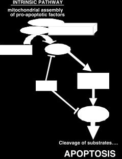
32 MITOCHONDRIA AND ACTIVATION OF CASPASE-9 IN MAMMALIAN CELLS APOPTOTIC STIMULI mitochondrion CYTOCHROME C APAF1 CASPASE-9 ACTIVATION CASPASE-9 CASP 3,6,7
33 MITOCHONDRIA AND ACTIVATION OF CASPASE-9 IN MAMMALIAN CELLS APOPTOTIC STIMULI mitochondrion WHERE DO PRO-AND ANTI-APOPTOTIC BCL2 MEMBERS FIT INTO THIS SCHEME?? CYTOCHROME C APAF1 (APOPTOSOME) CASPASE ACTIVATION CASPASE 9 CASP 3,6,7
34 THE BCL2 FAMILY AND CYTOCHROME C ARE RELEASED FROM MITOCHONDRIA ANTI-APOPTOTIC BCL2 MEMBERS ARE RESIDENT IN MITOCHONDRIA OVER-EXPRESSION OF ANTI-APOPTOTIC BCL2 MEMBERS (eg BCL2 AND BCLXL ) BLOCKS CYTOCHROME C RELEASE AND APOPTOSIS
35 MITOCHONDRIA AND APOPTOTIC DEATH -2 mitochondrion BCL2 BCL2 CYTOCHROME C APAF1 (APOPTOSOME) CASPASE 9 ACTIVATION CASPASE 3,6,7 ACTIVATION
36 HOW DO PRO-APOPTOTIC BCL2 FAMILY MEMBERS PROMOTE APOPTOSIS? THERE APPEARS TO BE A TWO STEP MECHANISM REQUIRING BOTH BAX FAMILY MEMBERS AND BH3-DOMAIN ONLY FAMILY MEMBERS
37 BOTH BAX AND BAK ARE REQUIRED FOR MITOCHONDRIALLY-DEPENDENT APOPTOSIS OVER-EXPRESSION OF PRO-APOPTOTIC BAX AND BAK MEMBERS CAUSES CYTOCHROME C RELEASE & DEATH BOTH ARE BH1-3 BAX FAMILY MEMBERS AND BOTH APPEAR TO BE REQUIRED FOR MANY CASES OF APOPTOSIS KNOCKOUT OF BAX AND BAK PROTECTS CELLS FROM DEATH
38 BAX AND BAK AND APOPTOTIC DEATH APOPTOTIC STIMULI BAX BAX mitochondrion BCL2 BAX BAK BCL2 BAX CYTOCHROME C APAF1 (APOPTOSOME) CASPASE 9 ACTIVATION CASPASE 3,6,7 ACTIVATION
39 HOW DO BAX/BAK INDUCE CYTOCHROME-C RELEASE AND APOPTOSIS? BAK APOPTOTIC STIMULI BAK BAK BAX BAK BAX BAK BAX BAX BAX BAK CYT- C BAX CYT- C PORE FORMATION IN MITOCHONDRIAL OUTER MEMBRANE RELEASE OF CYTOCHROME C
40 HOW DO BH3-ONLY MOLECULES CONTRIBUTE TO MITOCHONDRIAL-DEPENDENT APOPTOSIS? MITOCHONDRIAL-DEPENDENT APOPTOSIS REQUIRES BH3-ONLY PROTEINS AS WELL AS BAX/BAK OVER-EXPRESSION OF BH3-ONLY PROTEINS INDUCES APOPTOSIS, BUT NOT IN ABSENCE OF BAX OR BAK. SO THE LATTER APPEAR TO WORK DOWNSTREAM OF BH3 MOLECULES IN ABSENCE OF BH3-ONLY PROTEINS BAX AND BAK DO NOT CHANGE CONFORMATION AND FORM PORES THUS, APOPTOTIC DEATH VIA THE MITOCHONDRION REQUIRES BOTH BH3-ONLY PROTEINS AND BAX/BAK
41 HOW DO BAX/BAK AND BH3-ONLY PROTEINS COOPERATE TO INDUCE CYTOCHROME-C RELEASE AND APOPTOSIS? 1) BH3-ONLY PROTEINS DISPLACE BAX/BAK FROM BCL2 AND OTHER ANTI-APOPTOTIC FAMILY MEMBERS
42 HOW DO BH3-ONLY PROTEINS COOPERATE WITH BAX/BAK TO INDUCE CYTOCHROME-C RELEASE AND APOPTOSIS? BCL2 BAK APOPTOTIC STIMULI BAK BAX BAK BAX BAK BAK BCL2 BAX BH3-ONLY PROTEIN BCL2 BCL2 BAX BAX BAK CYT- C BAX CYT- C PORE FORMATION IN MITOCHONDRIAL OUTER MEMBRANE RELEASE OF CYTOCHROME C
43 THE RHEOSTAT MODEL OF CELL DEATH BCL2 BH3- BCL2ONLY BAK BCL2 BCL2 BCL2 BAX SURVIVAL BH3- BH3- ONLY BCL2ONLY BH3- ONLY BCL2 BH3- ONLY BCL2 BH3- BCL2 ONLY BCL2 BH3- ONLY BH3- ONLY BAX BAK DEATH
44 HOW ARE APOPTOTIC ACTIVITIES OF BH3 ONLY PROTEINS REGULATED? TRANSCRIPTIONAL CONTROL: EGL1, BIM, HRK, PUMA, NOXA PHOSPHORYLATION: BAD, BIM, BIK BAD AKT ON P BAD CYTOPLASMIC APOPTOTIC STIMULI AKT OFF BAD MITOCHONDRIAL TRANSLOCATION
45 REGULATION OF BH3 ONLY PROTEINS RELEVANT TO APOPTOSIS REGULATED SEQUESTRATION: BIM, Bmf Bmf Actin depolymerizing Conditions (Anoikis) Bmf MITOCHONDRIAL TRANSLOCATION DLC2 actin CLEAVAGE: BID Caspase 8,10 myr BID tbid MITOCHONDRIAL TRANSLOCATION
46 ADDITIONAL REGULATORS OF CELL DEATH IAPS - INHIBITOR OF APOPTOSIS PROTEINS

47 INHIBITOR OF APOPTOSIS PROTEINS Verhagen et al. Genome Biol. 2: , 2001
48 IAPS FAMILY OF PROTEINS THAT INHIBIT SELECT CASPASES (3,7,9) MULTIPLE FAMILY MEMBERS IN MAMMALS. ALSO PRESENT IN INSECTS AND VIRUSES. ALL HAVE BIR (BACULOVIRAL IAP REPEAT) DOMAINS THAT ARE REQUIRED FOR BINDING AND INHIBITING CASPASES SEVERAL (IAP1,2 AND XIAP) HAVE RING FINGERS AND E3 LIGASE ACTIVITY AND CAN LEAD TO DEGRADATION OF CASPASES AND OTHER PRO-APOPTOTIC MOLECULES
49 IAPS MAY FUNCTION AS A CHECK POINT BEFORE THE LAST IRREVERSIBLE STAGE OF DEATH LEVELS CAN BE UP-REGULATED BY GROWTH FACTORS (EG., GDNF) OR DOWN-REGULATED BY APOPTOTIC SIGNALS OVER-EXPRESSED IN SOME TUMORS - SO POTENTIAL CLINICAL TARGET
50 IAPS INHIBIT CASPASES AND APOPTOTIC DEATH APOPTOTIC STIMULI BIM BCL2 mitochondrion BIM BCL2 BAX BAX CYTOCHROME C APAF1 CASPASE 9 ACTIVATION CASPASE 3,7 ACTIVATION X XIAP ciap1,2
51 INTERACTIONS OF XIAP Bax BIRC4 XIAP BIR1 BIR2 BIR3 RING COOH Caspase-3, -7 Caspase-9 Rho-GDI vxiap binds to and inhibits caspases-3,-7 via the BIR2-linker domain vxiap binds to and inhibits caspase-9 via the BIR3 domain vxiap binds to Rho-GDI via the RING domain vxiap binds to and oligomerizes Bax via the RING domain

52 IAP LEVELS VARY IN MESOTHELIOMA CELL LINES AND AFFECT SENSITIVITY TO CHEMOTHERAPEUTICS Gordon, G. J. et al. Carcinogenesis : ;
53 ADDITIONAL REGULATORS OF CELL DEATH SMAC/DIABLO - INHIBITORS OF IAPS
54 SMAC/DIABLO INHIBITS IAPS APOPTOTIC STIMULI BIM BIM BCL2 mitochondrion BCL2 BAX BAX SMAC/DIABLO CYTOCHROME C APAF1 CASPASE 9 ACTIVATION CASPASE 3, 7 ACTIVATION X IAPs

55 SMAC DISPLACES IAPS FROM CASPASES XIAP BIR3 N-TERMINAL TETRA-PEPTIDE OF SMAC N-TERMINAL TETRA-PEPTIDE OF CASPASE 9 SMALL SUBUNIT
56 SMAC/DIABLO RELEASED FROM MITOCHONDRIA BY APOPTOTIC STIMULI BLOCKS IAPS FROM INHIBITING CASPASES: PRO-APOPTOTIC REQUIRED FOR DEATH IN AT LEAST SOME PARADIGMS. IN OTHERS, IT MAKES CELLS MORE SUCEPTIBLE TO DEATH
57 OMI/HTRA2 INHIBITS IAPS APOPTOTIC STIMULI BIM BIM mitochondrion BCL2 BAX BCL2 BAX OMI/HTRA2 CYTOCHROME C APAF1 CASPASE 9 ACTIVATION CASPASE 3,7 ACTIVATION X IAPs SER/THR PROTEASE
58 OMI/HTRA2 RELEASED FROM MITOCHONDRIA BY APOPTOTIC STIMULI IS A SERINE/THREONINE PROTEASE DEGRADES IAPS: PRO-APOPTOTIC MAKES CELLS MORE SUSCEPTIBLE TO DEATH UP-REGULATED BY p53
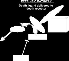
59 DEATH BY MURDER - RECEPTOR MEDIATED IN ADDITION TO SUICIDE (THE INTRINSIC APOPTOTIC MECHANISM), THERE IS ALSO A MAMMALIAN MECHANISM FOR MURDERING CELLS (THE EXTRINSIC APOPTOTIC PATHWAY) THE EXTRINSIC APOPTOTIC PATHWAY IS REGULATED BY A SERIES OF SPECIFIC DEATH-PROMOTING RECEPTORS AND LIGANDS. OCCUPATION OF THESE RECEPTORS BRINGS ABOUT ACTIVATION OF PATHWAYS THAT CULMINATE IN CELL DEATH.

60 THE RECEPTOR-MEDIATED PATHWAY OF APOPTOTIC DEATH DEATH DOMAIN TRAIL-R1 TRAIL-R2 CELL INTERIOR
61 DEATH PROMOTING RECEPTORS AND LIGANDS LIGAND TNFα FAS ligand TRAIL RECEPTOR TNFαR1 FAS TRAIL-R(DR-4 & DR-5)
62 THE RECEPTOR-MEDIATED PATHWAY OF APOPTOTIC DEATH = DD TNFα = DED TNFR1 FADD CASPASES 8,10 BID BAX e to ente tbid MITOCHONDRIAL PATHWAY CASPASE 3
63 CELLULAR ANTAGONISTS OF THE EXTRINSIC APOPTOTIC PATHWAY
64 ANTAGONISTS OF THE RECEPTOR-MEDIATED PATHWAY OF APOPTOTIC DEATH = DD = DED SODD TNFα TNFR FADD TNFα DECOY R (DcR3. TRID) c-flip X X BID Typ e to ente r text BAX tbid MITOCHONDRIAL PATHWAY CASPASE 3

65 ANTAGONISTS OF THE RECEPTOR-MEDIATED PATHWAY OF APOPTOTIC DEATH = DD TNFα TNFα = DED SODD TNFR FADD DECOY R (DcR3. TRID) c-flip X X BID tbid BAX MITOCHONDRIAL PATHWAY CASPASE 3
66 DEATH PROMOTING RECEPTORS AND LIGANDS-2 THE EXTRINSIC DEATH PATHWAY BEGINS WITH RECEPTOR-MEDIATED ACTIVATION OF INITIATOR CASPASES 8 AND/0R 10 THE PATHWAY CAN EITHER BYPASS MITOCHONDRIAL INVOLVEMENT IN DEATH OR CAN INVOLVE MITOCHONDRIA AS A MEANS OF AMPLIFICATION THE SAME CELL CAN EXPRESS BOTH DEATH RECEPTOR AND DEATH LIGAND SUSCEPTIBILITY IS SUBJECT TO REGULATION BY PATHWAY ANTAGONISTS

67 FLIP DOWN-REGULATION SENSITIZES SKBR3 BREAST CANCER CELL LINE TO TRAIL-INDUCED DEATH FROM Zang F, et al., BBRC 450: , 2014

68 PATHWAYS MEDIATING SENSITIZATION TO TRAIL BY DOWN-REGULATION OF MCL-1 Kim S et al. Cancer Res 2008;68: by American Association for Cancer Research
69 DEATH PROMOTING RECEPTORS AND LIGANDS-3 THE EXTRINSIC PATHWAY CONTRIBUTES TO DEATH IN A VARIETY OF CONTEXTS MANY TUMOR CELLS EXPRESS EXTRINSIC PATHWAY RECEPTORS, BUT ALSO OVER-EXPRESS PATHWAY ANTAGONISTS (50% of colon cancers have amplified DcR3 gene) EXPRESSION OF LIGAND AND RECEPTORS AS WELL AS OF PATHWAY ANTAGONISTS IS SUBJECT TO REGULATION
70 ROLE OF TRANSCRIPTION IN APOPTOSIS IN MANY, BUT NOT ALL PARADIGMS OF APOPTOTIC DEATH CELLS MUST SYNTHESIZE SPECIFIC GENES TO DIE THE PATHWAYS THAT REGULATE SUCH DEATH-ASSOCIATED GENES APPEAR TO BE ACTIVATED BY MECHANISMS THAT ARE INDEPENDENT OF MITOCHONDRIA. THE GENE PRODUCTS OF THESE PATHWAYS CAN ACT BOTH UPSTREAM AND DOWNSTREAM OF MITOCHONDRIA BH3-DOMAIN ONLY MOLECULES SEEM TO BE COMMON GENE TARGETS (eg. BIM IS REGULATED BY JUN, E2F, AND FORKHEAD)
71 TRANSCRIPTIONAL TARGETS OF P53 IN APOPTOSIS APOPTOTIC STIMULI (DNA DAMAGE) P53 PHOSPHORYLATION AND STABILIZATION E2F SIAH p53 PAC1 MAPK JNK/JUN BIM BAX BID PUMA NOXA APAF-1 Cell specific Stimulus specific CASPASE 6

72 POST-TRANSCRIPTIONAL REGULATION OF APOPTOSIS BY mirnas (APOPTOMIRS) Andrea Vecchione, and Carlo M Croce Endocr Relat Cancer 2010;17:F37-F Society for Endocrinology
73 WHAT SIGNALS KEEP CELL FROM DYING? ACTIVATION OF AKT/PKB & MAPK RAS RAF MEK SURVIVAL ERK Brazil DP, Hemmings BA.Trends Biochem Sci Nov;26(11):
74 EXAMPLES OF HOW AKT PROMOTES SURVIVAL BAD AKT P- BAD (cytoplasmic) mitochondria FKHR AKT P-FKHR (cytoplasmic) SURVIVAL nucleus MDM2 AKT P- MDM2 + p53 p53 inactive active p53
75 EXAMPLES OF HOW AKT PROMOTES SURVIVAL HEXOKINASE AKT P- HK PTP (mitochondria) Cytoplasm SURVIVAL IAP AKT TRANSCRIPTIONAL ACTIVATION IAP
76 EXAMPLES OF HOW RAF/ERKS PROMOTE SURVIVAL BAD RAF/ERKS P- BAD (cytoplasmic) mitochondria SURVIVAL BIM RAF/ERKS BIM INDUCTION Non-induced & destablized
77 MECHANISMS OF APOPTOSIS RESISTANCE-1 MUTATIONS OF CASPASES -MCF7 BREAST CA HAS NO CASPASE-3 EXPRESSION -DECREASED CASPASE-7 EXPRESSION IN COLON CA -HYPERMETHYLATION OF CASPASE-8 PROMOTER LOSS OF APAF-1 EXPRESSION IN MELANOMA
78 MECHANISMS OF APOPTOSIS RESISTANCE-2 INCREASED EXPRESSION OF IAPS -SURVIVIN IN NEUROBLASTOMA -ciap1/2 IN LUNG CA -ciap1 IN ESOPHAGEAL SQUAMOUS CELL CA -ciap1 INCREASES RESISTANCE TO CHEMOTHERAPY -XIAP IN OVARIAN CA
79 MECHANISMS OF APOPTOSIS RESISTANCE-3 INCREASED EXPRESSION OF BCL2 IN ALL, AML, CLL,MULTIPLE MYELOMA, PROSTATE CA, NEUROBLASTOMA DECREASED EXPRESSION OF BAX IN COLON CA, BREAST CA INCREASED EXPRESSION OF cflip IN BREAST CA
80 STRATEGIES FOR TARGETING CANCER INHIBITION OF OVEREXPRESSED ANTI- APOPTOTIC MOLECULES -e.g. BCL2, BH3 PROTEINS, SURVIVIN, OTHER IAPS ENHANCE RECEPTOR MEDIATED DEATH VIA TRAIL-R ENHANCE PRO-APOPTOTIC PATHWAYS -small molecule mimetic of SMAC/Diablo - stapled Bim TARGETING CANCER SPECIFIC APOPTOMIRS
 OF ANTI- APOPTOSIS FOR A PARTICULAR TUMOR WILL ENABLE DESIGN OF")
81 MAINTENANCE OF HOMEOSTASIS TIGHT REGULATION OF DEATH PATHWAYS AT SEVERAL LEVELS IS ESSENTIAL A BETTER UNDERSTANDING OF THE MOLECULAR MECHANISM(S) OF ANTI- APOPTOSIS FOR A PARTICULAR TUMOR WILL ENABLE DESIGN OF TARGETED THERAPIES
Mechanisms of Cell Death
 Mechanisms of Cell Death CELL DEATH AND FORMATION OF THE SEMICIRCULAR CANALS Carol M. Troy, MD PhD August 24, 2009 FROM: Fekete et al., Development 124: 2451 (1997) PHENOMENOLOGY OF CELL DEATH I. DEVELOPMENT
Mechanisms. Cell Death. Carol M. Troy, MD PhD August 24, 2009 PHENOMENOLOGY OF CELL DEATH I. DEVELOPMENT 8/20/2009
 Mechanisms of Cell Death Carol M. Troy, MD PhD August 24, 2009 PHENOMENOLOGY OF CELL DEATH I. DEVELOPMENT A. MORPHOGENESIS: SCULPTING/SHAPING STRUCTURES CREATION OF CAVITIES AND TUBES FUSION OF TISSUE
Cell Death & Trophic Factors II. Steven McLoon Department of Neuroscience University of Minnesota
 Cell Death & Trophic Factors II Steven McLoon Department of Neuroscience University of Minnesota 1 Remember? Neurotrophins are cell survival factors that neurons get from their target cells! There is a
Apoptosis: Death Comes for the Cell
 Apoptosis: Death Comes for the Cell Joe W. Ramos joeramos@hawaii.edu From Ingmar Bergman s The Seventh Seal 1 2 Mutations in proteins that regulate cell proliferation, survival and death can contribute
Apoptosis EXTRA REQUIREMETS
 Apoptosis Introduction (SLIDE 1) Organisms develop from a single cell, and in doing so an anatomy has to be created. This process not only involves the creation of new cells, but also the removal of cells
Apoptosis in Mammalian Cells
 Apoptosis in Mammalian Cells 7.16 2-10-05 Apoptosis is an important factor in many human diseases Cancer malignant cells evade death by suppressing apoptosis (too little apoptosis) Stroke damaged neurons
 http://cwp.embo.org/w09-13/index.html Roads to Ruin is s o t p o ap healthy cell autophagic cell death ne cr os is Thanks to Seamus Martin! Evolution of apoptosis signalling cascades Inhibitor Adopted
Death signaling. Color PDF file of handouts can be found at Wu lab web-page:
 Death signaling Hao Wu References 1. Wang, X. The expanding role of mitochondria in apoptosis. Genes Dev 15, 2922-2933. (2001). 2. Fesik, S. W. Insights into programmed cell death through structural biology.
Cell Survival Curves (Chap. 3)
") Cell Survival Curves (Chap. 3) Example of pedigress of EMT6 mouse cells Clone The in vitro cell survival curve Cell Survival Assay Dye exclusion assay: membrane integrity MTT assay: respiratory activity
Cell Survival Curves (Chap. 3) Example of pedigress of EMT6 mouse cells Clone The in vitro cell survival curve Cell Survival Assay Dye exclusion assay: membrane integrity MTT assay: respiratory activity
Mechanisms of Cell Death
 Mechanisms of Cell Death Etiology of cell death Major Factors Accidental Genetic Necrosis Apoptosis Necrosis: The sum of the morphologic changes that follow cell death in a living tissue or organ Apoptosis:
Bio-Plex Pro RBM Apoptosis Assays
 Acute Phase Apoptosis Cancer Cardiovascular Disease Cytokines Chemokines, Growth Factors Diabetes Gene Expression Genotyping Immunoglobulin Isotyping MicroRNA Expression Bio-Plex Pro RBM Apoptosis Assays
Monte Carlo study elucidates the type 1/type 2 choice in apoptotic death signaling in normal and cancer cells
 Cells 2013, 2, 1-x manuscripts; doi:10.3390/cells20x000x OPEN ACCESS cells ISSN 2073-4409 www.mdpi.com/journal/cells Article Monte Carlo study elucidates the type 1/type 2 choice in apoptotic death signaling
Programmed Cell Death
 Programmed Cell Death Dewajani Purnomosari Department of Histology and Cell Biology Faculty of Medicine Universitas Gadjah Mada d.purnomosari@ugm.ac.id What is apoptosis? a normal component of the development
Apoptosis and Carcinogenesis
 Death Becomes Us: Apoptosis and Carcinogenesis by Michele M. Cox Department of Biology, University of the Virgin Islands Part I What Is Cancer? Hi, Derek. Studying for your Cell and Molec final? asked
APOPTOSIS REGULATOR BCL-2
 Papers on Anthropology Apoptosis regulator XXII, 2013, BCL-2 pp. 63 67 P. Hussar, M. Žuravskaja, M. Kärner APOPTOSIS REGULATOR BCL-2 Piret Hussar 1, Maria Žuravskaja 2, Martin Kärner 2 1 Institute of Anatomy,
Mechanisms of Cell Death
 Mechanisms of Cell Death Etiology of cell death Major Factors Accidental Genetic Necrosis Apoptosis Necrosis: The sum of the morphologic changes that follow cell death in a living tissue or organ Apoptosis:
The Caspase System: a potential role in muscle proteolysis and meat quality? Tim Parr
 The Caspase System: a potential role in muscle proteolysis and meat quality? Tim Parr Caroline Kemp, Ron Bardsley,, Peter Buttery Division of Nutritional Sciences, School of Biosciences, University of
Bio 3411, Fall 2006, Lecture 19-Cell Death.
 Types of Cell Death Questions : Apoptosis (Programmed Cell Death) : Cell-Autonomous Stereotypic Rapid Clean (dead cells eaten) Necrosis : Not Self-Initiated Not Stereotypic Can Be Slow Messy (injury can
Embedded together: The life and death consequences of interaction of the Bcl-2 family with membranes
 DOI 10.1007/s10495-007-0746-4 Embedded together: The life and death consequences of interaction of the Bcl-2 family with membranes Brian Leber Jialing Lin David W. Andrews Published online: 15 February
IAPs. Structure of BIR2 Zn finger domain from XIAP
 IAPs The only known endogenous caspase inhibitors are members of a second family of proteins identified by apoptosis research, the IAP (inhibitor of apoptosis proteins) family. IAPs were originally described
Chang 1. Characterization of the Drosophila Ortholog of the Mammalian Anti-apoptotic Protein Aven
 Chang 1 Characterization of the Drosophila Ortholog of the Mammalian Anti-apoptotic Protein Aven Joy Chang Georgetown University Senior Digital Thesis May 1, 2006 Chang 2 Table of Contents BACKGROUND 4
A MATHEMATICAL MODEL FOR DYNAMIC SIMULATION OF APOPTOSIS PATHWAYS
 A MATHEMATICAL MODEL FOR DYNAMIC SIMULATION OF APOPTOSIS PATHWAYS Paolo Massari Department of Information Engineering Bioengineering University of Padova April 2014 Advisor: Gianna Maria Toolo Coadvisor:
Zool 3200: Cell Biology Exam 5 4/27/15
 Name: Trask Zool 3200: Cell Biology Exam 5 4/27/15 Answer each of the following short answer questions in the space provided, giving explanations when asked to do so. Circle the correct answer or answers
Major Factors. Accidental Genetic. Necrosis Apoptosis
 Mechanisms of Cell Death Etiology of cell death Major Factors Accidental Genetic Necrosis Apoptosis Necrosis: The sum of the morphologic changes that follow cell death in a living tissue or organ A t Apoptosis:
Proteases for Cell Suicide: Functions and Regulation of Caspases
 MICROBIOLOGY AND MOLECULAR BIOLOGY REVIEWS, Dec. 2000, p. 821 846 Vol. 64, No. 4 1092-2172/00/$04.00 0 Copyright 2000, American Society for Microbiology. All Rights Reserved. Proteases for Cell Suicide:
 Cell-cell communication in development Review of cell signaling, paracrine and endocrine factors, cell death pathways, juxtacrine signaling, differentiated state, the extracellular matrix, integrins, epithelial-mesenchymal
Validation of Petri Net Apoptosis Models Using P-Invariant Analysis
 2009 IEEE International Conference on Control and Automation Christchurch, New Zealand, December 9-11, 2009 WeMT5.1 Validation of Petri Net Apoptosis Models Using P-Invariant Analysis Ian Wee Jin Low,
Apoptosis: Death comes for the Cell. Apoptosis: Programmed Cell Death. Apoptosis. Necrosis
 poptosis: eath comes for the Cell Mutations in proteins that regulate cell proliferation, survival and death can contribute to oncogenesis Joe W. Ramos jramos@crch.hawaii.edu From Ingmar Bergman s The
TRAIL-induced apoptosis requires Bax-dependent mitochondrial release of Smac/DIABLO
 TRAIL-induced apoptosis requires Bax-dependent mitochondrial release of Smac/DIABLO Yibin Deng, Yahong Lin, and Xiangwei Wu 1 Huffington Center on Aging and Department of Molecular and Cellular Biology,
Massive loss of neurons in embryos occurs during normal development (!)
") Types of Cell Death Apoptosis (Programmed Cell Death) : Cell-Autonomous Stereotypic Rapid Clean (dead cells eaten) Necrosis : Not Self-Initiated Not Stereotypic Can Be Slow Messy (injury can spread) Apoptosis
Types of Cell Death Apoptosis (Programmed Cell Death) : Cell-Autonomous Stereotypic Rapid Clean (dead cells eaten) Necrosis : Not Self-Initiated Not Stereotypic Can Be Slow Messy (injury can spread) Apoptosis
International Journal of Pharma and Bio Sciences V1(2)2010 PETRI NET IMPLEMENTATION OF CELL SIGNALING FOR CELL DEATH
2010 PETRI NET IMPLEMENTATION OF CELL SIGNALING FOR CELL DEATH") SHRUTI JAIN *1, PRADEEP K. NAIK 2, SUNIL V. BHOOSHAN 1 1, 3 Department of Electronics and Communication Engineering, Jaypee University of Information Technology, Solan-173215, India 2 Department of Biotechnology
SHRUTI JAIN *1, PRADEEP K. NAIK 2, SUNIL V. BHOOSHAN 1 1, 3 Department of Electronics and Communication Engineering, Jaypee University of Information Technology, Solan-173215, India 2 Department of Biotechnology
Death Ligand- Death Receptor. Annexin V. Fas PI MKK7 PIP3. pro-caspase-3. Granzyme B. Caspase Activation. Bid. Bcl-2 Caspase-3 Caspase-12
 International Corporation life. science. discovery. { Apoptosis Apoptosis Kits International Corporation Apoptosis Signal Cascade and Detection Kits Annexin V Kits MEBCYTO Apoptosis Kit Annexin V Apoptosis
Biochimica et Biophysica Acta
 Biochimica et Biophysica Acta 1843 (2014) 2100 2113 Contents lists available at ScienceDirect Biochimica et Biophysica Acta journal homepage: www.elsevier.com/locate/bbamcr Review Regulating cell death
REGULATION OF VERTEBRATE NEURON DEATH IN DEVELOPMENT AND NEURODEGENERATION: ROLE OF TRANSCRIPTION LLOYD A. GREENE COLUMBIA UNIVERSITY NEW YORK, NY
 REGULATION OF VERTEBRATE NEURON DEATH IN DEVELOPMENT AND NEURODEGENERATION: ROLE OF TRANSCRIPTION LLOYD A. GREENE COLUMBIA UNIVERSITY NEW YORK, NY MASSIVE LOSS OF NEURONS DURING VERTEBRATE DEVELOPMENT
cellular division cell division cell cycle cell cycle kinases chapter 18-19
 cellular division chapter 18-19 cell division when? growth replacement of older cells production of specialized cells asexual reproduction sexual reproduction production of gametes prokaryotic binary fission
Exploring the Contextual Sensitivity of Factors that Determine Cellto-Cell Variability in Receptor-Mediated Apoptosis
 Exploring the Contextual Sensitivity of Factors that Determine Cellto-Cell Variability in Receptor-Mediated Apoptosis The Harvard community has made this article openly available. Please share how this
Apoptosis & Autophagy
 SPETSAI SUMMER SCHOOL 2010 Host Microbe Interactions Cellular Response to Infection: Apoptosis & Autophagy Christoph Dehio There are many ways to die Apoptosis: Historical perspective Process of programmed
Biol403 - Receptor Serine/Threonine Kinases
 Biol403 - Receptor Serine/Threonine Kinases The TGFβ (transforming growth factorβ) family of growth factors TGFβ1 was first identified as a transforming factor; however, it is a member of a family of structurally
Cytochrome c, cytochrome b 5 and electron transfer
 Cytochrome c, cytochrome b 5 and electron transfer Covalent bonds Cytochrome c is a major player in membrane associated electron transport systems in bacteria and mitochondria. Cytochrome c has two axial
TNFα 18hr. Control. CHX 18hr. TNFα+ CHX 18hr. TNFα: 18 18hr (KDa) PARP. Cleaved. Cleaved. Cleaved. Caspase3. Pellino3 shrna. Control shrna.
 PARP. Cleaved. Cleaved. Cleaved. Caspase3. Pellino3 shrna. Control shrna.") Survival ( %) a. TNFα 18hr b. Control sirna Pellino3 sirna TNFα: 18 18hr c. Control shrna Pellino3 shrna Caspase3 Actin Control d. Control shrna Pellino3 shrna *** 100 80 60 CHX 18hr 40 TNFα+ CHX 18hr
Survival ( %) a. TNFα 18hr b. Control sirna Pellino3 sirna TNFα: 18 18hr c. Control shrna Pellino3 shrna Caspase3 Actin Control d. Control shrna Pellino3 shrna *** 100 80 60 CHX 18hr 40 TNFα+ CHX 18hr
Biochimica et Biophysica Acta
 Biochimica et Biophysica Acta 1832 (2013) 453 474 Contents lists available at SciVerse ScienceDirect Biochimica et Biophysica Acta journal homepage: www.elsevier.com/locate/bbadis PSAP induces a unique
Game and Players: Mitochondrial Apoptosis and the Therapeutic Potential of Ursodeoxycholic Acid
 Curr. Issues Mol. Biol. 9: 123 139. Online journal at www.cimb.org Game and Players: Mitochondrial Apoptosis and the Therapeutic Potential of Ursodeoxycholic Acid Susana Solá 1, Márcia M. Aranha 1, Clifford
Signal Transduction. Dr. Chaidir, Apt
 Signal Transduction Dr. Chaidir, Apt Background Complex unicellular organisms existed on Earth for approximately 2.5 billion years before the first multicellular organisms appeared.this long period for
arxiv: v1 [q-bio.mn] 20 Dec 2012
![arxiv: v1 [q-bio.mn] 20 Dec 2012](/thumbs/94/121764302.jpg "arxiv: v1 [q-bio.mn] 20 Dec 2012") Boolean network-based model of the Bcl-2 family mediated MOMP regulation Tomas Tokar, Zdenko Turcan, Jozef Ulicny Department of Biophysics, University of P. J. Safarik, Jesenna 5, 040 01, Kosice, Slovakia
Boolean network-based model of the Bcl-2 family mediated MOMP regulation Tomas Tokar, Zdenko Turcan, Jozef Ulicny Department of Biophysics, University of P. J. Safarik, Jesenna 5, 040 01, Kosice, Slovakia
Analysis of retinoblastoma protein function in the regulation of apoptosis
 Analysis of retinoblastoma protein function in the regulation of apoptosis Analyse der Funktion des Retinoblastoma-Proteins in der Apoptose-Regulation Dissertation zur Erlangung des akademischen Grades
Types of biological networks. I. Intra-cellurar networks
 Types of biological networks I. Intra-cellurar networks 1 Some intra-cellular networks: 1. Metabolic networks 2. Transcriptional regulation networks 3. Cell signalling networks 4. Protein-protein interaction
cellular division cell division cell cycle cell cycle kinases chapter 18-19
 cellular division chapter 18-19 cell division when? growth replacement of older cells production of specialized cells asexual reproduction sexual reproduction production of gametes prokaryotic binary fission
Modeling a Snap-Action, Variable-Delay Switch Controlling Extrinsic Cell Death
 Modeling a Snap-Action, Variable-Delay Switch Controlling Extrinsic Cell Death John G. Albeck 1[, John M. Burke 1,2[, Sabrina L. Spencer 1,3, Douglas A. Lauffenburger 2,3, Peter K. Sorger 1,2* PLoS BIOLOGY
Chemical aspects of the cell. Compounds that induce cell proliferation, differentiation and death
 Chemical aspects of the cell Compounds that induce cell proliferation, differentiation and death Cell proliferation The cell cycle Chapter 17 Molecular Biology of the Cell 2 Cell cycle Chapter 17 Molecular
Journal Club & MSc Seminar
 IN THE NAME OF GOD Journal Club & MSc Seminar The relationship between apoptosis and microorganisms Superviser: Dr. Presented by: Mehri Sadeghi MSc Medical Microbiology What is it? Why is it important?
Drosophila Apoptosis and the Regulation of the Caspase Cascade
 Drosophila Apoptosis and the Regulation of the Caspase Cascade Kate Stafford March 18, 2005 Abstract The caspase cascade in Drosophila is controlled primarily by DIAP1 (Drosophila inhibitor of apoptosis),
Mechanisms of Caspase-3 Regulation in the Execution of Cell Death DISSERTATION
 Mechanisms of Caspase-3 Regulation in the Execution of Cell Death DISSERTATION Presented in Partial Fulfillment of the Requirements for the Degree Doctor of Philosophy in the Graduate School of The Ohio
Cell Cell Communication in Development
 Biology 4361 Developmental Biology Cell Cell Communication in Development June 25, 2008 Cell Cell Communication Concepts Cells in developing organisms develop in the context of their environment, including
Sig2GRN: A Software Tool Linking Signaling Pathway with Gene Regulatory Network for Dynamic Simulation
 Sig2GRN: A Software Tool Linking Signaling Pathway with Gene Regulatory Network for Dynamic Simulation Authors: Fan Zhang, Runsheng Liu and Jie Zheng Presented by: Fan Wu School of Computer Science and
A structural view of mitochondria-mediated apoptosis
 A structural view of mitochondria-mediated apoptosis Yigong Shi Mitochondria-mediated apoptosis plays a central role in animal development and tissue homeostasis, and its alteration results in a range
ADAM FAMILY. ephrin A INTERAZIONE. Eph ADESIONE? PROTEOLISI ENDOCITOSI B A RISULTATO REPULSIONE. reverse. forward
 ADAM FAMILY - a family of membrane-anchored metalloproteases that are known as A Disintegrin And Metalloprotease proteins and are key components in protein ectodomain shedding Eph A INTERAZIONE B ephrin
Reception The target cell s detection of a signal coming from outside the cell May Occur by: Direct connect Through signal molecules
 Why Do Cells Communicate? Regulation Cells need to control cellular processes In multicellular organism, cells signaling pathways coordinate the activities within individual cells that support the function
Cell-Cell Communication in Development
 Biology 4361 - Developmental Biology Cell-Cell Communication in Development October 2, 2007 Cell-Cell Communication - Topics Induction and competence Paracrine factors inducer molecules Signal transduction
Proteomics Analysis of Protein-Producing Chinese Hamster Ovary Cells. during Apoptosis in Prolonged Cultivation
 Proteomics Analysis of Protein-Producing Chinese Hamster Ovary Cells during Apoptosis in Prolonged Cultivation by Yi-Yun Wei A thesis presented to the University of Waterloo in fulfilment of the thesis
BET PROTACs Are More Broadly Effective Than BET Inhibitors. Dr. Kevin Coleman Arvinas, LLC New Haven, CT
 BET PROTACs Are More Broadly Effective Than BET Inhibitors Dr. Kevin Coleman Arvinas, LLC New Haven, CT 1 Disclosures I am an employee of and have an equity stake in Arvinas 2 Arvinas: The Protein Degradation
Growth factors and cell death. Urmas Arumäe Institute of Biotechnology
 Growth factors and cell death Urmas Arumäe Institute of Biotechnology 24.03.2011 Growth factors stimulate cell survival (among other activities). Or conversely: absence of the growth factors causes cell
Regulation and signaling. Overview. Control of gene expression. Cells need to regulate the amounts of different proteins they express, depending on
 Regulation and signaling Overview Cells need to regulate the amounts of different proteins they express, depending on cell development (skin vs liver cell) cell stage environmental conditions (food, temperature,
SUPPLEMENTARY INFORMATION
 DOI: 10.1038/ncb2362 Figure S1 CYLD and CASPASE 8 genes are co-regulated. Analysis of gene expression across 79 tissues was carried out as described previously [Ref: PMID: 18636086]. Briefly, microarray
Supplementary Materials
 Electronic Supplementary Material (ESI) for Integrative Biology. This journal is The Royal Society of Chemistry 2015 Predicting genetic interactions from Boolean models of biological networks Supplementary
Modeling heterogeneous responsiveness of intrinsic apoptosis pathway
 Ooi and Ma BMC Systems Biology 213, 7:65 http://www.biomedcentral.com/1752-59/7/65 RESEARCH ARTICLE OpenAccess Modeling heterogeneous responsiveness of intrinsic apoptosis pathway Hsu Kiang Ooi and Lan
Modelling intrinsic apoptosis
 Modelling intrinsic apoptosis In this part of the course we will model the steps that take place inside a cell when intrinsic apoptosis is triggered. The templates and instructions for preparing the models
Characterization of caspase regulation and Hepatitis B Virus induced cell death in the nematode Caenorhabditis elegans
 University of Colorado, Boulder CU Scholar Molecular, Cellular, and Developmental Biology Graduate Theses & Dissertations Molecular, Cellular, and Developmental Biology Spring 1-1-2011 Characterization
Proteome-wide High Throughput Cell Based Assay for Apoptotic Genes
 John Kenten, Doug Woods, Pankaj Oberoi, Laura Schaefer, Jonathan Reeves, Hans A. Biebuyck and Jacob N. Wohlstadter 9238 Gaither Road, Gaithersburg, MD 2877. Phone: 24.631.2522 Fax: 24.632.2219. Website:
CC2.04_t.txt The Caspase System and Muscle Degradation Tim Parr
 The Caspase System and Muscle Degradation Tim Parr [1]Our last speaker of the day is Dr. Tim Parr. Dr. Parr graduated in 1987 from the University of Southampton with a degree in physiology, biochemistry,
TO DIE OR TO DIFFERENTIATE: APOPTOTIC AND NON-APOPTOTIC ROLES OF DEATH MOLECULES IN DROSOPHILA MELANOGASTER
 TO DIE OR TO DIFFERENTIATE: APOPTOTIC AND NON-APOPTOTIC ROLES OF DEATH MOLECULES IN DROSOPHILA MELANOGASTER Thesis by Jun Ryul Huh In Partial Fulfillment of the Requirements for the Degree of Doctor of
Signaling to the Nucleus by an L-type Calcium Channel- Calmodulin Complex Through the MAP Kinase Pathway
 Signaling to the Nucleus by an L-type Calcium Channel- Calmodulin Complex Through the MAP Kinase Pathway Ricardo E. Dolmetsch, Urvi Pajvani, Katherine Fife, James M. Spotts, Michael E. Greenberg Science
CELL CYCLE AND GROWTH REGULATION
 CELL CYCLE AND GROWTH REGULATION The cell cycle is the set of stages through which a cell progresses from one division to the next. Interphase is the period between mitotic cell divisions; divided into
Direct Interaction between Survivin and Smac/DIABLO Is Essential for the Anti-apoptotic Activity of Survivin during Taxol-induced Apoptosis*
 THE JOURNAL OF BIOLOGICAL CHEMISTRY Vol. 278, No. 25, Issue of June 20, pp. 23130 23140, 2003 2003 by The American Society for Biochemistry and Molecular Biology, Inc. Printed in U.S.A. Direct Interaction
Introduction. Gene expression is the combined process of :
 1 To know and explain: Regulation of Bacterial Gene Expression Constitutive ( house keeping) vs. Controllable genes OPERON structure and its role in gene regulation Regulation of Eukaryotic Gene Expression
Model composition through model reduction: a combined model of CD95 and NF-κB signaling pathways
 Kutumova et al. BMC Systems Biology 2013, 7:13 RESEARCH ARTICLE Open Access Model composition through model reduction: a combined model of CD95 and NF-κB signaling pathways Elena Kutumova 1,2*, Andrei
ON/OFF and Beyond - A Boolean Model of Apoptosis
 Rebekka Schlatter 1 *, Kathrin Schmich 2, Ima Avalos Vizcarra 1,3, Peter Scheurich 4, Thomas Sauter 5, Christoph Borner 6, Michael Ederer 1,7, Irmgard Merfort 2, Oliver Sawodny 1 1 Institute for System
COMPUTER SIMULATION OF DIFFERENTIAL KINETICS OF MAPK ACTIVATION UPON EGF RECEPTOR OVEREXPRESSION
 COMPUTER SIMULATION OF DIFFERENTIAL KINETICS OF MAPK ACTIVATION UPON EGF RECEPTOR OVEREXPRESSION I. Aksan 1, M. Sen 2, M. K. Araz 3, and M. L. Kurnaz 3 1 School of Biological Sciences, University of Manchester,
Apoptosis and Dependence Receptors: A Molecular Basis for Cellular Addiction
 Physiol Rev 84: 411 430, 2004; 10.1152/physrev.00027.2003. Apoptosis and Dependence Receptors: A Molecular Basis for Cellular Addiction DALE E. BREDESEN, PATRICK MEHLEN, AND SHAHROOZ RABIZADEH The Buck
Cellular Mechanisms Controlling Caspase Activation and Function. Amanda B. Parrish, Christopher D. Freel, and Sally Kornbluth
 Cellular Mechanisms Controlling Caspase Activation and Function Amanda B. Parrish, Christopher D. Freel, and Sally Kornbluth Department of Pharmacology and Cancer Biology, Duke University School of Medicine,
P systems based Modelling of Cellular Signalling Pathways
 P systems based Modelling of Cellular Signalling Pathways Mario J. Pérez-Jiménez Research Group on Natural Computing Dpt. of Computer Science and Artificial Intelligence, University of Sevilla Avda. Reina
Exploring Apoptosis Signaling Using an Executable Rules-based Approach
 Exploring Apoptosis Signaling Using an Executable Rules-based Approach Carlos F. Lopez Jeremy L. Muhlich, John A. Bachman, Peter K. Sorger Harvard Medical School The battle ahead Weaknesses There is no
MAPK kinase kinase regulation of SAPK/JNK pathways
 MAPK kinase kinase regulation of SAPK/JNK pathways Lisa Stalheim and Gary L. Johnson Abstract SAPK/JNK members of the MAPK family are regulated by at least fourteen known MAPK kinase kinases (MKKKs). In
Death versus survival: functional interaction between the apoptotic and stress-inducible heat shock protein pathways
 Death versus survival: functional interaction between the apoptotic and stress-inducible heat shock protein pathways Helen M. Beere J Clin Invest. 2005;115(10):2633-2639. https://doi.org/10.1172/jci26471.
A C-terminus Mitochondrial-localization Region and BH3 Domain of Puma are Required for Apoptotic Function
 Western University Scholarship@Western Electronic Thesis and Dissertation Repository May 2013 A C-terminus Mitochondrial-localization Region and BH3 Domain of Puma are Required for Apoptotic Function Liz
Unraveling Apoptosis Signalling using Linear Control Methods: Linking the Loop Gain to Reverting the Decision to Undergo Apoptosis
 Preprints of the 9th International Symposium on Advanced Control of Chemical Processes The International Federation of Automatic Control TuPoster.3 Unraveling Apoptosis Signalling using Linear Control
Dissecting the Mechanisms of Direct Activation for Proapoptotic BAK and BAX
 Dissecting the Mechanisms of Direct Activation for Proapoptotic BAK and BAX The Harvard community has made this article openly available. Please share how this access benefits you. Your story matters.
Chem Lecture 10 Signal Transduction
 Chem 452 - Lecture 10 Signal Transduction 111202 Here we look at the movement of a signal from the outside of a cell to its inside, where it elicits changes within the cell. These changes are usually mediated
Kyung-Won LEE, a,b Hyun-Ju JUNG, c Hee-Juhn PARK, c Deog-Gon KIM, d Jin-Yong LEE, d and Kyung-Tae LEE*,a,b
 854 Biol. Pharm. Bull. 28(5) 854 859 (2005) Vol. 28, No. 5 b -D-Xylopyranosyl-(1 3)-b -D-glucuronopyranosyl Echinocystic Acid Isolated from the Roots of Codonopsis lanceolata Induces Caspase-Dependent
12/5/2014. The cell cycle and cell death. The cell cycle: cells duplicate their contents and divide
 The cell cycle and cell death The cell cycle: cells duplicate their contents and divide 1 The cell cycle may be divided into 4 phases Eucaryotic cell division: Mitosis (nuclear division) Cytokinesis (cell
MITOCHONDRIAL BIOGENESIS AND REDOX REGULATION
 MITOCHONDRIAL BIOGENESIS AND REDOX Claude A. Piantadosi, MD Professor of Medicine and Pathology Duke University Medical Center Durham, N.C. USA Objectives Provide an overview of the physiological and pathological
Gene Control Mechanisms at Transcription and Translation Levels
 Gene Control Mechanisms at Transcription and Translation Levels Dr. M. Vijayalakshmi School of Chemical and Biotechnology SASTRA University Joint Initiative of IITs and IISc Funded by MHRD Page 1 of 9
N-Bak: a neuron-specific splice variant of Bak with anti-apoptotic properties
 N-Bak: a neuron-specific splice variant of Bak with anti-apoptotic properties Yun-Fu Sun Institute of Biotechnology and Division of Animal Physiology, Department of Biosciences, Faculty of Science, University
The EGF Signaling Pathway! Introduction! Introduction! Chem Lecture 10 Signal Transduction & Sensory Systems Part 3. EGF promotes cell growth
 Chem 452 - Lecture 10 Signal Transduction & Sensory Systems Part 3 Question of the Day: Who is the son of Sevenless? Introduction! Signal transduction involves the changing of a cell s metabolism or gene
Chapter 4. Regulation of caspase-3 gene expression in MCF-7 cells by doxorubicin
 Chapter 4 Regulation of caspase-3 gene expression in MCF-7 cells by doxorubicin 4.1 Introduction The tumor suppressor p53 plays a major role in regulating the cellular response to various stresses and
Pathways of apoptosis and importance in development. Ciara Twomey, J.V. McCarthy *
 JCMM JCMM Apoptosis Review Series Available online at www.jcmm.ro www.jcmm.org J. Cell. Mol. Med. Vol 9, No 2, 2005 pp. 345-359 Pathways of apoptosis and importance in development Ciara Twomey, J.V. McCarthy
Examination paper for Bi3016 Molecular Cell Biology
 Department of Biology Examination paper for Bi3016 Molecular Cell Biology Academic contact during examination: Per Winge Phone: 99369359 Examination date: 20 th December 2017 Examination time (from-to):
Apoptosis regulators and their role in tumorigenesis
 Biochimica et Biophysica Acta 1551 (2001) F1^F37 www.bba-direct.com Review Apoptosis regulators and their role in tumorigenesis Martin Zo«rnig a; *, Anne-Odile Hueber b, Wiebke Baum a, Gerard Evan c b
Apoptosis was formally described and named
 Mechanisms of Action of Bcl-2 Family Proteins Aisha Shamas-Din 1, Justin Kale 1, Brian Leber 1,2, and David W. Andrews 1 1 Department of Biochemistry and Biomedical Sciences, McMaster University, Hamilton,
What are mitochondria?
 What are mitochondria? What are mitochondria? An intracellular organelle. There are 100 to 1000s of mitochondria/cell. Most mitochondria come from the mother. Mitochondria have their own DNA Mitochondria
Investigating the Mechanism of Nur77-Induced Apoptosis in T Cells
 University of Massachusetts Amherst ScholarWorks@UMass Amherst Masters Theses 1911 - February 2014 2012 Investigating the Mechanism of Nur77-Induced Apoptosis in T Cells Heather E. Fogarty University of