Supplementary Figure 1 Comparison of the temperature factors of the AR-LBD in the monomeric and homodimeric crystal forms.
|
|
|
- Silvester Martin
- 5 years ago
- Views:
Transcription

1
2 Supplementary Figure 1 Comparison of the temperature factors of the AR-LBD in the monomeric and homodimeric crystal forms. (a) The AR-LBD dimer is represented as a cartoon colored according to the average temperature factors (B-factors) of each residue. The left monomer is shown in the standard orientation, i.e. with helix H1 and the AF-2 groove facing the viewer. (b) A selection of 15 monomeric AR-LBD structures deposited in the PDB to date and refined to a similar resolution as the current crystal structure. The structures are identified by their PDB codes, and original papers describing these structures are cited in Supplementary References The regions of the protein structure with higher mobility/flexibility are highlighted with warmer colors, those that exhibit lower flexibility/higher stability with colder colors. (B factors were taken as deposited in the corresponding PDB entries, without further manipulations). Notice the overall stabilization of the helical bundle in the AR-LBD homodimer. By contrast, the monomeric form exhibits enhanced flexibility in the L1-3, L9-10 and L4-5 loops, as well as in helix H5, the C-terminal end of H9, and the start of H10.
3
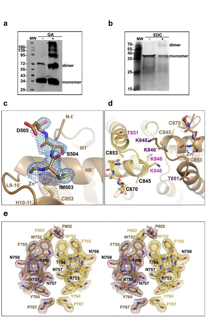
4 Supplementary Figure 2 Demonstration of AR-LBD dimer formation in solution and details of the dimer interface. (a) Results of the cross-linking of AR-LBD with glutaraldehyde. A Western blot is shown demonstrating rapid formation of an AR-LBD dimer, along with higher-order multimers. (b) Results of the incubation of AR-LBD with the zero-length crosslinker, EDC. Notice appearance of a faint band with a relative molecular mass corresponding to the AR-LBD dimer. (c) Closeup of the dimerization interface around residue C853 from monomer B. Notice the presence of extra electron density, which has been tentatively interpreted as a divalent cation coordinated by the Sγ atom, and in addition surrounded by a formylated peptide, probably stemming from recombinant AR-LBD itself. Notice also the close proximity of C687 residues from the two core monomers, B and C. The final electron density map contoured at 1σ is shown. (d) Post-translational modifications cluster around the L9-10 loop. Closeup of the AR-LBD dimer interface centered on the L9-10 loops. Note that residues K846 and K848 are known to be ubiquitinated 12, whereas phosphorylation of S792 and T851 has been reported 13. This is in addition to disulfide bridge formation between cysteine residues at positions 670 and 845 (See ref. 14 and our own unpublished observations). (e) Stereo closeup showing major interactions across the interface of the core dimer composed by the arbitrarily labeled molecules B (in yellow) and C (in brown). Electron density is shown as either a brown or yellow mesh contoured at 1σ.

5
6 Supplementary Figure 3 Predicted effects of PCa/AIS-linked mutations on AR-LBD structure and stability. Monomeric (a, d) and dimeric (b, e) AR-LBD structures are represented as cartoons colored light gray. Residues whose mutations have been associated with PCa (a, b) or AIS (d, e) are shown with all their non-hydrogen side chain atoms depicted as spheres, colored according to the predicted effect of the exchange on protein folding: dark green (more stable AR-LBD dimer), light green (increased monomer stability), salmon (local structural disorder of the monomer or less active/inactive dimer), or red (local or overall structural disorder of the AR-LBD monomer). Notice that mutations cluster at the dimer interface identified in the current crystal structure. (c, f) Consensus classification of the predicted effects of PCa or AIS-linked mutations on protein stability obtained with various independent computational tools.

7
8 Supplementary Figure 4 Comparison of the current AR-LBD homodimer with previous structures of steroid receptors. (a) Structure of the canonical ERα dimer, shown as a blue cartoon. The C-terminal end of helix H12 and the F-domain of AR (yellow; PDB: 1T7T), GR (light brown; PDB: 1M2Z), PR (salmon; PDB: 1A28) and MR (dark red; 2A3I) are overlaid onto the homodimeric ER structure. Notice that oxosteroid NRs cannot dimerize according to the canonical scheme if the F-domain is wrapped up around the LBD surface. (b) Overlay of the Cα traces of AR-LBD and GR-LBD (PDB code 1M2Z) homodimers. The two monomers are shown in yellow and gold and in gray and blue, respectively. Notice that the GR-LBD might easily adopt the symmetric head-to-head conformation seen in the current AR-LBD structure by a simple rotation towards the 2-fold axis (indicated with a curved arrow), accompanied by minor rearrangements of the L9-10 loop and of some interface side chains.

9
10 Supplementary Figure 5 Comparison of the AR-LBD with homo- and heterodimers of non-steroid nuclear receptors. Cartoon representation of the previously reported structures of the dimers of (a) GR-LBD (PDB code 1M2Z), (b) PR-LBD (1A28; notice that the AF-2 groove would be occluded in this arrangement, casting doubts about its physiological relevance), (c) TRβ (3D57), (d) HNF4α (4IQR), (e) RXRα-RARα (1DKF), and (f) PPARγ-RXRα (3E00). The individual domains are colored pale brown (GR), pink (PR), saddle brown (TRβ), purple (HNF4α), forest green (RXRα), olive (RARα), and lawn green (PPARγ). The corresponding ligands are shown as color-coded spheres (carbon, aquamarine blue; red, oxygen). Notice that the TRβ homodimer differs from the canonical dimers shown in panels (d) (f). The pathological relevance of point mutations that map to the C-terminal end of helix H5 and neighboring areas for generalized thyroid hormone resistance (GTHR) suggests that the TR-LBD homodimer might adopt an AR-like head-to-head conformation in solution.

11
12 Supplementary Figure 6 Uncropped scans of the Western blots shown in Figure 5d,e.

13 Supplementary Tables Supplementary Table 1 (ST1): Summary of BMOE-crosslinked peptides identified by mass spectrometry.
14 Supplementary Table 2 (ST2): Topologically equivalent interface residues in steroid receptors and in the TRβ.
15 Supplementary Table 3 (ST3): Bioinformatics analysis of AIS-associated mutations.
16 Supplementary Table 4 (ST4): Bioinformatics analysis of PCa-linked mutations.
17 Supplementary Table 5 (ST5): Functional data reported on PCa- and AIS-associated mutations clustering at the dimer interface.
18 Supplementary Notes The major implications of the current structure for disease-associated point mutations are summarized below. Mutations associated with androgen insensitivity syndromes: In addition to the large number of AIS-linked mutations that cluster at the core of the AR-LBD dimer interface, some variants affect the upper L5-6 and L7-8 loops (N759T, S760F/Y, R761S, M762T, Y764C/H, P767S/A and Q799E) or the lower area of the interface (V685I, C687R, D691V/E, D696N/Y/V/L and D768Y) (Figures 5a,c, and Supplementary Tables 3,5). Several of these AISassociated mutations may lead to local structural disorder of the AR monomer, which might explain their pathogenicity (e.g. W752R, R753P, S760F/Y, L763F and R856C) (Supplementary Figure 3). Most reported replacements, however, affect residues that are well exposed in the AR-LBD monomer and are predicted to be neutral or even favorable for its structure, but are involved in important inter-monomer contacts, and would therefore compromise the stability of the dimer (Supplementary Figure 3 and Supplementary Table 3). Mutations F755L and F755V, in particular, have an interesting correlation with AIS severity: the Phe Leu exchange is associated with a less severe PAIS phenotype, while introduction of the related aliphatic residue, Val, results in CAIS 15 (see also Supplementary Table 5). Although both leucine and valine would be equally well tolerated at this position in monomeric AR- LBD, inspection of the current structure reveals that a Leu side chain could still engage in vdw interactions with residue P802 from the adjacent monomer, albeit weaker than those formed by the bulkier wild-type Phe (Figure 2d). In contrast, a smaller Val side chain cannot participate in such interactions, and therefore the F755V mutant is predicted to remain in a monomeric state, disrupting biological activities of the receptor and explaining the complete female phenotype of carriers of this mutation. In a similar manner, loss of important H-bonds across the dimer axis would compromise dimer formation and/or stability (Figure 2e), which explains the deleterious impact of replacing residue R761 by Ser (reported in PAIS patients) and of the N757 exchange to Ser (MAIS/PAIS). Finally, impaired vdw interactions across the dimer interface (Figure 2d) are likely to underlie the CAIS phenotype in carriers of the P767A/S mutations. Indeed, we could demonstrate that the P767A variant interferes with dimer formation, in spite of retained ligand binding ability (see Figure 4c and main text for details). Mutations associated with metastatic prostate cancer: Some of these AR variants (in particular R847G, but also S760P and R841G) are predicted to destabilize the LBD monomer, while mutations that cluster in the central part of the interface are more preservative of structural integrity and ligand binding: G684S, G751S, Q799G, and I800T are predicted to stabilize the monomer. On the other hand, PCa-associated mutations that affect residues exposed on H5 (F755L, T756A, N757D and V758A/I), or neighboring areas (R761K, Y764C) are likely to be neutral regarding the structure of the monomer (Figures 5b,c, Supplementary Figure 3, and Supplementary Tables 4-5). For the exposed structure-preserving mutations T756A, N757D, V758I and Y764C, all bioinformatics tools predict enhanced dimerization (Supplementary Table 4). In the cases of T756A and Y764C the smaller size of the mutant side chains might facilitate a tighter dimer assembly. On the other hand, the V758I exchange is predicted to stabilize the AR-LBD dimer thanks to additional vdw interactions of the bulkier I758 side chain with residues M763 and Y764 from the neighboring monomer. Interestingly, mutation Y764C strongly enhanced the activity of the AR in our functional studies (Figure 5), which is in accordance with the general belief that overactive AR can lead to therapy resistance in advanced PCa 16.
19 Supplementary References 1 Sack, J. S. et al. Crystallographic structures of the ligand-binding domains of the androgen receptor and its T877A mutant complexed with the natural agonist dihydrotestosterone. Proc. Natl. Acad. Sci. U.S.A. 98, (2001). 2 Hur, E. et al. Recognition and accommodation at the androgen receptor coactivator binding interface. PLoS Biol. 2, e274 (2004). 3 Estébanez-Perpiñá, E. et al. The molecular mechanisms of coactivator utilization in ligand-dependent transactivation by the androgen receptor. J. Biol. Chem. 280, (2005). 4 He, B. et al. Structural basis for androgen receptor interdomain and coactivator interactions suggests a transition in nuclear receptor activation function dominance. Mol. Cell 16, (2004). 5 Bohl, C. E., Gao, W., Miller, D. D., Bell, C. E. & Dalton, J. T. Structural basis for antagonism and resistance of bicalutamide in prostate cancer. Proc. Natl. Acad. Sci. U. S. A. 102, (2005). 6 Salvati, M. E. et al. Structure based approach to the design of bicyclic-1hisoindole-1,3(2h)-dione based androgen receptor antagonists. Bioorg. Med. Chem. Lett. 15, (2005). 7 Sun, C. et al. Discovery of potent, orally-active, and muscle-selective androgen receptor modulators based on an N-aryl-hydroxybicyclohydantoin scaffold. J. Med. Chem. 49, (2006). 8 Wang, F. et al. Structure of the ligand-binding domain (LBD) of human androgen receptor in complex with a selective modulator LGD2226. Acta Crystallogr. Sect. F Struct. Biol. Cryst. Commun. 62, (2006). 9 Estébanez-Perpiñá, E. et al. A surface on the androgen receptor that allosterically regulates coactivator binding. Proc. Natl. Acad. Sci. U.S.A. 104, (2007). 10 Zhou, X. E. et al. Identification of SRC3/AIB1 as a preferred coactivator for Hhrmone-activated androgen receptor. J. Biol. Chem. 285, (2010). 11 Hsu, C.-L. et al. Identification of a new androgen receptor (AR) co-regulator BUD31 and related peptides to suppress wild-type and mutated AR-mediated prostate cancer growth via peptide screening and X-ray structure analysis. Mol. Oncol. 8, (2014). 12 Xu, K. et al. Regulation of androgen receptor transcriptional activity and specificity by RNF6-induced ubiquitination. Cancer Cell 15, (2009). 13 Lin, H.-K., Yeh, S., Kang, H.-Y. & Chang, C. Akt suppresses androgen-induced apoptosis by phosphorylating and inhibiting androgen receptor. Proc. Natl. Acad. Sci. U.S.A. 98, (2001).
20 14 Matias, P. M. et al. Structural evidence for ligand specificity in the binding domain of the human androgen receptor: Implications for pathogenic gene mutations J. Biol. Chem. 275, (2000). 15 Tadokoro, R., Bunch, T., Schwabe, J. W. R., Hughes, I. A. & Murphy, J. C. Comparison of the molecular consequences of different mutations at residue 754 and 690 of the androgen receptor (AR) and androgen insensitivity syndrome (AIS) phenotype. Clin. Endocrinol. 71, (2009). 16 Attard, G. et al. Prostate cancer. Lancet 387, (2016).
Supplementary Figure 1. Aligned sequences of yeast IDH1 (top) and IDH2 (bottom) with isocitrate
 and IDH2 (bottom) with isocitrate") SUPPLEMENTARY FIGURE LEGENDS Supplementary Figure 1. Aligned sequences of yeast IDH1 (top) and IDH2 (bottom) with isocitrate dehydrogenase from Escherichia coli [ICD, pdb 1PB1, Mesecar, A. D., and Koshland,
SUPPLEMENTARY FIGURE LEGENDS Supplementary Figure 1. Aligned sequences of yeast IDH1 (top) and IDH2 (bottom) with isocitrate dehydrogenase from Escherichia coli [ICD, pdb 1PB1, Mesecar, A. D., and Koshland,
SUPPLEMENTARY INFORMATION
 doi:10.1038/nature11085 Supplementary Tables: Supplementary Table 1. Summary of crystallographic and structure refinement data Structure BRIL-NOP receptor Data collection Number of crystals 23 Space group
Nature Structural and Molecular Biology: doi: /nsmb Supplementary Figure 1. Definition and assessment of ciap1 constructs.
 Supplementary Figure 1 Definition and assessment of ciap1 constructs. (a) ciap1 constructs used in this study are shown as primary structure schematics with domains colored as in the main text. Mutations
Cks1 CDK1 CDK1 CDK1 CKS1. are ice- lobe. conserved. conserved
 Cks1 d CKS1 Supplementary Figure 1 The -Cks1 crystal lattice. (a) Schematic of the - Cks1 crystal lattice. -Cks1 crystallizes in a lattice that contains c 4 copies of the t - Cks1 dimer in the crystallographic
Supplementary Figure 1. Biochemical and sequence alignment analyses the
 Supplementary Figure 1. Biochemical and sequence alignment analyses the interaction of OPTN and TBK1. (a) Analytical gel filtration chromatography analysis of the interaction between TBK1 CTD and OPTN(1-119).
Supplementary Tables and Figures
 Supplementary Tables Supplementary Tables and Figures Supplementary Table 1: Tumor types and samples analyzed. Supplementary Table 2: Genes analyzed here. Supplementary Table 3: Statistically significant
SUPPLEMENTARY INFORMATION
 doi:10.1038/nature17991 Supplementary Discussion Structural comparison with E. coli EmrE The DMT superfamily includes a wide variety of transporters with 4-10 TM segments 1. Since the subfamilies of the
SUPPLEMENTARY INFORMATION
 doi:10.1038/nature11054 Supplementary Fig. 1 Sequence alignment of Na v Rh with NaChBac, Na v Ab, and eukaryotic Na v and Ca v homologs. Secondary structural elements of Na v Rh are indicated above the
SUPPLEMENTARY INFORMATION
 SUPPLEMENTARY INFORMATION Structure of human carbamoyl phosphate synthetase: deciphering the on/off switch of human ureagenesis Sergio de Cima, Luis M. Polo, Carmen Díez-Fernández, Ana I. Martínez, Javier
Protein Structure. W. M. Grogan, Ph.D. OBJECTIVES
 Protein Structure W. M. Grogan, Ph.D. OBJECTIVES 1. Describe the structure and characteristic properties of typical proteins. 2. List and describe the four levels of structure found in proteins. 3. Relate
Nature Structural & Molecular Biology: doi: /nsmb Supplementary Figure 1
 Supplementary Figure 1 Crystallization. a, Crystallization constructs of the ET B receptor are shown, with all of the modifications to the human wild-type the ET B receptor indicated. Residues interacting
Preparing a PDB File
 Figure 1: Schematic view of the ligand-binding domain from the vitamin D receptor (PDB file 1IE9). The crystallographic waters are shown as small spheres and the bound ligand is shown as a CPK model. HO
SUPPLEMENTARY INFORMATION
 Figure S1. Secondary structure of CAP (in the camp 2 -bound state) 10. α-helices are shown as cylinders and β- strands as arrows. Labeling of secondary structure is indicated. CDB, DBD and the hinge are
Nuclear receptors. Lecture 1. Structure and function
 Nuclear receptors Lecture 1 Structure and function NR NR NR NR NR NR NR NR NR NR NR Let s start from a small fragment... (?) 49 General information Well-known nuclear receptor ligands Not well-known
Crystal Structure of Fibroblast Growth Factor 9 (FGF9) Reveals Regions. Implicated in Dimerization and Autoinhibition
 Reveals Regions. Implicated in Dimerization and Autoinhibition") JBC Papers in Press. Published on November 1, 2000 as Manuscript M006502200 Crystal Structure of Fibroblast Growth Factor 9 (FGF9) Reveals Regions Implicated in Dimerization and Autoinhibition 1 Copyright
JBC Papers in Press. Published on November 1, 2000 as Manuscript M006502200 Crystal Structure of Fibroblast Growth Factor 9 (FGF9) Reveals Regions Implicated in Dimerization and Autoinhibition 1 Copyright
Right click on the link and save the file on the disk (Save link target as...). Then execute this in the command window:
. Then execute this in the command window:") Läkemedelsutveckling ht 2005 Copyright 2005 Lars Brive Excercise Analysis of structures of protein ligand complexes In this excercise you will examine the geometrical features of six x ray structures in
Läkemedelsutveckling ht 2005 Copyright 2005 Lars Brive Excercise Analysis of structures of protein ligand complexes In this excercise you will examine the geometrical features of six x ray structures in
SUPPLEMENTARY INFORMATION
 Fig. 1 Influences of crystal lattice contacts on Pol η structures. a. The dominant lattice contact between two hpol η molecules (silver and gold) in the type 1 crystals. b. A close-up view of the hydrophobic
SUPPLEMENTARY INFORMATION
 Supplementary Results DNA binding property of the SRA domain was examined by an electrophoresis mobility shift assay (EMSA) using synthesized 12-bp oligonucleotide duplexes containing unmodified, hemi-methylated,
SUPPLEMENTARY INFORMATION
 doi:1.138/nature1737 Supplementary Table 1 variant Description FSEC - 2B12 a FSEC - 6A1 a K d (leucine) c Leucine uptake e K (wild-type like) K (Y18F) K (TS) K (TSY) K288A mutant, lipid facing side chain
SUPPLEMENTARY INFORMATION
 Supplementary materials Figure S1 Fusion protein of Sulfolobus solfataricus SRP54 and a signal peptide. a, Expression vector for the fusion protein. The signal peptide of yeast dipeptidyl aminopeptidase
Supplementary Figure 3 a. Structural comparison between the two determined structures for the IL 23:MA12 complex. The overall RMSD between the two
 Supplementary Figure 1. Biopanningg and clone enrichment of Alphabody binders against human IL 23. Positive clones in i phage ELISA with optical density (OD) 3 times higher than background are shown for
Table 1. Crystallographic data collection, phasing and refinement statistics. Native Hg soaked Mn soaked 1 Mn soaked 2
 Table 1. Crystallographic data collection, phasing and refinement statistics Native Hg soaked Mn soaked 1 Mn soaked 2 Data collection Space group P2 1 2 1 2 1 P2 1 2 1 2 1 P2 1 2 1 2 1 P2 1 2 1 2 1 Cell
Supplementary figure 1. Comparison of unbound ogm-csf and ogm-csf as captured in the GIF:GM-CSF complex. Alignment of two copies of unbound ovine
 Supplementary figure 1. Comparison of unbound and as captured in the GIF:GM-CSF complex. Alignment of two copies of unbound ovine GM-CSF (slate) with bound GM-CSF in the GIF:GM-CSF complex (GIF: green,
Transcription Regulation And Gene Expression in Eukaryotes UPSTREAM TRANSCRIPTION FACTORS
 Transcription Regulation And Gene Expression in Eukaryotes UPSTREAM TRANSCRIPTION FACTORS RG. Clerc March 26. 2008 UPSTREAM TRANSCRIPTION FACTORS Experimental approaches DNA binding domains (DBD) Transcription
SUPPLEMENTARY FIGURES
 SUPPLEMENTARY FIGURES Supplementary Figure 1 Protein sequence alignment of Vibrionaceae with either a 40-residue insertion or a 44-residue insertion. Identical residues are indicated by red background.
Structure and RNA-binding properties. of the Not1 Not2 Not5 module of the yeast Ccr4 Not complex
 Structure and RNA-binding properties of the Not1 Not2 Not5 module of the yeast Ccr4 Not complex Varun Bhaskar 1, Vladimir Roudko 2,3, Jerome Basquin 1, Kundan Sharma 4, Henning Urlaub 4, Bertrand Seraphin
NGF - twenty years a-growing
 NGF - twenty years a-growing A molecule vital to brain growth It is twenty years since the structure of nerve growth factor (NGF) was determined [ref. 1]. This molecule is more than 'quite interesting'
SUPPLEMENTARY INFORMATION
 Supplementary Table 1: Amplitudes of three current levels. Level 0 (pa) Level 1 (pa) Level 2 (pa) TrkA- TrkH WT 200 K 0.01 ± 0.01 9.5 ± 0.01 18.7 ± 0.03 200 Na * 0.001 ± 0.01 3.9 ± 0.01 12.5 ± 0.03 200
SUPPLEMENTARY INFORMATION
 Table of Contents Page Supplementary Table 1. Diffraction data collection statistics 2 Supplementary Table 2. Crystallographic refinement statistics 3 Supplementary Fig. 1. casic1mfc packing in the R3
Supplementary Materials for
 www.sciencesignaling.org/cgi/content/full/5/243/ra68/dc1 Supplementary Materials for Superbinder SH2 Domains Act as Antagonists of Cell Signaling Tomonori Kaneko, Haiming Huang, Xuan Cao, Xing Li, Chengjun
Table S1. Overview of used PDZK1 constructs and their binding affinities to peptides. Related to figure 1.
 Table S1. Overview of used PDZK1 constructs and their binding affinities to peptides. Related to figure 1. PDZK1 constru cts Amino acids MW [kda] KD [μm] PEPT2-CT- FITC KD [μm] NHE3-CT- FITC KD [μm] PDZK1-CT-
Sunhats for plants. How plants detect dangerous ultraviolet rays
 Sunhats for plants How plants detect dangerous ultraviolet rays Anyone who has ever suffered sunburn will know about the effects of too much ultraviolet (UV) radiation, in particular UV-B (from 280-315
Chapter 9 DNA recognition by eukaryotic transcription factors
 Chapter 9 DNA recognition by eukaryotic transcription factors TRANSCRIPTION 101 Eukaryotic RNA polymerases RNA polymerase RNA polymerase I RNA polymerase II RNA polymerase III RNA polymerase IV Function
Supplementary Information
 1 Supplementary Information Figure S1 The V=0.5 Harker section of an anomalous difference Patterson map calculated using diffraction data from the NNQQNY crystal at 1.3 Å resolution. The position of the
Supplementary Materials for
 advances.sciencemag.org/cgi/content/full/3/4/e1600663/dc1 Supplementary Materials for A dynamic hydrophobic core orchestrates allostery in protein kinases Jonggul Kim, Lalima G. Ahuja, Fa-An Chao, Youlin
Structure, mechanism and ensemble formation of the Alkylhydroperoxide Reductase subunits. AhpC and AhpF from Escherichia coli
 Structure, mechanism and ensemble formation of the Alkylhydroperoxide Reductase subunits AhpC and AhpF from Escherichia coli Phat Vinh Dip 1,#, Neelagandan Kamariah 2,#, Malathy Sony Subramanian Manimekalai
RNA Polymerase I Contains a TFIIF-Related DNA-Binding Subcomplex
 Molecular Cell, Volume 39 Supplemental Information RNA Polymerase I Contains a TFIIFRelated DNABinding Subcomplex Sebastian R. Geiger, Kristina Lorenzen, Amelie Schreieck, Patrizia Hanecker, Dirk Kostrewa,
SUPPLEMENTARY INFORMATION
 Dph2 SeMet (iron-free) # Dph2 (iron-free) Dph2-[4Fe-4S] Data collection Space group P2 1 2 1 2 1 P2 1 2 1 2 1 P2 1 2 1 2 1 Cell dimensions a, b, c (Å) 58.26, 82.08, 160.42 58.74, 81.87, 160.01 55.70, 80.53,
Gene Control Mechanisms at Transcription and Translation Levels
 Gene Control Mechanisms at Transcription and Translation Levels Dr. M. Vijayalakshmi School of Chemical and Biotechnology SASTRA University Joint Initiative of IITs and IISc Funded by MHRD Page 1 of 9
Supporting Information
 Supporting Information Superagonist, Full Agonist, Partial Agonist and Antagonist Actions of Arylguanidines at 5-Hydroxytryptamine-3 (5-HT 3 ) Subunit A Receptors Katie Alix, Shailesh Khatri, Philip D.
schematic diagram; EGF binding, dimerization, phosphorylation, Grb2 binding, etc.
 Lecture 1: Noncovalent Biomolecular Interactions Bioengineering and Modeling of biological processes -e.g. tissue engineering, cancer, autoimmune disease Example: RTK signaling, e.g. EGFR Growth responses
Supplementary Figure 1 Crystal packing of ClR and electron density maps. Crystal packing of type A crystal (a) and type B crystal (b).
 and type B crystal (b).") Supplementary Figure 1 Crystal packing of ClR and electron density maps. Crystal packing of type A crystal (a) and type B crystal (b). Crystal contacts at B-C loop are magnified and stereo view of A-weighted
Supplementary Figure 1 Crystal packing of ClR and electron density maps. Crystal packing of type A crystal (a) and type B crystal (b). Crystal contacts at B-C loop are magnified and stereo view of A-weighted
Structural characterization of NiV N 0 P in solution and in crystal.
 Supplementary Figure 1 Structural characterization of NiV N 0 P in solution and in crystal. (a) SAXS analysis of the N 32-383 0 -P 50 complex. The Guinier plot for complex concentrations of 0.55, 1.1,
Supporting Information. Structural and functional characterization of human and murine C5a anaphylatoxins
 Supporting Information Structural and functional characterization of human and murine C5a anaphylatoxins Janus Asbjørn Schatz-Jakobsen a, Laure Yatime a, Casper Larsen a, Steen Vang Petersen b, Andreas
Introduction to Comparative Protein Modeling. Chapter 4 Part I
 Introduction to Comparative Protein Modeling Chapter 4 Part I 1 Information on Proteins Each modeling study depends on the quality of the known experimental data. Basis of the model Search in the literature
SUPPLEMENTARY INFORMATION
 SUPPLEMENTARY INFORMATION doi:10.1038/nature11744 Supplementary Table 1. Crystallographic data collection and refinement statistics. Wild-type Se-Met-BcsA-B SmCl 3 -soaked EMTS-soaked Data collection Space
Supplementary Figure S1. Urea-mediated buffering mechanism of H. pylori. Gastric urea is funneled to a cytoplasmic urease that is presumably attached
 Supplementary Figure S1. Urea-mediated buffering mechanism of H. pylori. Gastric urea is funneled to a cytoplasmic urease that is presumably attached to HpUreI. Urea hydrolysis products 2NH 3 and 1CO 2
Nitrogenase MoFe protein from Clostridium pasteurianum at 1.08 Å resolution: comparison with the Azotobacter vinelandii MoFe protein
 Acta Cryst. (2015). D71, 274-282, doi:10.1107/s1399004714025243 Supporting information Volume 71 (2015) Supporting information for article: Nitrogenase MoFe protein from Clostridium pasteurianum at 1.08
Supplementary figure 1 Application of tmfret in LeuT. (a) To assess the feasibility of using tmfret for distance-dependent measurements in LeuT, a
 To assess the feasibility of using tmfret for distance-dependent measurements in LeuT, a") Supplementary figure 1 Application of tmfret in LeuT. (a) To assess the feasibility of using tmfret for distance-dependent measurements in LeuT, a series of tmfret-pairs comprised of single cysteine mutants
Supplementary figure 1 Application of tmfret in LeuT. (a) To assess the feasibility of using tmfret for distance-dependent measurements in LeuT, a series of tmfret-pairs comprised of single cysteine mutants
Signal Transduction Phosphorylation Protein kinases. Misfolding diseases. Protein Engineering Lysozyme variants
 Signal Transduction Phosphorylation Protein kinases Misfolding diseases Protein Engineering Lysozyme variants Cells and Signals Regulation The cell must be able to respond to stimuli Cellular activities
Supplementary Information
 Supplementary Information The direct role of selenocysteine in [NiFeSe] hydrogenase maturation and catalysis Marta C. Marques a, Cristina Tapia b, Oscar Gutiérrez-Sanz b, Ana Raquel Ramos a, Kimberly L.
T H E J O U R N A L O F G E N E R A L P H Y S I O L O G Y. jgp
 S u p p l e m e n ta l m at e r i a l jgp Lee et al., http://www.jgp.org/cgi/content/full/jgp.201411219/dc1 T H E J O U R N A L O F G E N E R A L P H Y S I O L O G Y S u p p l e m e n ta l D I S C U S
Supplementary Information. The Solution Structural Ensembles of RNA Kink-turn Motifs and Their Protein Complexes
 Supplementary Information The Solution Structural Ensembles of RNA Kink-turn Motifs and Their Protein Complexes Xuesong Shi, a Lin Huang, b David M. J. Lilley, b Pehr B. Harbury a,c and Daniel Herschlag
Nature Structural and Molecular Biology: doi: /nsmb Supplementary Figure 1
 Supplementary Figure 1 Quantitation of the binding of pro53 peptide to sorla Vps10p measured by the AP reporter assay. The graph shows tracings of the typical chromogenic AP reaction observed with AP-pro53
Supplementary materials. Crystal structure of the carboxyltransferase domain. of acetyl coenzyme A carboxylase. Department of Biological Sciences
 Supplementary materials Crystal structure of the carboxyltransferase domain of acetyl coenzyme A carboxylase Hailong Zhang, Zhiru Yang, 1 Yang Shen, 1 Liang Tong Department of Biological Sciences Columbia
Supplemental Information. The Mitochondrial Fission Receptor MiD51. Requires ADP as a Cofactor
 Structure, Volume 22 Supplemental Information The Mitochondrial Fission Receptor MiD51 Requires ADP as a Cofactor Oliver C. Losón, Raymond Liu, Michael E. Rome, Shuxia Meng, Jens T. Kaiser, Shu-ou Shan,
SI Text S1 Solution Scattering Data Collection and Analysis. SI references
 SI Text S1 Solution Scattering Data Collection and Analysis. The X-ray photon energy was set to 8 kev. The PILATUS hybrid pixel array detector (RIGAKU) was positioned at a distance of 606 mm from the sample.
SUPPLEMENTARY INFORMATION
 Supplementary Table 1: Data collection, phasing and refinement statistics ChbC/Ta 6 Br 12 Native ChbC Data collection Space group P4 3 2 1 2 P4 3 2 1 2 Cell dimensions a, c (Å) 132.75, 453.57 132.81, 452.95
Nature Structural & Molecular Biology: doi: /nsmb.3194
 Supplementary Figure 1 Mass spectrometry and solution NMR data for -syn samples used in this study. (a) Matrix-assisted laser-desorption and ionization time-of-flight (MALDI-TOF) mass spectrum of uniformly-
Structure and evolution of the spliceosomal peptidyl-prolyl cistrans isomerase Cwc27
 Acta Cryst. (2014). D70, doi:10.1107/s1399004714021695 Supporting information Volume 70 (2014) Supporting information for article: Structure and evolution of the spliceosomal peptidyl-prolyl cistrans isomerase
Viewing and Analyzing Proteins, Ligands and their Complexes 2
 2 Viewing and Analyzing Proteins, Ligands and their Complexes 2 Overview Viewing the accessible surface Analyzing the properties of proteins containing thousands of atoms is best accomplished by representing
Diphthamide biosynthesis requires a radical iron-sulfur enzyme. Pennsylvania State University, University Park, Pennsylvania 16802, USA
 Diphthamide biosynthesis requires a radical iron-sulfur enzyme Yang Zhang, 1,4 Xuling Zhu, 1,4 Andrew T. Torelli, 1 Michael Lee, 2 Boris Dzikovski, 1 Rachel Koralewski, 1 Eileen Wang, 1 Jack Freed, 1 Carsten
Docking with Water in the Binding Site using GOLD
 Docking with Water in the Binding Site using GOLD Version 2.0 November 2017 GOLD v5.6 Table of Contents Docking with Water in the Binding Site... 2 Case Study... 3 Introduction... 3 Provided Input Files...
Structural Perspectives on Drug Resistance
 Structural Perspectives on Drug Resistance Irene Weber Departments of Biology and Chemistry Molecular Basis of Disease Program Georgia State University Atlanta, GA, USA What have we learned from 20 years
SUPPLEMENTARY INFORMATION. doi: /nature07461
 Figure S1 Electrophysiology. a ph-activation of. Two-electrode voltage clamp recordings of Xenopus oocytes expressing in comparison to waterinjected oocytes. Currents were recorded at 40 mv. The ph of
Supplementary Information. Overlap between folding and functional energy landscapes for. adenylate kinase conformational change
 Supplementary Information Overlap between folding and functional energy landscapes for adenylate kinase conformational change by Ulrika Olsson & Magnus Wolf-Watz Contents: 1. Supplementary Note 2. Supplementary
Supporting Information
 Supporting Information Ottmann et al. 10.1073/pnas.0907587106 Fig. S1. Primary structure alignment of SBT3 with C5 peptidase from Streptococcus pyogenes. The Matchmaker tool in UCSF Chimera (http:// www.cgl.ucsf.edu/chimera)
SUPPLEMENTARY INFORMATION
 doi:10.1038/nature11991 Supplementary Figure 1 - Refinement strategy for PIC intermediate assemblies by negative stain EM. The cryo-negative stain structure of free Pol II 1 (a) was used as initial reference
Supplementary Information: Table S1. Potential energy and essential dynamics (ED) analysis of the MD simulations of the Bcl-2 complexes under study.
 analysis of the MD simulations of the Bcl-2 complexes under study.") Supplementary Information: Table S1. Potential energy and essential dynamics (ED) analysis of the MD simulations of the Bcl-2 complexes under study. Bcl2 and complex Potential energy (kj/mol) ED 2D projection
Supplementary Information: Table S1. Potential energy and essential dynamics (ED) analysis of the MD simulations of the Bcl-2 complexes under study. Bcl2 and complex Potential energy (kj/mol) ED 2D projection
Supplementary information
 Supplementary information The structural basis of modularity in ECF-type ABC transporters Guus B. Erkens 1,2, Ronnie P-A. Berntsson 1,2, Faizah Fulyani 1,2, Maria Majsnerowska 1,2, Andreja Vujičić-Žagar
Detailed description of overall and active site architecture of PPDC- 3dThDP, PPDC-2HE3dThDP, PPDC-3dThDP-PPA and PPDC- 3dThDP-POVA
 Online Supplemental Results Detailed description of overall and active site architecture of PPDC- 3dThDP, PPDC-2HE3dThDP, PPDC-3dThDP-PPA and PPDC- 3dThDP-POVA Structure solution and overall architecture
Gene regulation I Biochemistry 302. Bob Kelm February 25, 2005
 Gene regulation I Biochemistry 302 Bob Kelm February 25, 2005 Principles of gene regulation (cellular versus molecular level) Extracellular signals Chemical (e.g. hormones, growth factors) Environmental
Expanded View Figures
 The EMBO Journal Structure of a Dm peptide bound to the OT module Tobias Raisch et al Expanded View Figures A Hs Dm 262 297 685 8 HEAT HEAT MIF4G 9BD 1SHD 761 91 193 169 1152 1317 16 1376 1467 HEAT HEAT
Translation. A ribosome, mrna, and trna.
 Translation The basic processes of translation are conserved among prokaryotes and eukaryotes. Prokaryotic Translation A ribosome, mrna, and trna. In the initiation of translation in prokaryotes, the Shine-Dalgarno
Molecular Mechanisms in the Activation of Abscisic Acid Receptor PYR1
 Molecular Mechanisms in the Activation of Abscisic Acid Receptor PYR1 Lyudmyla Dorosh 1,2, Olesya A. Kharenko 3, Nandhakishore Rajagopalan 3, Michele C. Loewen 3,4, Maria Stepanova 1,2 * 1 National Research
Supplementary Information. The protease GtgE from Salmonella exclusively targets. inactive Rab GTPases
 Supplementary Information The protease GtgE from Salmonella exclusively targets inactive Rab GTPases Table of Contents Supplementary Figures... 2 Supplementary Figure 1... 2 Supplementary Figure 2... 3
Welcome to Class 21!
 Welcome to Class 21! Introductory Biochemistry! Lecture 21: Outline and Objectives l Regulation of Gene Expression in Prokaryotes! l transcriptional regulation! l principles! l lac operon! l trp attenuation!
Structural insights into Aspergillus fumigatus lectin specificity - AFL binding sites are functionally non-equivalent
 Acta Cryst. (2015). D71, doi:10.1107/s1399004714026595 Supporting information Volume 71 (2015) Supporting information for article: Structural insights into Aspergillus fumigatus lectin specificity - AFL
Protein Structure. Hierarchy of Protein Structure. Tertiary structure. independently stable structural unit. includes disulfide bonds
 Protein Structure Hierarchy of Protein Structure 2 3 Structural element Primary structure Secondary structure Super-secondary structure Domain Tertiary structure Quaternary structure Description amino
Lecture 2 and 3: Review of forces (ctd.) and elementary statistical mechanics. Contributions to protein stability
 and elementary statistical mechanics. Contributions to protein stability") Lecture 2 and 3: Review of forces (ctd.) and elementary statistical mechanics. Contributions to protein stability Part I. Review of forces Covalent bonds Non-covalent Interactions: Van der Waals Interactions
Lecture 2 and 3: Review of forces (ctd.) and elementary statistical mechanics. Contributions to protein stability Part I. Review of forces Covalent bonds Non-covalent Interactions: Van der Waals Interactions
Complex flexibility of the transforming growth factor
 Proc. Natl. Acad. Sci. USA Vol. 92, pp. 5406-5410, June 1995 Biophysics Complex flexibility of the transforming growth factor f3 superfamily (conformational analysis/protomer assembly) GANESH VENKATARAMAN*,
Structural biology and drug design: An overview
 Structural biology and drug design: An overview livier Taboureau Assitant professor Chemoinformatics group-cbs-dtu otab@cbs.dtu.dk Drug discovery Drug and drug design A drug is a key molecule involved
Ranjit P. Bahadur Assistant Professor Department of Biotechnology Indian Institute of Technology Kharagpur, India. 1 st November, 2013
 Hydration of protein-rna recognition sites Ranjit P. Bahadur Assistant Professor Department of Biotechnology Indian Institute of Technology Kharagpur, India 1 st November, 2013 Central Dogma of life DNA
Biophysics 490M Project
 Biophysics 490M Project Dan Han Department of Biochemistry Structure Exploration of aa 3 -type Cytochrome c Oxidase from Rhodobacter sphaeroides I. Introduction: All organisms need energy to live. They
Supplementary Figure 1 Crystal contacts in COP apo structure (PDB code 3S0R)
") Supplementary Figure 1 Crystal contacts in COP apo structure (PDB code 3S0R) Shown in cyan and green are two adjacent tetramers from the crystallographic lattice of COP, forming the only unique inter-tetramer
Supplementary Figure 1 Crystal contacts in COP apo structure (PDB code 3S0R) Shown in cyan and green are two adjacent tetramers from the crystallographic lattice of COP, forming the only unique inter-tetramer
SUPPLEMENTARY INFORMATION
 doi:10.1038/nature10955 Supplementary Figures Supplementary Figure 1. Electron-density maps and crystallographic dimer structures of the motor domain. (a f) Stereo views of the final electron-density maps
SUPPLEMENTARY INFORMATION
 SUPPLEMENTARY INFORMATION doi:10.1038/nature11524 Supplementary discussion Functional analysis of the sugar porter family (SP) signature motifs. As seen in Fig. 5c, single point mutation of the conserved
Plan. Day 2: Exercise on MHC molecules.
 Plan Day 1: What is Chemoinformatics and Drug Design? Methods and Algorithms used in Chemoinformatics including SVM. Cross validation and sequence encoding Example and exercise with herg potassium channel:
We used the PSI-BLAST program (http://www.ncbi.nlm.nih.gov/blast/) to search the
 to search the") SUPPLEMENTARY METHODS - in silico protein analysis We used the PSI-BLAST program (http://www.ncbi.nlm.nih.gov/blast/) to search the Protein Data Bank (PDB, http://www.rcsb.org/pdb/) and the NCBI non-redundant
SUPPLEMENTARY METHODS - in silico protein analysis We used the PSI-BLAST program (http://www.ncbi.nlm.nih.gov/blast/) to search the Protein Data Bank (PDB, http://www.rcsb.org/pdb/) and the NCBI non-redundant
Rex-Family Repressor/NADH Complex
 Kasey Royer Michelle Lukosi Rex-Family Repressor/NADH Complex Part A The biological sensing protein that we selected is the Rex-family repressor/nadh complex. We chose this sensor because it is a calcium
Physiochemical Properties of Residues
 Physiochemical Properties of Residues Various Sources C N Cα R Slide 1 Conformational Propensities Conformational Propensity is the frequency in which a residue adopts a given conformation (in a polypeptide)
Values are straight multiplications of WT affinities, so that 25 x WT is weaker binding and ½ WT is tighter binding. IPTG ph 7.4. IPTG ph 9.
 1 of 6 10/9/2012 1:05 PM nown functional effects of mutating hypothesized charged residues. Values are straight multiplications of WT affinities, so that 25 x WT is weaker binding and ½ WT is tighter binding.
SUPPLEMENTARY INFORMATION
 www.nature.com/nature 1 Figure S1 Sequence alignment. a Structure based alignment of the plgic of E. chrysanthemi (ELIC), the acetylcholine binding protein from the snail Lymnea stagnalis (AchBP, PDB code
Patrick: An Introduction to Medicinal Chemistry 5e Chapter 04
 01) Which of the following statements is not true about receptors? a. Most receptors are proteins situated inside the cell. b. Receptors contain a hollow or cleft on their surface which is known as a binding
Tracking Protein Allostery in Evolution
 Tracking Protein Allostery in Evolution Glycogen phosphorylase frees sugars to provide energy GP orthologs diverged 600,000,000 years can respond to transcription controls, metabolite concentrations and
Papers listed: Cell2. This weeks papers. Chapt 4. Protein structure and function. The importance of proteins
 1 Papers listed: Cell2 During the semester I will speak of information from several papers. For many of them you will not be required to read these papers, however, you can do so for the fun of it (and
TRI-Informatics (SW) and Department of Molecular Therapeutics (PG), The Scripps Research Institute, Scripps Florida, Jupiter, Florida, USA
 and Department of Molecular Therapeutics (PG), The Scripps Research Institute, Scripps Florida, Jupiter, Florida, USA") Nuclear Receptor Signaling The Open Access Journal of the Nuclear Receptor Signaling Atlas Mutual information identifies sequence positions conserved within the nuclear receptor superfamily: approach reveals
Nuclear Receptor Signaling The Open Access Journal of the Nuclear Receptor Signaling Atlas Mutual information identifies sequence positions conserved within the nuclear receptor superfamily: approach reveals
NMR study of complexes between low molecular mass inhibitors and the West Nile virus NS2B-NS3 protease
 University of Wollongong Research Online Faculty of Science - Papers (Archive) Faculty of Science, Medicine and Health 2009 NMR study of complexes between low molecular mass inhibitors and the West Nile
Apo and InsP 3 -bound crystal structures of the ligand-binding domain of an InsP 3 receptor
 Published as: Nat Struct Mol Biol. ; 18(10): 1172 1174. Apo and InsP 3 -bound crystal structures of the ligand-binding domain of an InsP 3 receptor Chun-Chi Lin 1,2, Kyuwon Baek 1,2, and Zhe Lu 1 1 Department
SUPPLEMENTARY INFORMATION
 doi:10.1038/nature12045 Supplementary Table 1 Data collection and refinement statistics. Native Pt-SAD X-ray source SSRF BL17U SPring-8 BL41XU Wavelength (Å) 0.97947 1.07171 Space group P2 1 2 1 2 1 P2