Supplementary Figures
|
|
|
- Logan Shepherd
- 5 years ago
- Views:
Transcription
 Life cycle of Platynereis dumerilii. (B-F) Axonal scaffolds and ciliated troches revealed by acetylated-tubulin antibody staining (cyan).")
1 Supplementary Figures Supplementary Fig. S1: Normal development and organization of the embryonic ventral nerve cord in Platynereis. (A) Life cycle of Platynereis dumerilii. (B-F) Axonal scaffolds and ciliated troches revealed by acetylated-tubulin antibody staining (cyan). Scale bars are 1
2 14, 17, 59 50µm. (B -F ) Schematic drawings of the developing CNS, based on published data and representing the expression of Pdu-soxB (light blue areas), a marker of naive neurectodermal cells; Pdu-elav (orange areas), an early marker of neuron differentiation; and Pdu-synaptotagmin (Pdu-syt; red areas) which is a late marker of neuron differentiation. At 24hpf, the neurectoderm (visualized by Pdu-SoxB expression) is still split into two bilateral domains separated by mesodermal midline cells. Between 24 and 33hpf, neurectodermal cells converge towards the midline resulting in a progressive posterior to anterior fusion (in a zipper-like fashion) of the two halves of the neurectoderm. The fusion is almost complete at 33hpf except in the region of the future mouth and is achieved by 40hpf. At both 24 and 33hpf, the ventral nerve cord (VNC) is composed of only a few primary neurons required for troches motility and swimming. They form two longitudinal connectives that connect the posterior serotoninergic neurons to the anterior prototroche ring nerve and will provide a support to guide the projections of neurons that will differentiate later. Massive neuron differentiation occurs between 40hpf and 72hpf as shown by the expansion of the expression domain of Pdu-elav, in a first time, and of Pdu-syt, in a second time. First commissural projections linking the two longitudinal connectives can be observed from 36-40hpf and their number increases rapidly during subsequent stages. Finally, the embryonic nervous system displays lateral projections that are visible from 48-55hpf and connect the ventral nerve cord to the developing peripheral nervous system. At 72hpf, these lateral projections have increased in number and form the three segmental nerve that innervate the developing parapods. Abbreviations: con = connectives, com = commissures, lat = lateral projections, seg = segmental nerves. 2
3 3
4 4
5 5
6 6
7 7
8 8
9 9
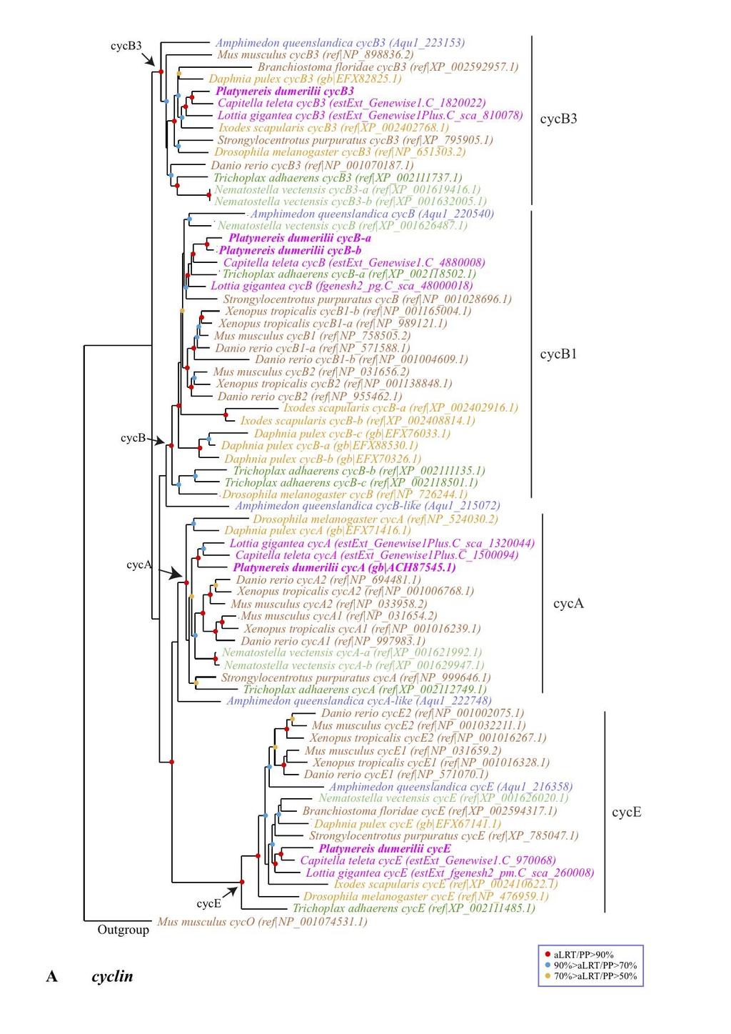
10 Supplementary Fig. S2: Orthology assignment of the newly cloned Platynereis genes. ML trees constructed with PhyML are shown. Statistical supports (alrt) are indicated on the nodes by color circles (color code is indicated in the figure). Nodes without color circles are not statistically supported. When possible, outgroups were used to root the phylogenetic trees; in the other cases, mid-branch rooting was used. Sequences were identified either by their NCBI accession number or the identifier given by the Joint Genome Institute when no accession numbers were available (in the case of Lottia, Capitella and Daphnia). (A) cyclin genes. We included in the analysis cyclin A, B, B3, and E sequences and used cyclin O as outgroup. Single Platynereis genes were found for each class, except for cyclin B where two genes were identified. (B) wntless genes. We found a single Platynereis gene that encodes a protein with the MIG-14_Wnt-bd domain which is specific to Wntless proteins. (C) axin genes. A single gene with high sequence similarity to known axin genes was found in 10
11 Platynereis. (D) flamingo genes. We cloned a single gene in Platynereis with high sequence similarity to Drosophila flamingo/starry night and vertebrates celsr genes. The Platynereis gene encodes a protein with several domains, cadherin repeats, Calcium-binding EGF-like domain, Laminin-type epidermal growth factor-like domain, and Hormone receptor domain, whose presence together is characteristic of Flamingo/Celsr proteins. (E) diego genes. We identified a Platynereis gene with moderate sequence similarity with Drosophila diego and their orthologs in vertebrates, ankyrin 6/diversin. We performed phylogenetic analyses with different samplings of ankyrin proteins, but failed to recover consistent phylogenetic trees (not shown). We however found that the cloned Platynereis gene strongly clusters with Drosophila diego and vertebrate ankyrin 6 genes. For sake of simplicity, we show here a simplified tree only comprising the sequences that cluster with the diego/ankyrin 6 group. Drosophila ankyrin was used as outgroup to root the tree. (F) prickle genes. We found a single Platynereis gene that show high sequence similarity with prickle genes and encodes a protein with PET (Prickle - Espinas-Testin) and Prickle-like LIM domains. Phylogenetic analysis was done using Drosophila paxilin as outgroup. (G) and (H) Rho and Rac genes. These genes belong to a large family that encode proteins characterized by a Rho domain. We identified two genes in Platynereis with high sequence similarities to Rho and Rac genes, respectively. We made a phylogenetic analysis with a large number of Rho and Rac like genes and found that the two Platynereis genes cluster with Drosophila and vertebrate Rac1/2 or Rho1/RhoA genes (not shown). For sake of simplicty, we only show here a subset of the sequences and two separated trees for the two subfamilies. Drosophila Cdc42 and mig-2-like were used as outgroups. (I) rok genes. A single Platynereis gene with high sequence similarity to rok genes and encoding a protein with a Rok-like PKc domain was identified. Phylogenetic analysis was performed with mouse MRCK genes as outgroup. (J) four-jointed genes. We identified two genes that encode proteins with a FAM20_c domain which is 11
12 specific of Four-jointed proteins. We failed to recover any four-jointed genes from non bilaterian species. Phylogenetic analysis showed the existence of two groups of four-jointed genes, one of which containing the Drosophila and vertebrate four-jointed genes as well as genes from most other species including Platynereis. The second group (the genes were named four-jointed-like) only comprises genes from a few species, including Platynereis. (K) and (L) fat and dachsous genes. We found two genes in Platynereis that encode atypical Cadherins and show high sequence similarity with fat and dachsous. 12
 WMISH for the PducyclinB-a (cycb-a) gene.")
13 Supplementary Fig. S3: Expression profile of Pdu-cycB-a in the neurectoderm of Platynereis larvae. Ventral views 24 to 72hpf larvae are shown. (A-E ) WMISH for the PducyclinB-a (cycb-a) gene. At all stages, the expression pattern of Pdu-cycB-a is highly similar to the profile of EdU incorporation showed in Figure 1. Scale bars are 50µm. 13

14 Supplementary Fig. S4: Cartoons summarizing the architecture of the neurectoderm of 55hpf Platynereis larvae. For the sake of clarity, only one half of the tissue is shown in transverse sections. Apical is up, lateral is on the left, and medial on the right. The dashed line demarcates the CNS from the PNS. At 55hpf the neurectoderm is composed of multiple concentric cell layers organized around the apical ventral midline. Regarding both their mitotic potential and the combination of genes that they express, the succession of these cell layers recapitulates the process of neurogenesis. This architecture suggests that extracellular signals produced by ventral midline cells regulate the balance between proliferation and differentiation of NPCs in a way that is reminiscent to that of the vertebrate neural tube. 14
 in DMSO (0.")
15 Supplementary Fig. S5: PNU treated embryos show defects similar to those observed in embryos treated with endo-iwr1. Ventral views of whole embryos and transverse sections through the neurectoderm are shown. See the cartoon in figure 1 for orientations. Embryos were incubated with PNU (5µM) in DMSO (0.05%) or in DMSO only (control group) from 33 to 55hpf. (A-B ) Antibody staining against acetylated tubulin (cyan) showing the axon scaffold of the VNC, coupled with 30min EdU (red) 15
16 incorporation at 55hpf showing the cell proliferation profile and Hoechst nuclear staining (blue). Embryos treated with PNU display an important reduction of the VNC, defects in axon guidance (arrowheads) and an extension of the cell proliferation profile. Abbreviations: con, longitudinal connectives; com, commissural axons; lat, lateral projections. (C) Graph showing the proportions of affected vs unaffected embryos in the control and treated groups (sample sizes are indicated on the graph). The experiment was replicated 3 times. All control embryos and 8% of treated ones are unaffected, 92% of treated embryos are affected. (D-G) WMISH that reveal the expression of several genes involved in the process of neurogenesis. Pdu-neurogenin expression is extended in treated embryos and suggests that the proliferating cells found throughout the whole neurectoderm are NPCs. By contrast, the reduction of Pdu-elav and Pdu-coe expression indicates a decrease in the production of neurons. These results show important similarities with those obtained using endo-iwr1 and therefore reinforce our conclusions about the role of the Wnt/β-catenin pathway in stimulating neuron differentiation. A strong reduction of Pdu-axin expression was also observed, similarly to what was found in endo-iwr1-treated embryos. Gene name abbreviations: Pdu-ngn, neurogenin; Pdu-coe, collier. Scale bars are 50µm for ventral views and 25µm for transverse views. 16

17 Supplementary Fig. S6: Pharmacological treatments altering Wnt/β-catenin pathway activity do not induce cell death in Platynereis embryos. Ventral views of whole embryos are shown. See the cartoon in figure 1 for orientations. A TUNEL protocol, revealing the presence of apoptotic cells has been adapted to Platynereis (see methods) and used on embryos treated with endo-iwr1 40µM, PNU µM and azakenpaullone 15µM from 33 to 55hpf, as well as on their respective controls incubated in DMSO. (A) A positive control embryo incubated in DNAseI shows a labelling of all cells following TUNEL assay. (B-D ) The amount of apoptotic cells observed in treated embryos was either equivalent or inferior to that observed in their control counterpart, suggesting that none of the treatments induced cell death in Platynereis embryos. Scale bars are 50µm. 17
 in DMSO (1.")
18 Supplementary Fig. S7: Constitutive activation of the Wnt/β-catenin pathway reduces cell proliferation and causes defects in VNC development. Ventral views of whole embryos and transverse sections through the neurectoderm are shown. See the cartoon in figure 1 for orientations. Embryos were incubated with azakenpaullone (15µM) in DMSO (1.5%) or in DMSO only (control group) from 33 to 55hpf. By inhibiting Glycogen-Synthase- Kinase-3 (GSK3), azakenpaullone prevents β-catenin degradation and therefore constitutively activates the Wnt/β-catenin pathway. (A-B ) Antibody staining against acetylated tubulin 18
19 (cyan) showing the axon scaffold of the VNC, coupled with 30min EdU (red) incorporation at 55hpf showing the cell proliferation profile and Hoechst nuclear staining (blue). Embryos treated with Azakenpaullone display an important reduction of the VNC, defects in axon guidance (arrowhead) and an almost complete absence of EdU labeled cells. Abbreviations: con, longitudinal connectives; com, commissural axons; lat, lateral projections. (C) Graph showing the proportions of affected vs unaffected embryos in the control and treated groups (sample sizes are indicated on the graph). The experiment was replicated 3 times. All control embryos are unaffected, and all treated embryos are affected. (D-G) WMISH revealing the expression of several genes involved in neurogenesis. Pdu-neurogenin expression is abolished in the neurectoderm of treated embryos. Together with the strong reduction of the number of EdU incorporating cells, this suggests that the constitutive activation of the Wnt/ -catenin pathway leads to the absence of proliferating NPCs. This effect is opposite to what is observed upon Wnt/β-catenin inhibition using endo-iwr1 or PNU The expression patterns of Pdu-elav and Pdu-coe are reduced, indicating a reduction in the number of neurons. The reduction of the number of both NPCs and neurons is puzzling and could be due to the premature differentiation of the NPCs that would have not stayed in a proliferative state long enough to produce a normal amount of neurons. Alternatively, the constitutive activation of the Wnt/ -catenin pathway may have forced the NPCs to exit the cell cycle while remaining in an undifferentiated state. We observed a very strong increase in the expression of Pdu-axin upon azakenpaullone treatment, reinforcing the hypothesis that the Wnt/β-catenin pathway positively regulates axin expression in Platynereis. Gene name abbreviations: Pdungn, neurogenin; Pdu-coe, collier. Scale bars are 50µm for ventral views and 25µm for transverse views. 19
Figure S1: Phylogenetic analysis of the Platynereis Notch pathway core components, Pdu-
 Electronic supplementary material legends from The Notch pathway in the annelid Platynereis: Insights into chaetogenesis and neurogenesis processes ; Eve Gazave, Quentin I. B. Lemaître and Guillaume Balavoine;
Nature Neuroscience: doi: /nn.2662
 Supplementary Figure 1 Atlastin phylogeny and homology. (a) Maximum likelihood phylogenetic tree based on 18 Atlastin-1 sequences using the program Quicktree. Numbers at internal nodes correspond to bootstrap
Role of Organizer Chages in Late Frog Embryos
 Ectoderm Germ Layer Frog Fate Map Frog Fate Map Role of Organizer Chages in Late Frog Embryos Organizer forms three distinct regions Notochord formation in chick Beta-catenin localization How does beta-catenin
SUPPLEMENTARY INFORMATION
 doi:10.1038/nature11589 Supplementary Figure 1 Ciona intestinalis and Petromyzon marinus neural crest expression domain comparison. Cartoon shows dorsal views of Ciona mid gastrula (left) and Petromyzon
Nervous system development in lecithotrophic larval and juvenile stages of the annelid Capitella teleta
 Meyer et al. Frontiers in Zoology (2015)12:15 DOI 10.1186/s12983-015-0108-y RESEARCH Open Access Nervous system development in lecithotrophic larval and juvenile stages of the annelid Capitella teleta
Neural development its all connected
 Neural development its all connected How do you build a complex nervous system? How do you build a complex nervous system? 1. Learn how tissue is instructed to become nervous system. Neural induction 2.
Cellular Neurobiology BIPN 140 Fall 2016 Problem Set #8
 Cellular Neurobiology BIPN 140 Fall 2016 Problem Set #8 1. Inductive signaling is a hallmark of vertebrate and mammalian development. In early neural development, there are multiple signaling pathways
Reading. Lecture VI. Making Connections 9/17/12. Bio 3411 Lecture VI. Making Connections. Bio 3411 Monday September 17, 2012
 Lecture VI. Making Connections Bio 3411 Monday September 17, 2012!! 1! Reading NEUROSCIENCE: 5 th ed, pp!507?536! 4 th ed, pp 577-609 Bentley, D., & Caudy, M. (1983). Nature, 304(5921), 62-65. Dickson,
Axon guidance I. Paul Garrity March 15, /9.013
 Axon guidance I Paul Garrity March 15, 2004 7.68/9.013 Neuronal Wiring: Functional Framework of the Nervous System Stretch reflex circuit Early theories of axonogenesis Schwann: many neurons link to form
Developmental Biology 3230 Midterm Exam 1 March 2006
 Name Developmental Biology 3230 Midterm Exam 1 March 2006 1. (20pts) Regeneration occurs to some degree to most metazoans. When you remove the head of a hydra a new one regenerates. Graph the inhibitor
16 The Cell Cycle. Chapter Outline The Eukaryotic Cell Cycle Regulators of Cell Cycle Progression The Events of M Phase Meiosis and Fertilization
 The Cell Cycle 16 The Cell Cycle Chapter Outline The Eukaryotic Cell Cycle Regulators of Cell Cycle Progression The Events of M Phase Meiosis and Fertilization Introduction Self-reproduction is perhaps
Conclusions. The experimental studies presented in this thesis provide the first molecular insights
 C h a p t e r 5 Conclusions 5.1 Summary The experimental studies presented in this thesis provide the first molecular insights into the cellular processes of assembly, and aggregation of neural crest and
Spatiotemporal regulation of nervous system development in the annelid Capitella teleta
 DOI 10.1186/s13227-017-0076-8 EvoDevo RESEARCH Spatiotemporal reguion of nervous system development in the annelid Capitella teleta Abhinav Sur 1, Craig R. Magie 2, Elaine C. Seaver 3 and Néva P. Meyer
Developmental Biology Lecture Outlines
 Developmental Biology Lecture Outlines Lecture 01: Introduction Course content Developmental Biology Obsolete hypotheses Current theory Lecture 02: Gametogenesis Spermatozoa Spermatozoon function Spermatozoon
Cells to Tissues. Peter Takizawa Department of Cell Biology
 Cells to Tissues Peter Takizawa Department of Cell Biology From one cell to ensembles of cells. Multicellular organisms require individual cells to work together in functional groups. This means cells
7.013 Problem Set
 7.013 Problem Set 5-2013 Question 1 During a summer hike you suddenly spot a huge grizzly bear. This emergency situation triggers a fight or flight response through a signaling pathway as shown below.
!!!!!!!! DB3230 Midterm 2 12/13/2013 Name:
 1. (10 pts) Draw or describe the fate map of a late blastula stage sea urchin embryo. Draw or describe the corresponding fate map of the pluteus stage larva. Describe the sequence of gastrulation events
Bio334 Neurobiology 1 Lecture 2
 Evolution of Bio334 Neurobiology 1 Lecture 2 1 Questions from the last lecture Patients with Broca s area lesions do have trouble writing Golgi Cajal debate reticular theory of the brain vs. individual
Axon Guidance. Multiple decision points along a growing axon s trajectory Different types of axon guidance cues:
 Axon Guidance Multiple decision points along a growing axon s trajectory Different types of axon guidance cues: Contact mediated - requires direct contact by growth cone Long range - growth cone responds
BIOLOGY - CLUTCH CH.32 - OVERVIEW OF ANIMALS.
 !! www.clutchprep.com Animals are multicellular, heterotrophic eukaryotes that feed by ingesting their food Most animals are diploid, and produce gametes produced directly by meiosis Animals lack cell
Cell-Cell Communication in Development
 Biology 4361 - Developmental Biology Cell-Cell Communication in Development June 23, 2009 Concepts Cell-Cell Communication Cells develop in the context of their environment, including: - their immediate
Supplementary Figure 1: Mechanism of Lbx2 action on the Wnt/ -catenin signalling pathway. (a) The Wnt/ -catenin signalling pathway and its
 The Wnt/ -catenin signalling pathway and its") Supplementary Figure 1: Mechanism of Lbx2 action on the Wnt/ -catenin signalling pathway. (a) The Wnt/ -catenin signalling pathway and its transcriptional activity in wild-type embryo. A gradient of canonical
Supplementary Figure 1: Mechanism of Lbx2 action on the Wnt/ -catenin signalling pathway. (a) The Wnt/ -catenin signalling pathway and its transcriptional activity in wild-type embryo. A gradient of canonical
Supplementary Materials for
 www.sciencesignaling.org/cgi/content/full/6/301/ra98/dc1 Supplementary Materials for Regulation of Epithelial Morphogenesis by the G Protein Coupled Receptor Mist and Its Ligand Fog Alyssa J. Manning,
Mesoderm Induction CBT, 2018 Hand-out CBT March 2018
 Mesoderm Induction CBT, 2018 Hand-out CBT March 2018 Introduction 3. Books This module is based on the following books: - 'Principles of Developement', Lewis Wolpert, et al., fifth edition, 2015 - 'Developmental
Sonic hedgehog (Shh) signalling in the rabbit embryo
 signalling in the rabbit embryo") Sonic hedgehog (Shh) signalling in the rabbit embryo In the first part of this thesis work the physical properties of cilia-driven leftward flow were characterised in the rabbit embryo. Since its discovery
Sonic hedgehog (Shh) signalling in the rabbit embryo In the first part of this thesis work the physical properties of cilia-driven leftward flow were characterised in the rabbit embryo. Since its discovery
Flamingo, a seven-pass transmembrane cadherin, cooperates with Netrin/Frazzled in Drosophila midline guidance
 Flamingo, a seven-pass transmembrane cadherin, cooperates with Netrin/Frazzled in Drosophila midline guidance Cristina Organisti1, Irina Hein1, Ilona C. Grunwald Kadow1* and Takashi Suzuki2* 1 Max Planck
Chapter 11. Development: Differentiation and Determination
 KAP Biology Dept Kenyon College Differential gene expression and development Mechanisms of cellular determination Induction Pattern formation Chapter 11. Development: Differentiation and Determination
MCDB 4777/5777 Molecular Neurobiology Lecture 29 Neural Development- In the beginning
 MCDB 4777/5777 Molecular Neurobiology Lecture 29 Neural Development- In the beginning Learning Goals for Lecture 29 4.1 Describe the contributions of early developmental events in the embryo to the formation
SUPPLEMENTARY INFORMATION
 DOI: 10.1038/ncb3267 Supplementary Figure 1 A group of genes required for formation or orientation of annular F-actin bundles and aecm ridges: RNAi phenotypes and their validation by standard mutations.
Unicellular: Cells change function in response to a temporal plan, such as the cell cycle.
 Spatial organization is a key difference between unicellular organisms and metazoans Unicellular: Cells change function in response to a temporal plan, such as the cell cycle. Cells differentiate as a
Introduction to Animals
 Introduction to Animals Characteristics of Animals multicellular Except for sponges, animal cells are arranged into tissues. Tissues are necessary to produce organs and organ systems. Tissues, organs,
9/4/2015 INDUCTION CHAPTER 1. Neurons are similar across phyla Thus, many different model systems are used in developmental neurobiology. Fig 1.
 INDUCTION CHAPTER 1 Neurons are similar across phyla Thus, many different model systems are used in developmental neurobiology Fig 1.1 1 EVOLUTION OF METAZOAN BRAINS GASTRULATION MAKING THE 3 RD GERM LAYER
MCN. Complex Genetic Interactions among Four Receptor Tyrosine Phosphatases Regulate Axon Guidance in Drosophila
 MCN Molecular and Cellular Neuroscience 17, 274 291 (2001) doi:10.1006/mcne.2000.0939, available online at http://www.idealibrary.com on Complex Genetic Interactions among Four Receptor Tyrosine Phosphatases
PRACTICE EXAM. 20 pts: 1. With the aid of a diagram, indicate how initial dorsal-ventral polarity is created in fruit fly and frog embryos.
 PRACTICE EXAM 20 pts: 1. With the aid of a diagram, indicate how initial dorsal-ventral polarity is created in fruit fly and frog embryos. No Low [] Fly Embryo Embryo Non-neural Genes Neuroectoderm Genes
7.013 Spring 2005 Problem Set 4
 MIT Department of Biology 7.013: Introductory Biology - Spring 2005 Instructors: Professor Hazel Sive, Professor Tyler Jacks, Dr. Claudette Gardel NAME TA 7.013 Spring 2005 Problem Set 4 FRIDAY April 8th,
Control and Integration. Nervous System Organization: Bilateral Symmetric Animals. Nervous System Organization: Radial Symmetric Animals
 Control and Integration Neurophysiology Chapters 10-12 Nervous system composed of nervous tissue cells designed to conduct electrical impulses rapid communication to specific cells or groups of cells Endocrine
Fig. S1. Proliferation and cell cycle exit are affected by the med mutation. (A,B) M-phase nuclei are visualized by a-ph3 labeling in wild-type (A)
 M-phase nuclei are visualized by a-ph3 labeling in wild-type (A)") Fig. S1. Proliferation and cell cycle exit are affected by the med mutation. (A,B) M-phase nuclei are visualized by a-ph3 labeling in wild-type (A) and mutant (B) 4 dpf retinae. The central retina of the
Fig. S1. Proliferation and cell cycle exit are affected by the med mutation. (A,B) M-phase nuclei are visualized by a-ph3 labeling in wild-type (A) and mutant (B) 4 dpf retinae. The central retina of the
MBios 401/501: Lecture 14.2 Cell Differentiation I. Slide #1. Cell Differentiation
 MBios 401/501: Lecture 14.2 Cell Differentiation I Slide #1 Cell Differentiation Cell Differentiation I -Basic principles of differentiation (p1305-1320) -C-elegans (p1321-1327) Cell Differentiation II
The majority of cells in the nervous system arise during the embryonic and early post
 Introduction Introduction The majority of cells in the nervous system arise during the embryonic and early post natal period. These cells are derived from population of neural stem cells first shown by
Name. Biology Developmental Biology Winter Quarter 2013 KEY. Midterm 3
 Name 100 Total Points Open Book Biology 411 - Developmental Biology Winter Quarter 2013 KEY Midterm 3 Read the Following Instructions: * Answer 20 questions (5 points each) out of the available 25 questions
Question Set # 4 Answer Key 7.22 Nov. 2002
 Question Set # 4 Answer Key 7.22 Nov. 2002 1) A variety of reagents and approaches are frequently used by developmental biologists to understand the tissue interactions and molecular signaling pathways
1. What are the three general areas of the developing vertebrate limb? 2. What embryonic regions contribute to the developing limb bud?
 Study Questions - Lecture 17 & 18 1. What are the three general areas of the developing vertebrate limb? The three general areas of the developing vertebrate limb are the proximal stylopod, zeugopod, and
Introduction Principles of Signaling and Organization p. 3 Signaling in Simple Neuronal Circuits p. 4 Organization of the Retina p.
 Introduction Principles of Signaling and Organization p. 3 Signaling in Simple Neuronal Circuits p. 4 Organization of the Retina p. 5 Signaling in Nerve Cells p. 9 Cellular and Molecular Biology of Neurons
Fig. S1. Expression pattern of moody-gal4 in third instar. Maximum projection illustrating a dissected moody-gal4>ngfp L3 larva stained for Repo
 Fig. S1. Expression pattern of moody-gal4 in third instar. Maximum projection illustrating a dissected moody-gal4>ngfp L3 larva stained for Repo (magenta), Fas2 (blue) and GFP (green) in overview (A) and
Supplemental table S7.
 Supplemental table S7. GO terms significantly enriched in significantly up-regulated genes of the microarray. K: number of genes from the input cluster in the given category. F: number of total genes in
Zool 3200: Cell Biology Exam 5 4/27/15
 Name: Trask Zool 3200: Cell Biology Exam 5 4/27/15 Answer each of the following short answer questions in the space provided, giving explanations when asked to do so. Circle the correct answer or answers
Revision Based on Chapter 25 Grade 11
 Revision Based on Chapter 25 Grade 11 Biology Multiple Choice Identify the choice that best completes the statement or answers the question. 1. A cell that contains a nucleus and membrane-bound organelles
NEURONS, SENSE ORGANS, AND NERVOUS SYSTEMS CHAPTER 34
 NEURONS, SENSE ORGANS, AND NERVOUS SYSTEMS CHAPTER 34 KEY CONCEPTS 34.1 Nervous Systems Are Composed of Neurons and Glial Cells 34.2 Neurons Generate Electric Signals by Controlling Ion Distributions 34.3
5- Semaphorin-Plexin-Neuropilin
 5- Semaphorin-Plexin-Neuropilin 1 SEMAPHORINS-PLEXINS-NEUROPILINS ligands receptors co-receptors semaphorins and their receptors are known signals for: -axon guidance -cell migration -morphogenesis -immune
Supplementary Figure 1 Schematic overview of ASTNs in neuronal migration. (a) Schematic of roles played by ASTNs 1 and 2. ASTN-1-mediated adhesions
 Schematic of roles played by ASTNs 1 and 2. ASTN-1-mediated adhesions") Supplementary Figure 1 Schematic overview of ASTNs in neuronal migration. (a) Schematic of roles played by ASTNs 1 and 2. ASTN-1-mediated adhesions undergo endocytosis into clathrin-coated vesicles dependent
Supplementary Figure 1 Schematic overview of ASTNs in neuronal migration. (a) Schematic of roles played by ASTNs 1 and 2. ASTN-1-mediated adhesions undergo endocytosis into clathrin-coated vesicles dependent
Drosophila melanogaster- Morphogen Gradient
 NPTEL Biotechnology - Systems Biology Drosophila melanogaster- Morphogen Gradient Dr. M. Vijayalakshmi School of Chemical and Biotechnology SASTRA University Joint Initiative of IITs and IISc Funded by
NN-A29932: Supplementary Data 1 Diversity and Wiring Variability of Olfactory Local Interneurons in the Drosophila Antennal Lobe
 NN-A29932: Supplementary Data 1 Diversity and Wiring Variability of Olfactory Local Interneurons in the Drosophila Antennal Lobe Ya-Hui Chou, Maria L. Spletter, Emre Yaksi, Jonathan C. S. Leong, Rachel
S1 Gene ontology (GO) analysis of the network alignment results
 analysis of the network alignment results") 1 Supplementary Material for Effective comparative analysis of protein-protein interaction networks by measuring the steady-state network flow using a Markov model Hyundoo Jeong 1, Xiaoning Qian 1 and
1 Supplementary Material for Effective comparative analysis of protein-protein interaction networks by measuring the steady-state network flow using a Markov model Hyundoo Jeong 1, Xiaoning Qian 1 and
Supplementary Information
 Supplementary Information MAP2/Hoechst Hyp.-AP ph 6.5 Hyp.-SD ph 7.2 Norm.-SD ph 7.2 Supplementary Figure 1. Mitochondrial elongation in cortical neurons by acidosis. Representative images of neuronal
Chapter 4 Evaluating a potential interaction between deltex and git in Drosophila: genetic interaction, gene overexpression and cell biology assays.
 Evaluating a potential interaction between deltex and git in Drosophila: genetic interaction, gene overexpression and cell biology assays. The data described in chapter 3 presented evidence that endogenous
Chapter 18 Lecture. Concepts of Genetics. Tenth Edition. Developmental Genetics
 Chapter 18 Lecture Concepts of Genetics Tenth Edition Developmental Genetics Chapter Contents 18.1 Differentiated States Develop from Coordinated Programs of Gene Expression 18.2 Evolutionary Conservation
Exam 1 ID#: October 4, 2007
 Biology 4361 Name: KEY Exam 1 ID#: October 4, 2007 Multiple choice (one point each) (1-25) 1. The process of cells forming tissues and organs is called a. morphogenesis. b. differentiation. c. allometry.
Exam 2 ID#: November 9, 2006
 Biology 4361 Name: KEY Exam 2 ID#: November 9, 2006 Multiple choice (one point each) Circle the best answer. 1. Inducers of Xenopus lens and optic vesicle include a. pharyngeal endoderm and anterior neural
 DOI: 10.1038/ncb2819 Gαi3 / Actin / Acetylated Tubulin Gαi3 / Actin / Acetylated Tubulin a a Gαi3 a Actin Gαi3 WT Gαi3 WT Gαi3 WT b b Gαi3 b Actin Gαi3 KO Gαi3 KO Gαi3 KO # # Figure S1 Loss of protein
Maternal Control of GermLayer Formation in Xenopus
 Maternal Control of GermLayer Formation in Xenopus The zygotic genome is activated at the mid-blastula transition mid-blastula fertilized egg Xenopus gastrulae early-gastrula 7 hrs 10 hrs control not VP
Neurochemistry 1. Nervous system is made of neurons & glia, as well as other cells. Santiago Ramon y Cajal Nobel Prize 1906
 Neurochemistry 1 Nervous system is made of neurons & glia, as well as other cells. Santiago Ramon y Cajal Nobel Prize 1906 How Many Neurons Do We Have? The human brain contains ~86 billion neurons and
7.06 Problem Set #4, Spring 2005
 7.06 Problem Set #4, Spring 2005 1. You re doing a mutant hunt in S. cerevisiae (budding yeast), looking for temperaturesensitive mutants that are defective in the cell cycle. You discover a mutant strain
The Midline Protein Regulates Axon Guidance by Blocking the Reiteration of Neuroblast Rows within the Drosophila Ventral Nerve Cord
 The Midline Protein Regulates Axon Guidance by Blocking the Reiteration of Neuroblast Rows within the Drosophila Ventral Nerve Cord Mary Ann Manavalan, Ivana Gaziova, Krishna Moorthi Bhat* Department of
BIO 115 SP 2012 Homework 1: Introduction to A&P and Chemistry Please note that this is not a required assignment but it is recommended.
 BIO 115 SP 2012 Homework 1: Introduction to A&P and Chemistry Please note that this is not a required assignment but it is recommended. 1. When a body is in the anatomical position, it is A. Standing erect,
and its origins G. E. Schneider 2009 Part 1: Introduction MIT 9.14 Class 1 Brain talk, and
 A sketch of the central nervous system and its origins G. E. Schneider 2009 Part 1: Introduction MIT 9.14 Class 1 Brain talk, and the ancient activities of brain cells 1. Introduction a) b) The plan for
Principles of Experimental Embryology
 Biology 4361 Developmental Biology Principles of Experimental Embryology June 16, 2008 Overview What forces affect embryonic development? The embryonic environment: external and internal How do forces
The Radiata-Bilateria split. Second branching in the evolutionary tree
 The Radiata-Bilateria split Second branching in the evolutionary tree Two very important characteristics are used to distinguish between the second bifurcation of metazoans Body symmetry Germinal layers
purpose of this Chapter is to highlight some problems that will likely provide new
 119 Chapter 6 Future Directions Besides our contributions discussed in previous chapters to the problem of developmental pattern formation, this work has also brought new questions that remain unanswered.
Systematic Screening of Drosophila Deficiency Mutations for Embryonic Phenotypes and Orphan Receptor Ligands
 Systematic Screening of Drosophila Deficiency Mutations for Embryonic Phenotypes and Orphan Receptor Ligands Ashley P. Wright 1, A. Nicole Fox 1, Karl G. Johnson 2, Kai Zinn 1 * 1 Division of Biology,
Developmental processes Differential gene expression Introduction to determination The model organisms used to study developmental processes
 Date Title Topic(s) Learning Outcomes: Sept 28 Oct 3 1. What is developmental biology and why should we care? 2. What is so special about stem cells and gametes? Developmental processes Differential gene
Attractive and repulsive functions of Slit are mediated by different receptors in the Drosophila trachea
 Development 129, 4941-4951 (2002) Printed in Great Britain The Company of Biologists Limited 2002 DEV7966 4941 Attractive and repulsive functions of Slit are mediated by different receptors in the Drosophila
b. The maximum binding will decrease.
 Cell Signaling Receptors are a. proteins that change conformation upon interaction with a stimulus b. genes that change expression in response to a stimulus c. phosphorylation cascades that control cellular
targets. clustering show that different complex pathway
 Supplementary Figure 1. CLICR allows clustering and activation of cytoplasmic protein targets. (a, b) Upon light activation, the Cry2 (red) and LRP6c (green) components co-cluster due to the heterodimeric
Bio 127 Section I Introduction to Developmental Biology. Cell Cell Communication in Development. Developmental Activities Coordinated in this Way
 Bio 127 Section I Introduction to Developmental Biology Cell Cell Communication in Development Gilbert 9e Chapter 3 It has to be EXTREMELY well coordinated for the single celled fertilized ovum to develop
The Fox/Forkhead transcription factor family of the hemichordate Saccoglossus kowalevskii
 The Fox/Forkhead transcription factor family of the hemichordate Saccoglossus kowalevskii Fritzenwanker et al. Fritzenwanker et al. EvoDevo 2014, 5:17 Fritzenwanker et al. EvoDevo 2014, 5:17 RESEARCH Open
SUPPLEMENTARY INFORMATION
 doi:10.1038/nature10923 Supplementary Figure 1 Ten-a and Ten-m antibody and cell type specificities. a c, Representative single confocal sections of a Drosophila NMJ stained with antibodies to Ten-a (red),
Supplementary Figure 1. Real time in vivo imaging of SG secretion. (a) SGs from Drosophila third instar larvae that express Sgs3-GFP (green) and
 SGs from Drosophila third instar larvae that express Sgs3-GFP (green) and") Supplementary Figure 1. Real time in vivo imaging of SG secretion. (a) SGs from Drosophila third instar larvae that express Sgs3-GFP (green) and Lifeact-Ruby (red) were imaged in vivo to visualize secretion
Supplementary Figure 1. Real time in vivo imaging of SG secretion. (a) SGs from Drosophila third instar larvae that express Sgs3-GFP (green) and Lifeact-Ruby (red) were imaged in vivo to visualize secretion
Paraxial and Intermediate Mesoderm
 Biology 4361 Paraxial and Intermediate Mesoderm December 6, 2007 Mesoderm Formation Chick Major Mesoderm Lineages Mesodermal subdivisions are specified along a mediolateral axis by increasing amounts of
Developmental Zoology. Ectodermal derivatives (ZOO ) Developmental Stages. Developmental Stages
 Developmental Stages. Developmental Stages") Developmental Zoology (ZOO 228.1.0) Ectodermal derivatives 1 Developmental Stages Ø Early Development Fertilization Cleavage Gastrulation Neurulation Ø Later Development Organogenesis Larval molts Metamorphosis
Developmental Zoology (ZOO 228.1.0) Ectodermal derivatives 1 Developmental Stages Ø Early Development Fertilization Cleavage Gastrulation Neurulation Ø Later Development Organogenesis Larval molts Metamorphosis
Elements of Bioinformatics 14F01 TP5 -Phylogenetic analysis
 Elements of Bioinformatics 14F01 TP5 -Phylogenetic analysis 10 December 2012 - Corrections - Exercise 1 Non-vertebrate chordates generally possess 2 homologs, vertebrates 3 or more gene copies; a Drosophila
Segment boundary formation in Drosophila embryos
 Segment boundary formation in Drosophila embryos Development 130, August 2003 Camilla W. Larsen, Elizabeth Hirst, Cyrille Alexandre and Jean Paul Vincent 1. Introduction: - Segment boundary formation:
Chapter 9. Benefits of Being Large. Levels of Organization in Organismal Complexity. Hierarchical Organization of Animal Complexity. Fig. 9.
 Copyright The McGraw-Hill Companies, Inc. Permission required for reproduction or display. Chapter 9 Architectural Pattern of an Animal Levels of Organization in Organismal Complexity Zoologists recognize
Three different fusions led to three basic ideas: 1) If one fuses a cell in mitosis with a cell in any other stage of the cell cycle, the chromosomes
 If one fuses a cell in mitosis with a cell in any other stage of the cell cycle, the chromosomes") Section Notes The cell division cycle presents an interesting system to study because growth and division must be carefully coordinated. For many cells it is important that it reaches the correct size
Section Notes The cell division cycle presents an interesting system to study because growth and division must be carefully coordinated. For many cells it is important that it reaches the correct size
Information processing. Divisions of nervous system. Neuron structure and function Synapse. Neurons, synapses, and signaling 11/3/2017
 Neurons, synapses, and signaling Chapter 48 Information processing Divisions of nervous system Central nervous system (CNS) Brain and a nerve cord Integration center Peripheral nervous system (PNS) Nerves
Lecture 7. Development of the Fruit Fly Drosophila
 BIOLOGY 205/SECTION 7 DEVELOPMENT- LILJEGREN Lecture 7 Development of the Fruit Fly Drosophila 1. The fruit fly- a highly successful, specialized organism a. Quick life cycle includes three larval stages
1 GO: regulation of cell size E-04 2 GO: negative regulation of cell growth GO:
 Table S2: The biological modulated by mir-5701 Sr. No Term Id 1 Term Name 2 Hit Gene Number 3 P-Value 4 1 GO:0008361 regulation of cell size 9 4.37E-04 2 GO:0030308 negative regulation of cell growth 8
Report. Functional Diversity of Robo Receptor Immunoglobulin Domains Promotes Distinct Axon Guidance Decisions
 Current Biology 20, 567 572, March 23, 2010 ª2010 Elsevier Ltd All rights reserved DOI 10.1016/j.cub.2010.02.021 Functional Diversity of Robo Receptor Immunoglobulin Domains Promotes Distinct Axon Guidance
Animal Origins and Evolution
 Animal Origins and Evolution Common Features of Animals multicellular heterotrophic motile Sexual reproduction, embryo Evolution of Animals All animals are multicellular and heterotrophic, which means
Nature Genetics: doi: /ng Supplementary Figure 1. The phenotypes of PI , BR121, and Harosoy under short-day conditions.
 Supplementary Figure 1 The phenotypes of PI 159925, BR121, and Harosoy under short-day conditions. (a) Plant height. (b) Number of branches. (c) Average internode length. (d) Number of nodes. (e) Pods
Wnt signaling in planarians: new answers to old questions
 Int. J. Dev. Biol. 56: 53-65 doi: 10.1387/ijdb.113451ma www.intjdevbiol.com Wnt signaling in planarians: new answers to old questions MARIA ALMUEDO-CASTILLO, MIQUEL SUREDA-GÓMEZ and TERESA ADELL* Department
Supplementary Figure 1
 Supplementary Figure 1 Single-cell RNA sequencing reveals unique sets of neuropeptides, transcription factors and receptors in specific types of sympathetic neurons (a) Dissection of paravertebral SGs
Organization of Vertebrate Body. Organization of Vertebrate Body
 The Animal Body and Principles of Regulation Chapter 43 There are four levels of organization: 1. Cells 2. Tissues 3. Organs 4. Organ systems Bodies of vertebrates are composed of different cell types
Regulation and signaling. Overview. Control of gene expression. Cells need to regulate the amounts of different proteins they express, depending on
 Regulation and signaling Overview Cells need to regulate the amounts of different proteins they express, depending on cell development (skin vs liver cell) cell stage environmental conditions (food, temperature,
RACK-1 Acts with Rac GTPase Signaling and UNC-115/ ablim in Caenorhabditis elegans Axon Pathfinding and Cell Migration
 RACK-1 Acts with Rac GTPase Signaling and UNC-115/ ablim in Caenorhabditis elegans Axon Pathfinding and Cell Migration Rafael S. Demarco, Erik A. Lundquist* Programs in Genetics and Molecular, Cellular,
The EGF Signaling Pathway! Introduction! Introduction! Chem Lecture 10 Signal Transduction & Sensory Systems Part 3. EGF promotes cell growth
 Chem 452 - Lecture 10 Signal Transduction & Sensory Systems Part 3 Question of the Day: Who is the son of Sevenless? Introduction! Signal transduction involves the changing of a cell s metabolism or gene
Supplementary Figure 1: To test the role of mir-17~92 in orthologous genetic model of ADPKD, we generated Ksp/Cre;Pkd1 F/F (Pkd1-KO) and Ksp/Cre;Pkd1
 and Ksp/Cre;Pkd1") Supplementary Figure 1: To test the role of mir-17~92 in orthologous genetic model of ADPKD, we generated Ksp/Cre;Pkd1 F/F (Pkd1-KO) and Ksp/Cre;Pkd1 F/F ;mir-17~92 F/F (Pkd1-miR-17~92KO) mice. (A) Q-PCR
Supplementary Figure 1: To test the role of mir-17~92 in orthologous genetic model of ADPKD, we generated Ksp/Cre;Pkd1 F/F (Pkd1-KO) and Ksp/Cre;Pkd1 F/F ;mir-17~92 F/F (Pkd1-miR-17~92KO) mice. (A) Q-PCR
Geert Geeven. April 14, 2010
 iction of Gene Regulatory Interactions NDNS+ Workshop April 14, 2010 Today s talk - Outline Outline Biological Background Construction of Predictors The main aim of my project is to better understand the
Conservation of arthropod midline netrin accumulation revealed with a cross-reactive antibody provides evidence for midline cell homology
 EVOLUTION & DEVELOPMENT 11:3, 260 268 (2009) DOI: 10.1111/j.1525-142X.2009.00328.x Conservation of arthropod midline netrin accumulation revealed with a cross-reactive antibody provides evidence for midline
Dendrites - receives information from other neuron cells - input receivers.
 The Nerve Tissue Neuron - the nerve cell Dendrites - receives information from other neuron cells - input receivers. Cell body - includes usual parts of the organelles of a cell (nucleus, mitochondria)
UNIVERSITY OF YORK BIOLOGY. Developmental Biology
 Examination Candidate Number: UNIVERSITY OF YORK BSc Stage 2 Degree Examinations 2017-18 Department: BIOLOGY Title of Exam: Developmental Biology Desk Number: Time allowed: 1 hour and 30 minutes Total