Introduction to Bioinformatics. Case Study
|
|
|
- Anthony Cooper
- 5 years ago
- Views:
Transcription

1 Case Study


2 Case 1 Case 2 How SIGIRR inhibit the TLR4 and 7 signaling pathways? Homology modeling of Tolllike receptor ectodomains.
3 Case 1 How SIGIRR inhibit the Toll-like receptors TLR4 and 7 signaling pathways?
4 Background : Structure of Toll-like receptors (TLRs) Ectodomain (ECD) Leucine-rich repeat (LRR) Transmembrane domain TIR domain TLRs belong to the Toll-like receptor/ interleukin-1 receptor (TLR/IL-1R) superfamily, which is defined by the presence of a conserved cytoplasmic Toll/interleukin-1 receptor (TIR) domain connected to an ectodomain through a single transmembrane stretch. Their ectodomains consist of leucinerich repeats (LRRs).
, lipopeptide, CpG DNA, flagellin, and double-")
5 TLR signaling pathways These LRRs provide a variety of structural frameworks for the binding of protein and non-protein ligands including lipopolysaccharide (LPS), lipopeptide, CpG DNA, flagellin, and double- /single-stranded RNA.
6 TLRs are capable of recognizing ligands in a dimer form. Determined crystal structures of TLR ECDligand-ECD complexes: human TLR2-1, mouse TLR3-3, human TLR4-4, mouse TLR2-6.
 was the first intracellular adaptor molecule characterized among all known adaptors in the TLR signaling.")
7 TIR DD Upon receptor activation, an intracellular TIR signaling complex is formed between the receptor and downstream adaptor TIR domains. MyD88 (Myeloid differentiation primary response protein 88) was the first intracellular adaptor molecule characterized among all known adaptors in the TLR signaling. It consists of an N-terminal death domain (DD) separated from its C-terminal TIR domain by a linker sequence. MyD88 also forms a dimer through DD-DD and TIR-TIR domain interactions when recruited to the receptor complex. MyD88 can recruit IRAK (IL-1RI-associated protein kinases) through its DD to continue signaling and, finally, to induce the nuclear factor-kb (NF-kB) leading to the expression of type I interferons.
, also known as TIR8, was initially identified as an Ig domain-containing receptor of the TLR/IL-1R superfamily.")
8 Leucine-rich repeats (LRRs) TLR SIGIRR (single immunoglobulin interleukin-1 receptorrelated molecule) Single immunoglobulin (Ig) Toll/interleukin-1 receptor (TIR) domain TIR domain 73 AA C-terminal tail SIGIRR (Single immunoglobulin interleukin-1 receptor-related molecule), also known as TIR8, was initially identified as an Ig domain-containing receptor of the TLR/IL-1R superfamily. Both the extracellular and intracellular domains of SIGIRR differ from those of other Ig domain-containing receptors, as its single extracellular Ig domain does not support ligand-binding. Its intracellular TIR domain cannot activate NF-kB. Moreover, the TIR domain of SIGIRR extends that of the typical TLR/IL-1R superfamily member by >73 amino acids at the C- terminal (C-tail).


9 mouse B6 lpr/lpr Sigirr +/+ mouse B6 lpr/lpr Sigirr -/- Lech et al., JEM, 2008 Instead, SIGIRR acts as an endogenous inhibitor for MyD88-dependent TLR and IL-1R signaling. This behavior was shown by over expression of SIGIRR in Jurkat or HepG2 cells which showed substantially reduced LPS, CpG DNA or IL-1-induced activation of NFkB. Thus, SIGIRR has attracted tremendous research interest because of its regulating function in cancerrelated inflammation and autoimmunity. For example, systemic lupus erythematosus (SLE, 系统性红斑狼疮 ) is caused by TLR7-mediated induction of type I interferons. Compared with wild type mice Sigirr-deficient mice develop excessive lymphoproliferation when introduced into the context of a lupus susceptibility gene. Although the significance of SIGIRR has been widely acknowledged, its inhibition mechanism remains unclear owing to a lack of structural information.
. bind to TLR4 inhibit signaling ΔN yes yes ΔC yes yes ΔTIR no no Fulllength yes Qin et al.")
10 Mutagenesis studies investigated three deletion mutants of SIGIRR: ΔN (lacking the extracellular Ig domain), ΔTIR (lacking the intracellular TIR domain) and ΔC (lacking the C-tail of the TIR domain with deletion of residues ). bind to TLR4 inhibit signaling ΔN yes yes ΔC yes yes ΔTIR no no Fulllength yes Qin et al., 2005 JBC yes The results showed that only the TIR domain (excluding the C-tail part) is necessary for SIGIRR to inhibit TLR4 signaling. Nevertheless, detailed structural interaction mechanisms of SIGIRR s TIR domain are still missing.
11 Hypothesis: SIGIRR blocks the molecular interface of TLR4 and MyD88 via its TIR domain Objective: to find a structural explanation for these TIR-TIR interactions. 1. Structure prediction of TIR domains of TLRs, MyD88 and SIGIRR. 2. Structure analysis/docking.
12 Step 1 : model construction Amino acid sequences of the target proteins, human TLR4, TLR7, MyD88, and SIGIRR were extracted from the NCBI protein database. Three-dimensional models of TLR4, TLR7, MyD88 and SIGIRR (without the C-tail) were constructed by homology modeling. Due to the homology of the target proteins, four common templates were obtained via BLAST search against the Protein Data Bank (PDB). They were TLR1 (1FYV), TLR2 (1FYW), TLR10 (2J67) and IL-1RAPL (1T3G). In the secondary structure-aided alignments for the homology modeling, the average target-template sequence similarity of TLR4, TLR7, MyD88 and SIGIRR was 51.7%, 50.4%, 44.5% and 42.7%, respectively Multiple sequence alignment of each target with the templates was generated with MUSCLE and analyzed with Jalview. Because the secondary structure of the TIR domain is composed of well-organized alternating β-strands and α-helixes, the alignments were adjusted manually according to the secondary structure information to improve the alignment quality. The secondary structure of each target was predicted by PSIPRED.
 on both sides. The loops are named by the letters of the secondary structure elements that they connect. For example, the BBloop connects β-strand B and α-helix B.")
13 Step 1 : model construction The resulting structures exhibit a typical TIR domain conformation in which a central five-stranded parallel β-sheet (βa- βe) is surrounded by a total of five α-helixes (αa αe) on both sides. The loops are named by the letters of the secondary structure elements that they connect. For example, the BBloop connects β-strand B and α-helix B. The structure of NSF-N was identified as a template for SIGIRR s C-tail through protein threading. To improve the model quality, ModLoop was used to rebuild the coordinates of the low quality loop regions. Finally, model quality assessment programs: ProQ, ModFOLD and MetaMQAP were used to evaluate the output candidate models and select the most reliable one. crystal structure of IL1-RAPL (1T3G)

14 Step 1 : model construction The BB-loop and αe of TLR4, TLR7 and MyD88, along with the BB-loop of SIGIRR, may be important to ensure binding specificity achieved by different combinations of TIRs during signaling.

15 Step 1 : model construction Surface charge distribution (APBS electrostatics) of BB-loop and αe were represented with red indicating areas of negative charge and blue indicating positive charge. Accordingly, all BB-loops can be divided into two self-complementary parts. The N-terminal (upper region of BB-loops) is negatively charged, whereas the C-terminal (lower region of BB-loops) is positively charged. The αes, by contrast, are predominantly positive.
16 Step 2 : protein-protein docking Unrestrained pairwise model docking included eight complexes of TIR domains: TLR4-TLR4, TLR7-TLR7, MyD88-MyD88, TLR4 dimer-myd88 dimer (tetramer), TLR7 dimer-myd88 dimer (tetramer), TLR4-SIGIRR, TLR7-SIGIRR and MyD88-SIGIRR. We used GRAMM-X and ZDOCK, which are widely accepted rigid-body protein-protein docking programs, to predict and assess the interactions between these complexes. The buried surface interaction area of dimer models were calculated with the protein interfaces, surfaces and assemblies service (PISA) at the European Bioinformatics Institute (EBI).
17 Step 3 : hypothesis model construction From a large number of docking results we established such a model of SIGIRR inhibiting the TLR7 signaling pathways.
18 Step 3 : hypothesis model construction From a large number of docking results and we established such a model of SIGIRR inhibiting the TLR7 signaling pathways.

19 Step 3 : hypothesis model construction From a large number of docking results and we established such a model of SIGIRR inhibiting the TLR7 signaling pathways. Lech et al., 2010 J. Pathol.


20 Step 3 : hypothesis model construction From a large number of docking results and we established such a model of SIGIRR inhibiting the TLR4 signaling pathways.
21 Step 4 : Conclusion In summary, we propose a residue-detailed structural framework of SIGIRR inhibiting the TLR4 and 7 signaling pathways. These results were obtained by computer modeling and are expected to facilitate efforts to design further site-directed mutagenesis experiments to clarity the regulatory role of SIGIRR in inflammatory and innate immune responses. Inhibition of the Toll-like receptors TLR4 and 7 signaling pathways by SIGIRR: a computational approach J. Struct. Biol., 2010, 169: IF: 4.06, SCI citation times: 5 Jing Gong, Tiandi Wei, Robert W. Stark, Ferdinand Jamitzky, Wolfgang M. Heckl, Hans-Joachim Anders, Maciej Lech and Shaila C. Röessle.
22 Case 2 Homology modeling of Toll-like receptor ectodomains

23 TLR sequences So far, there are about 3000 protein sequences of different TLRs from different species saved in primary protein databases. The number will continue growing.
 superfamily, which")

24 Background : Structure of Toll-like receptors (TLRs) Ectodomain (ECD) Leucine-rich repeat (LRR) Transmembrane domain TIR domain TLRs belong to the Toll-like receptor/ interleukin-1 receptor (TLR/IL-1R) superfamily, which is defined by the presence of a conserved cytoplasmic Toll/interleukin-1 receptor (TIR) domain connected to an ectodomain through a single transmembrane stretch. Their ectodomains consist of leucinerich repeats (LRRs).
25 LRR identification 22 LRR + 1 CT 6 LRR + 2 N/CT 6 LRR + 1 CT LRR identification ECD of human TLR3 23 LRRs + 2 N/CT LRRs 17 LRR + 2 N/CT 22 LRR
26 LRR identification LxxLxLxxNxLxxLxxxxFxxLxx PTNITVLNLTHNQLRRLPAANFTR PTNITVLNLTHNQLRRLPAANFTR NITVLNLTHNQLRRLPAANFTRY PTNITVLNLTHNQLRRLPAA NITVLNLTHNQLRRLPAANFTRY
 Variable Segment (VS) Inserted Segment")
27 TollML database Structural Motifs (3 Levels) Domains of each TLR Signal Peptide (SP) Ectodomain (ECD) Transmembrane Domain (TD) TIR Domain LRRs of each ECD 2734 sequences, 2011/08/01 Segments of each LRR Highly Conserved Segment (HCS) Variable Segment (VS) Inserted Segment (IS)

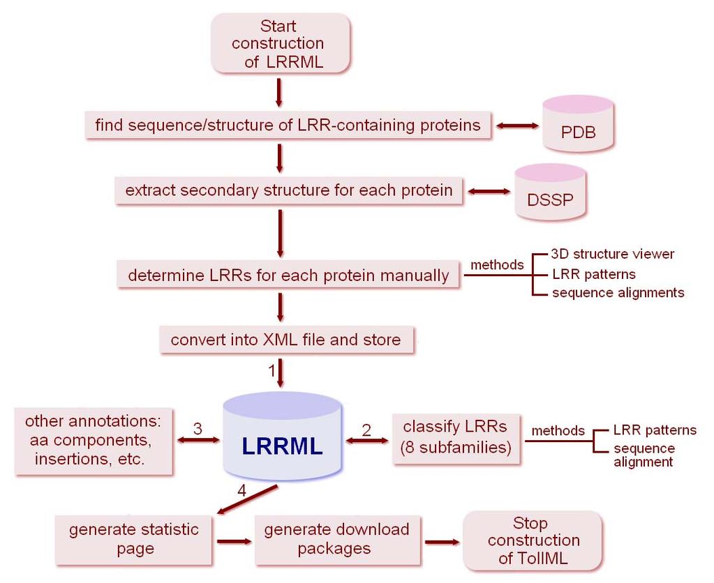
28 Construction pipeline


29 Domains LRRs Segments

30 LRR Finder main algorithm : a position-specific weight matrix of LRR motifs Position Amino acids Example: LPTNLTVLMLLHNQLRRLPAANFTRYSQLTSLDVGFNT % cutoff Yes No
31 Example: LPTNLTVLMLLHNQLRRLPAANFTRYSQLTSLDVGFNT Yes No No filter Sensitivity / Specificity Cutoff score Cutoff Sensitivity Specificity Spe. (filter)
32 This database is freely available at Any internet user can search and download data from the database, but only registered users can define and save labels for arbitrary entries. TollML: a database of toll-like receptor strutural motifs J. Mol. Model., 2010, 16(7): IF: 2.34, SCI citation times: 3 Jing Gong, Tiandi Wei, Ning Zhang, Ferdinand Jamitzky, Wolfgang M. Heckl, Shaila C. Rössle and Robert W. Stark


33 2010/11
34 Construction pipeline
35

36 Every LRR structure can be viewed with an online molecular viewer Jmol.

37 To simplify the homology modeling, the similarity search was implemented. It returns the structures of the most similar LRRs for a structure unknown LRR. At first, a global pairwise sequence alignment with sequence identity will be generated for the target LRR and each of the LRRs in the user selected set. Then, the most similar LRRs will be returned as template candidates, ranked by sequence identity.
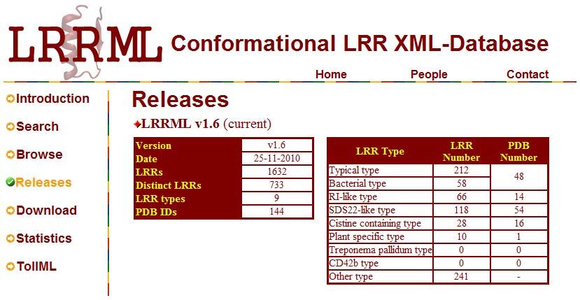
38 LRRML contains individual three-dimensional LRR structures with manual structural annotations. It presents useful sources for homology modeling and structural analysis of LRR proteins. This database is freely available at LRRML: a conformational database and an XML description of leucine-rich repeats (LRRs) BMC Struct. Biol., 2008, 8:47 IF: 3.06, SCI citation times: 3 Tiandi Wei, Jing Gong*, Ferdinand Jamitzky, Wolfgang M. Heckl, Robert W. Stark and Shaila C. Rössle *corresponding author

39 In mammalian, 13 TLRs have been identified. Protein sequences are available for a number of mammalian species. Using these sequences, a complete molecular phylogenetic analysis and a phylogenetic tree of the known TLRs were reported. According to this tree, mammalian TLRs can be divided into six subfamilies. TLR1, 2, 6 and 10 belong to the TLR1 subfamily. TLR3 constitutes the TLR3 subfamily. TLR4 constitutes the TLR4 subfamily and TLR5 constitutes the TLR5 subfamily. TLR7, 8 and 9 compose the TLR7 subfamily. TLR11, 12 and 13 belong to the TLR11 subfamily.
40 Since 2000 the crystal structure of human TLR3 ECD was firstly reported, four crystal structures of receptorligand complexes have been determined. They are : human TLR2-1 heterodimer, mouse TLR3 homodimer, human TLR4 homodimer, mouse TLR2-6 heterodimer.
41 TLR sequences ~3000 known TLR sequences Compared with the small number of crystal structures, there are about 3000 known protein sequences of different TLRs from different species. Because the X-ray crystallography remains timeconsuming and sometimes it is very difficult to crystallize proteins, computational methods can perform fast and large-scale structural predictions based on the sequences. Currently, the most accurate protein structure prediction method is homology modeling.
42 When applying the homology modeling on the TLR ectodomains, we encountered a problem. The sequence identity between the target and the full-length template(s), namely the aforementioned crystal structures, is much lower than 30% because of diverse numbers and arrangements of LRRs contained in the TLR ectodomains. This problem is also described by the phylogenetic tree. Thus we could not get a proper model. To solve this problem we developed an LRR template assembly approach with the help of both TollML and LRRML databases.

43 Flowchart of the LRR template assembly approach

44 Threading method Crystal structure Full-length templates LRR assembly TLR3 ECD

45 Superimposition of the model (blue) and crystal structure (orange) of TLR3 at the two ligand interaction regions. Global root mean square deviation: 1.96 Å and 1.90 Å.

46 If the root mean square deviation between a model and a structure is < 3 Å, the model is very good and can be used to perform liganddocking and molecular replacement. Zhang et al., 2009.

47 Average target-template sequence identity >= 45%

48 Superimposition of the model (green) and crystal structure (orange) of TLR6. Global root mean square deviation: 1.94 Å; ligand-binding region: 1.18 Å.
49 These models can be used to perform ligand-docking studies or to design mutagenesis experiments to investigate TLR ligand-binding mechanisms, and thus help to develop new TLR agonists and antagonists that have therapeutic significance for infectious diseases. A leucine-rich repeat assembly approach for homology modeling of human TLR5-10 and mouse TLR11-13 ectodomains. J. Mol. Model., 2011, 17(1):27-36 IF: 2.34, SCI citation times: 3 Tiandi Wei, Jing Gong*, Ferdinand Jamitzky, Wolfgang M. Heckl, Shaila C. Rössle and Robert W. Stark *corresponding author
50 Exam Thesis

51 Exam Thesis Topic : What can bioinformatics do for you? Language : English Word count : Deadline : 2011/11/30 Submit to : gongjing@sdu.edu.cn
52 Format : 1. The following word processor file formats are acceptable for the thesis: Microsoft Word (.doc) Rich text format (RTF) Portable document format (PDF) 2. You should choose a legible font and use double line-spacing. Your font should be no smaller than 11 pt font and no bigger than 12 pt font with standard margins. 3. Use hard returns only to end headings and paragraphs, not to rearrange lines. 4. All references must be numbered consecutively, in square brackets, in the order in which they are cited in the text, followed by any in tables or legends. 5. All pages should be numbered. 6. Greek and other special characters may be included. If you are unable to reproduce a particular special character, please type out the name of the symbol in full.


53 Thank you very much for your attention!
Introduction to Bioinformatics Introduction to Bioinformatics
 Dr. rer. nat. Gong Jing Cancer Research center Medicine School of Shandong University 2012.11.14 1 Case Study 2 Case 1 Case 2 How SIGIRR inhibit the TLR4 and 7 signaling pathways? Model Construction Tolllike
Introduction to Comparative Protein Modeling. Chapter 4 Part I
 Introduction to Comparative Protein Modeling Chapter 4 Part I 1 Information on Proteins Each modeling study depends on the quality of the known experimental data. Basis of the model Search in the literature
Building a Homology Model of the Transmembrane Domain of the Human Glycine α-1 Receptor
 Building a Homology Model of the Transmembrane Domain of the Human Glycine α-1 Receptor Presented by Stephanie Lee Research Mentor: Dr. Rob Coalson Glycine Alpha 1 Receptor (GlyRa1) Member of the superfamily
Week 10: Homology Modelling (II) - HHpred
 - HHpred") Week 10: Homology Modelling (II) - HHpred Course: Tools for Structural Biology Fabian Glaser BKU - Technion 1 2 Identify and align related structures by sequence methods is not an easy task All comparative
Week 10: Homology Modelling (II) - HHpred Course: Tools for Structural Biology Fabian Glaser BKU - Technion 1 2 Identify and align related structures by sequence methods is not an easy task All comparative
Structure-Function Relationship of Cytoplasmic and Nuclear IkB Proteins: An In Silico Analysis
 Structure-Function Relationship of Cytoplasmic and Nuclear IkB Proteins: An In Silico Analysis Balachandran Manavalan 1, Shaherin Basith 1, Yong-Min Choi 1, Gwang Lee 1,2, Sangdun Choi 1 * 1 Department
Nature Structural & Molecular Biology: doi: /nsmb Supplementary Figure 1
 Supplementary Figure 1 Crystallization. a, Crystallization constructs of the ET B receptor are shown, with all of the modifications to the human wild-type the ET B receptor indicated. Residues interacting
Bioinformatics. Dept. of Computational Biology & Bioinformatics
 Bioinformatics Dept. of Computational Biology & Bioinformatics 3 Bioinformatics - play with sequences & structures Dept. of Computational Biology & Bioinformatics 4 ORGANIZATION OF LIFE ROLE OF BIOINFORMATICS
COMP 598 Advanced Computational Biology Methods & Research. Introduction. Jérôme Waldispühl School of Computer Science McGill University
 COMP 598 Advanced Computational Biology Methods & Research Introduction Jérôme Waldispühl School of Computer Science McGill University General informations (1) Office hours: by appointment Office: TR3018
Basics of protein structure
 Today: 1. Projects a. Requirements: i. Critical review of one paper ii. At least one computational result b. Noon, Dec. 3 rd written report and oral presentation are due; submit via email to bphys101@fas.harvard.edu
CAP 5510 Lecture 3 Protein Structures
 CAP 5510 Lecture 3 Protein Structures Su-Shing Chen Bioinformatics CISE 8/19/2005 Su-Shing Chen, CISE 1 Protein Conformation 8/19/2005 Su-Shing Chen, CISE 2 Protein Conformational Structures Hydrophobicity
HMM applications. Applications of HMMs. Gene finding with HMMs. Using the gene finder
 HMM applications Applications of HMMs Gene finding Pairwise alignment (pair HMMs) Characterizing protein families (profile HMMs) Predicting membrane proteins, and membrane protein topology Gene finding
Homology models of the tetramerization domain of six eukaryotic voltage-gated potassium channels Kv1.1-Kv1.6
 Homology models of the tetramerization domain of six eukaryotic voltage-gated potassium channels Kv1.1-Kv1.6 Hsuan-Liang Liu* and Chin-Wen Chen Department of Chemical Engineering and Graduate Institute
We used the PSI-BLAST program (http://www.ncbi.nlm.nih.gov/blast/) to search the
 to search the") SUPPLEMENTARY METHODS - in silico protein analysis We used the PSI-BLAST program (http://www.ncbi.nlm.nih.gov/blast/) to search the Protein Data Bank (PDB, http://www.rcsb.org/pdb/) and the NCBI non-redundant
SUPPLEMENTARY METHODS - in silico protein analysis We used the PSI-BLAST program (http://www.ncbi.nlm.nih.gov/blast/) to search the Protein Data Bank (PDB, http://www.rcsb.org/pdb/) and the NCBI non-redundant
08/21/2017 BLAST. Multiple Sequence Alignments: Clustal Omega
 BLAST Multiple Sequence Alignments: Clustal Omega What does basic BLAST do (e.g. what is input sequence and how does BLAST look for matches?) Susan Parrish McDaniel College Multiple Sequence Alignments
Francisco Melo, Damien Devos, Eric Depiereux and Ernest Feytmans
 From: ISMB-97 Proceedings. Copyright 1997, AAAI (www.aaai.org). All rights reserved. ANOLEA: A www Server to Assess Protein Structures Francisco Melo, Damien Devos, Eric Depiereux and Ernest Feytmans Facultés
SUPPLEMENTARY INFORMATION
 doi:10.1038/nature17991 Supplementary Discussion Structural comparison with E. coli EmrE The DMT superfamily includes a wide variety of transporters with 4-10 TM segments 1. Since the subfamilies of the
Ch. 9 Multiple Sequence Alignment (MSA)
") Ch. 9 Multiple Sequence Alignment (MSA) - gather seqs. to make MSA - doing MSA with ClustalW - doing MSA with Tcoffee - comparing seqs. that cannot align Introduction - from pairwise alignment to MSA -
Ch. 9 Multiple Sequence Alignment (MSA) - gather seqs. to make MSA - doing MSA with ClustalW - doing MSA with Tcoffee - comparing seqs. that cannot align Introduction - from pairwise alignment to MSA -
Homology Modeling (Comparative Structure Modeling) GBCB 5874: Problem Solving in GBCB
 GBCB 5874: Problem Solving in GBCB") Homology Modeling (Comparative Structure Modeling) Aims of Structural Genomics High-throughput 3D structure determination and analysis To determine or predict the 3D structures of all the proteins encoded
Homology Modeling (Comparative Structure Modeling) Aims of Structural Genomics High-throughput 3D structure determination and analysis To determine or predict the 3D structures of all the proteins encoded
Biol403 - Receptor Serine/Threonine Kinases
 Biol403 - Receptor Serine/Threonine Kinases The TGFβ (transforming growth factorβ) family of growth factors TGFβ1 was first identified as a transforming factor; however, it is a member of a family of structurally
NGF - twenty years a-growing
 NGF - twenty years a-growing A molecule vital to brain growth It is twenty years since the structure of nerve growth factor (NGF) was determined [ref. 1]. This molecule is more than 'quite interesting'
Grundlagen der Bioinformatik Summer semester Lecturer: Prof. Daniel Huson
 Grundlagen der Bioinformatik, SS 10, D. Huson, April 12, 2010 1 1 Introduction Grundlagen der Bioinformatik Summer semester 2010 Lecturer: Prof. Daniel Huson Office hours: Thursdays 17-18h (Sand 14, C310a)
Comparing whole genomes
 BioNumerics Tutorial: Comparing whole genomes 1 Aim The Chromosome Comparison window in BioNumerics has been designed for large-scale comparison of sequences of unlimited length. In this tutorial you will
Examples of Protein Modeling. Protein Modeling. Primary Structure. Protein Structure Description. Protein Sequence Sources. Importing Sequences to MOE
 Examples of Protein Modeling Protein Modeling Visualization Examination of an experimental structure to gain insight about a research question Dynamics To examine the dynamics of protein structures To
Protein Structure Prediction II Lecturer: Serafim Batzoglou Scribe: Samy Hamdouche
 Protein Structure Prediction II Lecturer: Serafim Batzoglou Scribe: Samy Hamdouche The molecular structure of a protein can be broken down hierarchically. The primary structure of a protein is simply its
Protein Structure Prediction and Display
 Protein Structure Prediction and Display Goal Take primary structure (sequence) and, using rules derived from known structures, predict the secondary structure that is most likely to be adopted by each
Supplementary Figure 1. Aligned sequences of yeast IDH1 (top) and IDH2 (bottom) with isocitrate
 and IDH2 (bottom) with isocitrate") SUPPLEMENTARY FIGURE LEGENDS Supplementary Figure 1. Aligned sequences of yeast IDH1 (top) and IDH2 (bottom) with isocitrate dehydrogenase from Escherichia coli [ICD, pdb 1PB1, Mesecar, A. D., and Koshland,
SUPPLEMENTARY FIGURE LEGENDS Supplementary Figure 1. Aligned sequences of yeast IDH1 (top) and IDH2 (bottom) with isocitrate dehydrogenase from Escherichia coli [ICD, pdb 1PB1, Mesecar, A. D., and Koshland,
SUPPLEMENTARY INFORMATION
 SUPPLEMENTARY INFORMATION Structure of human carbamoyl phosphate synthetase: deciphering the on/off switch of human ureagenesis Sergio de Cima, Luis M. Polo, Carmen Díez-Fernández, Ana I. Martínez, Javier
5- Semaphorin-Plexin-Neuropilin
 5- Semaphorin-Plexin-Neuropilin 1 SEMAPHORINS-PLEXINS-NEUROPILINS ligands receptors co-receptors semaphorins and their receptors are known signals for: -axon guidance -cell migration -morphogenesis -immune
Measuring quaternary structure similarity using global versus local measures.
 Supplementary Figure 1 Measuring quaternary structure similarity using global versus local measures. (a) Structural similarity of two protein complexes can be inferred from a global superposition, which
Preparing a PDB File
 Figure 1: Schematic view of the ligand-binding domain from the vitamin D receptor (PDB file 1IE9). The crystallographic waters are shown as small spheres and the bound ligand is shown as a CPK model. HO
Homology Modeling. Roberto Lins EPFL - summer semester 2005
 Homology Modeling Roberto Lins EPFL - summer semester 2005 Disclaimer: course material is mainly taken from: P.E. Bourne & H Weissig, Structural Bioinformatics; C.A. Orengo, D.T. Jones & J.M. Thornton,
CS612 - Algorithms in Bioinformatics
 Fall 2017 Databases and Protein Structure Representation October 2, 2017 Molecular Biology as Information Science > 12, 000 genomes sequenced, mostly bacterial (2013) > 5x10 6 unique sequences available
Pymol Practial Guide
 Pymol Practial Guide Pymol is a powerful visualizor very convenient to work with protein molecules. Its interface may seem complex at first, but you will see that with a little practice is simple and powerful
Modeling for 3D structure prediction
 Modeling for 3D structure prediction What is a predicted structure? A structure that is constructed using as the sole source of information data obtained from computer based data-mining. However, mixing
Review. Membrane proteins. Membrane transport
 Quiz 1 For problem set 11 Q1, you need the equation for the average lateral distance transversed (s) of a molecule in the membrane with respect to the diffusion constant (D) and time (t). s = (4 D t) 1/2
Structure of the α-helix
 Structure of the α-helix Structure of the β Sheet Protein Dynamics Basics of Quenching HDX Hydrogen exchange of amide protons is catalyzed by H 2 O, OH -, and H 3 O +, but it s most dominated by base
Supplementary Figure 1 Schematic overview of ASTNs in neuronal migration. (a) Schematic of roles played by ASTNs 1 and 2. ASTN-1-mediated adhesions
 Schematic of roles played by ASTNs 1 and 2. ASTN-1-mediated adhesions") Supplementary Figure 1 Schematic overview of ASTNs in neuronal migration. (a) Schematic of roles played by ASTNs 1 and 2. ASTN-1-mediated adhesions undergo endocytosis into clathrin-coated vesicles dependent
Supplementary Figure 1 Schematic overview of ASTNs in neuronal migration. (a) Schematic of roles played by ASTNs 1 and 2. ASTN-1-mediated adhesions undergo endocytosis into clathrin-coated vesicles dependent
GCD3033:Cell Biology. Transcription
 Transcription Transcription: DNA to RNA A) production of complementary strand of DNA B) RNA types C) transcription start/stop signals D) Initiation of eukaryotic gene expression E) transcription factors
Giri Narasimhan. CAP 5510: Introduction to Bioinformatics. ECS 254; Phone: x3748
 CAP 5510: Introduction to Bioinformatics Giri Narasimhan ECS 254; Phone: x3748 giri@cis.fiu.edu www.cis.fiu.edu/~giri/teach/bioinfs07.html 2/15/07 CAP5510 1 EM Algorithm Goal: Find θ, Z that maximize Pr
Visualization of Macromolecular Structures
 Visualization of Macromolecular Structures Present by: Qihang Li orig. author: O Donoghue, et al. Structural biology is rapidly accumulating a wealth of detailed information. Over 60,000 high-resolution
EBI web resources II: Ensembl and InterPro. Yanbin Yin Spring 2013
 EBI web resources II: Ensembl and InterPro Yanbin Yin Spring 2013 1 Outline Intro to genome annotation Protein family/domain databases InterPro, Pfam, Superfamily etc. Genome browser Ensembl Hands on Practice
Introduction Molecular Structure Script Console External resources Advanced topics. JMol tutorial. Giovanni Morelli.
 Gen 19th, 2017 1 2 Create and edit Display and view Mesurament and labelling Surface and Orbitals 3 4 from Database Protein Enzyme Crystal Structure and Unit Cell 5 Symmetry Animation General information
SUPPLEMENTARY INFORMATION
 doi:10.1038/nature11085 Supplementary Tables: Supplementary Table 1. Summary of crystallographic and structure refinement data Structure BRIL-NOP receptor Data collection Number of crystals 23 Space group
Nature Structural and Molecular Biology: doi: /nsmb.2938
 Supplementary Figure 1 Characterization of designed leucine-rich-repeat proteins. (a) Water-mediate hydrogen-bond network is frequently visible in the convex region of LRR crystal structures. Examples
Bioinformatics: Investigating Molecular/Biochemical Evidence for Evolution
 Bioinformatics: Investigating Molecular/Biochemical Evidence for Evolution Background How does an evolutionary biologist decide how closely related two different species are? The simplest way is to compare
DATA ACQUISITION FROM BIO-DATABASES AND BLAST. Natapol Pornputtapong 18 January 2018
 DATA ACQUISITION FROM BIO-DATABASES AND BLAST Natapol Pornputtapong 18 January 2018 DATABASE Collections of data To share multi-user interface To prevent data loss To make sure to get the right things
Transmembrane Domains (TMDs) of ABC transporters
 of ABC transporters") Transmembrane Domains (TMDs) of ABC transporters Most ABC transporters contain heterodimeric TMDs (e.g. HisMQ, MalFG) TMDs show only limited sequence homology (high diversity) High degree of conservation
Transmembrane Domains (TMDs) of ABC transporters Most ABC transporters contain heterodimeric TMDs (e.g. HisMQ, MalFG) TMDs show only limited sequence homology (high diversity) High degree of conservation
Computational modeling of G-Protein Coupled Receptors (GPCRs) has recently become
 has recently become") Homology Modeling and Docking of Melatonin Receptors Andrew Kohlway, UMBC Jeffry D. Madura, Duquesne University 6/18/04 INTRODUCTION Computational modeling of G-Protein Coupled Receptors (GPCRs) has recently
Homology Modeling and Docking of Melatonin Receptors Andrew Kohlway, UMBC Jeffry D. Madura, Duquesne University 6/18/04 INTRODUCTION Computational modeling of G-Protein Coupled Receptors (GPCRs) has recently
Sequence analysis and comparison
 The aim with sequence identification: Sequence analysis and comparison Marjolein Thunnissen Lund September 2012 Is there any known protein sequence that is homologous to mine? Are there any other species
Neural Networks for Protein Structure Prediction Brown, JMB CS 466 Saurabh Sinha
 Neural Networks for Protein Structure Prediction Brown, JMB 1999 CS 466 Saurabh Sinha Outline Goal is to predict secondary structure of a protein from its sequence Artificial Neural Network used for this
BIOINFORMATICS LAB AP BIOLOGY
 BIOINFORMATICS LAB AP BIOLOGY Bioinformatics is the science of collecting and analyzing complex biological data. Bioinformatics combines computer science, statistics and biology to allow scientists to
7.06 Cell Biology EXAM #3 April 21, 2005
 7.06 Cell Biology EXAM #3 April 21, 2005 This is an open book exam, and you are allowed access to books, a calculator, and notes but not computers or any other types of electronic devices. Please write
Cross Discipline Analysis made possible with Data Pipelining. J.R. Tozer SciTegic
 Cross Discipline Analysis made possible with Data Pipelining J.R. Tozer SciTegic System Genesis Pipelining tool created to automate data processing in cheminformatics Modular system built with generic
Gene regulation I Biochemistry 302. Bob Kelm February 25, 2005
 Gene regulation I Biochemistry 302 Bob Kelm February 25, 2005 Principles of gene regulation (cellular versus molecular level) Extracellular signals Chemical (e.g. hormones, growth factors) Environmental
RANK. Alternative names. Discovery. Structure. William J. Boyle* SUMMARY BACKGROUND
 RANK William J. Boyle* Department of Cell Biology, Amgen, Inc., One Amgen Center Drive, Thousand Oaks, CA 91320-1799, USA * corresponding author tel: 805-447-4304, fax: 805-447-1982, e-mail: bboyle@amgen.com
1-D Predictions. Prediction of local features: Secondary structure & surface exposure
 1-D Predictions Prediction of local features: Secondary structure & surface exposure 1 Learning Objectives After today s session you should be able to: Explain the meaning and usage of the following local
Molecular Modeling Lecture 7. Homology modeling insertions/deletions manual realignment
 Molecular Modeling 2018-- Lecture 7 Homology modeling insertions/deletions manual realignment Homology modeling also called comparative modeling Sequences that have similar sequence have similar structure.
CAP 5510: Introduction to Bioinformatics CGS 5166: Bioinformatics Tools. Giri Narasimhan
 CAP 5510: Introduction to Bioinformatics CGS 5166: Bioinformatics Tools Giri Narasimhan ECS 254; Phone: x3748 giri@cis.fiu.edu www.cis.fiu.edu/~giri/teach/bioinff18.html Proteins and Protein Structure
Initiation of translation in eukaryotic cells:connecting the head and tail
 Initiation of translation in eukaryotic cells:connecting the head and tail GCCRCCAUGG 1: Multiple initiation factors with distinct biochemical roles (linking, tethering, recruiting, and scanning) 2: 5
Chapter 5. Proteomics and the analysis of protein sequence Ⅱ
 Proteomics Chapter 5. Proteomics and the analysis of protein sequence Ⅱ 1 Pairwise similarity searching (1) Figure 5.5: manual alignment One of the amino acids in the top sequence has no equivalent and
SUPPLEMENTARY INFORMATION
 SUPPLEMENTARY INFORMATION doi:10.1038/nature11524 Supplementary discussion Functional analysis of the sugar porter family (SP) signature motifs. As seen in Fig. 5c, single point mutation of the conserved
Prediction and Classif ication of Human G-protein Coupled Receptors Based on Support Vector Machines
 Article Prediction and Classif ication of Human G-protein Coupled Receptors Based on Support Vector Machines Yun-Fei Wang, Huan Chen, and Yan-Hong Zhou* Hubei Bioinformatics and Molecular Imaging Key Laboratory,
Protein Dynamics. The space-filling structures of myoglobin and hemoglobin show that there are no pathways for O 2 to reach the heme iron.
 Protein Dynamics The space-filling structures of myoglobin and hemoglobin show that there are no pathways for O 2 to reach the heme iron. Below is myoglobin hydrated with 350 water molecules. Only a small
EBI web resources II: Ensembl and InterPro
 EBI web resources II: Ensembl and InterPro Yanbin Yin http://www.ebi.ac.uk/training/online/course/ 1 Homework 3 Go to http://www.ebi.ac.uk/interpro/training.htmland finish the second online training course
Genomics and bioinformatics summary. Finding genes -- computer searches
 Genomics and bioinformatics summary 1. Gene finding: computer searches, cdnas, ESTs, 2. Microarrays 3. Use BLAST to find homologous sequences 4. Multiple sequence alignments (MSAs) 5. Trees quantify sequence
CMPS 6630: Introduction to Computational Biology and Bioinformatics. Structure Comparison
 CMPS 6630: Introduction to Computational Biology and Bioinformatics Structure Comparison Protein Structure Comparison Motivation Understand sequence and structure variability Understand Domain architecture
Can protein model accuracy be. identified? NO! CBS, BioCentrum, Morten Nielsen, DTU
 Can protein model accuracy be identified? Morten Nielsen, CBS, BioCentrum, DTU NO! Identification of Protein-model accuracy Why is it important? What is accuracy RMSD, fraction correct, Protein model correctness/quality
Online Protein Structure Analysis with the Bio3D WebApp
 Online Protein Structure Analysis with the Bio3D WebApp Lars Skjærven, Shashank Jariwala & Barry J. Grant August 13, 2015 (updated November 17, 2016) Bio3D1 is an established R package for structural bioinformatics
Algorithms in Bioinformatics FOUR Pairwise Sequence Alignment. Pairwise Sequence Alignment. Convention: DNA Sequences 5. Sequence Alignment
 Algorithms in Bioinformatics FOUR Sami Khuri Department of Computer Science San José State University Pairwise Sequence Alignment Homology Similarity Global string alignment Local string alignment Dot
Activation of a receptor. Assembly of the complex
 Activation of a receptor ligand inactive, monomeric active, dimeric When activated by growth factor binding, the growth factor receptor tyrosine kinase phosphorylates the neighboring receptor. Assembly
AP Biology Gene Regulation and Development Review
 AP Biology Gene Regulation and Development Review 1. What does the regulatory gene code for? 2. Is the repressor by default active/inactive? 3. What changes the repressor activity? 4. What does repressor
Transcription Regulation And Gene Expression in Eukaryotes UPSTREAM TRANSCRIPTION FACTORS
 Transcription Regulation And Gene Expression in Eukaryotes UPSTREAM TRANSCRIPTION FACTORS RG. Clerc March 26. 2008 UPSTREAM TRANSCRIPTION FACTORS Experimental approaches DNA binding domains (DBD) Transcription
Analysis and Prediction of Protein Structure (I)
") Analysis and Prediction of Protein Structure (I) Jianlin Cheng, PhD School of Electrical Engineering and Computer Science University of Central Florida 2006 Free for academic use. Copyright @ Jianlin Cheng
Analysis and Prediction of Protein Structure (I) Jianlin Cheng, PhD School of Electrical Engineering and Computer Science University of Central Florida 2006 Free for academic use. Copyright @ Jianlin Cheng
1. Protein Data Bank (PDB) 1. Protein Data Bank (PDB)
 1. Protein Data Bank (PDB)") Protein structure databases; visualization; and classifications 1. Introduction to Protein Data Bank (PDB) 2. Free graphic software for 3D structure visualization 3. Hierarchical classification of protein
Protein structure databases; visualization; and classifications 1. Introduction to Protein Data Bank (PDB) 2. Free graphic software for 3D structure visualization 3. Hierarchical classification of protein
RNA Synthesis and Processing
 RNA Synthesis and Processing Introduction Regulation of gene expression allows cells to adapt to environmental changes and is responsible for the distinct activities of the differentiated cell types that
Supporting Information
 Supporting Information Ottmann et al. 10.1073/pnas.0907587106 Fig. S1. Primary structure alignment of SBT3 with C5 peptidase from Streptococcus pyogenes. The Matchmaker tool in UCSF Chimera (http:// www.cgl.ucsf.edu/chimera)
User Guide for LeDock
 User Guide for LeDock Hongtao Zhao, PhD Email: htzhao@lephar.com Website: www.lephar.com Copyright 2017 Hongtao Zhao. All rights reserved. Introduction LeDock is flexible small-molecule docking software,
Zool 3200: Cell Biology Exam 5 4/27/15
 Name: Trask Zool 3200: Cell Biology Exam 5 4/27/15 Answer each of the following short answer questions in the space provided, giving explanations when asked to do so. Circle the correct answer or answers
Molecular Cell Biology 5068 In Class Exam 2 November 8, 2016
 Molecular Cell Biology 5068 In Class Exam 2 November 8, 2016 Exam Number: Please print your name: Instructions: Please write only on these pages, in the spaces allotted and not on the back. Write your
Advanced Certificate in Principles in Protein Structure. You will be given a start time with your exam instructions
 BIRKBECK COLLEGE (University of London) Advanced Certificate in Principles in Protein Structure MSc Structural Molecular Biology Date: Thursday, 1st September 2011 Time: 3 hours You will be given a start
Bahnson Biochemistry Cume, April 8, 2006 The Structural Biology of Signal Transduction
 Name page 1 of 6 Bahnson Biochemistry Cume, April 8, 2006 The Structural Biology of Signal Transduction Part I. The ion Ca 2+ can function as a 2 nd messenger. Pick a specific signal transduction pathway
ALL LECTURES IN SB Introduction
 1. Introduction 2. Molecular Architecture I 3. Molecular Architecture II 4. Molecular Simulation I 5. Molecular Simulation II 6. Bioinformatics I 7. Bioinformatics II 8. Prediction I 9. Prediction II ALL
BA, BSc, and MSc Degree Examinations
 Examination Candidate Number: Desk Number: BA, BSc, and MSc Degree Examinations 2017-8 Department : BIOLOGY Title of Exam: Molecular Biology and Biochemistry Part I Time Allowed: 1 hour and 30 minutes
Lipniacki 2004 Ground Truth
 Abstract Lipniacki 2004 Ground Truth The two-feedback-loop regulatory module of nuclear factor kb (NF-kB) signaling pathway is modeled by means of ordinary differential equations. signaling pathway: https://en.wikipedia.org/wiki/signaling_pathway
Supporting Online Material for
 www.sciencemag.org/cgi/content/full/309/5742/1868/dc1 Supporting Online Material for Toward High-Resolution de Novo Structure Prediction for Small Proteins Philip Bradley, Kira M. S. Misura, David Baker*
The majority of cells in the nervous system arise during the embryonic and early post
 Introduction Introduction The majority of cells in the nervous system arise during the embryonic and early post natal period. These cells are derived from population of neural stem cells first shown by
Goals. Structural Analysis of the EGR Family of Transcription Factors: Templates for Predicting Protein DNA Interactions
 Structural Analysis of the EGR Family of Transcription Factors: Templates for Predicting Protein DNA Interactions Jamie Duke 1,2 and Carlos Camacho 3 1 Bioengineering and Bioinformatics Summer Institute,
SUPPLEMENTARY FIGURES. Structure of the cholera toxin secretion channel in its. closed state
 SUPPLEMENTARY FIGURES Structure of the cholera toxin secretion channel in its closed state Steve L. Reichow 1,3, Konstantin V. Korotkov 1,3, Wim G. J. Hol 1$ and Tamir Gonen 1,2$ 1, Department of Biochemistry
Minireview: Molecular Structure and Dynamics of Drug Targets
 Prague Medical Report / Vol. 109 (2008) No. 2 3, p. 107 112 107) Minireview: Molecular Structure and Dynamics of Drug Targets Dahl S. G., Sylte I. Department of Pharmacology, Institute of Medical Biology,
CSCE555 Bioinformatics. Protein Function Annotation
 CSCE555 Bioinformatics Protein Function Annotation Why we need to do function annotation? Fig from: Network-based prediction of protein function. Molecular Systems Biology 3:88. 2007 What s function? The
Table 1. Crystallographic data collection, phasing and refinement statistics. Native Hg soaked Mn soaked 1 Mn soaked 2
 Table 1. Crystallographic data collection, phasing and refinement statistics Native Hg soaked Mn soaked 1 Mn soaked 2 Data collection Space group P2 1 2 1 2 1 P2 1 2 1 2 1 P2 1 2 1 2 1 P2 1 2 1 2 1 Cell
Module: Sequence Alignment Theory and Applications Session: Introduction to Searching and Sequence Alignment
 Module: Sequence Alignment Theory and Applications Session: Introduction to Searching and Sequence Alignment Introduction to Bioinformatics online course : IBT Jonathan Kayondo Learning Objectives Understand
In-Depth Assessment of Local Sequence Alignment
 2012 International Conference on Environment Science and Engieering IPCBEE vol.3 2(2012) (2012)IACSIT Press, Singapoore In-Depth Assessment of Local Sequence Alignment Atoosa Ghahremani and Mahmood A.
RNA and Protein Structure Prediction
 RNA and Protein Structure Prediction Bioinformatics: Issues and Algorithms CSE 308-408 Spring 2007 Lecture 18-1- Outline Multi-Dimensional Nature of Life RNA Secondary Structure Prediction Protein Structure
β1 Structure Prediction and Validation
 13 Chapter 2 β1 Structure Prediction and Validation 2.1 Overview Over several years, GPCR prediction methods in the Goddard lab have evolved to keep pace with the changing field of GPCR structure. Despite
CHAPTER 1 THE STRUCTURAL BIOLOGY OF THE FGF19 SUBFAMILY
 CHAPTER 1 THE STRUCTURAL BIOLOGY OF THE FGF19 SUBFAMILY Andrew Beenken and Moosa Mohammadi* Department of Pharmacology, New York University School of Medicine, New York, New York, USA. *Corresponding Author:
ICM-Chemist-Pro How-To Guide. Version 3.6-1h Last Updated 12/29/2009
 ICM-Chemist-Pro How-To Guide Version 3.6-1h Last Updated 12/29/2009 ICM-Chemist-Pro ICM 3D LIGAND EDITOR: SETUP 1. Read in a ligand molecule or PDB file. How to setup the ligand in the ICM 3D Ligand Editor.
Assignment A02: Geometry Definition: File Formats, Redundant Coordinates, PES Scans
 Assignment A02: Geometry Definition: File Formats, Redundant Coordinates, PES Scans In Assignments A00 and A01, you familiarized yourself with GaussView and G09W, you learned the basics about input (GJF)
Genome Annotation Project Presentation
 Halogeometricum borinquense Genome Annotation Project Presentation Loci Hbor_05620 & Hbor_05470 Presented by: Mohammad Reza Najaf Tomaraei Hbor_05620 Basic Information DNA Coordinates: 527,512 528,261
Supporting Information
 Supporting Information Manning et al. 1.173/pnas.8131415 SI Text RM1 Motif. This Monosiga-specific motif of 22 aa is repeated 8 13 times in the extracellular regions of two RTKA s, one RTKG, and 4 other