Fig. 1. Stereo images showing (A) the best fit of the atomic model for F actin and the F actin map obtained by cryo-em and image analysis, and (B) goo
|
|
|
- Anabel Nichols
- 5 years ago
- Views:
Transcription
1

2
3


4
5

6
7
8

9

10
11
12

13
14

15

16
 the best fit of the atomic model for F actin and the F actin map obtained by cryo-em and image analysis, and (B) good correspondence between the location of Cys374 and a")
17 Fig. 1. Stereo images showing (A) the best fit of the atomic model for F actin and the F actin map obtained by cryo-em and image analysis, and (B) good correspondence between the location of Cys374 and a gold cluster label (monomaleimide undecagold) which was attached to Cys374 and then localized by cryo-em and difference analysis (18). The location of Cys374 is indicated by a space-filling model. The atomic model for F actin was obtained by model building and refinement with the use of the atomic coordinates for the actin monomer (13) together with lowangle fiber diffraction data (16). The EM data for F actin and the Cys374 localization were those described in (18). The F actin model and the EM map were fit together by changing the phase origin of the EM map until optimal correspondence between the model and map was achieved. The optimal phase origin shift was also applied to the undecagold difference map before display. The final position of the atomic model within the low resolution map was confirmed both by the general correspondence between the gross features of the model and the molecular envelope and by the position of Cys374. Figures 1 and 2 were prepared from a plot file generated from the molecular graphics program FRODO (19)
 the best fit of the F actin model and the S1 x-ray structure in the molecular envelope of S1 (A2)-decorated F actin obtained by cryo-em and image analysis (18) and (B) the")
18 Fig. 2. Stereo images showing (A) the best fit of the F actin model and the S1 x-ray structure in the molecular envelope of S1 (A2)-decorated F actin obtained by cryo-em and image analysis (18) and (B) the good agreement between the location of the essential light chain (A2) and the corresponding difference density. (C) An a-carbon plot of five actin monomers and one molecule of S1. Samples for EM were prepared as described (17, 18). Cryo-EM and image analyses were carried out as described with some modifications (17, 18). Filament stretches of 30 to 32 crossovers were analyzed. As these were generally curved, they were computationally straightened. Prior to processing the helical filaments, density gradients in the images were removed (17, 18). The final data set was the average of 20 near and far side data sets from 10 filaments and represents averaging of about 2950 asymmetric units. Data on 22 layer lines extending to a nominal resolution of about -27 A were used to calculate the three-dimensional map. No adjustments were made to the data to compensate for the effects of the electron microscope contrast transfer function (ctf). In the data presented, the phases are unaffected by the ctf, however the amplitudes at very low resolution are underemphasized. The F actin and S1 (A2)-decorated F actin maps were brought to the same phase origin by a realspace correlation method (18). Figures 2C; 3, B and C; and 4, A and B, were prepared with the molecular graphics program MOLSCRIPT (47).
 The envelope derived from cryo-em shows the surface feature identified as the NH2-terminal domain of skeletal muscle S1 (20).")
19 Fig. 3. Enlarged views of the model of the myosin head and its interaction with actin. (A) The envelope derived from cryo-em shows the surface feature identified as the NH2-terminal domain of skeletal muscle S1 (20). Attempts to rotate the head to align the 13 barrel and the surface feature result in a misfit for the rest of the molecule. In that this barrel projects away from the rest of the molecule it may adopt different positions relative to the head, which may account for the lack of exact correspondence with the EM data. (B and C) The interaction of myosin with actin viewed from two orientations revealing the details of the actomyosin interface and the relation between the active site and the actin binding site. The secondary structural elements in the myosin heavy chain are color coded according to their position in the primary sequence (14). The three tryptic fragments are represented in different colors (27, 48, 49). These are the NH2-terminal 25-kD, the central 50-kD, and one of the COOH-terminal 20-kD fragments, colored in green, red, and blue, respectively. The 50-kD and the 20-kD fragments have been shown to interact with actin (27, 50). In (B) the equivalent positions of residues crosslinked in chicken gizzard heavy meromyosin are indicated. In the actomyosin complex has been rotated 90 relative to (B) and shows the position of the nucleotide binding pocket relative to the actinmyosin interface.

20 Fig. 4. Close-up views of the actomyosin interface. (A) Interaction between actin and myosin viewed along the thin filament axis toward the M line. This stereo reveals the relation between the narrow cleft that divides the 50-kD region of the myosin head and actin. The phosphate binding loop, as indicated by the sulfate ion, lies above the start of the cleft. (B) A few of the residues on actin and myosin located in the interface in the current model. Given the expected conformational change in myosin when it binds to actin and the errors in the modeling process, it is inappropriate to consider the exact interaction between the residues. However, it is compelling that this orientation places exposed hydrophobic residues on both actin and myosin in the same

21 Fig. 5. The contractile cycle incorporating structural features of the myosin head and their proposed involvement in the cycle. Actin is represented as a sphere. In the near axial third of the myosin head, the narrow cleft that splits the 50-kD segment of the myosin heavy chain sequence into two domains is for simplicity represented as a horizontal gap perpendicular to the filament axis. In the model, this cleft lies at an angle of ~300 to the filament axis and the opening and closing of the cleft would not be evident from this view. The representation of the nucleotide-bound state and its associated conformational change relative to the xray structure of myosin is conceptual in nature.
ParM filament images were extracted and from the electron micrographs and
 Supplemental methods Outline of the EM reconstruction: ParM filament images were extracted and from the electron micrographs and straightened. The digitized images were corrected for the phase of the Contrast
Introduction: actin and myosin
 Introduction: actin and myosin Actin Myosin Myosin V and actin 375 residues Found in all eukaryotes Polymeric Forms track for myosin Many other cellular functions 36 nm pseudo-helical repeat Catalytic
The ideal fiber pattern exhibits 4-quadrant symmetry. In the ideal pattern the fiber axis is called the meridian, the perpendicular direction is
 Fiber diffraction is a method used to determine the structural information of a molecule by using scattering data from X-rays. Rosalind Franklin used this technique in discovering structural information
According to the diagram, which of the following is NOT true?
 Instructions: Review Chapter 44 on muscular-skeletal systems and locomotion, and then complete the following Blackboard activity. This activity will introduce topics that will be covered in the next few
Direct Modeling of X-Ray Diffraction Pattern from Skeletal Muscle in Rigor
 1082 Biophysical Journal Volume 83 August 2002 1082 1097 Direct Modeling of X-Ray Diffraction Pattern from Skeletal Muscle in Rigor Natalia A. Koubassova and A. K. Tsaturyan Institute of Mechanics, Lomonosov
Structure, mechanism and ensemble formation of the Alkylhydroperoxide Reductase subunits. AhpC and AhpF from Escherichia coli
 Structure, mechanism and ensemble formation of the Alkylhydroperoxide Reductase subunits AhpC and AhpF from Escherichia coli Phat Vinh Dip 1,#, Neelagandan Kamariah 2,#, Malathy Sony Subramanian Manimekalai
PHYSIOLOGY CHAPTER 9 MUSCLE TISSUE Fall 2016
 PHYSIOLOGY CHAPTER 9 MUSCLE TISSUE Fall 2016 2 Chapter 9 Muscles and Muscle Tissue Overview of Muscle Tissue types of muscle: are all prefixes for muscle Contractility all muscles cells can Smooth & skeletal
Nitrogenase MoFe protein from Clostridium pasteurianum at 1.08 Å resolution: comparison with the Azotobacter vinelandii MoFe protein
 Acta Cryst. (2015). D71, 274-282, doi:10.1107/s1399004714025243 Supporting information Volume 71 (2015) Supporting information for article: Nitrogenase MoFe protein from Clostridium pasteurianum at 1.08
Muscle regulation and Actin Topics: Tropomyosin and Troponin, Actin Assembly, Actin-dependent Movement
 1 Muscle regulation and Actin Topics: Tropomyosin and Troponin, Actin Assembly, Actin-dependent Movement In the last lecture, we saw that a repeating alternation between chemical (ATP hydrolysis) and vectorial
Lecture 13, 05 October 2004 Chapter 10, Muscle. Vertebrate Physiology ECOL 437 University of Arizona Fall instr: Kevin Bonine t.a.
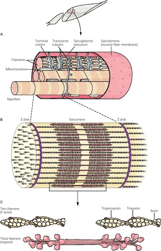
 Lecture 13, 05 October 2004 Chapter 10, Muscle Vertebrate Physiology ECOL 437 University of Arizona Fall 2004 instr: Kevin Bonine t.a.: Nate Swenson Vertebrate Physiology 437 18 1. Muscle A. Sarcomere
Introduction to Comparative Protein Modeling. Chapter 4 Part I
 Introduction to Comparative Protein Modeling Chapter 4 Part I 1 Information on Proteins Each modeling study depends on the quality of the known experimental data. Basis of the model Search in the literature
Structural Studies of the Flexible Filamentous Plant Viruses
 Structural Studies of the Flexible Filamentous Plant Viruses Caitlin Azzo Dr. Gerald Stubbs BSCI 296 [4] Honors Thesis April 23, 2014 The Plant Viruses of Interest In the Potyviridae family: Wheat streak
SUPPLEMENTARY INFORMATION
 SUPPLEMENTARY INFORMATION Structure of human carbamoyl phosphate synthetase: deciphering the on/off switch of human ureagenesis Sergio de Cima, Luis M. Polo, Carmen Díez-Fernández, Ana I. Martínez, Javier
Supplementary Materials for
 advances.sciencemag.org/cgi/content/full/1/9/e1500511/dc1 Supplementary Materials for Contractility parameters of human -cardiac myosin with the hypertrophic cardiomyopathy mutation R403Q show loss of
SUPPLEMENTARY INFORMATION
 Supplementary Results DNA binding property of the SRA domain was examined by an electrophoresis mobility shift assay (EMSA) using synthesized 12-bp oligonucleotide duplexes containing unmodified, hemi-methylated,
Nature Structural & Molecular Biology: doi: /nsmb Supplementary Figure 1
 Supplementary Figure 1 Cryo-EM structure and model of the C. thermophilum 90S preribosome. a, Gold standard FSC curve showing the average resolution of the 90S preribosome masked and unmasked (left). FSC
Orientational degeneracy in the presence of one alignment tensor.
 Orientational degeneracy in the presence of one alignment tensor. Rotation about the x, y and z axes can be performed in the aligned mode of the program to examine the four degenerate orientations of two
Modelling Muscle Contraction a multiscale approach
 Porto Ercole, M&MKT 2016 Multiscale Systems from Particles to Continuum: Modelling and Computation Modelling Muscle Contraction a multiscale approach Giovanni Naldi Dipartimento di Matematica ``F. Enriques
Structure and RNA-binding properties. of the Not1 Not2 Not5 module of the yeast Ccr4 Not complex
 Structure and RNA-binding properties of the Not1 Not2 Not5 module of the yeast Ccr4 Not complex Varun Bhaskar 1, Vladimir Roudko 2,3, Jerome Basquin 1, Kundan Sharma 4, Henning Urlaub 4, Bertrand Seraphin
the axons of the nerve meet with the muscle cell.
 Steps to Contraction 1. A nerve impulse travels to the neuromuscular junction on a muscle cell. The neuromuscular junction is the point where the axons of the nerve meet with the muscle cell. 2. Ach is
SUPPLEMENTARY INFORMATION
 Supplementary Table 1: Data collection, phasing and refinement statistics ChbC/Ta 6 Br 12 Native ChbC Data collection Space group P4 3 2 1 2 P4 3 2 1 2 Cell dimensions a, c (Å) 132.75, 453.57 132.81, 452.95
Possible mechanisms for initiating macroscopic left-right asymmetry in developing organisms
 Possible mechanisms for initiating macroscopic left-right asymmetry in developing organisms Chris Henley, Ricky Chachra, Jimmy Shen Cornell U. [Support: U.S. Dept. of Energy] APS March Meeting, Mar. 2,
SUPPLEMENTARY INFORMATION
 doi:10.1038/nature12045 Supplementary Table 1 Data collection and refinement statistics. Native Pt-SAD X-ray source SSRF BL17U SPring-8 BL41XU Wavelength (Å) 0.97947 1.07171 Space group P2 1 2 1 2 1 P2
Electronic Supplementary Information (ESI) for Chem. Commun. Unveiling the three- dimensional structure of the green pigment of nitrite- cured meat
 for Chem. Commun. Unveiling the three- dimensional structure of the green pigment of nitrite- cured meat") Electronic Supplementary Information (ESI) for Chem. Commun. Unveiling the three- dimensional structure of the green pigment of nitrite- cured meat Jun Yi* and George B. Richter- Addo* Department of Chemistry
Electronic Supplementary Information (ESI) for Chem. Commun. Unveiling the three- dimensional structure of the green pigment of nitrite- cured meat Jun Yi* and George B. Richter- Addo* Department of Chemistry
THE UNIVERSITY OF MANITOBA. PAPER NO: 409 LOCATION: Fr. Kennedy Gold Gym PAGE NO: 1 of 6 DEPARTMENT & COURSE NO: CHEM 4630 TIME: 3 HOURS
 PAPER NO: 409 LOCATION: Fr. Kennedy Gold Gym PAGE NO: 1 of 6 DEPARTMENT & COURSE NO: CHEM 4630 TIME: 3 HOURS EXAMINATION: Biochemistry of Proteins EXAMINER: J. O'Neil Section 1: You must answer all of
Part 3 - Image Formation
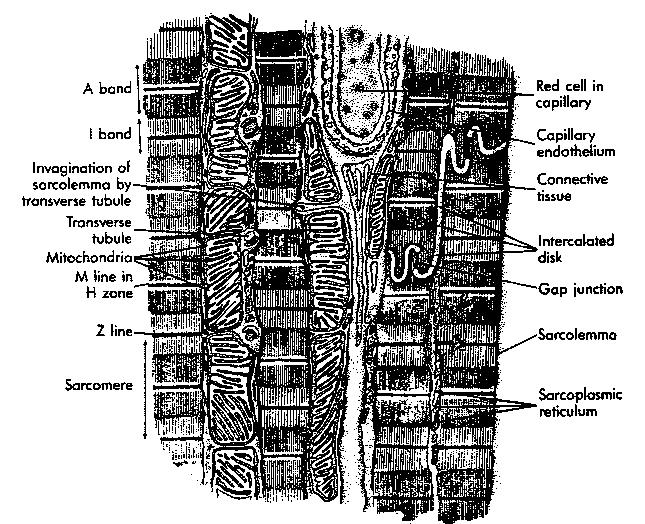
 Part 3 - Image Formation Three classes of scattering outcomes Types of electron microscopes Example SEM image: fly nose Example TEM image: muscle Skeletal muscle. Cell and Tissue Ultrastructure Mercer
Conformationally Variable Single Particles Heterogeneity in the real world
 Conformationally Variable Single Particles Heterogeneity in the real world Stan Burgess University of Leeds, UK Workshop on Advanced Topics in EM Structure Determination: Challenging Molecules November
Chapter 9 DNA recognition by eukaryotic transcription factors
 Chapter 9 DNA recognition by eukaryotic transcription factors TRANSCRIPTION 101 Eukaryotic RNA polymerases RNA polymerase RNA polymerase I RNA polymerase II RNA polymerase III RNA polymerase IV Function
Three-dimensional structure of a viral genome-delivery portal vertex
 Three-dimensional structure of a viral genome-delivery portal vertex Adam S. Olia 1, Peter E. Prevelige Jr. 2, John E. Johnson 3 and Gino Cingolani 4 1 Department of Biological Sciences, Purdue University,
UNIT 6 THE MUSCULAR SYSTEM
 UNIT 6 THE MUSCULAR SYSTEM I. Functions of Muscular System A. Produces Movement Internal vs. External «locomotion & manipulation «circulate blood & maintain blood pressure «move fluids, food, baby B. Maintaining
SUPPLEMENTARY INFORMATION
 SUPPLEMENTARY INFORMATION doi:10.1038/nature11744 Supplementary Table 1. Crystallographic data collection and refinement statistics. Wild-type Se-Met-BcsA-B SmCl 3 -soaked EMTS-soaked Data collection Space
SUPPLEMENTARY INFORMATION
 doi:10.108/nature11899 Supplementar Table 1. Data collection and refinement statistics (+TPMP, native) (-TPMP, native) (+TPMP, recombinant) (MgCl ) (MgSO ) Data collection Space group C P 1 C P 1 1 P 1
Supplemental Information for: Mechanical properties of a complete microtubule revealed through molecular dynamics simulation
 Supplemental Information for: Mechanical properties of a complete microtubule revealed through molecular dynamics simulation avid Wells and Aleksei Aksimentiev Water placement and histidine protonation
SUPPLEMENTARY INFORMATION. doi: /nature07461
 Figure S1 Electrophysiology. a ph-activation of. Two-electrode voltage clamp recordings of Xenopus oocytes expressing in comparison to waterinjected oocytes. Currents were recorded at 40 mv. The ph of
Supporting Information
 Supporting Information Ottmann et al. 10.1073/pnas.0907587106 Fig. S1. Primary structure alignment of SBT3 with C5 peptidase from Streptococcus pyogenes. The Matchmaker tool in UCSF Chimera (http:// www.cgl.ucsf.edu/chimera)
Multimedia : Fibronectin and Titin unfolding simulation movies.
 I LECTURE 21: SINGLE CHAIN ELASTICITY OF BIOMACROMOLECULES: THE GIANT PROTEIN TITIN AND DNA Outline : REVIEW LECTURE #2 : EXTENSIBLE FJC AND WLC... 2 STRUCTURE OF MUSCLE AND TITIN... 3 SINGLE MOLECULE
Muscle tissue. Types. Functions. Cardiac, Smooth, and Skeletal
 Types Cardiac, Smooth, and Skeletal Functions movements posture and body position Support soft tissues Guard openings body temperature nutrient reserves Muscle tissue Special Characteristics of Muscle
Identification of Functionally Important Residues of Arp2/3 Complex by Analysis of Homology Models from Diverse Species
 doi:10.1016/j.jmb.2003.12.017 J. Mol. Biol. (2004) 336, 551 565 Identification of Functionally Important Residues of Arp2/3 Complex by Analysis of Homology Models from Diverse Species Christopher C. Beltzner
Table 1. Crystallographic data collection, phasing and refinement statistics. Native Hg soaked Mn soaked 1 Mn soaked 2
 Table 1. Crystallographic data collection, phasing and refinement statistics Native Hg soaked Mn soaked 1 Mn soaked 2 Data collection Space group P2 1 2 1 2 1 P2 1 2 1 2 1 P2 1 2 1 2 1 P2 1 2 1 2 1 Cell
R-factor-fit R-factor-non-fit
 Supplementary Figures and Legends R-factor-fit R-factor-non-fit R-factor-non-fit (overall) Supplementary figure 1 The areas where the R-factor-fit and the R-factor-non-fit were calculated. The edges of
Supplementary Information
 1 Supplementary Information Figure S1 The V=0.5 Harker section of an anomalous difference Patterson map calculated using diffraction data from the NNQQNY crystal at 1.3 Å resolution. The position of the
SUPPLEMENTARY FIGURES
 SUPPLEMENTARY FIGURES Supplementary Figure 1 Protein sequence alignment of Vibrionaceae with either a 40-residue insertion or a 44-residue insertion. Identical residues are indicated by red background.
Structural Resolution of the Initiation of Actomyosin's Force Generating Stroke
 Structural Resolution of the Initiation of Actomyosin's Force Generating Stroke Yang Zhenhui for the Degree of Doctor of Philosophy Supervisor: András Málnási-Csizmadia PhD. Structural Biochemistry Doctoral
Unit 2: Cells Guided Reading Questions (60 pts total)
") Name: AP Biology Biology, Campbell and Reece, 7th Edition Adapted from chapter reading guides originally created by Lynn Miriello Chapter 6 A Tour of the Cell Unit 2: Cells Guided Reading Questions (60
Name: AP Biology Biology, Campbell and Reece, 7th Edition Adapted from chapter reading guides originally created by Lynn Miriello Chapter 6 A Tour of the Cell Unit 2: Cells Guided Reading Questions (60
Viewing and Analyzing Proteins, Ligands and their Complexes 2
 2 Viewing and Analyzing Proteins, Ligands and their Complexes 2 Overview Viewing the accessible surface Analyzing the properties of proteins containing thousands of atoms is best accomplished by representing
A New Approach to EM of Helical Polymers Yields New Insights
 A New Approach to EM of Helical Polymers Yields New Insights Helical polymers are ubiquitous in biology Actin,, microtubules, intermediate filaments, thick filaments, viruses, bacteriophage,, flagella,
Examples of Protein Modeling. Protein Modeling. Primary Structure. Protein Structure Description. Protein Sequence Sources. Importing Sequences to MOE
 Examples of Protein Modeling Protein Modeling Visualization Examination of an experimental structure to gain insight about a research question Dynamics To examine the dynamics of protein structures To
NOTE: LOOK ON MY WEBSITE FOR THE MUSCLE LABELING POWER POINT/PDF Part I. Identify the parts of the neuron that are labeled below.
 Anatomy & Physiology Nervous System Part I 2/26/16 NOTE: LOOK ON MY WEBSITE FOR THE MUSCLE LABELING POWER POINT/PDF Part I. Identify the parts of the neuron that are labeled below. 1. 2. 3. 5. 4. 6. Part
The Structure and Functions of Proteins
 Wright State University CORE Scholar Computer Science and Engineering Faculty Publications Computer Science and Engineering 2003 The Structure and Functions of Proteins Dan E. Krane Wright State University
Supplementary Figure 1. Aligned sequences of yeast IDH1 (top) and IDH2 (bottom) with isocitrate
 and IDH2 (bottom) with isocitrate") SUPPLEMENTARY FIGURE LEGENDS Supplementary Figure 1. Aligned sequences of yeast IDH1 (top) and IDH2 (bottom) with isocitrate dehydrogenase from Escherichia coli [ICD, pdb 1PB1, Mesecar, A. D., and Koshland,
SUPPLEMENTARY FIGURE LEGENDS Supplementary Figure 1. Aligned sequences of yeast IDH1 (top) and IDH2 (bottom) with isocitrate dehydrogenase from Escherichia coli [ICD, pdb 1PB1, Mesecar, A. D., and Koshland,
Supplementary Figure 1. Biochemical and sequence alignment analyses the
 Supplementary Figure 1. Biochemical and sequence alignment analyses the interaction of OPTN and TBK1. (a) Analytical gel filtration chromatography analysis of the interaction between TBK1 CTD and OPTN(1-119).
1. Protein Data Bank (PDB) 1. Protein Data Bank (PDB)
 1. Protein Data Bank (PDB)") Protein structure databases; visualization; and classifications 1. Introduction to Protein Data Bank (PDB) 2. Free graphic software for 3D structure visualization 3. Hierarchical classification of protein
Protein structure databases; visualization; and classifications 1. Introduction to Protein Data Bank (PDB) 2. Free graphic software for 3D structure visualization 3. Hierarchical classification of protein
Introduction to" Protein Structure
 Introduction to" Protein Structure Function, evolution & experimental methods Thomas Blicher, Center for Biological Sequence Analysis Learning Objectives Outline the basic levels of protein structure.
Acto-myosin: from muscles to single molecules. Justin Molloy MRC National Institute for Medical Research LONDON
 Acto-myosin: from muscles to single molecules. Justin Molloy MRC National Institute for Medical Research LONDON Energy in Biological systems: 1 Photon = 400 pn.nm 1 ATP = 100 pn.nm 1 Ion moving across
From x-ray crystallography to electron microscopy and back -- how best to exploit the continuum of structure-determination methods now available
 From x-ray crystallography to electron microscopy and back -- how best to exploit the continuum of structure-determination methods now available Scripps EM course, November 14, 2007 What aspects of contemporary
BIOMECHANICS 3 Origins and consequences of forces in biological systems
 BIOMECHANICS 3 Origins and consequences of forces in biological systems MOLECULAR MECHANISMS OF BIOLOGICAL MOVEMENT AT THE LEVELOF ORGANISMS MOLECULAR BASIS OF MUSCLE CONTRACTION DR. BEÁTA BUGYI - BIOPHYSICS
Structure and evolution of the spliceosomal peptidyl-prolyl cistrans isomerase Cwc27
 Acta Cryst. (2014). D70, doi:10.1107/s1399004714021695 Supporting information Volume 70 (2014) Supporting information for article: Structure and evolution of the spliceosomal peptidyl-prolyl cistrans isomerase
History of 3D Electron Microscopy and Helical Reconstruction
 T H E U N I V E R S I T Y of T E X A S S C H O O L O F H E A L T H I N F O R M A T I O N S C I E N C E S A T H O U S T O N History of 3D Electron Microscopy and Helical Reconstruction For students of HI
Cks1 CDK1 CDK1 CDK1 CKS1. are ice- lobe. conserved. conserved
 Cks1 d CKS1 Supplementary Figure 1 The -Cks1 crystal lattice. (a) Schematic of the - Cks1 crystal lattice. -Cks1 crystallizes in a lattice that contains c 4 copies of the t - Cks1 dimer in the crystallographic
Procheck output. Bond angles (Procheck) Structure verification and validation Bond lengths (Procheck) Introduction to Bioinformatics.
 Structure verification and validation Bond lengths (Procheck) Introduction to Bioinformatics.") Structure verification and validation Bond lengths (Procheck) Introduction to Bioinformatics Iosif Vaisman Email: ivaisman@gmu.edu ----------------------------------------------------------------- Bond
Structure verification and validation Bond lengths (Procheck) Introduction to Bioinformatics Iosif Vaisman Email: ivaisman@gmu.edu ----------------------------------------------------------------- Bond
Use of Stable Analogs of Myosin ATPase Intermediates for Kinetic Studies of the Weak Binding of Myosin Heads to F-Actin
 Biochemistry (Moscow), Vol. 64, No. 8, 1999, pp. 875-882. Translated from Biokhimiya, Vol. 64, No. 8, 1999, pp. 1043-1051. Original Russian Text Copyright 1999 by Rostkova, Moiseeva, Teplova, Nikolaeva,
SUPPLEMENTARY INFORMATION
 Supplementary Table 1: Amplitudes of three current levels. Level 0 (pa) Level 1 (pa) Level 2 (pa) TrkA- TrkH WT 200 K 0.01 ± 0.01 9.5 ± 0.01 18.7 ± 0.03 200 Na * 0.001 ± 0.01 3.9 ± 0.01 12.5 ± 0.03 200
Nature Structural & Molecular Biology: doi: /nsmb Supplementary Figure 1
 Supplementary Figure 1 Crystallization. a, Crystallization constructs of the ET B receptor are shown, with all of the modifications to the human wild-type the ET B receptor indicated. Residues interacting
Announcements. Primary (1 ) Structure. Lecture 7 & 8: PROTEIN ARCHITECTURE IV: Tertiary and Quaternary Structure
 Structure. Lecture 7 & 8: PROTEIN ARCHITECTURE IV: Tertiary and Quaternary Structure") Announcements TA Office Hours: Brian Eckenroth Monday 3-4 pm Thursday 11 am-12 pm Lecture 7 & 8: PROTEIN ARCHITECTURE IV: Tertiary and Quaternary Structure Margaret Daugherty Fall 2003 Homework II posted
Announcements TA Office Hours: Brian Eckenroth Monday 3-4 pm Thursday 11 am-12 pm Lecture 7 & 8: PROTEIN ARCHITECTURE IV: Tertiary and Quaternary Structure Margaret Daugherty Fall 2003 Homework II posted
Preparing a PDB File
 Figure 1: Schematic view of the ligand-binding domain from the vitamin D receptor (PDB file 1IE9). The crystallographic waters are shown as small spheres and the bound ligand is shown as a CPK model. HO
Basics of protein structure
 Today: 1. Projects a. Requirements: i. Critical review of one paper ii. At least one computational result b. Noon, Dec. 3 rd written report and oral presentation are due; submit via email to bphys101@fas.harvard.edu
Patrick: An Introduction to Medicinal Chemistry 5e Chapter 04
 01) Which of the following statements is not true about receptors? a. Most receptors are proteins situated inside the cell. b. Receptors contain a hollow or cleft on their surface which is known as a binding
The Potassium Ion Channel: Rahmat Muhammad
 The Potassium Ion Channel: 1952-1998 1998 Rahmat Muhammad Ions: Cell volume regulation Electrical impulse formation (e.g. sodium, potassium) Lipid membrane: the dielectric barrier Pro: compartmentalization
From Amino Acids to Proteins - in 4 Easy Steps
 From Amino Acids to Proteins - in 4 Easy Steps Although protein structure appears to be overwhelmingly complex, you can provide your students with a basic understanding of how proteins fold by focusing
Lipid Regulated Intramolecular Conformational Dynamics of SNARE-Protein Ykt6
 Supplementary Information for: Lipid Regulated Intramolecular Conformational Dynamics of SNARE-Protein Ykt6 Yawei Dai 1, 2, Markus Seeger 3, Jingwei Weng 4, Song Song 1, 2, Wenning Wang 4, Yan-Wen 1, 2,
Building a Homology Model of the Transmembrane Domain of the Human Glycine α-1 Receptor
 Building a Homology Model of the Transmembrane Domain of the Human Glycine α-1 Receptor Presented by Stephanie Lee Research Mentor: Dr. Rob Coalson Glycine Alpha 1 Receptor (GlyRa1) Member of the superfamily
Department of Physics, University at Buffalo, Buffalo, NY INTRODUCTION
 proteins STRUCTURE O FUNCTION O BIOINFORMATICS Coarse-grained modeling of conformational transitions underlying the processive stepping of myosin V dimer along filamentous actin Wenjun Zheng* Department
SUPPLEMENTARY INFORMATION
 www.nature.com/nature 1 Figure S1 Sequence alignment. a Structure based alignment of the plgic of E. chrysanthemi (ELIC), the acetylcholine binding protein from the snail Lymnea stagnalis (AchBP, PDB code
Packing of Secondary Structures
 7.88 Lecture Notes - 4 7.24/7.88J/5.48J The Protein Folding and Human Disease Professor Gossard Retrieving, Viewing Protein Structures from the Protein Data Base Helix helix packing Packing of Secondary
Structural determinants of cooperativity in acto-myosin interactions
 Vol. 49 No. 4/2002 805 812 QUARTERLY Rewiev Structural determinants of cooperativity in acto-myosin interactions Joanna Moraczewska Kazimierz Wielki University of Bydgoszcz, Institute of Biology and Environmental
Full wwpdb X-ray Structure Validation Report i
 Full wwpdb X-ray Structure Validation Report i Mar 8, 2018 08:34 pm GMT PDB ID : 1RUT Title : Complex of LMO4 LIM domains 1 and 2 with the ldb1 LID domain Authors : Deane, J.E.; Ryan, D.P.; Maher, M.J.;
Supplementary Materials for
 www.sciencesignaling.org/cgi/content/full/5/243/ra68/dc1 Supplementary Materials for Superbinder SH2 Domains Act as Antagonists of Cell Signaling Tomonori Kaneko, Haiming Huang, Xuan Cao, Xing Li, Chengjun
4 Examples of enzymes
 Catalysis 1 4 Examples of enzymes Adding water to a substrate: Serine proteases. Carbonic anhydrase. Restrictions Endonuclease. Transfer of a Phosphoryl group from ATP to a nucleotide. Nucleoside monophosphate
LS1a Fall 2014 Problem Set #2 Due Monday 10/6 at 6 pm in the drop boxes on the Science Center 2 nd Floor
 LS1a Fall 2014 Problem Set #2 Due Monday 10/6 at 6 pm in the drop boxes on the Science Center 2 nd Floor Note: Adequate space is given for each answer. Questions that require a brief explanation should
A conserved P-loop anchor limits the structural dynamics that mediate. nucleotide dissociation in EF-Tu.
 Supplemental Material for A conserved P-loop anchor limits the structural dynamics that mediate nucleotide dissociation in EF-Tu. Evan Mercier 1,2, Dylan Girodat 1, and Hans-Joachim Wieden 1 * 1 Alberta
SUPPLEMENTARY INFORMATION
 Figure S1. Secondary structure of CAP (in the camp 2 -bound state) 10. α-helices are shown as cylinders and β- strands as arrows. Labeling of secondary structure is indicated. CDB, DBD and the hinge are
Myosin Motors: The Chemical Restraints Imposed by ATP
 2 Myosin Motors: The Chemical Restraints Imposed by ATP I. Rayment and J. Allingham University of Wisconsin, Department of Biochemistry, Madison, Wisconsin, USA Most molecular motors use ATP. This is not
Crystal Structure of Fibroblast Growth Factor 9 (FGF9) Reveals Regions. Implicated in Dimerization and Autoinhibition
 Reveals Regions. Implicated in Dimerization and Autoinhibition") JBC Papers in Press. Published on November 1, 2000 as Manuscript M006502200 Crystal Structure of Fibroblast Growth Factor 9 (FGF9) Reveals Regions Implicated in Dimerization and Autoinhibition 1 Copyright
JBC Papers in Press. Published on November 1, 2000 as Manuscript M006502200 Crystal Structure of Fibroblast Growth Factor 9 (FGF9) Reveals Regions Implicated in Dimerization and Autoinhibition 1 Copyright
Modeling. EC-Coupling and Contraction
 Bioeng 6460 Electrophysiology and Bioelectricity Modeling of EC-Coupling and Contraction Frank B. Sachse fs@cvrti.utah.edu Overview Quiz Excitation-Contraction Coupling Anatomy Cross Bridge Binding Coupling
Macromolecular X-ray Crystallography
 Protein Structural Models for CHEM 641 Fall 07 Brian Bahnson Department of Chemistry & Biochemistry University of Delaware Macromolecular X-ray Crystallography Purified Protein X-ray Diffraction Data collection
Supporting information to: Time-resolved observation of protein allosteric communication. Sebastian Buchenberg, Florian Sittel and Gerhard Stock 1
 Supporting information to: Time-resolved observation of protein allosteric communication Sebastian Buchenberg, Florian Sittel and Gerhard Stock Biomolecular Dynamics, Institute of Physics, Albert Ludwigs
Sequence analysis and comparison
 The aim with sequence identification: Sequence analysis and comparison Marjolein Thunnissen Lund September 2012 Is there any known protein sequence that is homologous to mine? Are there any other species
Protein Dynamics. The space-filling structures of myoglobin and hemoglobin show that there are no pathways for O 2 to reach the heme iron.
 Protein Dynamics The space-filling structures of myoglobin and hemoglobin show that there are no pathways for O 2 to reach the heme iron. Below is myoglobin hydrated with 350 water molecules. Only a small
Time-dependence of key H-bond/electrostatic interaction distances in the sirna5-hago2 complexes... Page S14
 Supporting Information Probing the Binding Interactions between Chemically Modified sirnas and Human Argonaute 2 Using Microsecond Molecular Dynamics Simulations S. Harikrishna* and P. I. Pradeepkumar*
There and back again A short trip to Fourier Space. Janet Vonck 23 April 2014
 There and back again A short trip to Fourier Space Janet Vonck 23 April 2014 Where can I find a Fourier Transform? Fourier Transforms are ubiquitous in structural biology: X-ray diffraction Spectroscopy
Sec. 2.1 Filaments in the cell 21 PART I - RODS AND ROPES
 Sec. 2.1 Filaments in the cell 21 PART I - RODS AND ROPES Sec. 2.1 Filaments in the cell 22 CHAPTER 2 - POLYMERS The structural elements of the cell can be broadly classified as filaments or sheets, where
Metabolism: Energy and Enzymes. February 24 th, 2012
 Metabolism: Energy and Enzymes February 24 th, 2012 1 Outline Forms of Energy Laws of Thermodynamics Metabolic Reactions ATP Metabolic Pathways Energy of Activation Enzymes Photosynthesis Cellular Respiration
Pymol Practial Guide
 Pymol Practial Guide Pymol is a powerful visualizor very convenient to work with protein molecules. Its interface may seem complex at first, but you will see that with a little practice is simple and powerful
Name: Date: Period: Biology Notes: Biochemistry Directions: Fill this out as we cover the following topics in class
 Name: Date: Period: Biology Notes: Biochemistry Directions: Fill this out as we cover the following topics in class Part I. Water Water Basics Polar: part of a molecule is slightly, while another part
Supplemental Data SUPPLEMENTAL FIGURES
 Supplemental Data CRYSTAL STRUCTURE OF THE MG.ADP-INHIBITED STATE OF THE YEAST F 1 C 10 ATP SYNTHASE Alain Dautant*, Jean Velours and Marie-France Giraud* From Université Bordeaux 2, CNRS; Institut de
Three-Dimensional Electron Microscopy of Macromolecular Assemblies
 Three-Dimensional Electron Microscopy of Macromolecular Assemblies Joachim Frank Wadsworth Center for Laboratories and Research State of New York Department of Health The Governor Nelson A. Rockefeller
Unit 2: Cells Guided Reading Questions (55 pts total)
") Name: AP Biology Biology, Campbell and Reece, 7th Edition Adapted from chapter reading guides originally created by Lynn Miriello Chapter 6 A Tour of the Cell Unit 2: Cells Guided Reading Questions (55
Name: AP Biology Biology, Campbell and Reece, 7th Edition Adapted from chapter reading guides originally created by Lynn Miriello Chapter 6 A Tour of the Cell Unit 2: Cells Guided Reading Questions (55
Nature Structural & Molecular Biology: doi: /nsmb Supplementary Figure 1
 Supplementary Figure 1 Chemical structure of LPS and LPS biogenesis in Gram-negative bacteria. a. Chemical structure of LPS. LPS molecule consists of Lipid A, core oligosaccharide and O-antigen. The polar
Supporting Information
 Supporting Information Oxaliplatin binding to human copper chaperone Atox1 and protein dimerization Benny D. Belviso, 1 Angela Galliani, 2 Alessia Lasorsa, 2 Valentina Mirabelli, 1,3 Rocco Caliandro, 1
Sarcomere Lattice Geometry Influences Cooperative Myosin Binding in Muscle
 Sarcomere Lattice Geometry Influences Cooperative Myosin Binding in Muscle Bertrand C. W. Tanner 1, Thomas L. Daniel 2, Michael Regnier 1* 1 Department of Bioengineering, University of Washington, Seattle,