A conserved P-loop anchor limits the structural dynamics that mediate. nucleotide dissociation in EF-Tu.
|
|
|
- Norah Leonard
- 6 years ago
- Views:
Transcription
1 Supplemental Material for A conserved P-loop anchor limits the structural dynamics that mediate nucleotide dissociation in EF-Tu. Evan Mercier 1,2, Dylan Girodat 1, and Hans-Joachim Wieden 1 * 1 Alberta RNA Research and Training Institute, Department of Chemistry and Biochemistry, University of Lethbridge, Lethbridge, AB T1K 3M4, Canada 2 Current address: Department of Physical Biochemistry, Max-Planck Institute for Biophysical Chemistry, Göttingen, Germany
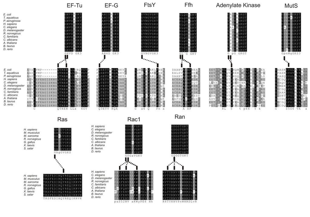
2 FIGURE LEGENDS Figure S1. Eyring plots of mant-gtp dissociation from EF-Tu variants. Mant-GTP dissociation rate constants (k off ) were determined at different temperatures. Data points represent rates obtained from fitting individual traces in replicate experiments; wild type EF-Tu ( ), H22G ( ), M112G ( ), M112A ( ), M112L ( ). Figure S2. The P-loop forms a helical turn in simulation of EF-Tu apo M112A. P-loop backbone conformations are shown for wild type (A&F), H22G (B&G), M112G (C&H), M112A (D&I), and M112L (E&J) after 10ns of simulation of EF-Tu GTP (A-E) or EF-Tu apo (F-J). Figure S3. Interactions between the two N-terminal P-loop amino acids and helix C are conserved in P-loop NTPases. Multiple sequence alignments of the G-proteins EF-Tu, EF-G, FtsY, Ffh, Ras, Rac1, and Ran as well as the ATPases adenylate kinase and MutS were performed in ClustalW 1,2. The P-loop (top) and helix C (bottom) alignments are shown for each protein with amino acids coloured according to conservation (black: 100%, grey: >80%, light grey: >60%). Interactions between amino acids of the P-loop and helix C, shown as dashed lines, were identified in crystal structures of each G-protein bound to a non-hydrolyzable GTP analogue. The PDBIDs for the structures investigated were as follows: EF-Tu:1EFT, EF-G: 2BV3, FtsY: 2Q9B, Ffh: 2CO4, Adenylate Kinase: 1AKE, MutS: 1E3M, Ras: 3L8Z, Rac1: 1MH1, Ran: 1IBR. Interacting amino acids were defined by close approach (<3.5Å; <3.8Å for FtsY and MutS) of heavy atoms. Figure S4. Root mean-squared deviation (RMSD) of backbone atoms in EF-Tu during molecular dynamics simulations. (a) Comparison of variability of EF-Tu simulations with GTP Mg 2+ bound in the nucleotide-binding pocket. Based on this, simulations (10ns) were performed with (b) and without (c) GTP Mg + bound in the nucleotide-binding pocket of EF- Tu WT and respective variants. Each simulation was carried out with a 0.5 fs time step at 300K in an NPT ensemble; conformations were sampled every 0.5ps for RMSD analysis. The traces shown are wild type (blue), H22G (black), M112L (red), M112G (green), and M112A (purple) EF-Tu Figure S5. Relative total energies of EF-Tu during 10 ns molecular dynamics simulations. Comparison of EF-Tu WT and variants (a) free and (b) with GTP Mg 2+ bound in the nucleotidebinding pocket.
3 FIGURES Figure S1. Figure S2.
4 Figure S3.
5 Figure S4.
6 Figure S5. a b
7 Table S1. Distances between N-H bonds of the P-loop backbone and oxygen atoms of GTP phosphates based on different MD simulation segments. Distance EF-Tu wt GTP EF-Tu wt GTP EF-Tu wt GTP EF-Tu EF-Tu Measured 6-10ns 6-40ns 10-40ns H22G GTP H22G GTP 6-20ns N-H O γ2 (2.3 ± 0.3) Å (2.3 ± 0.3) Å (2.3 ± 0.3) Å (1.96 ± 0.21) Å (1.97 ± 0.20) Å N-H O β1 (3.1 ± 0.3) Å (3.0 ± 0.3) Å (3.0 ± 0.3) Å (1.97 ± 0.17) Å (1.96 ± 0.17) Å N-H O β1 (2.19 ± 0.19) Å (2.18 ± 0.19) Å (2.18 ± 0.19) Å (1.97 ± 0.19) Å (2.04 ± 0.23) Å N-H O α3 (2.63 ± 0.23) Å (2.61 ± 0.23) Å (2.61 ± 0.23) Å (2.55 ± 0.25) Å (2.48 ± 0.25) Å N-H O β1 (1.97 ± 0.14) Å (1.98 ± 0.14) Å (1.98 ± 0.14) Å (2.01 ± 0.15) Å (2.03 ± 0.15) Å N-H Thr25 O β2 (1.95 ± 0.12) Å (1.95 ± 0.12) Å (1.95 ± 0.12) Å (1.98 ± 0.12) Å (1.98 ± 0.13) Å Table S2. Probability of P-loop amino acids undergoing backbone conformational changes during steered MD. Probabilities (in percent) were computed based on the fractions of SMD simulations in which the respective amino acid occupied multiple backbone conformations. Amino Acid wt H22G M112G M112A M112L Gly His Val Thr
8 Table S3. Summary of hot P-loop amino acids in MD simulations of all EF-Tu variants. Amino acids listed for SMD were hot in 20% or more of SMD simulations. wild type H22G M112G M112A M112L K D /nm a 70 ± ± ± ± ± 100 k 20 C / s -1 b ± ± ± ± ± TΔS 20 C b /kjmol ± 1-29 ± 2-45 ± 5-44 ± 3-40 ± 1 EF-Tu GTP hot amino acids Gly18 His19 Gly18 His19 SMD hot amino acids EF-Tu Apo hot amino acids Gly22 Gly22 a b computed from kinetic analysis of the EF-Tu GTP complex computed for EF-Tu GTP dissociation References 1 Thompson, J. D., Higgins, D. G. & Gibson, T. J. CLUSTAL W: improving the sensitivity of progressive multiple sequence alignment through sequence weighting, positionspecific gap penalties and weight matrix choice. Nucleic Acids Res. 22, (1994). 2 Larkin, M. A. et al. Clustal W and Clustal X version 2.0. Bioinformatics 23, (2007).
Supplementary Materials for
 advances.sciencemag.org/cgi/content/full/3/5/e1601684/dc1 Supplementary Materials for Directional mechanical stability of Bacteriophage ϕ29 motor s 3WJ-pRNA: Extraordinary robustness along portal axis
Supplementary figure 1. Comparison of unbound ogm-csf and ogm-csf as captured in the GIF:GM-CSF complex. Alignment of two copies of unbound ovine
 Supplementary figure 1. Comparison of unbound and as captured in the GIF:GM-CSF complex. Alignment of two copies of unbound ovine GM-CSF (slate) with bound GM-CSF in the GIF:GM-CSF complex (GIF: green,
SUPPLEMENTARY INFORMATION
 Supplementary materials Figure S1 Fusion protein of Sulfolobus solfataricus SRP54 and a signal peptide. a, Expression vector for the fusion protein. The signal peptide of yeast dipeptidyl aminopeptidase
SUPPLEMENTARY MATERIAL. Supplementary material and methods:
 Electronic Supplementary Material (ESI) for Catalysis Science & Technology. This journal is The Royal Society of Chemistry 2015 SUPPLEMENTARY MATERIAL Supplementary material and methods: - Computational
Time-dependence of key H-bond/electrostatic interaction distances in the sirna5-hago2 complexes... Page S14
 Supporting Information Probing the Binding Interactions between Chemically Modified sirnas and Human Argonaute 2 Using Microsecond Molecular Dynamics Simulations S. Harikrishna* and P. I. Pradeepkumar*
SUPPLEMENTARY FIGURES
 SUPPLEMENTARY FIGURES Supplementary Figure 1 Protein sequence alignment of Vibrionaceae with either a 40-residue insertion or a 44-residue insertion. Identical residues are indicated by red background.
SUPPLEMENTARY INFORMATION
 Supplementary Results DNA binding property of the SRA domain was examined by an electrophoresis mobility shift assay (EMSA) using synthesized 12-bp oligonucleotide duplexes containing unmodified, hemi-methylated,
Structure and evolution of the spliceosomal peptidyl-prolyl cistrans isomerase Cwc27
 Acta Cryst. (2014). D70, doi:10.1107/s1399004714021695 Supporting information Volume 70 (2014) Supporting information for article: Structure and evolution of the spliceosomal peptidyl-prolyl cistrans isomerase
Supplementary Information. The protease GtgE from Salmonella exclusively targets. inactive Rab GTPases
 Supplementary Information The protease GtgE from Salmonella exclusively targets inactive Rab GTPases Table of Contents Supplementary Figures... 2 Supplementary Figure 1... 2 Supplementary Figure 2... 3
Introduction to the Ribosome Overview of protein synthesis on the ribosome Prof. Anders Liljas
 Introduction to the Ribosome Molecular Biophysics Lund University 1 A B C D E F G H I J Genome Protein aa1 aa2 aa3 aa4 aa5 aa6 aa7 aa10 aa9 aa8 aa11 aa12 aa13 a a 14 How is a polypeptide synthesized? 2
SUPPLEMENTARY INFORMATION
 SUPPLEMENTARY INFORMATION doi:10.1038/nature11524 Supplementary discussion Functional analysis of the sugar porter family (SP) signature motifs. As seen in Fig. 5c, single point mutation of the conserved
Analysis of correlated mutations in Ras G-domain
 www.bioinformation.net Volume 13(6) Hypothesis Analysis of correlated mutations in Ras G-domain Ekta Pathak * Bioinformatics Department, MMV, Banaras Hindu University. Ekta Pathak - E-mail: ektavpathak@gmail.com;
Supporting Information How does Darunavir prevent HIV-1 protease dimerization?
 Supporting Information How does Darunavir prevent HIV- protease dimerization? Danzhi Huang and Amedeo Caflisch a Department of Biochemistry University of Zürich, Winterthurerstrasse 9 CH-7 Zürich, Switzerland
SUPPLEMENTARY INFORMATION
 doi:1.138/nature1737 Supplementary Table 1 variant Description FSEC - 2B12 a FSEC - 6A1 a K d (leucine) c Leucine uptake e K (wild-type like) K (Y18F) K (TS) K (TSY) K288A mutant, lipid facing side chain
Supplementary Figure 1. Aligned sequences of yeast IDH1 (top) and IDH2 (bottom) with isocitrate
 and IDH2 (bottom) with isocitrate") SUPPLEMENTARY FIGURE LEGENDS Supplementary Figure 1. Aligned sequences of yeast IDH1 (top) and IDH2 (bottom) with isocitrate dehydrogenase from Escherichia coli [ICD, pdb 1PB1, Mesecar, A. D., and Koshland,
SUPPLEMENTARY FIGURE LEGENDS Supplementary Figure 1. Aligned sequences of yeast IDH1 (top) and IDH2 (bottom) with isocitrate dehydrogenase from Escherichia coli [ICD, pdb 1PB1, Mesecar, A. D., and Koshland,
Tex 25mer ssrna Binding Stoichiometry
 Figure S. Determination of Tex:2nt ssrna binding stoichiometry using fluorescence polarization. Fluorescein labeled RNA was held at a constant concentration 2-fold above the K d. Tex protein was titrated
Biophysics 490M Project
 Biophysics 490M Project Dan Han Department of Biochemistry Structure Exploration of aa 3 -type Cytochrome c Oxidase from Rhodobacter sphaeroides I. Introduction: All organisms need energy to live. They
Mechanical Proteins. Stretching imunoglobulin and fibronectin. domains of the muscle protein titin. Adhesion Proteins of the Immune System
 Mechanical Proteins F C D B A domains of the muscle protein titin E Stretching imunoglobulin and fibronectin G NIH Resource for Macromolecular Modeling and Bioinformatics Theoretical Biophysics Group,
Structure Investigation of Fam20C, a Golgi Casein Kinase
 Structure Investigation of Fam20C, a Golgi Casein Kinase Sharon Grubner National Taiwan University, Dr. Jung-Hsin Lin University of California San Diego, Dr. Rommie Amaro Abstract This research project
Equilibrated atomic models of outward-facing P-glycoprotein and effect of ATP binding on structural dynamics (Supplementary Information)
") Equilibrated atomic models of outward-facing P-glycoprotein and effect of ATP binding on structural dynamics (Supplementary Information) Lurong Pan 1 and Stephen G. Aller 2 * 1,2 Department of Pharmacology
Equilibrated atomic models of outward-facing P-glycoprotein and effect of ATP binding on structural dynamics (Supplementary Information) Lurong Pan 1 and Stephen G. Aller 2 * 1,2 Department of Pharmacology
Supporting Information
 Supporting Information Allosteric communication disrupted by small molecule binding to the Imidazole glycerol phosphate synthase protein-protein interface. Ivan Rivalta*,#, George P. Lisi #, Ning-Shiuan
Secondary and sidechain structures
 Lecture 2 Secondary and sidechain structures James Chou BCMP201 Spring 2008 Images from Petsko & Ringe, Protein Structure and Function. Branden & Tooze, Introduction to Protein Structure. Richardson, J.
Protein synthesis II Biochemistry 302. Bob Kelm February 25, 2004
 Protein synthesis II Biochemistry 302 Bob Kelm February 25, 2004 Two idealized views of the 70S ribosomal complex during translation 70S cavity Fig. 27.25 50S tunnel View with 30S subunit in front, 50S
SUPPLEMENTARY INFORMATION
 doi:10.1038/nature11054 Supplementary Fig. 1 Sequence alignment of Na v Rh with NaChBac, Na v Ab, and eukaryotic Na v and Ca v homologs. Secondary structural elements of Na v Rh are indicated above the
I690/B680 Structural Bioinformatics Spring Protein Structure Determination by NMR Spectroscopy
 I690/B680 Structural Bioinformatics Spring 2006 Protein Structure Determination by NMR Spectroscopy Suggested Reading (1) Van Holde, Johnson, Ho. Principles of Physical Biochemistry, 2 nd Ed., Prentice
Section 7. Junaid Malek, M.D.
 Section 7 Junaid Malek, M.D. RNA Processing and Nomenclature For the purposes of this class, please do not refer to anything as mrna that has not been completely processed (spliced, capped, tailed) RNAs
Supporting information to: Time-resolved observation of protein allosteric communication. Sebastian Buchenberg, Florian Sittel and Gerhard Stock 1
 Supporting information to: Time-resolved observation of protein allosteric communication Sebastian Buchenberg, Florian Sittel and Gerhard Stock Biomolecular Dynamics, Institute of Physics, Albert Ludwigs
Supplemental data for
 Supplemental data for A Real-Time Guanine Nucleotide Exchange Assay using NMR: Activation of RhoA by PDZ- RhoGEF. Geneviève M.C. Gasmi-Seabrook 1,3, Christopher B. Marshall 1,3, Melissa Cheung 1,3, Bryan
Supporting Information
 Supporting Information Micelle-Triggered b-hairpin to a-helix Transition in a 14-Residue Peptide from a Choline-Binding Repeat of the Pneumococcal Autolysin LytA HØctor Zamora-Carreras, [a] Beatriz Maestro,
Goals. Structural Analysis of the EGR Family of Transcription Factors: Templates for Predicting Protein DNA Interactions
 Structural Analysis of the EGR Family of Transcription Factors: Templates for Predicting Protein DNA Interactions Jamie Duke 1,2 and Carlos Camacho 3 1 Bioengineering and Bioinformatics Summer Institute,
SUPPLEMENTARY INFORMATION
 Figure S1. Secondary structure of CAP (in the camp 2 -bound state) 10. α-helices are shown as cylinders and β- strands as arrows. Labeling of secondary structure is indicated. CDB, DBD and the hinge are
Computational engineering of cellulase Cel9A-68 functional motions through mutations in its linker region. WT 1TF4 (crystal) -90 ERRAT PROVE VERIFY3D
 -90 ERRAT PROVE VERIFY3D") Electronic Supplementary Material (ESI) for Physical Chemistry Chemical Physics. This journal is the Owner Societies 218 Supplementary Material: Computational engineering of cellulase Cel9-68 functional
Electronic Supplementary Material (ESI) for Physical Chemistry Chemical Physics. This journal is the Owner Societies 218 Supplementary Material: Computational engineering of cellulase Cel9-68 functional
Impact of the crystallization condition on importin-β conformation
 Supporting information Volume 72 (2016) Supporting information for article: Impact of the crystallization condition on importin-β conformation Marcel J. Tauchert, Clément Hémonnot, Piotr Neumann, Sarah
Introduction: actin and myosin
 Introduction: actin and myosin Actin Myosin Myosin V and actin 375 residues Found in all eukaryotes Polymeric Forms track for myosin Many other cellular functions 36 nm pseudo-helical repeat Catalytic
The Potassium Ion Channel: Rahmat Muhammad
 The Potassium Ion Channel: 1952-1998 1998 Rahmat Muhammad Ions: Cell volume regulation Electrical impulse formation (e.g. sodium, potassium) Lipid membrane: the dielectric barrier Pro: compartmentalization
Supplementary Figure 3 a. Structural comparison between the two determined structures for the IL 23:MA12 complex. The overall RMSD between the two
 Supplementary Figure 1. Biopanningg and clone enrichment of Alphabody binders against human IL 23. Positive clones in i phage ELISA with optical density (OD) 3 times higher than background are shown for
Secondary Structure. Bioch/BIMS 503 Lecture 2. Structure and Function of Proteins. Further Reading. Φ, Ψ angles alone determine protein structure
 Bioch/BIMS 503 Lecture 2 Structure and Function of Proteins August 28, 2008 Robert Nakamoto rkn3c@virginia.edu 2-0279 Secondary Structure Φ Ψ angles determine protein structure Φ Ψ angles are restricted
Ranjit P. Bahadur Assistant Professor Department of Biotechnology Indian Institute of Technology Kharagpur, India. 1 st November, 2013
 Hydration of protein-rna recognition sites Ranjit P. Bahadur Assistant Professor Department of Biotechnology Indian Institute of Technology Kharagpur, India 1 st November, 2013 Central Dogma of life DNA
Type II Kinase Inhibitors Show an Unexpected Inhibition Mode against Parkinson s Disease-Linked LRRK2 Mutant G2019S.
 Type II Kinase Inhibitors Show an Unexpected Inhibition Mode against Parkinson s Disease-Linked LRRK2 Mutant G219S. Min Liu@&*, Samantha A. Bender%*, Gregory D Cuny@, Woody Sherman, Marcie Glicksman@ Soumya
Structure, mechanism and ensemble formation of the Alkylhydroperoxide Reductase subunits. AhpC and AhpF from Escherichia coli
 Structure, mechanism and ensemble formation of the Alkylhydroperoxide Reductase subunits AhpC and AhpF from Escherichia coli Phat Vinh Dip 1,#, Neelagandan Kamariah 2,#, Malathy Sony Subramanian Manimekalai
of the Guanine Nucleotide Exchange Factor FARP2
 Structure, Volume 21 Supplemental Information Structural Basis for Autoinhibition of the Guanine Nucleotide Exchange Factor FARP2 Xiaojing He, Yi-Chun Kuo, Tyler J. Rosche, and Xuewu Zhang Inventory of
SUPPLEMENTARY INFORMATION
 doi:10.1038/nature10955 Supplementary Figures Supplementary Figure 1. Electron-density maps and crystallographic dimer structures of the motor domain. (a f) Stereo views of the final electron-density maps
Structural and mechanistic insight into the substrate. binding from the conformational dynamics in apo. and substrate-bound DapE enzyme
 Electronic Supplementary Material (ESI) for Physical Chemistry Chemical Physics. This journal is the Owner Societies 215 Structural and mechanistic insight into the substrate binding from the conformational
Supporting Information
 Supporting Information Decoding Allosteric Networks in Biocatalysts: Rational Approach to Therapies and Biotechnologies Johannes T. Cramer 1,2, Jana I. Führing 1, Petra Baruch 2, Christian Brütting 3,
Experimental and Computational Mutagenesis to Investigate the. Positioning of a General Base within an Enzyme Active Site
 Experimental and Computational Mutagenesis to Investigate the Positioning of a General Base within an Enzyme Active Site Jason P. Schwans, Philip Hanoian, Benjamin J. Lengerich, Fanny Sunden, Ana Gonzalez
Protein synthesis I Biochemistry 302. February 17, 2006
 Protein synthesis I Biochemistry 302 February 17, 2006 Key features and components involved in protein biosynthesis High energy cost (essential metabolic activity of cell Consumes 90% of the chemical energy
Evolution of Translation: Dynamics of Recognition in RNA:Protein Complexes
 Evolution of Translation: Dynamics of Recognition in RNA:Protein Complexes Zaida (Zan) Luthey-Schulten Dept. Chemistry, Beckman Institute, Biophysics, Institute of Genomics Biology, & Physics NIH Workshop
Effekt des Antibiotikums Kirromycin auf den Elongations Faktor Tu untersucht mit Molekulardynamik-Simulationen
 Master s Thesis Effekt des Antibiotikums Kirromycin auf den Elongations Faktor Tu untersucht mit Molekulardynamik-Simulationen Effect of the antibiotic kirromycin on elongation factor Tu studied through
Introduction to" Protein Structure
 Introduction to" Protein Structure Function, evolution & experimental methods Thomas Blicher, Center for Biological Sequence Analysis Learning Objectives Outline the basic levels of protein structure.
Molecular dynamics simulation of Aquaporin-1. 4 nm
 Molecular dynamics simulation of Aquaporin-1 4 nm Molecular Dynamics Simulations Schrödinger equation i~@ t (r, R) =H (r, R) Born-Oppenheimer approximation H e e(r; R) =E e (R) e(r; R) Nucleic motion described
What makes a good graphene-binding peptide? Adsorption of amino acids and peptides at aqueous graphene interfaces: Electronic Supplementary
 Electronic Supplementary Material (ESI) for Journal of Materials Chemistry B. This journal is The Royal Society of Chemistry 21 What makes a good graphene-binding peptide? Adsorption of amino acids and
Signal Transduction Phosphorylation Protein kinases. Misfolding diseases. Protein Engineering Lysozyme variants
 Signal Transduction Phosphorylation Protein kinases Misfolding diseases Protein Engineering Lysozyme variants Cells and Signals Regulation The cell must be able to respond to stimuli Cellular activities
Engineering an Mg 2 Site to Replace a Structurally Conserved Arginine in the Catalytic Center of Histidyl-tRNA Synthetase by Computer Experiments
 PROTEINS: Structure, Function, and Genetics 32:362 380 (1998) Engineering an Mg 2 Site to Replace a Structurally Conserved Arginine in the Catalytic Center of Histidyl-tRNA Synthetase by Computer Experiments
What binds to Hb in addition to O 2?
 Reading: Ch5; 158-169, 162-166, 169-174 Problems: Ch5 (text); 3,7,8,10 Ch5 (study guide-facts); 1,2,3,4,5,8 Ch5 (study guide-apply); 2,3 Remember Today at 5:30 in CAS-522 is the second chance for the MB
Structure of EF-G ribosome complex in a pre-translocation state
 Structure of EF-G ribosome complex in a pre-translocation state Yun Chen, Shu Feng, Veerendra Kumar, Rya Ero, Yong-Gui Gao Supplementary Figure 1 Supplementary Figure 1 Conformational change of the stalk
Structure of the quaternary complex between SRP, SR, and translocon bound to the translating ribosome
 Structure of the quaternary complex between SRP, SR, and translocon bound to the translating ribosome Ahmad Jomaa 1, Yu-Hsien Hwang Fu 2, Daniel Boehringer 1, Marc Leibundgut 1, Shu-ou Shan 2, and Nenad
SUPPLEMENTARY INFORMATION
 SUPPLEMENTARY INFORMATION doi:10.1038/nature11539 Supplementary Figure 1 Schematic representation of plant (A) and mammalian (B) P 2B -ATPase domain organization. Actuator (A-), nucleotide binding (N-),
Supporting Information
 Electronic Supplementary Material (ESI) for Physical Chemistry Chemical Physics. This journal is the Owner Societies 2016 Supporting Information Lipid molecules can induce an opening of membrane-facing
Tu 1,*, , Sweden
 Supplementary Material Computational studiess of the binding profile of phosphoinositide PtdIns(,4,5)P with the pleckstrin homology domain d of an oomycetee cellulose synthase Guanglin Kuang 1, Vincent
Structural insights into energy regulation of light-harvesting complex from spinach CP29
 SUPPLEMENTARY INFORMATION Structural insights into energy regulation of light-harvesting complex from spinach CP29 Xiaowei Pan 1, Mei Li 1, Tao Wan 1,2, Longfei Wang 1,2, Chenjun Jia 1,2, Zhiqiang Hou
Hands-on Course in Computational Structural Biology and Molecular Simulation BIOP590C/MCB590C. Course Details
 Hands-on Course in Computational Structural Biology and Molecular Simulation BIOP590C/MCB590C Emad Tajkhorshid Center for Computational Biology and Biophysics Email: emad@life.uiuc.edu or tajkhors@uiuc.edu
Bahnson Biochemistry Cume, April 8, 2006 The Structural Biology of Signal Transduction
 Name page 1 of 6 Bahnson Biochemistry Cume, April 8, 2006 The Structural Biology of Signal Transduction Part I. The ion Ca 2+ can function as a 2 nd messenger. Pick a specific signal transduction pathway
NUCLEOTIDE BINDING ENZYMES
 NUCLEOTIDE BINDING ENZYMES The Rossmann fold Relationship between sequence, structure and function. Anna Casas, Júlia Gasull and Nerea Vega Index 1. Introduction: Adenine nucleotides 1. The most commonly
Nature Structural & Molecular Biology: doi: /nsmb Supplementary Figure 1
 Supplementary Figure 1 Resonance assignment and NMR spectra for hairpin and duplex A 6 constructs. (a) 2D HSQC spectra of hairpin construct (hp-a 6 -RNA) with labeled assignments. (b) 2D HSQC or SOFAST-HMQC
LACTOFERRIN: ANALYSIS OF THE STRUCTURE PROFILE
 Chemistry Journal of Moldova. General, Industrial and Ecological Chemistry. 2014, 9(2), 99-106 LACTOFERRIN: ANALYSIS OF THE STRUCTURE PROFILE Lilia Anghel Institute of Chemistry of the Academy of Sciences
SUPPLEMENTARY INFORMATION. doi: /nature07461
 Figure S1 Electrophysiology. a ph-activation of. Two-electrode voltage clamp recordings of Xenopus oocytes expressing in comparison to waterinjected oocytes. Currents were recorded at 40 mv. The ph of
Supplementary Figures for Tong et al.: Structure and function of the intracellular region of the plexin-b1 transmembrane receptor
 Supplementary Figures for Tong et al.: Structure and function of the intracellular region of the plexin-b1 transmembrane receptor Figure S1. Plexin-B1 GAP segments are homologous to RasGAPs. Sequence alignment
Molecular dynamics simulations of anti-aggregation effect of ibuprofen. Wenling E. Chang, Takako Takeda, E. Prabhu Raman, and Dmitri Klimov
 Biophysical Journal, Volume 98 Supporting Material Molecular dynamics simulations of anti-aggregation effect of ibuprofen Wenling E. Chang, Takako Takeda, E. Prabhu Raman, and Dmitri Klimov Supplemental
SUPPLEMENTARY INFORMATION
 doi:10.1038/nature11085 Supplementary Tables: Supplementary Table 1. Summary of crystallographic and structure refinement data Structure BRIL-NOP receptor Data collection Number of crystals 23 Space group
Supplementary Materials for
 www.sciencesignaling.org/cgi/content/full/5/243/ra68/dc1 Supplementary Materials for Superbinder SH2 Domains Act as Antagonists of Cell Signaling Tomonori Kaneko, Haiming Huang, Xuan Cao, Xing Li, Chengjun
Supplementary Materials for
 advances.sciencemag.org/cgi/content/full/3/4/e1600663/dc1 Supplementary Materials for A dynamic hydrophobic core orchestrates allostery in protein kinases Jonggul Kim, Lalima G. Ahuja, Fa-An Chao, Youlin
FW 1 CDR 1 FW 2 CDR 2
 Supplementary Figure 1 Supplementary Figure 1: Interface of the E9:Fas structure. The two interfaces formed by V H and V L of E9 with Fas are shown in stereo. The Fas receptor is represented as a surface
Supporting Information
 Supporting Information Boehr et al. 10.1073/pnas.0914163107 SI Text Materials and Methods. R 2 relaxation dispersion experiments. 15 NR 2 relaxation dispersion data measured at 1 H Larmor frequencies of
SUPPLEMENTARY INFORMATION
 doi:10.1038/nature17991 Supplementary Discussion Structural comparison with E. coli EmrE The DMT superfamily includes a wide variety of transporters with 4-10 TM segments 1. Since the subfamilies of the
Introduction to Comparative Protein Modeling. Chapter 4 Part I
 Introduction to Comparative Protein Modeling Chapter 4 Part I 1 Information on Proteins Each modeling study depends on the quality of the known experimental data. Basis of the model Search in the literature
Homology models of the tetramerization domain of six eukaryotic voltage-gated potassium channels Kv1.1-Kv1.6
 Homology models of the tetramerization domain of six eukaryotic voltage-gated potassium channels Kv1.1-Kv1.6 Hsuan-Liang Liu* and Chin-Wen Chen Department of Chemical Engineering and Graduate Institute
Computational Modeling of Protein Kinase A and Comparison with Nuclear Magnetic Resonance Data
 Computational Modeling of Protein Kinase A and Comparison with Nuclear Magnetic Resonance Data ABSTRACT Keyword Lei Shi 1 Advisor: Gianluigi Veglia 1,2 Department of Chemistry 1, & Biochemistry, Molecular
GENETICS - CLUTCH CH.11 TRANSLATION.
 !! www.clutchprep.com CONCEPT: GENETIC CODE Nucleotides and amino acids are translated in a 1 to 1 method The triplet code states that three nucleotides codes for one amino acid - A codon is a term for
Details of Protein Structure
 Details of Protein Structure Function, evolution & experimental methods Thomas Blicher, Center for Biological Sequence Analysis Anne Mølgaard, Kemisk Institut, Københavns Universitet Learning Objectives
Table 1. Crystallographic data collection, phasing and refinement statistics. Native Hg soaked Mn soaked 1 Mn soaked 2
 Table 1. Crystallographic data collection, phasing and refinement statistics Native Hg soaked Mn soaked 1 Mn soaked 2 Data collection Space group P2 1 2 1 2 1 P2 1 2 1 2 1 P2 1 2 1 2 1 P2 1 2 1 2 1 Cell
Effects of Chemical Exchange on NMR Spectra
 Effects of Chemical Exchange on NMR Spectra Chemical exchange refers to any process in which a nucleus exchanges between two or more environments in which its NMR parameters (e.g. chemical shift, scalar
Table S1. Overview of used PDZK1 constructs and their binding affinities to peptides. Related to figure 1.
 Table S1. Overview of used PDZK1 constructs and their binding affinities to peptides. Related to figure 1. PDZK1 constru cts Amino acids MW [kda] KD [μm] PEPT2-CT- FITC KD [μm] NHE3-CT- FITC KD [μm] PDZK1-CT-
Protein synthesis I Biochemistry 302. Bob Kelm February 23, 2004
 Protein synthesis I Biochemistry 302 Bob Kelm February 23, 2004 Key features of protein synthesis Energy glutton Essential metabolic activity of the cell. Consumes 90% of the chemical energy (ATP,GTP).
Sensitive NMR Approach for Determining the Binding Mode of Tightly Binding Ligand Molecules to Protein Targets
 Supporting information Sensitive NMR Approach for Determining the Binding Mode of Tightly Binding Ligand Molecules to Protein Targets Wan-Na Chen, Christoph Nitsche, Kala Bharath Pilla, Bim Graham, Thomas
Crystal Structure of a camp-dependent Protein Kinase Mutant at 1.26 Å: New Insights into the Catalytic Mechanism
 doi:10.1016/j.jmb.2003.11.044 J. Mol. Biol. (2004) 336, 473 487 Crystal Structure of a camp-dependent Protein Kinase Mutant at 1.26 Å: New Insights into the Catalytic Mechanism Jie Yang 1, Lynn F. Ten
NB-DNJ/GCase-pH 7.4 NB-DNJ+/GCase-pH 7.4 NB-DNJ+/GCase-pH 4.5
 SUPPLEMENTARY TABLES Suppl. Table 1. Protonation states at ph 7.4 and 4.5. Protonation states of titratable residues in GCase at ph 7.4 and 4.5. Histidine: HID, H at δ-nitrogen; HIE, H at ε-nitrogen; HIP,
Supporting Information
 S1 Supporting Information Unraveling the Reaction Mechanism of Enzymatic C5-Cytosine Methylation of DNA. A Combined Molecular Dynamics and QM/MM Study of Wild Type and Gln119 Variant Juan Aranda 1, Kirill
ومن أحياها Translation 2. Translation 2. DONE BY :Nisreen Obeidat
 Translation 2 DONE BY :Nisreen Obeidat Page 0 Prokaryotes - Shine-Dalgarno Sequence (2:18) What we're seeing here are different portions of sequences of mrna of different promoters from different bacterial
Journal of Pharmacology and Experimental Therapy-JPET#172536
 A NEW NON-PEPTIDIC INHIBITOR OF THE 14-3-3 DOCKING SITE INDUCES APOPTOTIC CELL DEATH IN CHRONIC MYELOID LEUKEMIA SENSITIVE OR RESISTANT TO IMATINIB Manuela Mancini, Valentina Corradi, Sara Petta, Enza
Supplementary information
 Supplementary information The structural basis of modularity in ECF-type ABC transporters Guus B. Erkens 1,2, Ronnie P-A. Berntsson 1,2, Faizah Fulyani 1,2, Maria Majsnerowska 1,2, Andreja Vujičić-Žagar
Mechanical Proteins. Stretching imunoglobulin and fibronectin. domains of the muscle protein titin. Adhesion Proteins of the Immune System
 Mechanical Proteins F C D B A domains of the muscle protein titin E Stretching imunoglobulin and fibronectin G NIH Resource for Macromolecular Modeling and Bioinformatics Theoretical Biophysics Group,
Ch. 2 BASIC CHEMISTRY. Copyright 2010 Pearson Education, Inc.
 Ch. 2 BASIC CHEMISTRY Matter and Composition of Matter Definition: Anything that has mass and occupies space Matter is made up of elements An element cannot be broken down by ordinary chemical means Atoms
Lattice protein models
 Lattice protein models Marc R. Roussel epartment of Chemistry and Biochemistry University of Lethbridge March 5, 2009 1 Model and assumptions The ideas developed in the last few lectures can be applied
NMR Structure of the HWE Kinase Associated Response Regulator Sma0114 in Its Activated State
 pubs.acs.org/biochemistry NMR Structure of the HWE Kinase Associated Response Regulator Sma0114 in Its Activated State Sarah R. Sheftic, Emma White, Daniel J. Gage, and Andrei T. Alexandrescu* Department
β1 Structure Prediction and Validation
 13 Chapter 2 β1 Structure Prediction and Validation 2.1 Overview Over several years, GPCR prediction methods in the Goddard lab have evolved to keep pace with the changing field of GPCR structure. Despite
Nature Structural & Molecular Biology: doi: /nsmb Supplementary Figure 1
 Supplementary Figure 1 Chemical structure of LPS and LPS biogenesis in Gram-negative bacteria. a. Chemical structure of LPS. LPS molecule consists of Lipid A, core oligosaccharide and O-antigen. The polar
L718Q mutant EGFR escapes covalent inhibition by stabilizing. a non-reactive conformation of the lung cancer drug. osimertinib
 Electronic Supplementary Material (ESI) for Chemical Science. This journal is The Royal Society of Chemistry 2018 Electronic Supplementary Information (ESI) for L718Q mutant EGFR escapes covalent inhibition
Supporting Information Converter domain mutations in myosin alter structural kinetics and motor function. Hershey, PA, MN 55455, USA
 Supporting Information Converter domain mutations in myosin alter structural kinetics and motor function Laura K. Gunther 1, John A. Rohde 2, Wanjian Tang 1, Shane D. Walton 1, William C. Unrath 1, Darshan
Supporting Information for. Models for the Metal Transfer Complex of the N-terminal Region of CusB and. CusF
 Supporting Information for Models for the Metal Transfer Complex of the N-terminal Region of CusB and CusF Melek N. Ucisik, Dhruva K. Chakravorty, and Kenneth M. Merz Jr. * Department of Chemistry and
SUPPLEMENTARY FIGURES. Structure of the cholera toxin secretion channel in its. closed state
 SUPPLEMENTARY FIGURES Structure of the cholera toxin secretion channel in its closed state Steve L. Reichow 1,3, Konstantin V. Korotkov 1,3, Wim G. J. Hol 1$ and Tamir Gonen 1,2$ 1, Department of Biochemistry