GFP WxTP-GFP WxTP-GFP. stromules. DsRed WxTP-DsRed WxTP-DsRed. stromules
|
|
|
- Estella Owens
- 5 years ago
- Views:
Transcription


 GFP- and WxTP-GFP- bombarded cells.")


 and a cooled")


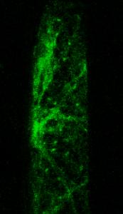
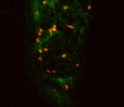
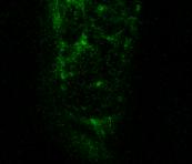
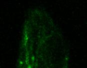
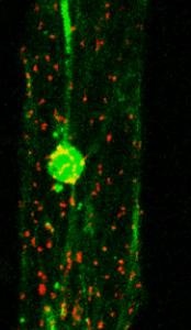
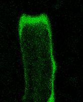
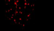
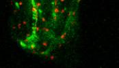
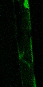
1 Supplemental Data. Kitajima et al. (2009). The rice -amylase glycoprotein is targeted from the Golgi apparatus through the secretory pathway to the plastids. A GFP WxTP-GFP WxTP-GFP stromules stromules DsRed WxTP-DsRed WxTP-DsRed stromules Supplemental Figure 1. Fluorescence images of onion epidermal cells expressing either GFP, DsRed, WxTP-GFP or WxTP-DsRed. (A) GFP- and WxTP-GFP- bombarded cells. () DsRed- and WxTP-DsRed- bombarded cells. The epidermal cells were incubated on 0.6% gelrite with 2,4-D-free Murashige-Skoog (MS) medium for 24 h at 25 C in darkness. Images of GFP and DsRed were observed using a X-61 microscope (Olympus, Tokyo, Japan) and a cooled CoolSnap-fx CCD camera (Photometrics). An Hg lamp was used to excite the fluorescent proteins. GFP fluorescence was obtained by 470 to 490-nm excitation with 510 to 550-nm detection, and that of DsRed was by 520 to 550-nm excitation with 580-nm detection. Deconvolution was carried out using Lumina Vision imaging software (Mitani, Tokyo, Japan). In a whole cell, 15 to 20 images per cell, from the top to middle of the cell, every 1 to 2 μm, were taken and combined into one image. All of the images presented are stacks. The right-hand panels represent the fluorescent plastids in close-up. Plastids with stromules were similarly visualized using WxTP-GFP and WxTP-DsRed. Left panels, bars = 100 μm; Right panels, bars = 10 μm.


 (A) or mt-gfp")

. Images were obtained")


2 GFP DsRed merged GFP DsRed merged Supplemental Figure 2. Fluorescence images of onion cells simultaneously expressing WxTP- DsRed and per-gfp (the peroxisomal targeting signal 2 fused with GFP) (A) or mt-gfp (the presequence of the subunit of Arabidopsis F1-ATPase fused with GFP) (). Images were obtained as described in the legend for Supplemental Figure 1. The results show that the distribution of WxTP-DsRed is distinguishable from those of per-gfp and mt-gfp, with the peroxisomal and mitochondrial markers not overlapping with the plastid marker. ars = 100 μm


3 AmyI-1 AmyII-3 AmyII-4 AmyII-5 AmyII-6 tid/gfp total (%) GFP plast Supplemental Figure 3. Distinct localization of various Amy isoforms in onion epidermal cells. (A) Simultaneous expression of WxTP-DsRed and either AmyI-1-, AmyII-3-, AmyII-4-, AmyII-5- or AmyII- 6-GFP was carried out. The merged images were constructed as described in Figure 3. ars = 100 m. () Statistical ti ti evaluation of the plastidial l localization li of different Amy isoforms. Ratios of the fluorescence intensity of GFP in the plastidial area to GFP in the whole cell (GFP plastid /GFP total ) (%) were determined. Values show the means s.d. (n = 4).

,")
 on")


 was")


4 C D Supplemental Figure 4. Effects of ARF1(T31N), ARF1(Q71L) and SAR1(H74L) on the localization of various organelle markers. Simultaneous expression of the organelle marker and either ARF1(T31N), ARF1(Q71L) or SAR1(H74L) was carried out in onion cells. (A) trans-golgi marker (ST-mRFP), () plastidial marker (WxTP-GFP), (C) peroxisomal marker (per-gfp), (D) mitochondrial marker (mt- GFP). Twenty to thirty images per cell, from the top to middle of the cell, every 1 2 μm, were taken and combined into one image. ars = 100 m.
 GFP ()")


5 (A) AmyI-1(1-369)-GFP AmyI-1(1-369) GFP () AmyII-6(1-266):AmyI-1( )-GFP AmyII-6(1-266) ( ) GGG GFP (C) AmyII-6(1-266):AmyI-1( )-GFP AmyII-6(1-266) ( ) GGG GFP D 80 C GFP Plastid /GFP total (%) GFP DsRed merged Supplemental Figure 5. Plastid targeting abilities of AmyII-6:AmyI-1 chimera proteins. The AmyII-6:AmyI-1 chimera proteins showed little targeting to the plastids in onion cells. The merged images were constructed as described in Figure 3. (A) AmyI-1(1-369)-GFP. () AmyII-6(1-266):AmyI-1( )-GFP. (C) AmyII-6(1-266):AmyI-1( )-GFP. ars = 100 m. (D) Quantitative results. Ratios of the fluorescence intensity of GFP in the plastidial area to GFP in the whole cell (GFP plastid /GFP total ) (%) were determined. Values show the means s.d. (n = 4).
- and AmyI-1( 370 428)-GFP in cells")

 GFP alone, () AmyI-1( 101 428) GFP, (C) AmyI-1 ( 370 428) GFP and (D)")
6 GFP Chlorophyll merged C D Supplemental Figure 6. Plastid targeting abilities of AmyI-1( )- and AmyI-1( )-GFP in cells of transgenic rice plants. The stable transformant cells were sectioned with a vibratome to 25 mm thickness, and immediately observed by means of confocal laser scanning microscopy. (A) GFP alone, () AmyI-1( ) GFP, (C) AmyI-1 ( ) GFP and (D) AmyI-1-GFP. Left, GFP fluorescence; middle, chlorophyll autofluorescence; right, merged image. ars = 5 m.
7 Supplemental Table 1: Primer sequences for PCR-amplification Amplified DNA DNA template Primer sequences (Plasmid constructed) WxTP pwcw 5 -ctggatccatgtcggctctcaccacg-3 (pwxtp-gfp, pwxtp-dsred) 5 -tatggatccggcaggggggaggccaccgag-3 AmyI-1 pamyi-1 5 -atcgggatccatggtgaacaaacacttctt-3 (pamyi-1-gfp) 5 -aaggatccgcttttctcccagattgcgt-3 AmyII-3 pamyii-3 5 -aaggatccatgggcaagcaccatgtcac-3 (pamyii-3-gfp) 5 -ttggtaccatagtgctttctaccggca-3 AmyII-4 pamyii-4 5 -atcgggatccatgaagaacaccagcagc-3 (pamyii-4-gfp) 5 -aaggatcctaggtgccgccccgccgggac-3 AmyII-5 pamyii-5 5 -aaggatccatggcaaagcgcatagcctc-3 (pamyii-5-gfp) 5 -ttggatccatagtggtgccgccctgcagg-3 AmyII-6 pamyii-6 5 -taggatccatggcaaagcattccaccac-3 (pamyii-6-gfp) 5 -atggatccgtggcgccgccctgctggaac-3 AmyI-1( ) pamyi-1 5 -atcgggatccatggtgaacaaacacttctt-3 (pamyi-1( )-GFP) 5 -aaggatccgttgaatccctgaaacaggac-3 AmyI-1( ) pamyi-1 5 -atcgggatccatggtgaacaaacacttctt-3 (pamyi-1( )-GFP) 5 -aaggatccctcgatcagcgacttgagct-3 AmyI-1( ) pamyi-1 5 -atcgggatccatggtgaacaaacacttctt-3 (pamyi-1( )-GFP) 5 -aaggatccccacgcgtcgaagccgatgtc-3 AmyI-1( ) pamyi-1 5 -atcgggatccatggtgaacaaacacttctt-3 (pamyi-1( )-GFP) 5 -taggatcccccgatcatgccgggcgcctt-3 AmyI-1( ) pamyi-1 5 -atcgggatccatggtgaacaaacacttctt-3 (pamyi-1( )-GFP) 5 -aaggtacccggccaccacccgatcatgcc-3 AmyI-1( ) pamyi-1 5 -atcgggatccatggtgaacaaacacttctt-3 (pamyi-1( )-GFP) 5 -aaggtacccaaatggtcgtagaagatgca-3 AmyI-1( ) pamyi-1 5 -atcgggatccatggtgaacaaacacttctt-3 (pamyi-1( )-GFP) 5 -aaggatccctgccggtttctgattgaca-3 AmyI-1(W302A) pamyi-1 5 -gcgcccggcatgatcgggtgggcgccggccaaggcgacgacc-3 (pamyi-1(w302a)-gfp) 5 -ggtcgtcgccttggccggcgcccacccgatcatgccgggcgc-3 AmyI-1(W302L) pamyi-1 5 -atgatcgggtggctgccggccaaggcg-3 (pamyi-1(w302l)-gfp) 5 -cgccttggccggcagccacccgatcat-3 AmyI-1(T307V) pamyi-1 5 -ccggccaaggcggtgaccttcgtcgac-3 (pamyi-1(t307v)-gfp) 5 -gtcgacgaaggtcaccgccttggccgg-3 AmyI-1(G354N) pamyi-1 5 -ttcttcgattggaatctcaaggtggtg-3 (pamyi-1(g354n)-gfp) 5 -ctcctccttgagattccaatcgaagaa-3 AmyI-1( )(Gly) 4 pamyi-1 5 -aaggatccgtcgatcgtgtcggcggcgcc-3 (pamyi-1( )-gfp ) 5 -ataggtacccccgccgcccccctgccggtttctgattga-3 AmyI-1( )(Gly) 4 pamyi-1 5 -aaggatccctccgcggcgaggacggcaag-3 (pamyi-1( )-gfp ) 5 -ataggtacccccgccgcccccctgccggtttctgattga-3 35S-(amHI to Apa1)-AmyII-6 pamyii-6-gfp 5 -ggggactctagagggcccatggcaaagcat-3 (p35s-(amhi to Apa1)-AmyII-6-GFP) 5 -atgctttgccatgggccctctagagtcccc-3 35S-(amHI to Apa1)-AmyII-6(1-266) p35s-(amhi to Apa1)-AmyII-6-GFP 5 -gcgcaagcttagattagccttttcaatttc-3 (pamyii-6(1-266)/amyi-1( )gfp 5 -taaggatcccgatgcagggccaccga-3 35S-(amHI to Apa1)-AmyII-6(1-266) p35s-(amhi to Apa1)-AmyII-6-GFP 5 -gcgcaagcttagattagccttttcaatttc-3 (pamyii-6(1-266)/amyi-1( )gfp 5 -taaggatcccgatgcagggccaccga-3 1
Supplemental Data. Gao et al. (2012). Plant Cell /tpc
. Plant Cell /tpc") Supplemental Figure 1. Plant EMP Proteins. (A) The Accession numbers of the 12 EMP members from Arabidopsis. (B) Phylogenetic analysis of EMP proteins from Arabidopsis, human and yeast using the Mac Vector
Supplemental Figure 1. Plant EMP Proteins. (A) The Accession numbers of the 12 EMP members from Arabidopsis. (B) Phylogenetic analysis of EMP proteins from Arabidopsis, human and yeast using the Mac Vector
Last time: Obtaining information from a cloned gene
 Last time: Obtaining information from a cloned gene Objectives: 1. What is the biochemical role of the gene? 2. Where and when is the gene expressed (transcribed)? 3. Where and when is the protein made?
Supplementary information
 Supplementary information Statistical organelle dissection of Arabidopsis guard cells using image database LIPS Takumi Higaki *, Natsumaro Kutsuna, Yoichiroh Hosokawa, Kae Akita, Kazuo Ebine, Takashi Ueda,
Waithe et al Supplementary Figures
 Waithe et al Supplementary Figures Supplementary Figure 1 Expression and properties of WT and W391A mutant YFP- Ca V 2.2. A Immunoblot using Ca V 2.2 Ab for untransfected cells (UT, lane 1), YFP-Ca V 2.2
Reconstructing Mitochondrial Evolution?? Morphological Diversity. Mitochondrial Diversity??? What is your definition of a mitochondrion??
 Reconstructing Mitochondrial Evolution?? What is your definition of a mitochondrion?? Morphological Diversity Mitochondria as we all know them: Suprarenal gland Liver cell Plasma cell Adrenal cortex Mitochondrial
Transitivity-dependent and transitivity-independent cell-to-cell movement of RNA
 Himber et al. Transitivity-dependent and transitivity-independent cell-to-cell movement of RNA silencing SUPPLEMENTAL MATERIAL Supplemental material 1. Short-range movement of GFP silencing affects a nearly
SUPPLEMENTARY INFORMATION
 DOI: 10.1038/ncb2647 Figure S1 Other Rab GTPases do not co-localize with the ER. a, Cos-7 cells cotransfected with an ER luminal marker (either KDEL-venus or mch-kdel) and mch-tagged human Rab5 (mch-rab5,
T H E J O U R N A L O F C E L L B I O L O G Y
 T H E J O U R N A L O F C E L L B I O L O G Y Supplemental material Eisner et al., http://www.jcb.org/cgi/content/full/jcb.201312066/dc1 Figure S1. Mitochondrial continuity in adult skeletal muscle. (A)
Supplementary Figure 1. Real time in vivo imaging of SG secretion. (a) SGs from Drosophila third instar larvae that express Sgs3-GFP (green) and
 SGs from Drosophila third instar larvae that express Sgs3-GFP (green) and") Supplementary Figure 1. Real time in vivo imaging of SG secretion. (a) SGs from Drosophila third instar larvae that express Sgs3-GFP (green) and Lifeact-Ruby (red) were imaged in vivo to visualize secretion
Supplementary Figure 1. Real time in vivo imaging of SG secretion. (a) SGs from Drosophila third instar larvae that express Sgs3-GFP (green) and Lifeact-Ruby (red) were imaged in vivo to visualize secretion
Supplemental material
 Supplemental material THE JOURNAL OF CELL BIOLOGY Mourier et al., http://www.jcb.org/cgi/content/full/jcb.201411100/dc1 Figure S1. Size and mitochondrial content in Mfn1 and Mfn2 knockout hearts. (A) Body
Supplemental Data. Chen and Thelen (2010). Plant Cell /tpc
. Plant Cell /tpc") Supplemental Data. Chen and Thelen (2010). Plant Cell 10.1105/tpc.109.071837 1 C Total 5 kg 20 kg 100 kg Transmission Image 100 kg soluble pdtpi-gfp Plastid (PDH-alpha) Mito (PDH-alpha) GFP Image vector
Supplemental Data. Chen and Thelen (2010). Plant Cell 10.1105/tpc.109.071837 1 C Total 5 kg 20 kg 100 kg Transmission Image 100 kg soluble pdtpi-gfp Plastid (PDH-alpha) Mito (PDH-alpha) GFP Image vector
Supplemental Data. Perrella et al. (2013). Plant Cell /tpc
. Plant Cell /tpc") Intensity Intensity Intensity Intensity Intensity Intensity 150 50 150 0 10 20 50 C 150 0 10 20 50 D 0 10 20 Distance (μm) 50 20 40 E 50 F 0 10 20 50 0 15 30 Distance (μm) Supplemental Figure 1: Co-localization
Intensity Intensity Intensity Intensity Intensity Intensity 150 50 150 0 10 20 50 C 150 0 10 20 50 D 0 10 20 Distance (μm) 50 20 40 E 50 F 0 10 20 50 0 15 30 Distance (μm) Supplemental Figure 1: Co-localization
23-. Shoot and root development depend on ratio of IAA/CK
 Balance of Hormones regulate growth and development Environmental factors regulate hormone levels light- e.g. phototropism gravity- e.g. gravitropism temperature Mode of action of each hormone 1. Signal
GFP GAL bp 3964 bp
 Supplemental Data. Møller et al. (2009) Shoot Na + exclusion and increased salinity tolerance engineered by cell type-specific alteration of Na + transport in Arabidopsis Supplemental Figure 1. Salt-sensitive
GFP-TARGETED MITOCHONDRIA SHOW HETEROGENEITY OF SIZE, MORPHOLOGY, AND DYNAMICS IN TRANSGENIC NICOTIANA TABACUM L. PLANTS IN VIVO
 Int. J. Plant Sci. 165(6):949 955. 2004. Ó 2004 by The University of Chicago. All rights reserved. 1058-5893/2004/16506-0003$15.00 GFP-TARGETED MITOCHONDRIA SHOW HETEROGENEITY OF SIZE, MORPHOLOGY, AND
Photonic multilayer structure of Begonia chloroplasts enhances photosynthetic efficiency
 Photonic multilayer structure of Begonia chloroplasts enhances photosynthetic efficiency Supplementary Figure. S1 Location and context of iridoplasts and chloroplasts in Begonia. a, TEM image of mesophyll
13-3. Synthesis-Secretory pathway: Sort lumenal proteins, Secrete proteins, Sort membrane proteins
 13-3. Synthesis-Secretory pathway: Sort lumenal proteins, Secrete proteins, Sort membrane proteins Molecular sorting: specific budding, vesicular transport, fusion 1. Why is this important? A. Form and
Supplementary Information for. Single-cell dynamics of the chromosome replication and cell division cycles in mycobacteria
 Supplementary Information for Single-cell dynamics of the chromosome replication and cell division cycles in mycobacteria Isabella Santi 1 *, Neeraj Dhar 1, Djenet Bousbaine 1, Yuichi Wakamoto, John D.
Supplemental Data. Yamamoto et al. (2016). Plant Cell /tpc WT + HCO 3. slac1-4/δnc #5 + HCO 3 WT + ABA. slac1-4/δnc #5 + ABA
. Plant Cell /tpc WT + HCO 3. slac1-4/δnc #5 + HCO 3 WT + ABA. slac1-4/δnc #5 + ABA") I (pa) I (pa) A WT + HCO 3 B 2 V (mv) 15 1 5 5 2 slac14/δnc #5 + HCO 3 4 6 WT (n = 6) 8 WT + HCO 3 (n = 7) slac14/δnc #5 (n = 6) slac14/δnc #5 + HCO 3 (n = 1) C WT + ABA D 2 V (mv) 15 1 5 5 2 slac14/δnc
I (pa) I (pa) A WT + HCO 3 B 2 V (mv) 15 1 5 5 2 slac14/δnc #5 + HCO 3 4 6 WT (n = 6) 8 WT + HCO 3 (n = 7) slac14/δnc #5 (n = 6) slac14/δnc #5 + HCO 3 (n = 1) C WT + ABA D 2 V (mv) 15 1 5 5 2 slac14/δnc
SUPPLEMENTARY INFORMATION
 GP2 Type I-piliated bacteria FAE M cell M cell pocket idc T cell mdc Generation of antigenspecific T cells Induction of antigen-specific mucosal immune response Supplementary Figure 1 Schematic diagram
Correct Targeting of Plant ARF GTPases Relies on Distinct Protein Domains
 Traffic 2008; 9: 103 120 Blackwell Munksgaard # 2007 The Authors Journal compilation # 2007 Blackwell Publishing Ltd doi: 10.1111/j.1600-0854.2007.00671.x Correct Targeting of Plant ARF GTPases Relies
SUPPLEMENTARY INFORMATION
 SUPPLEMENTARY INFORMATION doi:10.1038/nature12791 Supplementary Figure 1 (1/3) WWW.NATURE.COM/NATURE 1 RESEARCH SUPPLEMENTARY INFORMATION Supplementary Figure 1 (2/3) 2 WWW.NATURE.COM/NATURE SUPPLEMENTARY
Supplemental Data. Fernández-Calvo et al. Plant Cell. (2011) /tpc
 /tpc") Supplemental Data. Fernández-Calvo et al. Plant Cell. (2011). 10.1105/tpc.110.080788 Supplemental Figure S1. Phylogenetic tree of MYC2-related proteins from Arabidopsis and other plants. Phenogram representation
Supplemental Data. Fernández-Calvo et al. Plant Cell. (2011). 10.1105/tpc.110.080788 Supplemental Figure S1. Phylogenetic tree of MYC2-related proteins from Arabidopsis and other plants. Phenogram representation
Unit 1 Cell Biology Topic 1: Cell Structure
 Unit 1 Cell Biology Topic 1: Cell Structure Lesson 1.1.1 I will know I am successful if I can: 1. Label all parts of plant and animal cells and state their functions 2. State the differences between plant
Ultrafast Dynamics and Single Particle Spectroscopy of Au-CdSe Nanorods
 Supporting Information Ultrafast Dynamics and Single Particle Spectroscopy of Au-CdSe Nanorods G. Sagarzazu a, K. Inoue b, M. Saruyama b, M. Sakamoto b, T. Teranishi b, S. Masuo a and N. Tamai a a Department
Supplementary Figure 1.
 Supplementary Figure 1. Characterisation of IHG-1 overexpressing and knockdown cell lines. (A) Total cellular RNA was prepared from HeLa cells stably overexpressing IHG-1 or mts-ihg-1. IHG-1 mrna was quantified
Guided Reading Activities
 Name Period Chapter 4: A Tour of the Cell Guided Reading Activities Big Idea: Introduction to the Cell Answer the following questions as you read Modules 4.1 4.4: 1. A(n) uses a beam of light to illuminate
Practical course 1. Microscopy
 Cellular and Molecular Biology Practicum 1 Practical course 1. Microscopy Name and surname Exercise 1. Prepare a part of plant tissue, for example a part of the leaf of Elodea canadensis by putting it
** * * * Col-0 cau1 CAU1. Actin2 CAS. Actin2. Supplemental Figure 1. CAU1 affects calcium accumulation.
 Ca 2+ ug g -1 DW Ca 2+ ug g -1 DW Ca 2+ ug g -1 DW Supplemental Data. Fu et al. Plant Cell. (213). 1.115/tpc.113.113886 A 5 4 3 * Col- cau1 B 4 3 2 Col- cau1 ** * * ** C 2 1 25 2 15 1 5 Shoots Roots *
Eukaryotic microorganisms and viruses
 Eukaryotic microorganisms and viruses 2 1 Heribert Cypionka What is the main difference between the prokaryotic and the eukaryotic cell? Compartimentation >> Separation of reaction rooms >> More complex
Identification of factors involved in Xylem Cell Differentiation Aarush Mohit Mittal 1, 2
 Identification of factors involved in Xylem Cell Differentiation Aarush Mohit Mittal 1, 2 1 Department of Biological Sciences and Bio-Engineering, Indian Institute of Technology, Kanpur, India 2 Department
Bio 111 Study Guide Chapter 6 Tour of the Cell
 Bio 111 Study Guide Chapter 6 Tour of the Cell BEFORE CLASS: Reading: Read the whole chapter from p. 93-121, mostly skimming Concept 6.1 on microscopy. Figure 6.8 on pp. 100-101 is really helpful in showing
CHAPTER 2 The Cell: An Overview
 CHAPTER 2 The Cell: An Overview MULTIPLE CHOICE 1. Which plant tissue did the first observed cells come from? a. cork b. pollen c. a maple leaf d. human skin ANS: A PTS: 1 DIF: Easy REF: p. 25 TOP: 2.0
Supplementary Figure 1. Phenotype of the HI strain.
 Supplementary Figure 1. Phenotype of the HI strain. (A) Phenotype of the HI and wild type plant after flowering (~1month). Wild type plant is tall with well elongated inflorescence. All four HI plants
Chapter 6 A Tour of the Cell
 Chapter 6 A Tour of the Cell The cell is the basic unit of life Although cells differ substantially from one another, they all share certain characteristics that reflect a common ancestry and remind us
Supp- Figure 2 Confocal micrograph of N. benthamiana tissues transiently expressing 35S:YFP-PDCB1. PDCB1 was targeted to plasmodesmata (twin punctate
 Supplemental Data. Simpson et al. (009). n rabidopsis GPI-anchor plasmodesmal neck protein with callosebinding activity and potential to regulate cell-to-cell trafficking. 5 0 stack Supp- Figure Confocal
SUPPLEMENTARY INFORMATION
 PRC2 represses dedifferentiation of mature somatic cells in Arabidopsis Momoko Ikeuchi 1 *, Akira Iwase 1 *, Bart Rymen 1, Hirofumi Harashima 1, Michitaro Shibata 1, Mariko Ohnuma 1, Christian Breuer 1,
Cover Page. The handle holds various files of this Leiden University dissertation
 Cover Page The handle http://hdl.handle.net/1887/41480 holds various files of this Leiden University dissertation Author: Tleis, Mohamed Title: Image analysis for gene expression based phenotype characterization
Figure S1: Extracellular nicotinic acid, but not tryptophan, is sufficient to maintain
 SUPPLEMENTAL INFORMATION Supplemental Figure Legends Figure S1: Extracellular nicotinic acid, but not tryptophan, is sufficient to maintain mitochondrial NAD +. A) Extracellular tryptophan, even at 5 µm,
Auxin is not asymmetrically distributed in initiating Arabidopsis leaves. *Author for correspondence: Marcus G Heisler
 1 2 3 4 5 6 7 8 9 10 11 12 13 14 15 16 17 18 19 20 21 22 23 24 25 26 27 28 29 30 31 32 Auxin is not asymmetrically distributed in initiating Arabidopsis leaves Neha Bhatia 1 and Marcus G. Heisler 1* Affiliations
Engineering & construction of a Bio-photo-generator
 Engineering & construction of a Bio-photo-generator Roy I. Pinhassi 4, Gadi Schuster 1, Noam Adir 2 and Avner Rotchild 4 1. Faculty of Biology 2. Schulich Faculty of Chemistry 3. Department of Materials
CELL THEORY & FUNCTION
 CELL THEORY & FUNCTION DISCOVERY OF THE CELL Can t see cells, so who knew they existed? Discovered after the microscope was invented. Mid 1600s when scientists began using microscopes Robert Hooke
Division Ave. High School AP Biology
 Tour of the Cell 1 Types of cells Prokaryote bacteria cells - no organelles - organelles Eukaryote animal cells Eukaryote plant cells Why organelles? Specialized structures u specialized functions cilia
Supplemental Data. Wang et al. (2014). Plant Cell /tpc
. Plant Cell /tpc") Supplemental Figure1: Mock and NPA-treated tomato plants. (A) NPA treated tomato (cv. Moneymaker) developed a pin-like inflorescence (arrowhead). (B) Comparison of first and second leaves from mock and
Supplemental Figure1: Mock and NPA-treated tomato plants. (A) NPA treated tomato (cv. Moneymaker) developed a pin-like inflorescence (arrowhead). (B) Comparison of first and second leaves from mock and
MITOHEALTH Nordic Centre of Excellence Workshop in: Mitochondrial function and metabolic diseases
 MITOHEALTH Nordic Centre of Excellence Workshop in: Mitochondrial function and metabolic diseases Quantitative multi-parameter microscopy of mitochondrial function in living cells Werner J.H. Koopman,
Ron et al SUPPLEMENTAL DATA
 Ron et al SUPPLEMENTAL DATA Hairy root transformation using Agrobacterium rhizogenes as a tool for exploring cell type-specific gene expression and function using tomato as a model Mily Ron, Kaisa Kajala,
Using fs-laser pulses to selectively kill specific cells inside tomato meristems Investigation of leaf patterning
 Using fs-laser pulses to selectively kill specific cells inside tomato meristems Investigation of leaf patterning Dominik Marti, Yamini Deb, Cris Kuhlemeier, Martin Frenz Institute of Applied Physics Institute
Chapter 4 A Tour of the Cell*
 Chapter 4 A Tour of the Cell* *Lecture notes are to be used as a study guide only and do not represent the comprehensive information you will need to know for the exams. The Fundamental Units of Life Cells
Apicoplast. Apicoplast - history. Treatments and New drug targets
 Treatments and New drug targets What is the apicoplast? Where does it come from? How are proteins targeted to the organelle? How does the organelle replicate? What is the function of the organelle? - history
Chapter 6: A Tour of the Cell
 AP Biology Reading Guide Fred and Theresa Holtzclaw Chapter 6: A Tour of the Cell Name Period Chapter 6: A Tour of the Cell Concept 6.1 To study cells, biologists use microscopes and the tools of biochemistry
Detection of Single Photon Emission by Hanbury-Brown Twiss Interferometry
 Detection of Single Photon Emission by Hanbury-Brown Twiss Interferometry Greg Howland and Steven Bloch May 11, 009 Abstract We prepare a solution of nano-diamond particles on a glass microscope slide
Chapter 4 Active Reading Guide A Tour of the Cell
 Name: AP Biology Mr. Croft Chapter 4 Active Reading Guide A Tour of the Cell Section 1 1. The study of cells has been limited by their small size, and so they were not seen and described until 1665, when
DEVELOPMENT OF THREE-DIMENSIONAL MEASUREMENT METHOD FOR ICE CRYSTALS IN FROZEN LIQUID FOODS
 DEVELOPMENT OF THREE-DIMENSIONAL MEASUREMENT METHOD FOR ICE CRYSTALS IN FROZEN LIQUID FOODS SHIGEAKI UENO, YASUYUKI SAGARA, KEN-ICHI KUDOH* and TOSHIRO HIGUCHI* Department of Global Agricultural Sciences,
7.06 Cell Biology EXAM #3 KEY
 7.06 Cell Biology EXAM #3 KEY May 2, 2006 This is an OPEN BOOK exam, and you are allowed access to books, a calculator, and notes BUT NOT computers or any other types of electronic devices. Please write
Supporting Information. Fine spatiotemporal control of nitric oxide release by infrared pulse-laser irradiation of a photo-labile donor
 Supporting Information Fine spatiotemporal control of nitric oxide release by infrared pulse-laser irradiation of a photo-labile donor Hidehiko Nakagawa, Kazuhiro Hishikawa, Kei Eto, Naoya Ieda, Tomotaka
Simultaneous intracellular chloride and ph measurements using a GFPbased
 nature methods Simultaneous intracellular chloride and ph measurements using a GFPbased sensor Daniele Arosio, Fernanda Ricci, Laura Marchetti, Roberta Gualdani, Lorenzo Albertazzi & Fabio Beltram Supplementary
To study cells, biologists use microscopes and the tools of biochemistry [2].
![To study cells, biologists use microscopes and the tools of biochemistry [2].](/thumbs/84/91204507.jpg "To study cells, biologists use microscopes and the tools of biochemistry [2].") GUIDED READING - Ch. 6 - THE CELL NAME: Please print out these pages and HANDWRITE the answers directly on the printouts. Typed work or answers on separate sheets of paper will not be accepted. Importantly,
GUIDED READING - Ch. 6 - THE CELL NAME: Please print out these pages and HANDWRITE the answers directly on the printouts. Typed work or answers on separate sheets of paper will not be accepted. Importantly,
Supporting Information
 Supporting Information Fleissner et al. 10.1073/pnas.0907039106 Fig. S1. (A) MAK-2-GFP localized to CATs tips is not bound by membrane. his-3::pccg1 mak-2-gfp; mak-2 strain labeled with membrane dye FM4
Supplemental Figures S1 S5
 Beyond reduction of atherosclerosis: PON2 provides apoptosis resistance and stabilizes tumor cells Ines Witte (1), Sebastian Altenhöfer (1), Petra Wilgenbus (1), Julianna Amort (1), Albrecht M. Clement
Chapter 6: A Tour of the Cell
 Chapter 6: A Tour of the Cell 1. The study of cells has been limited by their small size, and so they were not seen and described until 1665, when Robert Hooke first looked at dead cells from an oak tree.
Organelle Structure and function
 Organelle Structure and function Organelles Molecules Cellular function Ch 5: Cells, the working units of life Ch 27: The origin and diversification of Eukaryotes Discussion Summary: Week 2 Cell Biology
Supplemental material
 Supplemental material THE JOURNAL OF CELL BIOLOGY Civelekoglu-Scholey et al., http://www.jcb.org/cgi/content/full/jcb.200908150/dc1 Figure S1. Spindle dynamics in Drosophila embryos and in vitro gliding
Femtosecond laser microfabrication in. Prof. Dr. Cleber R. Mendonca
 Femtosecond laser microfabrication in polymers Prof. Dr. Cleber R. Mendonca laser microfabrication focus laser beam on material s surface laser microfabrication laser microfabrication laser microfabrication
Complete the table by stating the function associated with each organelle. contains the genetic material.... lysosome ribosome... Table 6.
 1 (a) Table 6.1 gives the functions of certain organelles in a eukaryotic cell. Complete the table by stating the function associated with each organelle. The first row has been completed for you. Organelle
Heather Currinn, Benjamin Guscott, Zita Balklava, Alice Rothnie and Thomas Wassmer*
 Online Resources APP controls the formation of PI(3,5)P 2 vesicles through its binding of the PIKfyve complex. Cellular and Molecular Life Sciences Heather Currinn, Benjamin Guscott, Zita Balklava, Alice
Supporting Online Material
 1 Stomatal Patterning and Differentiation by Synergistic Interactions of Receptor Kinases Elena D. Shpak, Jessica Messmer McAbee, Lynn Jo Pillitteri, and Keiko U. Torii Supporting Online Material Material
The Discovery of the Cell
 The Discovery of the Cell The Discovery of the Cell Because there were no instruments to make cells visible, the existence of cells was unknown for most of human history. This changed with the invention
Zimmerman AP Biology CBHS South Name Chapter 7&8 Guided Reading Assignment 1) What is resolving power and why is it important in biology?
 What is resolving power and why is it important in biology?") Zimmerman AP Biology CBHS South Name Chapter 7&8 Guided Reading Assignment 1) What is resolving power and why is it important in biology? 2) How does an electron microscope work and what is the difference
Zimmerman AP Biology CBHS South Name Chapter 7&8 Guided Reading Assignment 1) What is resolving power and why is it important in biology? 2) How does an electron microscope work and what is the difference
The Basic Unit of Life Copyright Amy Brown Science Stuff
 Cell Structure and Function The Basic Unit of Life Copyright Amy Brown Science Stuff The Discovery of the Cell Robert Hooke looked at thin slices of cork (plant cells) under the microscope. Named it a
A. The Cell: The Basic Unit of Life. B. Prokaryotic Cells. D. Organelles that Process Information. E. Organelles that Process Energy
 The Organization of Cells A. The Cell: The Basic Unit of Life Lecture Series 4 The Organization of Cells B. Prokaryotic Cells C. Eukaryotic Cells D. Organelles that Process Information E. Organelles that
Overview: The Fundamental Units of Life Concept 6.1: Biologists use microscopes and the tools of biochemistry to study cells Microscopy
 Overview: The Fundamental Units of Life All organisms are made of cells The cell is the simplest collection of matter that can be alive Cell structure is correlated to cellular function All cells are related
Ethylene is critical to the maintenance of primary root growth and Fe. homeostasis under Fe stress in Arabidopsis
 Ethylene is critical to the maintenance of primary root growth and Fe homeostasis under Fe stress in Arabidopsis Guangjie Li, Weifeng Xu, Herbert J. Kronzucker, Weiming Shi * Supplementary Data Supplementary
Maria V. Yamburenko, Yan O. Zubo, Radomíra Vanková, Victor V. Kusnetsov, Olga N. Kulaeva, Thomas Börner
 ABA represses the transcription of chloroplast genes Maria V. Yamburenko, Yan O. Zubo, Radomíra Vanková, Victor V. Kusnetsov, Olga N. Kulaeva, Thomas Börner Supplementary data Supplementary tables Table
Supplemental Materials Molecular Biology of the Cell
 Supplemental Materials Molecular iology of the Cell Figure S1 Krüger et al. Arabidopsis Plasmodium H. sapiens* 1) Xenopus* 1) Drosophila C.elegans S.cerevisae S.pombe L.major T.cruzi T.brucei DCP5 CITH
Dual-Domain, Dual-Targeting Organellar Protein Presequences in Arabidopsis Can Use Non-AUG Start Codons
 The Plant Cell, Vol. 17, 2805 2816, October 2005, www.plantcell.org ª 2005 American Society of Plant Biologists Dual-Domain, Dual-Targeting Organellar Protein Presequences in Arabidopsis Can Use Non-AUG
Supplementary Materials for
 www.sciencesignaling.org/cgi/content/full/9/452/ra106/dc1 Supplementary Materials for Stem-piped light activates phytochrome B to trigger light responses in Arabidopsis thaliana roots Hyo-Jun Lee, Jun-Ho
UNIT 3 CP BIOLOGY: Cell Structure
 UNIT 3 CP BIOLOGY: Cell Structure Page CP: CHAPTER 3, Sections 1-3; HN: CHAPTER 7, Sections 1-2 Standard B-2: The student will demonstrate an understanding of the structure and function of cells and their
AS Biology Summer Work 2015
 AS Biology Summer Work 2015 You will be following the OCR Biology A course and in preparation for this you are required to do the following for September 2015: Activity to complete Date done Purchased
SIR MICHELANGELO REFALO CENTRE FOR FURTHER STUDIES VICTORIA GOZO
 SIR MICHELANGELO REFALO CENTRE FOR FURTHER STUDIES VICTORIA GOZO Half-Yearly Exam 2013 Subject: BIOLOGY Level: INT 1 st Yr Time: 2hrs Name: Course: Year: SECTION A: Answer ALL questions in this section
Plant mitochondrial dynamics
 Plant mitochondrial dynamics Peroxisome Chloroplast Nucleus Mitochondria from Alberts et al. 1994 Living Arabidopsis leaf 10 µm Logan & Leaver (2000) Journal of Experimental Botany, 51: 865-871 5 µm S.
A. The Cell: The Basic Unit of Life. B. Prokaryotic Cells. C. Eukaryotic Cells. D. Organelles that Process Information
 The Organization of Cells A. The Cell: The Basic Unit of Life Lecture Series 4 The Organization of Cells B. Prokaryotic Cells C. Eukaryotic Cells D. Organelles that Process Information E. Organelles that
EXPRESSION OF THE FIS2 PROMOTER IN ARABIDOPSIS THALIANA
 EXPRESSION OF THE FIS2 PROMOTER IN ARABIDOPSIS THALIANA Item Type text; Electronic Thesis Authors Bergstrand, Lauren Janel Publisher The University of Arizona. Rights Copyright is held by the author. Digital
Agrobacterium-derived cytokinin influences plastid morphology and starch accumulation in Nicotiana benthamiana during transient assays
 Erickson et al. BMC Plant Biology 2014, 14:127 RESEARCH ARTICLE Open Access Agrobacterium-derived cytokinin influences plastid morphology and starch accumulation in Nicotiana benthamiana during transient
Supporting Online Material for
 www.sciencemag.org/cgi/content/full/1121356/dc1 Supporting Online Material for Polar PIN Localization Directs Auxin Flow in Plants Justyna Wiśniewska, Jian Xu, Daniela Seifertová, Philip B. Brewer, Kamil
The Blue Two Photon Fluorescence Metal Cluster Probe. Precisely Marking Cell Nuclei of Two Cell Lines
 Electronic Supplementary Information The Blue Two Photon Fluorescence Metal Cluster Probe Precisely Marking Cell Nuclei of Two Cell Lines Yaling Wang, a, Yanyan Cui, a, Ru Liu, a Yueteng Wei, a Xinglu
Nanoscopy with Focused Light
 Nanoscopy with Focused Light Stefan W. Hell Max Planck Institute for Biophysical Chemistry Department of NanoBiophotonics Göttingen & German Cancer Research Center (DKFZ) Optical Nanoscopy Division Heidelberg
Supplementary Figure 1
 Supplementary Figure 1 Supplementary Figure 1. HSP21 expression in 35S:HSP21 and hsp21 knockdown plants. (a) Since no T- DNA insertion line for HSP21 is available in the publicly available T-DNA collections,
History of Cell Theory. Organization of Life
 History of Cell Theory Robert Hooke first observed cells while examining cork under the microscope (mid- 1600 s) Anton van Leeuwenhoek first observed microscopic organisms in pond water, as well as blood
Visualization of Multicolored in vivo Organelle Markers for Co-Localization Studies in
 828 Mol. Cells 2017; 40(11): 828-836 Molecules and Cells Minireview Visualization of Multicolored in vivo Organelle Markers for Co-Localization Studies in Oryza sativa Sarmina Dangol 1,3, Raksha Singh
Supplementary Figure 1. SDS-PAGE analysis of GFP oligomer variants with different linkers. Oligomer mixtures were applied to a PAGE gel containing
 Supplementary Figure 1. SDS-PAGE analysis of GFP oligomer variants with different linkers. Oligomer mixtures were applied to a PAGE gel containing 0.1% SDS without boiling. The gel was analyzed by a fluorescent
From Gene to Protein
 From Gene to Protein Gene Expression Process by which DNA directs the synthesis of a protein 2 stages transcription translation All organisms One gene one protein 1. Transcription of DNA Gene Composed
Growth and development of Arabidopsis thaliana under single-wavelength red
 1 Supplementary Information 2 3 4 Growth and development of Arabidopsis thaliana under single-wavelength red and blue laser light 5 6 7 8 Authors Amanda Ooi 1 *, Aloysius Wong 1 *, Tien Khee Ng 2, Claudius
10/1/2014. Chapter Explain why the cell is considered to be the basic unit of life.
 Chapter 4 PSAT $ by October by October 11 Test 3- Tuesday October 14 over Chapter 4 and 5 DFA- Monday October 20 over everything covered so far (Chapters 1-5) Review on Thursday and Friday before 1. Explain
Class IX: Biology Chapter 5: The fundamental unit of life. Chapter Notes. 1) In 1665, Robert Hooke first discovered and named the cells.
 In 1665, Robert Hooke first discovered and named the cells.") Class IX: Biology Chapter 5: The fundamental unit of life. Key learnings: Chapter Notes 1) In 1665, Robert Hooke first discovered and named the cells. 2) Cell is the structural and functional unit of all
Class IX: Biology Chapter 5: The fundamental unit of life. Key learnings: Chapter Notes 1) In 1665, Robert Hooke first discovered and named the cells. 2) Cell is the structural and functional unit of all
T H E J O U R N A L O F C E L L B I O L O G Y
 T H E J O U R N A L O F C E L L B I O L O G Y Supplemental material Breker et al., http://www.jcb.org/cgi/content/full/jcb.201301120/dc1 Figure S1. Single-cell proteomics of stress responses. (a) Using
Nature Genetics: doi: /ng Supplementary Figure 1. The phenotypes of PI , BR121, and Harosoy under short-day conditions.
 Supplementary Figure 1 The phenotypes of PI 159925, BR121, and Harosoy under short-day conditions. (a) Plant height. (b) Number of branches. (c) Average internode length. (d) Number of nodes. (e) Pods
Cell Structure. Lab Exercise 6. Contents. Objectives. Introduction
 Lab Exercise Cell Structure Contents Objectives 1 Introduction 1 Activity.1 Cellular Structures 2 Activity.2 Matching Exercise 2 Activity.3 Identify Organelles 2 Resutls Section 3 Objectives - Identify
Clicker Question. Clicker Question
 Which organelle provides a cell with protection? A. Mitochondria B. Cell membrane C. Nucleus D. Chloroplast This organelle uses sunlight in order to make glucose. A. Chloroplast B. Mitochondria C. Golgi
CHEM Outline (Part 15) - Luminescence 2013
 - Luminescence 2013") CHEM 524 -- Outline (Part 15) - Luminescence 2013 XI. Molecular Luminescence Spectra (Chapter 15) Kinetic process, competing pathways fluorescence, phosphorescence, non-radiative decay Jablonski diagram
CHEM 524 -- Outline (Part 15) - Luminescence 2013 XI. Molecular Luminescence Spectra (Chapter 15) Kinetic process, competing pathways fluorescence, phosphorescence, non-radiative decay Jablonski diagram
Stress Effects on Myosin Mutant Root Length in Arabidopsis thaliana
 University of Tennessee, Knoxville Trace: Tennessee Research and Creative Exchange University of Tennessee Honors Thesis Projects University of Tennessee Honors Program 5-2011 Stress Effects on Myosin