MITOHEALTH Nordic Centre of Excellence Workshop in: Mitochondrial function and metabolic diseases
|
|
|
- Dale Ralf Paul
- 6 years ago
- Views:
Transcription


1 MITOHEALTH Nordic Centre of Excellence Workshop in: Mitochondrial function and metabolic diseases Quantitative multi-parameter microscopy of mitochondrial function in living cells Werner J.H. Koopman, PhD. Dept. of Biochemistry NCMLS Radboud University Nijmegen Medical Centre Nijmegen, The Netherlands
2 Quantitative life cell technology currently running at our department FLUORESCENT PROTEINS -GFP et al. (dynamics) - GFP-protein et al. (protein dynamics) FLUORESCENT CHEMICAL REPORTERS - Rhodamine 123 (dynamics) - TMRM et al. ( ψ, dynamics, PTP) - Mitotrackers t (dynamics) -rogfp1 (thiol redox status) t - Rhod-2 et al. (mito Ca 2+ ) - Fura-2 et al. (cyto Ca 2+ ) - BCECF et al. (cyto ph) - phluorin (mito ph) - D1ER (ER Ca 2+ ) - Pericam (Ca 2+ ) - Photo-activatable Dendra2 (mito/protein dynamics) -Hydroethidine / MitoSOX RED (superoxide) - CM-DCFDA (ROS) - ER tracker (dynamics) - C11 BODIPY (lipid peroxidation) - NAD(P)H autofluorescence - FADH 2 autofluorescence BIOLUMINESCENCE - Targeted aequorin (Ca 2+ ) - Targeted luciferase (ATP) SOFTWARE - Image analysis: Image Pro Plus Metamorph Imaris IMAGING APPROACHES & DATA HANDLING Volocity - Multispectral confocal laser scanning microscopy ImageJ - Videorate confocal laser scanning microscopy - Numerical analysis: - CCD camera videomicroscopy Origin Pro - Bioluminescence recording (PMT) Excel - Fluorescence correlation spectroscopy - Modeling: - FRAP/FLIP microscopy - Curve fitting - Mathematical cell/data modeling MATLAB/Simulink - Figure preparation CorelDraw
3 Fluorescence: Jablonski diagram Fluorescence Bleaching Higher energy and vibration state Lowest singlet excited state t Absorpt tion Absorpt tion Irreversible oxidation (bleaching) Ground state
 E = h f =")
4 Fluorescence: Stokes shift George Gabriel Stokes ( ) E = h f = h λ


5 The green fluorescent protein family Shaner et al., J. Cell Sci., 2007


6 The green fluorescent protein family Shaner et al., J. Cell Sci., 2007


7 Protein tagging with GFP Wang et al., Annu. Rev. Biomed. Eng., 2008
8 Getting the fluorescent proteins in the cell: Using the Gateway system (Invitrogen)
9 Fluorescence imaging principle Dichroic mirror Barrier filter Light source Fluorescent sensor measurement Exciter filter Transmis ssion Objective Wavelength (nm) Optimal excitation a elength Optimal emission Specimen wavelength wavelength Adapted from: and


10 XYZ-resolution of various imaging approaches Jaiswal & Simon, Nature Chem. Biol., 2007 Koopman et al., AJP, 2008
11 Not images but numbers : The key paradigm for quantitative image analysis Rubbish Image processing and analysis Rubbish SO FOCUS ON: -Optimal cell quality - Optimal reporter selection - Optimal staining approach - Optimal image acquisition iti Optimal image quality Best quality numerical data
12 What is a good life cell image? The easy answer: That depends on the question you want to address GENERAL RECOMMENDATIONS - Keep your cells happy at all time - Avoid photobleaching / damage: Correct fluorophore, lamp/laser intensity - Use the correct objective: Magnification, numerical aperture, coverslip correction! - Acquire a low noise image with a good signal-to-noise noise ratio: signal averaging - Use an optically aligned microscopy system MORE SPECIFIC RECOMMENDATIONS: I want to measure the intensity of a signal: - Keep your settings identical between experiments - Cell thickness changes? - Ratio imaging possible? I want to measure co-localization - No optical pixel shift (optical quality of the imaging system) - No bleed through between wavelengths (fluorophore choice, spectral unmixing) - How to calculate? (currently the best: The Manders Algorithm)


13 Which fluorescent cation to choose? ph sensitive Binds to proteins Koopman et al., Methods, 2008
14 Acquiring the optimal image: Black-level and intensity gain
15 Stability of the TMRM signal [TMRM] TMRM Leakage TMRM Illumination TMRM Illumination Distelmaier et al., Cytometry A, 2008
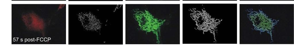
16 Automated quantification of mitochondrial parameters
17 Automated quantification of mitochondrial parameters: Algorithm Koopman et al., 2005, AJP Cell Phys.; Koopman et al., 2006, Cytometry A; Koopman et al., 2007, AJP Cell Phys.; Komen et al., 2007, Cell Mol. Life Sci.; Distelmaier et al., 2008, Cytometry A; Koopman et al., 2008, Methods; Eisenberg et al., 2008, Hum Mol. Gen.; Mortiboys et al., (in press) Ann. Neurol.


18 Automated quantification of mitochondrial parameters
19 How does a spatial filter work?

 and")


20 Meaning of aspect ratio (AR) and formfactor (F) Koopman et al., 2005a, AJP Cell Phys.


21 Effect of noise in the images Koopman et al., 2006, Cytometry A
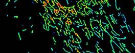
22 Branching mitogram Class Objects% Objects Branching µm Koopman et al., 2005a, AJP Cell Phys. Koopman et al., 2005b, AJP Cell Phys. Koopman et al., 2006, Cytometry A
23 Spatial visualization of mitochondrial area and structure 15 µm 15 µm Mitochondrial area Degree of branching Distelmaier et al. (submitted)
24 Automated determination of mitochondrial position

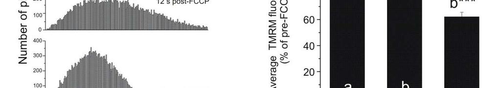
25 Quantification of Ψ during more depolarized conditions Distelmaier et al.,cytometry, 2007


26 Human complex I deficiency: ψ is depolarized Distelmaier et al. (Submitted)
 2D pre-processing")
 Background subtraction (4)")
 3D stack calculation")
 A/V ratio - Shape - Connectivity")
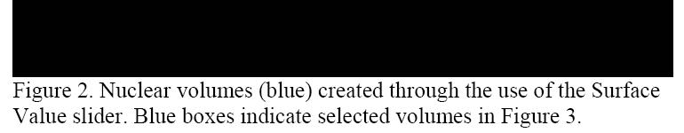
27 BD pathway 855 R123-stained life cells: 2D Z-Stack Optimize contrast (1) 2D pre-processing 3D post-processing Median filter 3x3, S1, P1(2) Optimize contrast (5) Background subtraction (4) - Avg min/max intensity it -Z= No autosharp (3) 3D stack calculation 3D-Stack (6) Quantification - Volume (V) - Surface area (A) A/V ratio - Shape - Connectivity (4D) - Dynamics (4D) - Signaling (4D)
28
29 The next challenge: Quantifying mitochondrial shape and function in multiple dimensions Dimensionality of the data - Spatial dimensions (x,y,z) - Time (t) - Multiple reporters (n 1) - Multiple parameters from same reporter (n 1) Quantification - Volume (V), Surface area (A), A/V ratio - Shape - Colocalization (4D) - Connectivity (4D): Intra/inter mitochondrial, with ER - Dynamics, 3D tracking (4D) - Signaling (4D) Example - Mitochondria stained (GFP), ER stained (RFP): 2 Dimensions - Make 3D stacks in time: 4 Dimensions - Determine co-localization and shape 3 Dimensions


30 Quantifying mitochondrial shape and function in higher dimensions: The right software? Media Cybernetics, 3D Constructor software


31 Quantifying mitochondrial shape and function in higher dimensions: The right software? Media Cybernetics, 3D Constructor software


32 Photoconvertible FPs PA-GFP PS-CFP2 Kaede KikGR Eos Dendra2 Dronpa mtfp0.7 KFP1 Shaner et al., J. Cell Sci., 2007


33 Dendra2 photoconversion mechanism Neutral state Anionic (green) state Red state Red state Anionic (green) state Chudakov et al., Nature Protocols, 2007
Fluorescence Resonance Energy Transfer (FRET) Microscopy
 Microscopy") Fluorescence Resonance Energy Transfer () Microscopy Mike Lorenz Optical Technology Development mlorenz@mpi-cbg.de -FLM course, May 2009 What is fluorescence? Stoke s shift Fluorescence light is always
Fluorescence Resonance Energy Transfer () Microscopy Mike Lorenz Optical Technology Development mlorenz@mpi-cbg.de -FLM course, May 2009 What is fluorescence? Stoke s shift Fluorescence light is always
Mitochondrial Dynamics Is a Distinguishing Feature of Skeletal Muscle Fiber Types and Regulates Organellar Compartmentalization
 Cell Metabolism Supplemental Information Mitochondrial Dynamics Is a Distinguishing Feature of Skeletal Muscle Fiber Types and Regulates Organellar Compartmentalization Prashant Mishra, Grigor Varuzhanyan,
FROM LOCALIZATION TO INTERACTION
 EPFL SV PTBIOP FROM LOCALIZATION TO INTERACTION BIOP COURSE 2015 COLOCALIZATION TYPICAL EXAMPLE EPFL SV PTBIOP Vinculin Alexa568 Actin Alexa488 http://www.olympusconfocal.com/applications/colocalization.html
Supplemental Figures S1 S5
 Beyond reduction of atherosclerosis: PON2 provides apoptosis resistance and stabilizes tumor cells Ines Witte (1), Sebastian Altenhöfer (1), Petra Wilgenbus (1), Julianna Amort (1), Albrecht M. Clement
Co-localization, FRET
 Co-localization, FRET Last class FRAP Diffusion This class Co-localization Correlation FRET Co-localization Can you infer function of protein from it s intracellular location How do you measure if two
Supplemental material
 Supplemental material THE JOURNAL OF CELL BIOLOGY Mourier et al., http://www.jcb.org/cgi/content/full/jcb.201411100/dc1 Figure S1. Size and mitochondrial content in Mfn1 and Mfn2 knockout hearts. (A) Body
4) Please cite Dagda et al J Biol Chem 284: , for any publications or presentations resulting from use or modification of the macro.
 Please cite Dagda et al J Biol Chem 284: , for any publications or presentations resulting from use or modification of the macro.") Supplement Figure S1. Algorithmic quantification of mitochondrial morphology in SH- SY5Y cells treated with known fission/fusion mediators. Parental SH-SY5Y cells were transiently transfected with an empty
Supplement Figure S1. Algorithmic quantification of mitochondrial morphology in SH- SY5Y cells treated with known fission/fusion mediators. Parental SH-SY5Y cells were transiently transfected with an empty
High photostability and enhanced fluorescence of gold nanoclusters by silver doping-supporting information
 High photostability and enhanced fluorescence of gold nanoclusters by silver doping-supporting information Size measurements Figure S1 P2 FTIR measurements Figure S2 P2 XPS measurements Figure S3 P3 Photo-physical
Fluorescence Workshop UMN Physics June 8-10, 2006 Basic Spectroscopic Principles Joachim Mueller
 Fluorescence Workshop UMN Physics June 8-10, 2006 Basic Spectroscopic Principles Joachim Mueller Fluorescence, Light, Absorption, Jablonski Diagram, and Beer-Law First stab at a definition: What is fluorescence?
SUPPLEMENTARY INFORMATION
 Cell viability rate 0.8 0.6 0 0.05 0.1 0.2 0.3 0.4 0.5 0.7 1 Exposure duration (s) Supplementary Figure 1. Femtosecond laser could disrupt and turn off GFP without photons at 473 nm and keep cells alive.
Multiphoton Imaging and Spectroscopy in Cell and Tissue Biophysics. J Moger and C P Winlove
 Multiphoton Imaging and Spectroscopy in Cell and Tissue Biophysics J Moger and C P Winlove Relating Structure to Function Biochemistry Raman microspectrometry Surface enhanced Raman spectrometry (SERS)
Confocal Microscopy Imaging of Single Emitter Fluorescence and Hanbury Brown and Twiss Photon Antibunching Setup
 1 Confocal Microscopy Imaging of Single Emitter Fluorescence and Hanbury Brown and Twiss Photon Antibunching Setup Abstract Jacob Begis The purpose of this lab was to prove that a source of light can be
Practical applications of TIRF microscopy
 Practical applications of TIRF microscopy Evgeny Pryazhnikov University of Helsinki, Neuroscience Center Functional and Morphological Plasticity of the Tripartite Synapse Vesicular release ATP Perisynaptic
BMB Class 17, November 30, Single Molecule Biophysics (II)
") BMB 178 2018 Class 17, November 30, 2018 15. Single Molecule Biophysics (II) New Advances in Single Molecule Techniques Atomic Force Microscopy Single Molecule Manipulation - optical traps and tweezers
BMB 178 2018 Class 17, November 30, 2018 15. Single Molecule Biophysics (II) New Advances in Single Molecule Techniques Atomic Force Microscopy Single Molecule Manipulation - optical traps and tweezers
Measuring Colocalization within Fluorescence Microscopy Images
 from photonics.com: 03/01/2007 http://www.photonics.com/article.aspx?aid=39341 Measuring Colocalization within Fluorescence Microscopy Images Two-color fluorescence-based methods are uncovering molecular
Single-Molecule Methods I - in vitro
 Single-Molecule Methods I - in vitro Bo Huang Macromolecules 2014.03.10 F 1 -ATPase: a case study Membrane ADP ATP Rotation of the axle when hydrolyzing ATP Kinosita group, 1997-2005 Single Molecule Methods
Visualize and Measure Nanoparticle Size and Concentration
 NTA : Nanoparticle Tracking Analysis Visualize and Measure Nanoparticle Size and Concentration 30 Apr 2015 NanoSight product range LM 10 series NS300 series NS500 series Dec 13 34 www.nanosight.com NanoSight
Supporting Information
 Supporting Information Wang et al. 10.1073/pnas.0804871105 SI Materials and Methods Cell Culture and Transfection. Human neuroblastoma M17 cells were grown as described before (1). Transfection was performed
Supplementary Information
 Supplementary Information MAP2/Hoechst Hyp.-AP ph 6.5 Hyp.-SD ph 7.2 Norm.-SD ph 7.2 Supplementary Figure 1. Mitochondrial elongation in cortical neurons by acidosis. Representative images of neuronal
Chapter 6 Photoluminescence Spectroscopy
 Chapter 6 Photoluminescence Spectroscopy Course Code: SSCP 4473 Course Name: Spectroscopy & Materials Analysis Sib Krishna Ghoshal (PhD) Advanced Optical Materials Research Group Physics Department, Faculty
GFP WxTP-GFP WxTP-GFP. stromules. DsRed WxTP-DsRed WxTP-DsRed. stromules
 Supplemental Data. Kitajima et al. (2009). The rice -amylase glycoprotein is targeted from the Golgi apparatus through the secretory pathway to the plastids. A GFP WxTP-GFP WxTP-GFP stromules stromules
Reconstructing Mitochondrial Evolution?? Morphological Diversity. Mitochondrial Diversity??? What is your definition of a mitochondrion??
 Reconstructing Mitochondrial Evolution?? What is your definition of a mitochondrion?? Morphological Diversity Mitochondria as we all know them: Suprarenal gland Liver cell Plasma cell Adrenal cortex Mitochondrial
T H E J O U R N A L O F C E L L B I O L O G Y
 T H E J O U R N A L O F C E L L B I O L O G Y Supplemental material Eisner et al., http://www.jcb.org/cgi/content/full/jcb.201312066/dc1 Figure S1. Mitochondrial continuity in adult skeletal muscle. (A)
Visualization of Xe and Sn Atoms Generated from Laser-Produced Plasma for EUV Light Source
 3rd International EUVL Symposium NOVEMBER 1-4, 2004 Miyazaki, Japan Visualization of Xe and Sn Atoms Generated from Laser-Produced Plasma for EUV Light Source H. Tanaka, A. Matsumoto, K. Akinaga, A. Takahashi
protein biology cell imaging Automated imaging and high-content analysis
 protein biology cell imaging Automated imaging and high-content analysis Automated imaging and high-content analysis Thermo Fisher Scientific offers a complete portfolio of imaging platforms, reagents
SUPPLEMENTARY INFORMATION
 DOI: 10.1038/NPHOTON.2013.97 Supplementary Information Far-field Imaging of Non-fluorescent Species with Sub-diffraction Resolution Pu Wang et al. 1. Theory of saturated transient absorption microscopy
Chemistry 524--Final Exam--Keiderling May 4, :30 -?? pm SES
 Chemistry 524--Final Exam--Keiderling May 4, 2011 3:30 -?? pm -- 4286 SES Please answer all questions in the answer book provided. Calculators, rulers, pens and pencils are permitted. No open books or
(i.e. what you should be able to answer at end of lecture)
") Today s Announcements 1. Test given back next Wednesday 2. HW assigned next Wednesday. 3. Next Monday 1 st discussion about Individual Projects. Today s take-home lessons (i.e. what you should be able
Today s Announcements 1. Test given back next Wednesday 2. HW assigned next Wednesday. 3. Next Monday 1 st discussion about Individual Projects. Today s take-home lessons (i.e. what you should be able
bio-molecular studies Physical methods in Semmelweis University Osváth Szabolcs
 Physical methods in bio-molecular studies Osváth Szabolcs Semmelweis University szabolcs.osvath@eok.sote.hu Light emission and absorption spectra Stokes shift is the difference (in wavelength or frequency
CD Basis Set of Spectra that is used is that derived from comparing the spectra of globular proteins whose secondary structures are known from X-ray
 CD Basis Set of Spectra that is used is that derived from comparing the spectra of globular proteins whose secondary structures are known from X-ray crystallography An example of the use of CD Modeling
Measuring Mitochondrial Membrane Potential with JC-1 Using the Cellometer Vision Image Cytometer
 Measuring Mitochondrial Membrane Potential with JC-1 Using the Cellometer Vision Image Cytometer Nexcelom Bioscience LLC. 360 Merrimack Street, Building 9 Lawrence, MA 01843 T: 978.327.5340 F: 978.327.5341
Computational Biology, Part 24 Clustering and Unmixing of Subcellular Patterns
 Computational Biology, Part 24 Clustering and Unmixing of Subcellular Patterns Robert F. Murphy Copyright 1996, 1999, 2000-2009. All rights reserved. Unsupervised Learning to Identify High-Resolution Protein
Rice/TCU REU on Computational Neuroscience. Fundamentals of Molecular Imaging
 Rice/TCU REU on Computational Neuroscience Fundamentals of Molecular Imaging June 3, 2008 Neal Waxham 713-500-5621 m.n.waxham@uth.tmc.edu Objectives Brief discussion of optical resolution and lasers as
Fluorescence imaging microscopy studies on single molecule diffusion and photophysical dynamics
 Institut für Biophysik Fachrichtung für Physik Fakultät für Mathematik und Naturwissenschaften Technische Universität Dresden K Fluorescence imaging microscopy studies on single molecule diffusion and
Supplementary Figure 1.
 Supplementary Figure 1. Characterisation of IHG-1 overexpressing and knockdown cell lines. (A) Total cellular RNA was prepared from HeLa cells stably overexpressing IHG-1 or mts-ihg-1. IHG-1 mrna was quantified
DEVELOPMENT OF NANO PARTICLE SIZING SYSTEM USING FLUORESCENCE POLARIZATION
 XX IMEKO World Congress Metrology for Green Growth September 9 14, 2012, Busan, Republic of Korea DEVELOPMENT OF NANO PARTICLE SIZING SYSTEM USING FLUORESCENCE POLARIZATION Terutake Hayashi, Masaki Michihata,
SCI. Scientific Computing International. Scientific Computing International. FilmTek. Raising Thin Film Metrology Performance to a New Level
 FilmTek Raising Thin Film Metrology Performance to a New Level 1 Through Silicon Via (TSV) Metrology FilmTek TM TM TSV TSV Metrology Advantages Measure high aspect ratio TSV structures (up to 30:1) Measure
Laboratory 3: Confocal Microscopy Imaging of Single Emitter Fluorescence and Hanbury Brown, and Twiss Setup for Photon Antibunching
 Laboratory 3: Confocal Microscopy Imaging of Single Emitter Fluorescence and Hanbury Brown, and Twiss Setup for Photon Antibunching Jonathan Papa 1, * 1 Institute of Optics University of Rochester, Rochester,
Fluorescence Polarization Anisotropy FPA
 Fluorescence Polarization Anisotropy FPA Optics study of light Spectroscopy = light interacts the study of the interaction between matter & electro-magnetic radiation matter Spectroscopy Atomic Spectroscopy
Fluorescence Spectroscopy
 Fluorescence Spectroscopy Thomas Schmidt Department of Biophysics Leiden University, The Netherlands tschmidt@biophys.leidenuniv.nl www.biophys.leidenuniv.nl/research/fvl Biophysical Structural Biology
Enhancement of Exciton Transport in Porphyrin. Aggregate Nanostructures by Controlling. Hierarchical Self-Assembly
 Electronic Supplementary Material (ESI) for Nanoscale. This journal is The Royal Society of Chemistry 2018 Supporting Information for Enhancement of Exciton Transport in Porphyrin Aggregate Nanostructures
Two-photon single-beam particle trapping of active micro-spheres
 Two-photon single-beam particle trapping of active micro-spheres Dru Morrish, Xiaosong Gan and Min Gu * Centre for Mirco-Photonics, School of Biophysical Sciences and Electrical Engineering, Swinburne
Multiphoton microscopy
 Multiphoton microscopy Joonas Holmi ELEC October 6, 2016 Multiphoton microscopy 1. 2. 3. 4. Multiphoton microscopy 2/14 Intro: Multiphoton microscopy Nonlinear optical characterization method Pulsed laser
Instrumental Limitations for High Absorbance Measurements in Noise
 TECHNICAL NOTE Instrumental Limitations for High Absorbance Measurements in Noise UV/Vis/NIR Spectroscopy Author: Jeffrey L. Taylor, Ph.D. PerkinElmer, Inc. Shelton, CT Spectrophotometer Energy Using a
Effects of Temperature and Concentration on the Rate of Photo-bleaching of Erythrosine in Water
 Supporting Information for: Effects of Temperature and Concentration on the Rate of Photo-bleaching of Erythrosine in Water Joshua K. G. Karlsson, Owen J. Woodford, Roza Al-Aqar and Anthony Harriman* Molecular
Optics and Spectroscopy
 Introduction to Optics and Spectroscopy beyond the diffraction limit Chi Chen 陳祺 Research Center for Applied Science, Academia Sinica 2015Apr09 1 Light and Optics 2 Light as Wave Application 3 Electromagnetic
χ (3) Microscopic Techniques
 Microscopic Techniques") χ (3) Microscopic Techniques Quan Wang Optical Science and Engineering University of New Mexico Albuquerque, NM 87131 Microscopic techniques that utilize the third order non-linearality (χ (3) ) of the
χ (3) Microscopic Techniques Quan Wang Optical Science and Engineering University of New Mexico Albuquerque, NM 87131 Microscopic techniques that utilize the third order non-linearality (χ (3) ) of the
Homo-FRET detection in wide-field, steady-state microscopy, with mcherry as labeling fluorophore
 University Utrecht Debey Institute, Biophysics Homo-FRET detection in wide-field, steady-state microscopy, with mcherry as labeling fluorophore Author: Margriet Oomen Supervisors: prof. dr. H.C. Gerritsen
Electronic Supplementary Information
 Electronic Supplementary Material (ESI) for Journal of Materials Chemistry C. This journal is The Royal Society of Chemistry 2017 Electronic Supplementary Information Polymorphism and microcrystal shape
Single Molecule Spectroscopy and Imaging
 Single Molecule Spectroscopy and Imaging Ingo Gregor, Thomas Dertinger, Iris von der Hocht, Jan Sykora, Luru Dai, Jörg Enderlein Institute for Biological Information Processing 1 Forschungszentrum Jülich
Chemistry 2. Molecular Photophysics
 Chemistry 2 Lecture 12 Molecular Photophysics Assumed knowledge Electronic states are labelled using their spin multiplicity with singlets having all electron spins paired and triplets having two unpaired
Supplementary Materials
 Supplementary Materials Sample characterization The presence of Si-QDs is established by Transmission Electron Microscopy (TEM), by which the average QD diameter of d QD 2.2 ± 0.5 nm has been determined
Optical Spectroscopy. Steady State and Time Dependent Fluorescence Measurements. Kai Wen Teng. October 8 th PHYS 403 Fall 2013
 Optical Spectroscopy Steady State and Time Dependent Fluorescence Measurements Kai Wen Teng October 8 th 2013 PHYS 403 Fall 2013 EM Spectrum of molecules Rotational Energy Infrared Vibrational Energy Near
Fluorescence fluctuation microscopy: FRAP and FCS
 Fluorescence fluctuation microscopy: FRAP and FCS Confocal laser scanning fluorescence microscope for the mobility analysis in living cells Ensemble measurements of protein mobility and interactions pre
SUPPLEMENTARY INFORMATION
 DOI: 10.1038/ncb2215 Figure S1 Number of egfp-vps4a bursts versus cellular expression levels. The total number of egfp-vps4a bursts, counted at the end of each movie (frame 2000, after 1h 28 min) are plotted
Neurobiology Biomed 509 Ion Channels
 eurobiology Biomed 509 Ion hannels References: Luo 2.4, 2.4 2., (3.20 3.2), M 2.8 I. ores vs. carriers In order for hydrophilic ions to transit the hydrophobic membrane, they need to have a specific pathway
Brownian Motion and single particle tracking
 Brownian Motion and single particle tracking Lab Course B2 in the Lab NU115 Prof. Dr. Dieter Braun Sommer-Semester 2015 Figure 1: Fluorescent particles under a fluorescence microscope and their tracks
Fluorescence (Notes 16)
") Fluorescence - 2014 (Notes 16) XV 74 Jablonski diagram Where does the energy go? Can be viewed like multistep kinetic pathway 1) Excite system through A Absorbance S 0 S n Excite from ground excited singlet
Fluorescence - 2014 (Notes 16) XV 74 Jablonski diagram Where does the energy go? Can be viewed like multistep kinetic pathway 1) Excite system through A Absorbance S 0 S n Excite from ground excited singlet
Fluorescence polarisation, anisotropy FRAP
 Fluorescence polarisation, anisotropy FRAP Reminder: fluorescence spectra Definitions! a. Emission sp. b. Excitation sp. Stokes-shift The difference (measured in nm) between the peak of the excitation
Refractive index determination of single sub micrometer vesicles in suspension using dark field microscopy
 Refractive index determination of single sub micrometer vesicles in suspension using dark field microscopy Edwin van der Pol 1,2 Frank Coumans 1,2, Anita Böing 2, Auguste Sturk 2, Rienk Nieuwland 2, and
Combining High Resolution Optical and Scanning Probe Microscopy
 Combining High Resolution Optical and Scanning Probe Microscopy Fernando Vargas WITec, Ulm, Germany www.witec.de Company Background Foundation 1997 by O. Hollricher, J. Koenen, K. Weishaupt WITec = Wissenschaftliche
Modular Microscope Accessory
 Modular Microscope Accessory Modular Microscope Accessory SAMPLE STRUCTURE OBSERVATION DHR MMA Key Features Compact, modular design that directly installs to the DHR frame for easy alignment and minimal
Fluorescence Spectroscopy
 Fluorescence Spectroscopy Steady State and Time Dependent Fluorescence Measurements Kai Wen Teng PHYS 403 Fall 15 EM Spectrum of molecules Rotational Energy Infrared Vibrational Energy Near Infrared Electronic
Nature Protocols: doi: /nprot Supplementary Figure 1
 Supplementary Figure 1 Photographs of the 3D-MTC device and the confocal fluorescence microscopy. I: The system consists of a Leica SP8-Confocal microscope (with an option of STED), a confocal PC, a 3D-MTC
Solution set for EXAM IN TFY4265/FY8906 Biophysical microtechniques
 ENGLISH NORWEGIAN UNIVERSITY OF SCIENCE AND TECHNOLOGY DEPARTMENT OF PHYSICS Contact during exam: Magnus Borstad Lilledahl Telefon: 73591873 (office) 92851014 (mobile) Solution set for EXAM IN TFY4265/FY8906
Nanoparticle Toxicity Assessment in a Bacterial Model
 Nanoparticle Toxicity Assessment in a Bacterial Model Ian L. Gunsolus, Dehong Hu, Cosmin Mihai, William B. Chrisler, Samuel E. Lohse, Marco D. Torelli, Robert J. Hamers, Catherine J. Murphy, Galya Orr,
Visualizing the bi-directional electron transfer in a Schottky junction consisted of single CdS nanoparticles and a planar gold film
 Electronic Supplementary Material (ESI) for Chemical Science. This journal is The Royal Society of Chemistry 2017 Electronic Supplementary Information Visualizing the bi-directional electron transfer in
For more information, please contact: or +1 (302)
") Introduction Graphene Raman Analyzer: Carbon Nanomaterials Characterization Dawn Yang and Kristen Frano B&W Tek Carbon nanomaterials constitute a variety of carbon allotropes including graphene, graphene
Introduction Graphene Raman Analyzer: Carbon Nanomaterials Characterization Dawn Yang and Kristen Frano B&W Tek Carbon nanomaterials constitute a variety of carbon allotropes including graphene, graphene
Ultrafast Dynamics and Single Particle Spectroscopy of Au-CdSe Nanorods
 Supporting Information Ultrafast Dynamics and Single Particle Spectroscopy of Au-CdSe Nanorods G. Sagarzazu a, K. Inoue b, M. Saruyama b, M. Sakamoto b, T. Teranishi b, S. Masuo a and N. Tamai a a Department
Lecture 3: Light absorbance
 Lecture 3: Light absorbance Perturbation Response 1 Light in Chemistry Light Response 0-3 Absorbance spectrum of benzene 2 Absorption Visible Light in Chemistry S 2 S 1 Fluorescence http://www.microscopyu.com
Advanced Spectroscopy Laboratory
 Advanced Spectroscopy Laboratory - Raman Spectroscopy - Emission Spectroscopy - Absorption Spectroscopy - Raman Microscopy - Hyperspectral Imaging Spectroscopy FERGIELAB TM Raman Spectroscopy Absorption
Laser induced fluorescence
 Report experiment 1 by 4-7 February 22, id#471917 Summary Several measurements are made around laser induced fluorescence (LIF). First the system was calibrated. The resolution of the spectrograph was
single-molecule fluorescence resonance energy transfer
 single-molecule fluorescence resonance energy transfer (2) determing the Förster radius: quantum yield, donor lifetime, spectral overlap, anisotropy michael börsch 26/05/2004 1 fluorescence (1) absorbance
Introduction to Nanoparticle Tracking Analysis (NTA) Measurement Principle of ZetaView
 Measurement Principle of ZetaView") Technical Note Nanoparticle Tracking Key words: Introduction to Nanoparticle Tracking Analysis (NTA) Measurement Principle of ZetaView Particle characterization, Nanoparticle Tracking Analysis (NTA), Brownian
Technical Note Nanoparticle Tracking Key words: Introduction to Nanoparticle Tracking Analysis (NTA) Measurement Principle of ZetaView Particle characterization, Nanoparticle Tracking Analysis (NTA), Brownian
Rapid hyperspectral, vibrationally resonant sum-frequency generation microscopy
 Rapid hyperspectral, vibrationally resonant sum-frequency generation microscopy Adam M. Hanninen a and Eric O. Potma b a Department of Astronomy and Physics, University of California, Irvine, CA 92697,
EUV Reflectivity measurements on Acktar Sample Magic Black
 Report EUV Reflectivity measurements on Acktar Sample Magic Black S. Döring, Dr. K. Mann Laser-Laboratorium Göttingen e.v. October 28, 2011 Contents 1 Introduction 3 2 Setup 3 3 Measurements 4 4 Conclusion
SUPPLEMENTARY INFORMATION
 SUPPLEMENTARY INFORMATION Electroluminescence from a single nanotube-molecule-nanotube junction Christoph W. Marquardt, Sergio Grunder, Alfred Błaszczyk, Simone Dehm, Frank Hennrich, Hilbert v. Löhneysen,
Femtosecond laser microfabrication in. Prof. Dr. Cleber R. Mendonca
 Femtosecond laser microfabrication in polymers Prof. Dr. Cleber R. Mendonca laser microfabrication focus laser beam on material s surface laser microfabrication laser microfabrication laser microfabrication
Correlative Raman Imaging of Polymeric Materials
 APPLICATION NOTE Correlative Raman Imaging of Polymeric Materials WITec GmbH, Lise-Meitner-Str. 6, 89081 Ulm, Germany phone+49 (0) 731 140 700, fax +49 (0) 731 140 70 200 info@witec.de, www.witec.de Characterization
Selected measurements with FluoTime 300
 Selected measurements with FluoTime 300 Sebastian Tannert, Peter Kapusta, Felix Koberling, Manoel Veiga, Steffen Rüttinger Uwe Ortmann, Matthias Patting, Marcus Sackrow, Michael Wahl, Rainer Erdmann 12th
Fluorescence Excitation and Emission Fundamentals
 Fluorescence Excitation and Emission Fundamentals Fluorescence is a member of the ubiquitous luminescence family of processes in which susceptible molecules emit light from electronically excited states
The design of an integrated XPS/Raman spectroscopy instrument for co-incident analysis
 The design of an integrated XPS/Raman spectroscopy instrument for co-incident analysis Tim Nunney The world leader in serving science 2 XPS Surface Analysis XPS +... UV Photoelectron Spectroscopy UPS He(I)
for XPS surface analysis
 Thermo Scientific Avantage XPS Software Powerful instrument operation and data processing for XPS surface analysis Avantage Software Atomic Concentration (%) 100 The premier software for surface analysis
Advanced Fluorescence Microscopy I: Fluorescence (Foster) Resonance Energy Transfer
 Resonance Energy Transfer") Advanced Fluorescence Microscopy I: Fluorescence (Foster) Resonance Energy Transfer 3.0 Pax 2.5 2.0 1200 800 400 GFP- Pax GFP-Pax + FATmCherry FAT FAT 1.5 Lifetime (ns) 0 1500 2000 2500 3000 Average lifetime
Advanced Fluorescence Microscopy I: Fluorescence (Foster) Resonance Energy Transfer 3.0 Pax 2.5 2.0 1200 800 400 GFP- Pax GFP-Pax + FATmCherry FAT FAT 1.5 Lifetime (ns) 0 1500 2000 2500 3000 Average lifetime
LABORATORY OF ELEMENTARY BIOPHYSICS
 LABORATORY OF ELEMENTARY BIOPHYSICS Experimental exercises for III year of the First cycle studies Field: Applications of physics in biology and medicine Specialization: Molecular Biophysics Fluorescence
Fundamentals of nanoscience
 Fundamentals of nanoscience Spectroscopy of nano-objects Mika Pettersson 1. Non-spatially resolved spectroscopy Traditionally, in spectroscopy, one is interested in obtaining information on the energy
Chapter 15 Molecular Luminescence Spectrometry
 Chapter 15 Molecular Luminescence Spectrometry Two types of Luminescence methods are: 1) Photoluminescence, Light is directed onto a sample, where it is absorbed and imparts excess energy into the material
Transmission Electron Microscopy
 L. Reimer H. Kohl Transmission Electron Microscopy Physics of Image Formation Fifth Edition el Springer Contents 1 Introduction... 1 1.1 Transmission Electron Microscopy... 1 1.1.1 Conventional Transmission
δf / δx = σ F (N 2 -N 1 ) ΔF~N 2 -N 1
 ΔF~N 2 -N 1") LASER Light Amplification by Stimulated Emission of Radiation BASIC PROPERTIES O LASER RADIATION Spontaneous emission Incoherence in time Incoherence in space Polychromatic light Small energy density Non-polarized
LASER Light Amplification by Stimulated Emission of Radiation BASIC PROPERTIES O LASER RADIATION Spontaneous emission Incoherence in time Incoherence in space Polychromatic light Small energy density Non-polarized
Femtosecond laser applied to biophotonics. Prof. Cleber R. Mendonca
 Femtosecond laser applied to biophotonics Prof. Cleber R. Mendonca introduction short pulse duration ö high intensity (even at low energy) introduction how short is a femtosecond pulse? 1fs= 10-15 s introduction
Low-Frequency Raman Spectra of Carbon Nanotubes Measured with an Astigmatism-Free Schmidt-Czerny-Turner Spectrograph
 Low-Frequency Raman Spectra of Carbon Nanotubes Measured with an Astigmatism-Free Schmidt-Czerny-Turner Spectrograph Abstract Traditional Czerny-Turner (CT) spectrographs suffer from the optical aberration
Signal and Noise Modeling in Confocal Laser Scanning Fluorescence Microscopy
 Signal and Noise Modeling in Confocal Laser Scanning Fluorescence Microscopy Gerlind Herberich 1, Reinhard Windoffer 2, Rudolf E. Leube 2, and Til Aach 1 1 Institute of Imaging and Computer Vision, RWTH
Frontiers in CardioVascular Biology
 Frontiers in CardioVascular Biology Young Investigator Award Mitofusin 2 controls calcium transmission between the SR and mitochondria and regulates the bioenergetic feedback response in cardiac myocytes
Fluorescence Spectroscopy
 Fluorescence Spectroscopy Frequency and time dependent emission Emission and Excitation fluorescence spectra Stokes Shift: influence of molecular vibrations and solvent Time resolved fluorescence measurements
Administrative details:
 Administrative details: Anything from your side? www.photonics.ethz.ch 1 Where do we stand? Optical imaging: Focusing by a lens Angular spectrum Paraxial approximation Gaussian beams Method of stationary
Redox cycling. basic concepts of redox biology
 Redox cycling basic concepts of redox biology, MD PhD Division of Biochemistry Medical Biochemistry and Biophysics Karolinska Institutet Stockholm, Sweden Elias.Arner@ki.se What is redox? Redox: Reduction
Detection of Single Photon Emission by Hanbury-Brown Twiss Interferometry
 Detection of Single Photon Emission by Hanbury-Brown Twiss Interferometry Greg Howland and Steven Bloch May 11, 009 Abstract We prepare a solution of nano-diamond particles on a glass microscope slide
Nonlinear Ultra-fast Focal-point Optics (NUFO) for Biology - Nonlinear Microscopy and Manipulation of Intracellular Structures -
 for Biology - Nonlinear Microscopy and Manipulation of Intracellular Structures -") LASERLAB Foresight Workshop and Users Meeting Trends of Laser Applications in Biology and Biomedicine Creta Maris beach hotel, Heraklion, Crete Greece, October 23-24, 2008 Nonlinear Ultra-fast Focal-point
LASERLAB Foresight Workshop and Users Meeting Trends of Laser Applications in Biology and Biomedicine Creta Maris beach hotel, Heraklion, Crete Greece, October 23-24, 2008 Nonlinear Ultra-fast Focal-point
Edge Radiation IR end-station at ESRF
 X-TIP Workshop Coupling of Synchrotron Radiation IR and X-rays with Tip based Scanning Probe Microscopies 16-18 November 2005 Edge Radiation IR end-station at ESRF Jean Susini European Synchrotron Radiation
Plasma Spectroscopy in ISTTOK
 Plasma Spectroscopy in ISTTOK J. Figueiredo 1, R. B. Gomes 1, T. Pereira 1, H. Fernandes 1, A. Sharakovski 2 1 Associação EURATOM/IST, Centro de Fusão Nuclear, IST, 1049-001 Lisboa, Portugal 2 Association