Combining High Resolution Optical and Scanning Probe Microscopy
|
|
|
- Dinah Amberlynn Dickerson
- 5 years ago
- Views:
Transcription


1 Combining High Resolution Optical and Scanning Probe Microscopy Fernando Vargas WITec, Ulm, Germany

2 Company Background Foundation 1997 by O. Hollricher, J. Koenen, K. Weishaupt WITec = Wissenschaftliche Instrumente und Technologie = Scientific Instruments and Technology WITec today established company with world-wide distribution US branch office since 2002: WITec Instruments 3D confocal image of a phone card chip
3 Company Background


4 Company Background Headquarters Ulm, Germany US office Urbana, IL

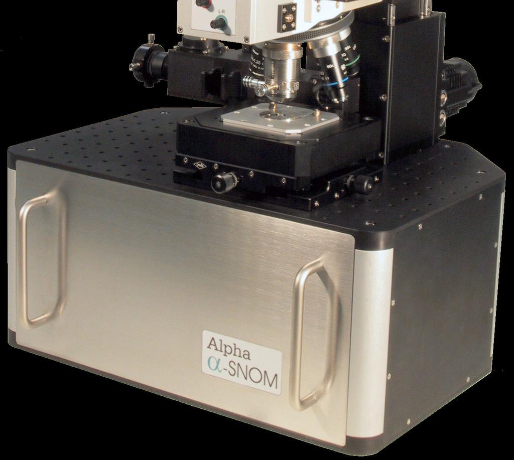
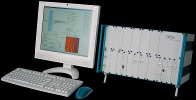
5 Products Scanning Probe Microscopes and Accessories High Resolution Optical Microscopes

6 Atomic Force Microscopy (AFM) position sensitive photo diode laser diode piezoscanner Binnig, Quate, Gerber IBM Lab Rüschlikon 1986 AFM tip cantilever probe sample


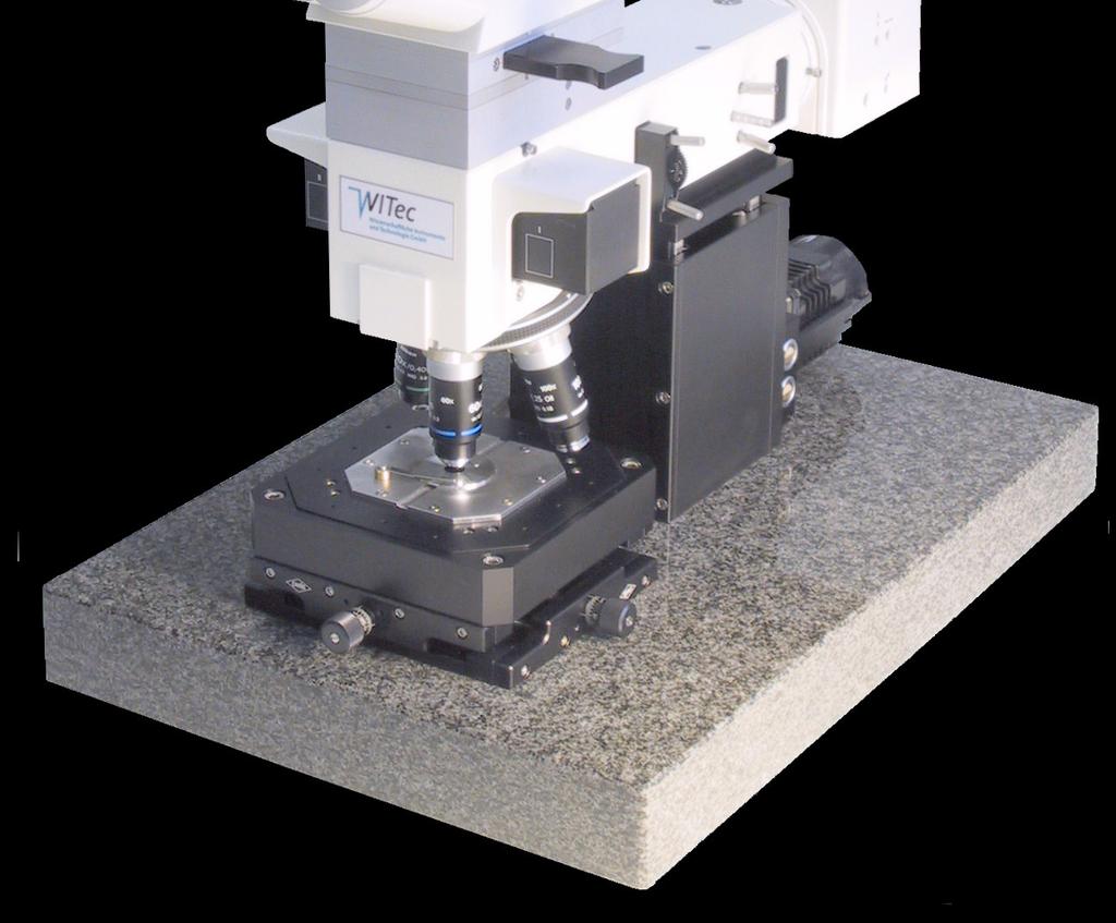
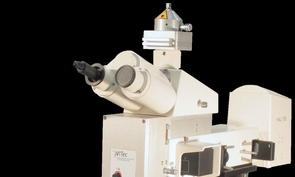
7 end-cap AFM Probe AFM Objective


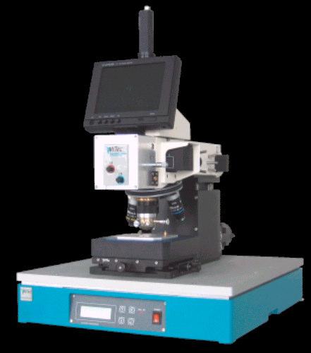
8 Mercury 100 AFM


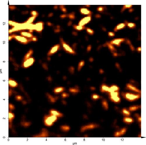
9 HeLa Cells human epithelial carcinoma cells fluoroalcane macromolecules 40x40 microns 400x400 nm


10 MFM - Magnetic Force Microscopy MFM 20 microns scan range Topography MFM 5 microns scan range

11 Pulsed Force Mode free cantilever oscillation


12 Pulsed Force Mode AFM SBS-PMMA polymer blend Topography Stiffness Adhesion 10x10x0.08 µm 3
13 Confocal Raman Microscopy Confocal Microscopy Raman Spectroscopy Confocal Raman Microscopy Examples
14 confocal microscopy detector pinhole classical Laser beamsplitter confocal sample objective focal plane - much smaller background - 3-D information - slightly higher resolution possible
15 Raman Spectroscopy
16 Raman effect: - Some of the incidents photons are able to excite (or anhilate) vibrational quantum states in molecules of the sample. - Each molecule has characteristics vibrational quantums states which are possible to be stimulated.
17 Raman spectrum of Aspirin (acetylsalicylacid) => chemical, but NO spacial resolution
18 Confocal Microscope + Raman Spectrometer => Imaging with chemical sensitivity
19 main problem in Raman microscopy: signal intensity max. intensity: 1-10 mw at the sample 5 2 1mW, 0,5µm spot => 4x10 W/cm time: 128x128 pixel image, 1s/pixel integration time => s = 4,5 h!!! => short integration times needed (ð 100ms / pixel!!!) => system must have highest efficiency
20 CCD APD multi mode fiber video camera spectrometer single mode fiber Super Notch filter holographic beam splitter laser coupler objective XYZ stage
21 Application examples
 samples prepared by A.")
22 biological samples (living) epithelium cells of rat in physiological buffer (RR1022 virus transfected) samples prepared by A. Rück, ILM, Ulm
23 epithelium cells of rat in physiological buffer 100 x 100 pixel = spectra objective: Nikon 60x, NA = 1.0 water 532 nm excitation, 10 mw 100 ms / spectrum => 17 min. single spectrum, 0.1 s integration time CH 2 Proteins: 1450/cm 1660/cm Lipids: 1070/cm 1300/cm 1440/cm / water
 Intensity in the CH 2 streching")
 100 x 100 pixel =")

24 epithelium cells of rat in physiological buffer Video Raman (CH 2) Intensity in the CH 2 streching band ( / cm) Peak position of the CH 2 streching band ( / cm) 100 x 100 pixel = spectra objective: Nikon 60x, NA = 1.0 water 532 nm excitation, 10 mw 100 ms / spectrum => 17 min.
25 epithelium cells of rat in physiological buffer - determination of basic spectra - fit of measured spectra with linear combination of basic spectra
26 epithelium cells of rat in physiological buffer
27 epithelium cells of rat in physiological buffer nucleus endoplasmatic reticulum nucleoli cytoplasm nuclear membrane mitochondriae
, 532nm.")
28 inner coating, Tropicana orange juice container, polymer, different layers 50µm x 100µm scan, 200 x 120= spectra, 50ms. 100 x objective (NA= 0.9), 532nm.

29 Vickers indent (Si) Topography shift of Si Raman line

30 Carbon nanotubes AFM Raman
Correlative Raman Imaging of Polymeric Materials
 APPLICATION NOTE Correlative Raman Imaging of Polymeric Materials WITec GmbH, Lise-Meitner-Str. 6, 89081 Ulm, Germany phone+49 (0) 731 140 700, fax +49 (0) 731 140 70 200 info@witec.de, www.witec.de Characterization
Laboratory 3&4: Confocal Microscopy Imaging of Single-Emitter Fluorescence and Hanbury Brown and Twiss setup for Photon Antibunching
 Laboratory 3&4: Confocal Microscopy Imaging of Single-Emitter Fluorescence and Hanbury Brown and Twiss setup for Photon Antibunching Jose Alejandro Graniel Institute of Optics University of Rochester,
General concept and defining characteristics of AFM. Dina Kudasheva Advisor: Prof. Mary K. Cowman
 General concept and defining characteristics of AFM Dina Kudasheva Advisor: Prof. Mary K. Cowman Overview Introduction History of the SPM invention Technical Capabilities Principles of operation Examples
Scanning Probe Microscopy. Amanda MacMillan, Emmy Gebremichael, & John Shamblin Chem 243: Instrumental Analysis Dr. Robert Corn March 10, 2010
 Scanning Probe Microscopy Amanda MacMillan, Emmy Gebremichael, & John Shamblin Chem 243: Instrumental Analysis Dr. Robert Corn March 10, 2010 Scanning Probe Microscopy High-Resolution Surface Analysis
MSE 321 Structural Characterization
 Auger Spectroscopy Auger Electron Spectroscopy (AES) Scanning Auger Microscopy (SAM) Incident Electron Ejected Electron Auger Electron Initial State Intermediate State Final State Physical Electronics
Industrial Diode Lasers. High-resolution Raman microscopy. Bernhard Wondra, Harald Rossmeier and Thomas Hellerer TOPTICA Photonics AG
 Appl-1006 Industrial Diode Lasers High-resolution Raman microscopy Bernhard Wondra, Harald Rossmeier and Thomas Hellerer TOPTICA Photonics AG Raman microscopy faces a bright future Modern quality assurance
Multiphoton Imaging and Spectroscopy in Cell and Tissue Biophysics. J Moger and C P Winlove
 Multiphoton Imaging and Spectroscopy in Cell and Tissue Biophysics J Moger and C P Winlove Relating Structure to Function Biochemistry Raman microspectrometry Surface enhanced Raman spectrometry (SERS)
Detection of Single Photon Emission by Hanbury-Brown Twiss Interferometry
 Detection of Single Photon Emission by Hanbury-Brown Twiss Interferometry Greg Howland and Steven Bloch May 11, 009 Abstract We prepare a solution of nano-diamond particles on a glass microscope slide
Clark Atlanta University Center for Surface Chemistry and Catalysis Instrument Capabilities
 Center for Surface Chemistry and Catalysis Instrument Capabilities For information contact: Dr. Eric Mintz Research Center for Science and Technology Clark Atlanta University Atlanta, Georgia 30314 Phone:
Laboratory 3: Confocal Microscopy Imaging of Single Emitter Fluorescence and Hanbury Brown, and Twiss Setup for Photon Antibunching
 Laboratory 3: Confocal Microscopy Imaging of Single Emitter Fluorescence and Hanbury Brown, and Twiss Setup for Photon Antibunching Jonathan Papa 1, * 1 Institute of Optics University of Rochester, Rochester,
Confocal Microscope Imaging of Single-Emitter Fluorescence and Photon Antibunching
 Confocal Microscope Imaging of Single-Emitter Fluorescence and Photon Antibunching By Dilyana Mihaylova Abstract The purpose of this lab is to study different types of single emitters including quantum
Optics and Spectroscopy
 Introduction to Optics and Spectroscopy beyond the diffraction limit Chi Chen 陳祺 Research Center for Applied Science, Academia Sinica 2015Apr09 1 Light and Optics 2 Light as Wave Application 3 Electromagnetic
Contents. What is AFM? History Basic principles and devices Operating modes Application areas Advantages and disadvantages
 Contents What is AFM? History Basic principles and devices Operating modes Application areas Advantages and disadvantages Figure1: 2004 Seth Copen Goldstein What is AFM? A type of Scanning Probe Microscopy
Module 26: Atomic Force Microscopy. Lecture 40: Atomic Force Microscopy 3: Additional Modes of AFM
 Module 26: Atomic Force Microscopy Lecture 40: Atomic Force Microscopy 3: Additional Modes of AFM 1 The AFM apart from generating the information about the topography of the sample features can be used
I. Proteomics by Mass Spectrometry 1. What is an internal standard and what does it accomplish analytically?
 Name I. Proteomics by Mass Spectrometry 1. What is an internal standard and what does it accomplish analytically? Internal standards are standards added intentionally to all samples, standards and blanks.
Atomic and molecular interactions. Scanning probe microscopy.
 Atomic and molecular interactions. Scanning probe microscopy. Balázs Kiss Nanobiotechnology and Single Molecule Research Group, Department of Biophysics and Radiation Biology 27. November 2013. 2 Atomic
Confocal Microscopy Imaging of Single Emitter Fluorescence and Hanbury Brown and Twiss Photon Antibunching Setup
 1 Confocal Microscopy Imaging of Single Emitter Fluorescence and Hanbury Brown and Twiss Photon Antibunching Setup Abstract Jacob Begis The purpose of this lab was to prove that a source of light can be
Scanning Probe Microscopy. EMSE-515 F. Ernst
 Scanning Probe Microscopy EMSE-515 F. Ernst 1 Literature 2 3 Scanning Probe Microscopy: The Lab on a Tip by Ernst Meyer,Ans Josef Hug,Roland Bennewitz 4 Scanning Probe Microscopy and Spectroscopy : Theory,
CHARACTERIZATION of NANOMATERIALS KHP
 CHARACTERIZATION of NANOMATERIALS Overview of the most common nanocharacterization techniques MAIN CHARACTERIZATION TECHNIQUES: 1.Transmission Electron Microscope (TEM) 2. Scanning Electron Microscope
Università degli Studi di Bari "Aldo Moro"
 Università degli Studi di Bari "Aldo Moro" Table of contents 1. Introduction to Atomic Force Microscopy; 2. Introduction to Raman Spectroscopy; 3. The need for a hybrid technique Raman AFM microscopy;
LAB 3: Confocal Microscope Imaging of single-emitter fluorescence. LAB 4: Hanbury Brown and Twiss setup. Photon antibunching. Roshita Ramkhalawon
 LAB 3: Confocal Microscope Imaging of single-emitter fluorescence LAB 4: Hanbury Brown and Twiss setup. Photon antibunching Roshita Ramkhalawon PHY 434 Department of Physics & Astronomy University of Rochester
CNPEM Laboratório de Ciência de Superfícies
 Investigating electrical charged samples by scanning probe microscopy: the influence to magnetic force microscopy and atomic force microscopy phase images. Carlos A. R. Costa, 1 Evandro M. Lanzoni, 1 Maria
Solution set for EXAM IN TFY4265/FY8906 Biophysical microtechniques
 ENGLISH NORWEGIAN UNIVERSITY OF SCIENCE AND TECHNOLOGY DEPARTMENT OF PHYSICS Contact during exam: Magnus Borstad Lilledahl Telefon: 73591873 (office) 92851014 (mobile) Solution set for EXAM IN TFY4265/FY8906
Kavli Workshop for Journalists. June 13th, CNF Cleanroom Activities
 Kavli Workshop for Journalists June 13th, 2007 CNF Cleanroom Activities Seeing nm-sized Objects with an SEM Lab experience: Scanning Electron Microscopy Equipment: Zeiss Supra 55VP Scanning electron microscopes
Lecture 4 Scanning Probe Microscopy (SPM)
") Lecture 4 Scanning Probe Microscopy (SPM) General components of SPM; Tip --- the probe; Cantilever --- the indicator of the tip; Tip-sample interaction --- the feedback system; Scanner --- piezoelectric
Lecture 4 Scanning Probe Microscopy (SPM) General components of SPM; Tip --- the probe; Cantilever --- the indicator of the tip; Tip-sample interaction --- the feedback system; Scanner --- piezoelectric
Basic Laboratory. Materials Science and Engineering. Atomic Force Microscopy (AFM)
") Basic Laboratory Materials Science and Engineering Atomic Force Microscopy (AFM) M108 Stand: 20.10.2015 Aim: Presentation of an application of the AFM for studying surface morphology. Inhalt 1.Introduction...
Basic Laboratory Materials Science and Engineering Atomic Force Microscopy (AFM) M108 Stand: 20.10.2015 Aim: Presentation of an application of the AFM for studying surface morphology. Inhalt 1.Introduction...
MSE 321 Structural Characterization
 Auger Spectroscopy Auger Electron Spectroscopy (AES) Scanning Auger Microscopy (SAM) Incident Electron Ejected Electron Auger Electron Initial State Intermediate State Final State Physical Electronics
Nanoscale Chemical Imaging with Photo-induced Force Microscopy
 OG2 BCP39nm_0062 PiFM (LIA1R)Fwd 500 279.1 µv 375 250 nm 500 375 250 125 0 nm 125 219.0 µv Nanoscale Chemical Imaging with Photo-induced Force Microscopy 0 Thomas R. Albrecht, Derek Nowak, Will Morrison,
SUPPLEMENTARY INFORMATION
 DOI: 10.1038/NPHOTON.2013.97 Supplementary Information Far-field Imaging of Non-fluorescent Species with Sub-diffraction Resolution Pu Wang et al. 1. Theory of saturated transient absorption microscopy
Ultrafast Dynamics and Single Particle Spectroscopy of Au-CdSe Nanorods
 Supporting Information Ultrafast Dynamics and Single Particle Spectroscopy of Au-CdSe Nanorods G. Sagarzazu a, K. Inoue b, M. Saruyama b, M. Sakamoto b, T. Teranishi b, S. Masuo a and N. Tamai a a Department
Lab 3 and 4: Single Photon Source
 Lab 3 and 4: Single Photon Source By: Justin Deuro, December 10 th, 2009 Abstract We study methods of single photon emission by exciting single colloidal quantum dot (QD) samples. We prepare the single
Other SPM Techniques. Scanning Probe Microscopy HT10
 Other SPM Techniques Scanning Near-Field Optical Microscopy (SNOM) Scanning Capacitance Microscopy (SCM) Scanning Spreading Resistance Microscopy (SSRM) Multiprobe techniques Electrostatic Force Microscopy,
Instrumentation and Operation
 Instrumentation and Operation 1 STM Instrumentation COMPONENTS sharp metal tip scanning system and control electronics feedback electronics (keeps tunneling current constant) image processing system data
Lab 3-4 : Confocal Microscope Imaging of Single-Emitter Fluorescence and Hanbury-Brown and Twiss Set Up, Photon Antibunching
 Lab 3-4 : Confocal Microscope Imaging of Single-Emitter Fluorescence and Hanbury-Brown and Twiss Set Up, Photon Antibunching Mongkol Moongweluwan 1 1 Department of Physics and Astronomy, University of
Characterization Tools
 Lectures in Nanoscience & Technology Characterization Tools K. Sakkaravarthi Department of Physics National Institute of Technology Tiruchirappalli 620 015 Tamil Nadu India sakkaravarthi@nitt.edu ksakkaravarthi.weebly.com
Vibrational imaging and microspectroscopies based on coherent anti-stokes Raman scattering (CARS)
") Vibrational imaging and microspectroscopies based on coherent anti-stokes Raman scattering (CARS) by Andreas Volkmer Universität Stuttgart 3 rd Institute of Physics, University of Stuttgart, Pfaffenwaldring
Vibrational imaging and microspectroscopies based on coherent anti-stokes Raman scattering (CARS) by Andreas Volkmer Universität Stuttgart 3 rd Institute of Physics, University of Stuttgart, Pfaffenwaldring
Scanning Tunneling Microscopy
 Scanning Tunneling Microscopy Scanning Direction References: Classical Tunneling Quantum Mechanics Tunneling current Tunneling current I t I t (V/d)exp(-Aφ 1/2 d) A = 1.025 (ev) -1/2 Å -1 I t = 10 pa~10na
Optical Properties of CdSe Colloidal Quantum Dots and NV-Nanodiamonds
 Optical Properties of CdSe Colloidal Quantum Dots and NV-Nanodiamonds James MacNeil and Madhu Ashok University of Rochester The Institute of Optics Submitted to Dr. Svetlana Lukishova on 11/20/2013 Abstract:
Two-photon single-beam particle trapping of active micro-spheres
 Two-photon single-beam particle trapping of active micro-spheres Dru Morrish, Xiaosong Gan and Min Gu * Centre for Mirco-Photonics, School of Biophysical Sciences and Electrical Engineering, Swinburne
Nanoscopy with Focused Light
 Nanoscopy with Focused Light Stefan W. Hell Max Planck Institute for Biophysical Chemistry Department of NanoBiophotonics Göttingen & German Cancer Research Center (DKFZ) Optical Nanoscopy Division Heidelberg
Reducing dimension. Crystalline structures
 Reducing dimension 2D surfaces, interfaces and quantum wells 1D carbon nanotubes, quantum wires and conducting polymers 0D nanocrystals, nanoparticles, lithographically patterned quantum dots Crystalline
Lorentz Contact Resonance for viscoelastic measurements of polymer blends
 The nanoscale spectroscopy company The world leader in nanoscale IR spectroscopy Lorentz Contact Resonance for viscoelastic measurements of polymer blends Lorentz Contact Resonance (LCR) reliably compares
bio-molecular studies Physical methods in Semmelweis University Osváth Szabolcs
 Physical methods in bio-molecular studies Osváth Szabolcs Semmelweis University szabolcs.osvath@eok.sote.hu Light emission and absorption spectra Stokes shift is the difference (in wavelength or frequency
Electronic Supplementary Information
 Electronic Supplementary Material (ESI) for Journal of Materials Chemistry C. This journal is The Royal Society of Chemistry 2017 Electronic Supplementary Information Polymorphism and microcrystal shape
SOLID STATE PHYSICS PHY F341. Dr. Manjuladevi.V Associate Professor Department of Physics BITS Pilani
 SOLID STATE PHYSICS PHY F341 Dr. Manjuladevi.V Associate Professor Department of Physics BITS Pilani 333031 manjula@bits-pilani.ac.in Characterization techniques SEM AFM STM BAM Outline What can we use
Atomic Force Microscopy imaging and beyond
 Atomic Force Microscopy imaging and beyond Arif Mumtaz Magnetism and Magnetic Materials Group Department of Physics, QAU Coworkers: Prof. Dr. S.K.Hasanain M. Tariq Khan Alam Imaging and beyond Scanning
Lorentz Contact Resonance for viscoelastic measurements of polymer blends
 The world leader in nanoscale IR spectroscopy for viscoelastic measurements of polymer blends (LCR) reliably compares viscoleastic properties with nanoscale spatial resolution With no moving parts in the
Infrared Spectroscopy: Identification of Unknown Substances
 Infrared Spectroscopy: Identification of Unknown Substances Suppose a white powder is one of the four following molecules. How can they be differentiated? H N N H H H H Na H H H H H A technique that is
Characterization of MEMS Devices
 MEMS: Characterization Characterization of MEMS Devices Prasanna S. Gandhi Assistant Professor, Department of Mechanical Engineering, Indian Institute of Technology, Bombay, Recap Fabrication of MEMS Conventional
RamanStation 400: a Versatile Platform for SERS Analysis
 FIELD APPLICATION REPORT Raman Spectroscopy Author: Dean H. Brown PerkinElmer, Inc. Shelton, CT USA RamanStation 400 RamanStation 400: a Versatile Platform for SERS Analysis Introduction Surface Enhanced
Nanoelectronics 09. Atsufumi Hirohata Department of Electronics. Quick Review over the Last Lecture
 Nanoelectronics 09 Atsufumi Hirohata Department of Electronics 13:00 Monday, 12/February/2018 (P/T 006) Quick Review over the Last Lecture ( Field effect transistor (FET) ): ( Drain ) current increases
Gold nanothorns macroporous silicon hybrid structure: a simple and ultrasensitive platform for SERS
 Supporting Information Gold nanothorns macroporous silicon hybrid structure: a simple and ultrasensitive platform for SERS Kamran Khajehpour,* a Tim Williams, b,c Laure Bourgeois b,d and Sam Adeloju a
Scanning Tunneling Microscopy
 Scanning Tunneling Microscopy References: 1. G. Binnig, H. Rohrer, C. Gerber, and Weibel, Phys. Rev. Lett. 49, 57 (1982); and ibid 50, 120 (1983). 2. J. Chen, Introduction to Scanning Tunneling Microscopy,
Confocal Microscope Imaging of Single emitter fluorescence and Observing Photon Antibunching Using Hanbury Brown and Twiss setup. Lab.
 Submitted for the partial fulfilment of the course PHY 434 Confocal Microscope Imaging of Single emitter fluorescence and Observing Photon Antibunching Using Hanbury Brown and Twiss setup Lab. 3 and 4
Scanning Probe Microscopy (SPM)
") http://ww2.sljus.lu.se/staff/rainer/spm.htm Scanning Probe Microscopy (FYST42 / FAFN30) Scanning Probe Microscopy (SPM) overview & general principles March 23 th, 2018 Jan Knudsen, room K522, jan.knudsen@sljus.lu.se
http://ww2.sljus.lu.se/staff/rainer/spm.htm Scanning Probe Microscopy (FYST42 / FAFN30) Scanning Probe Microscopy (SPM) overview & general principles March 23 th, 2018 Jan Knudsen, room K522, jan.knudsen@sljus.lu.se
Scanning Probe Microscopy (SPM)
") Scanning Probe Microscopy (SPM) Scanning Tunneling Microscopy (STM) --- G. Binnig, H. Rohrer et al, (1982) Near-Field Scanning Optical Microscopy (NSOM) --- D. W. Pohl (1982) Atomic Force Microscopy (AFM)
Scanning Probe Microscopy (SPM) Scanning Tunneling Microscopy (STM) --- G. Binnig, H. Rohrer et al, (1982) Near-Field Scanning Optical Microscopy (NSOM) --- D. W. Pohl (1982) Atomic Force Microscopy (AFM)
High Resolution Laser Microscopy: a fascinating method to explore the molecular world
 High Resolution Laser Microscopy: a fascinating method to explore the molecular world Alfred J. Meixner Physical and Theoretical Chemistry Laboratory University of Siegen Single-molecule spectroscopy and
FEM-SIMULATIONS OF VIBRATIONS AND RESONANCES OF STIFF AFM CANTILEVERS
 FEM-SIMULATIONS OF VIBRATIONS AND RESONANCES OF STIFF AFM CANTILEVERS Kai GENG, Ute RABE, Sigrun HIRSEKORN Fraunhofer Institute for Nondestructive Testing (IZFP); Saarbrücken, Germany Phone: +49 681 9302
Nanoacoustics II Lecture #4 Brillouin scattering
 Nanoacoustics II Lecture #4 Brillouin scattering Dr. Ari Salmi www.helsinki.fi/yliopisto 29.3.2018 1 Last lecture key points Phonons propagate in liquids but not in gases Shear phonons have a minimum frequency
Anti-Bunching from a Quantum Dot
 Anti-Bunching from a Quantum Dot Gerardo I. Viza 1, 1 Department of Physics and Astronomy, University of Rochester, Rochester, NY 14627 We study the nature of non-classical single emitter light experimentally
requency generation spectroscopy Rahul N
 requency generation spectroscopy Rahul N 2-11-2013 Sum frequency generation spectroscopy Sum frequency generation spectroscopy (SFG) is a technique used to analyze surfaces and interfaces. SFG was first
Tracking Iodide and Bromide Ion Segregation in Mixed Halide Lead Perovskites during Photoirradiation
 Supporting Information Tracking Iodide and Bromide Ion Segregation in Mixed Halide Lead Perovskites during Photoirradiation Seog Joon Yoon, 1,2 Sergiu Draguta, 2 Joseph S. Manser, 1,3 Onise Sharia, 3 William
Joshua S. Geller. Department of Physics and Astronomy, University of Rochester, Rochester NY, 14627
 LAB 3-4, PHY434. Single Photon Source: Confocal Microscope Imaging of Single-Emitter Fluorescence and Hanbury Brown and Twiss setup for Photon Antibunching Measurements Joshua S. Geller Department of Physics
The effects of probe boundary conditions and propagation on nano- Raman spectroscopy
 The effects of probe boundary conditions and propagation on nano- Raman spectroscopy H. D. Hallen,* E. J. Ayars** and C. L. Jahncke*** * Physics Department, North Carolina State University, Raleigh, NC
Ecole Franco-Roumaine : Magnétisme des systèmes nanoscopiques et structures hybrides - Brasov, Modern Analytical Microscopic Tools
 1. Introduction Solid Surfaces Analysis Group, Institute of Physics, Chemnitz University of Technology, Germany 2. Limitations of Conventional Optical Microscopy 3. Electron Microscopies Transmission Electron
Single-Molecule Methods I - in vitro
 Single-Molecule Methods I - in vitro Bo Huang Macromolecules 2014.03.10 F 1 -ATPase: a case study Membrane ADP ATP Rotation of the axle when hydrolyzing ATP Kinosita group, 1997-2005 Single Molecule Methods
DOWNLOAD OR READ : INFRARED AND RAMAN SPECTROSCOPY CONCEPTS AND APPLICATIONS PDF EBOOK EPUB MOBI
 DOWNLOAD OR READ : INFRARED AND RAMAN SPECTROSCOPY CONCEPTS AND APPLICATIONS PDF EBOOK EPUB MOBI Page 1 Page 2 infrared and raman spectroscopy concepts and applications infrared and raman spectroscopy
nano-ftir: Material Characterization with Nanoscale Spatial Resolution
 neaspec presents: neasnom microscope nano-ftir: Material Characterization with Nanoscale Spatial Resolution AMC Workshop 2017 6th of June Dr. 2017 Tobias Gokus Company neaspec GmbH leading experts of nanoscale
AFM Imaging In Liquids. W. Travis Johnson PhD Agilent Technologies Nanomeasurements Division
 AFM Imaging In Liquids W. Travis Johnson PhD Agilent Technologies Nanomeasurements Division Imaging Techniques: Scales Proteins 10 nm Bacteria 1μm Red Blood Cell 5μm Human Hair 75μm Si Atom Spacing 0.4nm
Measurements of interaction forces in (biological) model systems
 model systems") Measurements of interaction forces in (biological) model systems Marina Ruths Department of Chemistry, UMass Lowell What can force measurements tell us about a system? Depending on the technique, we might
Measurements of interaction forces in (biological) model systems Marina Ruths Department of Chemistry, UMass Lowell What can force measurements tell us about a system? Depending on the technique, we might
Bringing optics into the nanoscale a double-scanner AFM brings advanced optical experiments within reach
 Bringing optics into the nanoscale a double-scanner AFM brings advanced optical experiments within reach Beyond the diffraction limit The resolution of optical microscopy is generally limited by the diffraction
MSE640: Advances in Investigation of Intermolecular & Surface Forces
 MSE640: Advances in Investigation of Forces Course Title Advances in investigation of Intermolecular & surface forces Course Code MSE640 Credit Hours 3 Pre-requisites (if any) MSE507, MSE508 or equivalent
Laser Tweezers and Other Advanced Physical Methods
 Phys 6715 - Biomedical Physics Laser Tweezers and Other Advanced Physical Methods Yong-qing Li, PhD Department of Physics, East Carolina University Greenville, NC 27858, USA Email: liy@ecu.edu 1 Optical
An Introduction to: Light
 An Introduction to: Light Created by Anna Opitz July 2007 Why is light important? Light allows us to see. Light carries information from our surroundings to our eyes and brain. Light enables us to communicate
Damage to Molecular Solids Irradiated by X-ray Laser Beam
 WDS'11 Proceedings of Contributed Papers, Part II, 247 251, 2011. ISBN 978-80-7378-185-9 MATFYZPRESS Damage to Molecular Solids Irradiated by X-ray Laser Beam T. Burian, V. Hájková, J. Chalupský, L. Juha,
Quantum Optics and Quantum Information Laboratory
 Quantum Optics and Quantum Information Laboratory OPT 253, Fall 2011 Institute of Optics University of Rochester Instructor: Dr. Lukishova Jonathan Papa Contents Lab 1: Entanglement and Bell s Inequalities
Program Operacyjny Kapitał Ludzki SCANNING PROBE TECHNIQUES - INTRODUCTION
 Program Operacyjny Kapitał Ludzki SCANNING PROBE TECHNIQUES - INTRODUCTION Peter Liljeroth Department of Applied Physics, Aalto University School of Science peter.liljeroth@aalto.fi Projekt współfinansowany
Visualizing the bi-directional electron transfer in a Schottky junction consisted of single CdS nanoparticles and a planar gold film
 Electronic Supplementary Material (ESI) for Chemical Science. This journal is The Royal Society of Chemistry 2017 Electronic Supplementary Information Visualizing the bi-directional electron transfer in
The design of an integrated XPS/Raman spectroscopy instrument for co-incident analysis
 The design of an integrated XPS/Raman spectroscopy instrument for co-incident analysis Tim Nunney The world leader in serving science 2 XPS Surface Analysis XPS +... UV Photoelectron Spectroscopy UPS He(I)
The Blue Two Photon Fluorescence Metal Cluster Probe. Precisely Marking Cell Nuclei of Two Cell Lines
 Electronic Supplementary Information The Blue Two Photon Fluorescence Metal Cluster Probe Precisely Marking Cell Nuclei of Two Cell Lines Yaling Wang, a, Yanyan Cui, a, Ru Liu, a Yueteng Wei, a Xinglu
Lecture 0. NC State University
 Chemistry 736 Lecture 0 Overview NC State University Overview of Spectroscopy Electronic states and energies Transitions between states Absorption and emission Electronic spectroscopy Instrumentation Concepts
1. Fabrication. Lukáš Ondič a, Marian Varga a, Karel Hruška a, Jan Fait a,b and Peter Kapusta c
 Supporting information to Enhanced Extraction of Silicon-Vacancy Centers Light Emission Using Bottom-Up Engineered Polycrystalline Diamond Photonic Crystal Slabs Lukáš Ondič a, Marian Varga a, Karel Hruška
Survey on Laser Spectroscopic Techniques for Condensed Matter
 Survey on Laser Spectroscopic Techniques for Condensed Matter Coherent Radiation Sources for Small Laboratories CW: Tunability: IR Visible Linewidth: 1 Hz Power: μw 10W Pulsed: Tunabality: THz Soft X-ray
Labs 3-4: Single-photon Source
 Labs 3-4: Single-photon Source Lab. 3. Confocal fluorescence microscopy of single-emitter Lab. 4. Hanbury Brown and Twiss setup. Fluorescence antibunching 1 Labs 3-4: Single-photon Source Efficiently produces
STM: Scanning Tunneling Microscope
 STM: Scanning Tunneling Microscope Basic idea STM working principle Schematic representation of the sample-tip tunnel barrier Assume tip and sample described by two infinite plate electrodes Φ t +Φ s =
Fundamentals of nanoscience
 Fundamentals of nanoscience Spectroscopy of nano-objects Mika Pettersson 1. Non-spatially resolved spectroscopy Traditionally, in spectroscopy, one is interested in obtaining information on the energy
Vibrational imaging of tablets by epi-detected stimulated Raman scattering microscopy
 Vibrational imaging of tablets by epi-detected stimulated Raman scattering microscopy Supplementary Information Mikhail N. Slipchenko, a Hongtao Chen, b David R. Ely, c Yookyung Jung, d M. Teresa Carvajal,*
Electromagnetic Radiation and Scientific Instruments. PTYS April 1, 2008
 Electromagnetic Radiation and Scientific Instruments PTYS 206-2 April 1, 2008 Announcements Deep Impact 6 PM Wednesday Night Pizza, no beer Watch at home if you can t watch here. It will be discussed in
Morphology-dependent resonance induced by two-photon excitation in a micro-sphere trapped by a femtosecond pulsed laser
 Morphology-dependent resonance induced by two-photon excitation in a micro-sphere trapped by a femtosecond pulsed laser Dru Morrish, Xiaosong Gan and Min Gu Centre for Micro-Photonics, School of Biophysical
Improving nano-scale imaging of of intergrated micro-raman/afm systems using negativestiffness
 See vibration isolation technology @ www.minusk.com?pdf) Electronic Products and Technology - May 2014 Improving nano-scale imaging of of intergrated micro-raman/afm systems using negativestiffness vibration
Scanning Force Microscopy
 Scanning Force Microscopy Roland Bennewitz Rutherford Physics Building 405 Phone 398-3058 roland.bennewitz@mcgill.ca Scanning Probe is moved along scan lines over a sample surface 1 Force Microscopy Data
Supplementary Figure 1 Comparison of single quantum emitters on two type of substrates:
 Supplementary Figure 1 Comparison of single quantum emitters on two type of substrates: a, Photoluminescence (PL) spectrum of localized excitons in a WSe 2 monolayer, exfoliated onto a SiO 2 /Si substrate
Administrative details:
 Administrative details: Anything from your side? www.photonics.ethz.ch 1 Where do we stand? Optical imaging: Focusing by a lens Angular spectrum Paraxial approximation Gaussian beams Method of stationary
Vibrational Spectroscopies. C-874 University of Delaware
 Vibrational Spectroscopies C-874 University of Delaware Vibrational Spectroscopies..everything that living things do can be understood in terms of the jigglings and wigglings of atoms.. R. P. Feymann Vibrational
Characterization of Materials with a Combined AFM/Raman Microscope
 Application Note 089 short Characterization of Materials with a Combined AFM/Raman Microscope Marko Surtchev 1, Sergei Magonov 1 and Mark Wall 2 1 NT-MDT America, Tempe, AZ U.S.A. 2 Thermo Fisher Scientific,
Laser heating of noble gas droplet sprays: EUV source efficiency considerations
 Laser heating of noble gas droplet sprays: EUV source efficiency considerations S.J. McNaught, J. Fan, E. Parra and H.M. Milchberg Institute for Physical Science and Technology University of Maryland College
Analyzer for Determination of the Composition of Deposits in Nuclear Power Plants
 A Portable Remote Chemical Analyzer for Determination of the Composition of Deposits in Nuclear Power Plants Jack Roberts 1, 1) Symphotic TII Corporation 1 Introduction A large electricity producer identified
TEOS characterization of 2D materials from graphene to TMDCs
 Marc Chaigneau Yoshito Okuno, Andrey Krayev, Filippo Fabbri HORIBA Scientific AIST-NT Inc. IMEM-CNR Institute TEOS characterization of 2D materials from graphene to TMDCs 30-03-2017 Graphene2017 2015 2017
Characterization of MEMS Devices
 MEMS: Characterization Characterization of MEMS Devices Prasanna S. Gandhi Assistant Professor, Department of Mechanical Engineering, Indian Institute of Technology, Bombay, Recap Characterization of MEMS
Quantum and Nano Optics Laboratory. Jacob Begis Lab partners: Josh Rose, Edward Pei
 Quantum and Nano Optics Laboratory Jacob Begis Lab partners: Josh Rose, Edward Pei Experiments to be Discussed Lab 1: Entanglement and Bell s Inequalities Lab 2: Single Photon Interference Labs 3 and 4: