Single Molecule Spectroscopy and Imaging
|
|
|
- Aubrey Butler
- 6 years ago
- Views:
Transcription


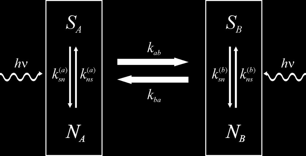
1 Single Molecule Spectroscopy and Imaging Ingo Gregor, Thomas Dertinger, Iris von der Hocht, Jan Sykora, Luru Dai, Jörg Enderlein Institute for Biological Information Processing 1 Forschungszentrum Jülich


2 Motivation Distribution functions of molecular parameters (photo-physics, enzymatic activity, binding affinity) Cellular and molecular biology studies (cell signaling, membrane dynamics) Ultra-sensitive chemical analysis (drug screening, medical diagnostics)
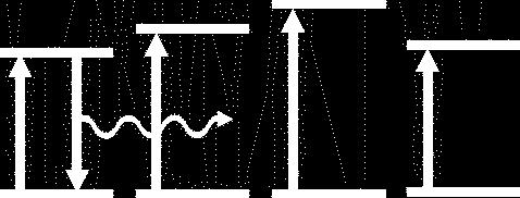
3 Jablonski Scheme of Fluorescence Photobleaching S1 Excitation T1 Fluorescence Emission S0
4 Main challenge of single molecule detection: Raman and Rayleigh scattering High-efficient optical filters Minimizing detection volume Long wavelength dyes Background ~ V Background ~ λ-4
5 Absoprtion Spectra of Standard Dyes and Autofluorescent Biomolecules Furan η Coumarine Fluorescein Rhodamine Oxazine Cyanine fl Thyrosin Coproporphyrine / Protoporphyrine Tryptophan Elastin Chlorophyll Collagen Flavins Wavelength (nm) Courtesy: Christoph Zander Uni GH Siegen
6 Fluorescence Correlation Spectroscopy

7 Confocal Fluorescence Microscopy


8 Principle of Confocal Detection Objective Dichroic mirror Tube lens Confocal aperture Towards detector

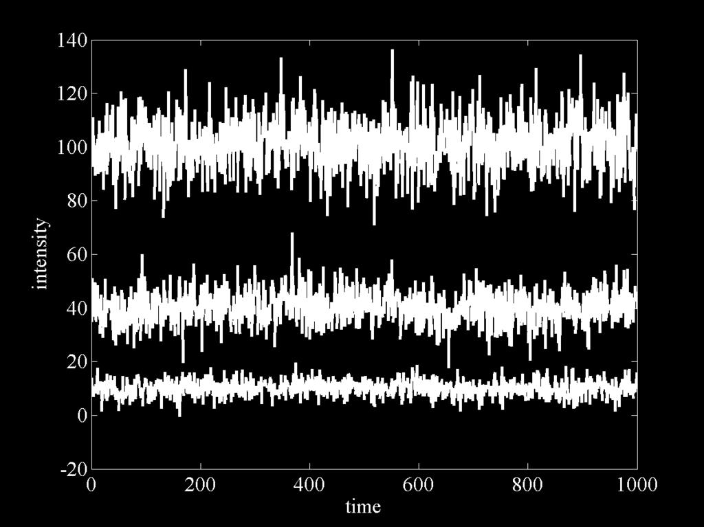
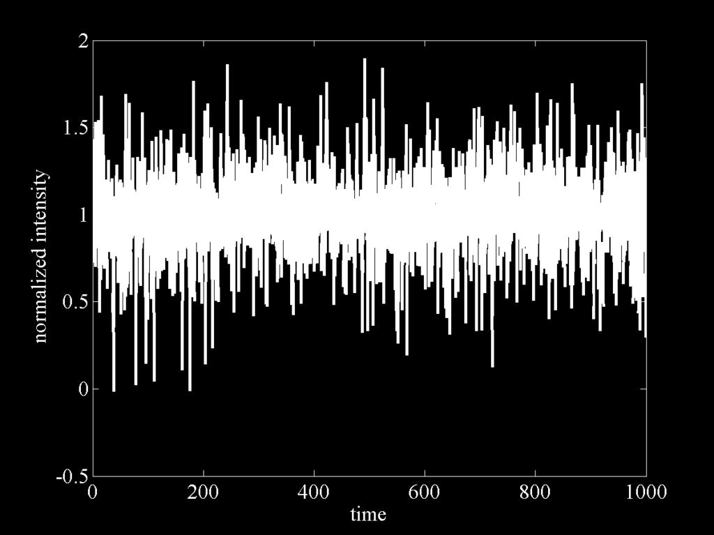
9 Fluorescence Intensity Fluctuations
10 Fluorescence Intensity Fluctuations: Autocorrelation

11 Fluorescence Intensity Fluctuations: Autocorrelation
12 Fluorescence Intensity Fluctuations: Autocorrelation

13 Structure of an autocorrelation curve

14 Example: Measured FCS curves of yellow fluorescent protein

15 Amplitude of an autocorrelation curve

16 Normalized amplitude of an autocorrelation curve

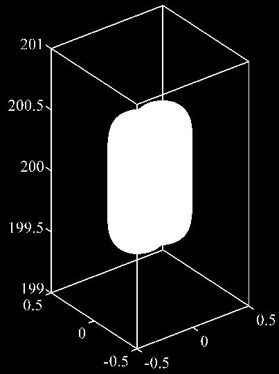
17 Ideal molecule detection function Molecule detection function (1/e2 isosurface) NA = 1.2 wd = 3 mm tubelens = 180 mm n0 = 1.33 λex = 635 nm ω = 4.9 mm focus pos. = 10 µm λem = 670 nm magn. = 60 pinhole radius = 50 µm

18 Cover-slide thickness deviation

19 Refractive index mismatch
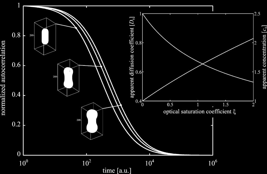
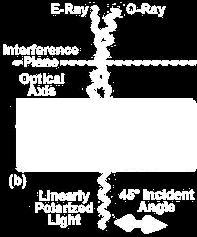
20 Optical saturation
21 Intensity dependence of FCS (Alexa633) 30 µw 100 µw 300 µw 1 autocorrelation [a.u.] time [s]
22 Pulsed versus cw-excitation (Alexa633) 2.4 x 10⁻⁶ pulsed 635 nm cw 647 nm 2 2 apparent diffusion [cm /s] cw excitation power [µw]


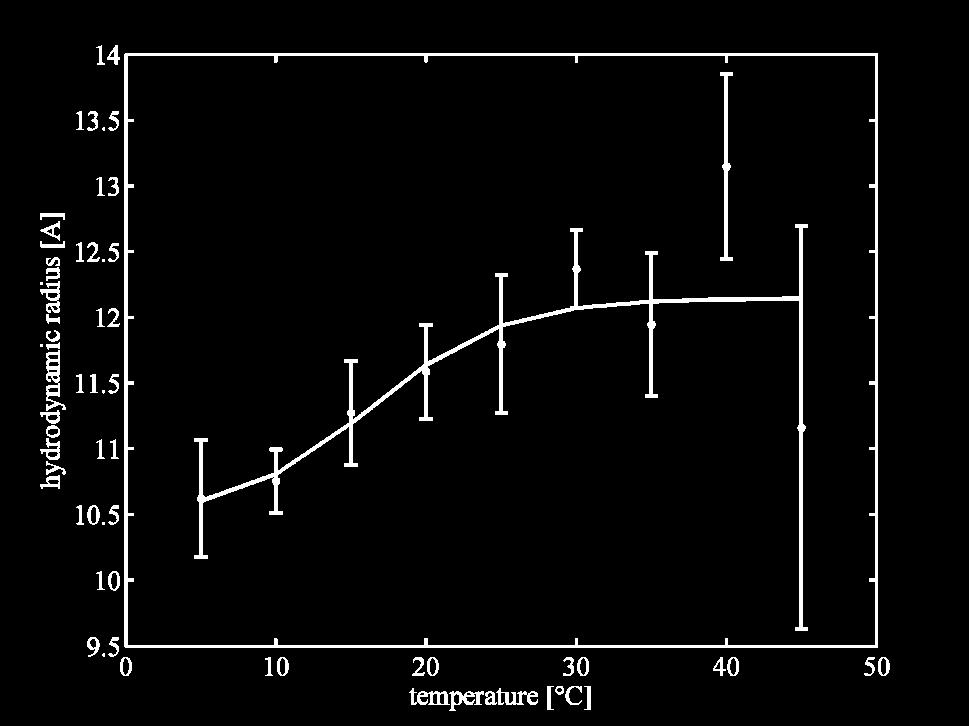
23 Laser beam width and detection volume


24 2-focus confocal system
25 Time-tagged time-resolved mode of photon counting Frequency Fluorescence decay curve Data: t1 Laser pulse Decay time (ns) 15 t2 t3 t4 t5 t6 t7 t8 τ1 τ2 τ3 τ4 τ5 τ6 τ7 τ8
26 PIE: Pulsed interleaved excitation A Photon counts [a.u.] B A 0 5 B Time [ns] 20 25


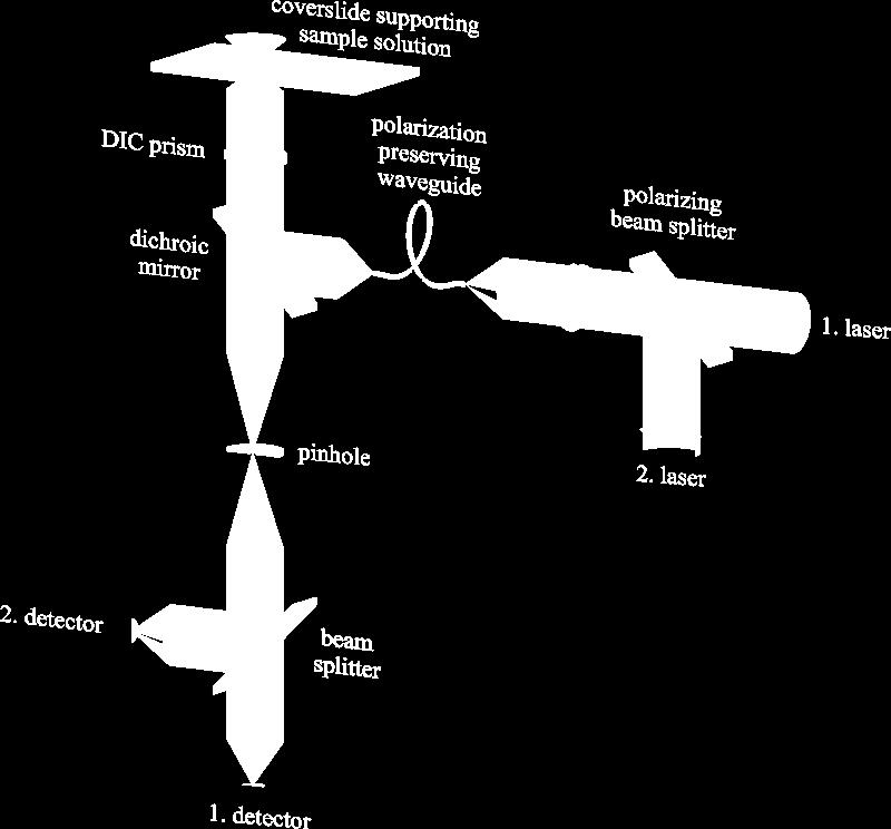
27 Absolute FCS: two mutually shifted detection volumes

28 2fFCS of Atto655 in GdHCl: refractive index dependence

29 2fFCS: optical saturation dependence

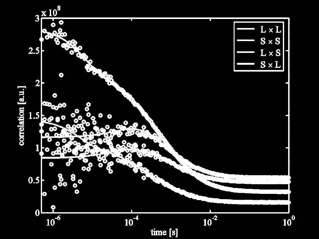
30 Hard application of 2fFCS: 2+ Ca -binding of Calmodulin
31 2+ Ca -binding of Calmodulin: Hydrodynamic radius

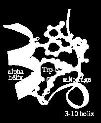
32 Protein folding/unfolding: Tryptophan cage

 Intensity")
33 Measuring fast conformational fluctuations of biomolecules Time scale of interest: nanoseconds up to milliseconds Probes: Förster resonance energy transfer Electron transfer Reporter: (i) Intensity (ii) Lifetime
34 Tryptophan induced fluorescence quenching of dye Atto I0/I τ/τ0 10 N 1.0 N O Trp [mm] O OH 60 N
35 Conformational dynamics of small peptide k+ k hν hν k0 k+ k k0
36 Conformational dynamics of small peptide (binding epitope of p53-antibody) 1 k + = 120 ns 1 k = 267 ns Pexc = 4 mw Pexc = 400 µw
37 Time-tagged time-resolved mode of photon counting Frequency Fluorescence decay curve Data: t1 Laser pulse Decay time (ns) 15 t2 t3 t4 t5 t6 t7 t8 τ1 τ2 τ3 τ4 τ5 τ6 τ7 τ8

38 FLCS Fluorescence lifetime correlation spectroscopy

39 FLCS Fluorescence lifetime correlation spectroscopy

40 FLCS: Working principle

41 FLCS Fluorescence lifetime correlation spectroscopy

42 Bi-exponential lifetime of a Cy5-streptavidin conjugate
43 FLCS of Cy5-Streptavidin
44 FLCS of Cy5-Streptavidin
45 FLCS of Cy5-Streptavidin 1.2 µ s τ = 1.7 ns τ = 0.7 ns A > 90 % A < 10 % 0.91 µ s 0.23 µ s 0.23 µ s 3.5 µ s 3.5 µ s 1.2 µ s dark state dark state 0.91 µ s
46 Single Molecule Imaging


47 Fluorescing molecule as an electric dipole Negative charge Amplitude Orientation Positive charge


48 The electric dipole: Near field, far field, and virtual photons Oscillating dipole is surrounded by virtual photons that are damped with increasing distance from the dipole. During return to the ground state, a propagating photons is emitted carrying away the excited state energy.


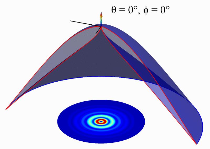
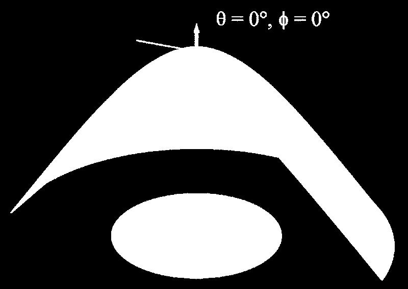
49 Angular distribution of emission Angular distribution of emitted radiation is given by the classical sin2θ law. In the quantum mechanical picture, the classical angular distribution of radiation corresponds to a probability of emitting a photon into a given direction.

50 Tunneling of evanescent modes into optically denser medium: Vertical dipole case upper medium n1 = 1.33 lower medium n2 = 1.33
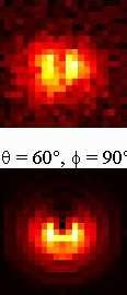
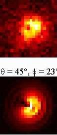
51 Tunneling of evanescent modes into optically denser medium: Vertical dipole case

52 Emission into glass from a fluorescent molecule crossing a water/glass interface

53 Lifetime of fluorescent molecule crossing a water/glass interface

54 Collection efficiency of oil immersion microscope objective

55 Angular distribution of single molecules on glass surface
56 Defocused imaging of single molecules Microscope Table Oil Immersion 1.4 NA, 100 x PiFoc Dichroic Mirror Emission Filter Excitation/ Polarization Filter Tube Lens CCD KrAr nm
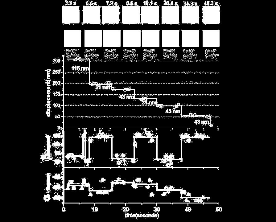
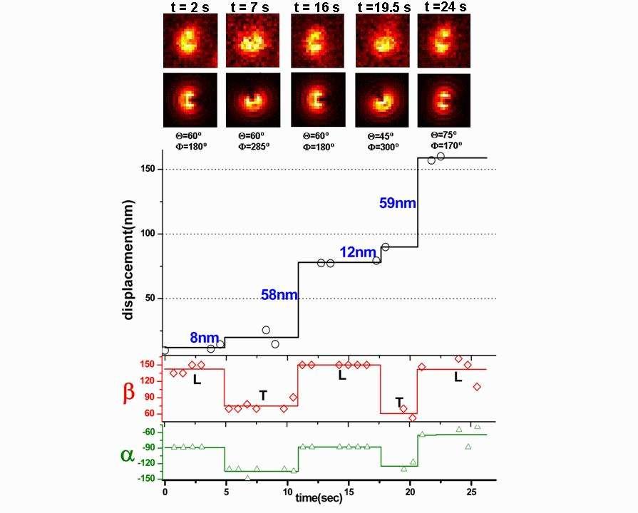
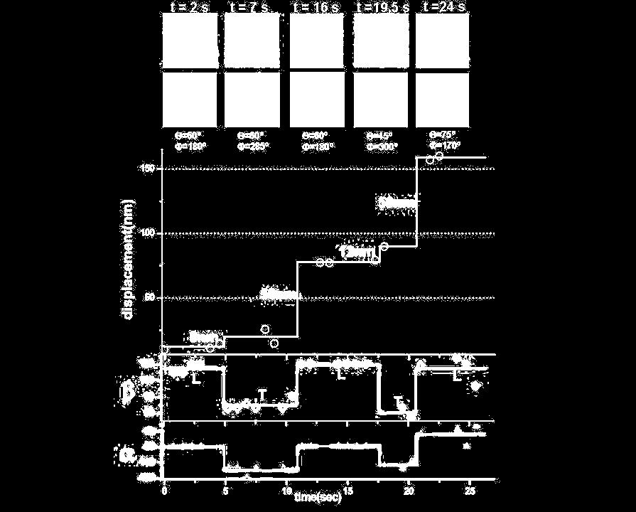
57 Theoretically calculated patterns


58 Defocused imaging of single molecules: pattern matching


59 Emission dipole hopping in a perylene tetrachromophore


60 Emission dipole hopping in a perylene tetrachromophore

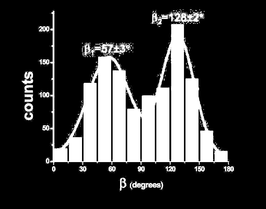
61 Rotational diffusion of molecules Measurement: Hiroshi Uji-i

62 Rotational diffusion of molecules
63 Symmetric top Brownian rotator = D D cos Θ t = cos φ cos ψ sin φ sin ψ cos θ π 2π 2π t = d θ d φ d ψ sin θg ( φ, θ, ψ, t ) ( cos φ cos ψ sin φ sin ψ cos θ ) =e cos 2 Θ = t ( 2 D + ) t D t 1 ( 6 D + 4 ) t + e + e D + t 3 12 D + t 1 12 D +9 t = e( ) + e( ) + e( ) t D t 3 ( 6 D + 4 ) t 1 ( 20 D + 4 ) t 1 ( 20 D +16 ) t cos 4 Θ = + e 6 D t + e + e + e + e t cos3 Θ

64 Rotational diffusion of molecules: Correlation analysis D << D

65 Motor proteins: myosin V along actin
66 Myosin V moving along actin filament 1.45 oil immersion objective 160 x magnification 10 ms exposure time / frame defocusing 500 nm Measurement by Erdal Toprak, UIUC

67 Myosin motion and reorientation

68 Myosin motion and reorientation

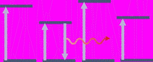
69 Myosin motion and reorientation N = 97 molecules 1151 tilting events
70 Myosin motion and reorientation We observe that there is a consistent fluctuation of β between two well defined angles as myosin V steps. This is consistent with the lever arm hypothesis. Unlike β, the change in α shows no consistent or recognizable pattern which is an evidence for diffusional binding of myosin V.
 Confocal Laser Scanning Microscope (CLSM) (A tribute to microscopy pioneer Antoni van")
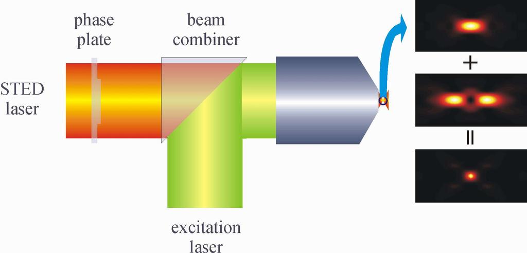
71 Superresolution microscopy: Overcoming Abbe's resolution limit Fluorophore distribution (bar = 1µm) Confocal Laser Scanning Microscope (CLSM) (A tribute to microscopy pioneer Antoni van Leeuwenhoek)

72 intensity Spatial resolution limit of standard light microscopy position [µm]


73 Lateral resolution limit of standard light microscopy: Abbe's equation λ 2n.sinθ θ objective N.A. = n.sinθ

74 Laser Scanning Confocal Microscopy (LSCM) LSCM with deconvolution is completely equivalent in resolution power and photon usage with structured illumination microscopy laser beam objective PSF


75 Axial resolution limit of standard light microscopy e ik0 z +e k z,θ = θ ik z,θ z 2 = 2 + 2cos ( k0 k z,θ ) z n cos θ λ n k0 = λ objective λ n ( 1 cos θ )


76 4π microscopy standing wave generation by counter-propagating focusing of two coherent laser beams λ λ = n ( 1 cos θ4 π ) 2n laser beam 1st objective PSF 2nd objective laser beam
77 Back to basics: Physics of fluorescence Photobleaching S1 Excitation T1 Fluorescence Emission S0

78 Ground state depletion microscopy: Using saturation of the excited state
79 Stimulated Emission S1 STE Excitation S0 Fluorescence Emission

80 Stimulated Emission Depletion Microscopy excitation laser PSF STED laser

81 Stimulated Emission Depletion Microscopy
82 Stimulated Emission Depletion Microscopy

83 Temporal behavior of ground state depletion after sudden switch-on of excitation
84 µ s µ s 3.2 µ s 6.4 µ s x [µ m] rel. amplitude 0.4 µ s 320 nm 0.2 µ s 160 nm nm 0.1 µ s 40 nm 1 0 nm 0.0 µ s rel. amplitude Converting temporal into spatial information: Dynamic Saturation Optical Microscopy time [µ s] 8 10

85 Potential realization of Dynamic Saturation Optical Microscopy:

86 Potential realization of Dynamic Saturation Optical Microscopy:

87 Dynamic Saturation Optical Microscopy: Point spread function
88 Theoretical estimate of DSOM performance Fluorophore distribution (bar = 1µm) DSOM Confocal Laser Scanning Microscope (CLSM) DSOM + Bessel beam

89 Complex photophysics of Alexa647 Alexa 647

90 Combining DSOM and FCS Alexa 647
91 Ground state depletion into triplet state S1 T1 Excitation Fluorescence Emission S0 f ( r) = a ( r) 1 + τa ( r ) τkisc f ( r ) s ( r, t ) = + exp k ph + τkisc f ( r ) t k ph + τkisc f ( r ) k ph + τkisc f ( r ) k ph { }
92 Ground state depletion into metastable state (switchable chromophores) S1 Excitation M Fluorescence Emission S0 s ( r, t ) = exp { τktrans f ( r ) t} f ( r) = a ( r) 1 + τa ( r )
93 Ground state depletion into first excited state S1 Excitation Fluorescence Emission S0 s ( r, t ) = a ( r) { { 1 1 exp τ + a ( r ) t 1 τ + a( r) }}
94 Summary of DSOM Relatively simple: one laser only employing a standard CLSM pure electronic data evaluation relatively robust against aberration can be combined with 4π or other techniques Drawback: resolution enhancement limited to ca. 5 times

95 Publications available at

 Hiroshi Uji-i, Johan Hofkens (Katholieke Universiteit Leuven) Erdal")
96 Acknowledgements/Cooperations Ingo Gregor Digambara Patra Jan Sykora Luru Dai Thomas Dertinger Iris von der Hocht Jörg Fitter Thomas Gensch Benjamin Kaupp (FZ Jülich) Markus Sauer (Univ. Bielefeld) Hiroshi Uji-i, Johan Hofkens (Katholieke Universiteit Leuven) Erdal Toprak, Paul Selvin (Univ. Illinois Urbana-Champaign)
Single-Molecule Methods I - in vitro
 Single-Molecule Methods I - in vitro Bo Huang Macromolecules 2014.03.10 F 1 -ATPase: a case study Membrane ADP ATP Rotation of the axle when hydrolyzing ATP Kinosita group, 1997-2005 Single Molecule Methods
Optics and Spectroscopy
 Introduction to Optics and Spectroscopy beyond the diffraction limit Chi Chen 陳祺 Research Center for Applied Science, Academia Sinica 2015Apr09 1 Light and Optics 2 Light as Wave Application 3 Electromagnetic
Dual-Focus Fluorescence Correlation Spectroscopy
 Application Note Dual-Focus Fluorescence Correlation Spectroscopy Thomas Dertinger, University of California Los Angeles, USA Benjamin Ewers, Benedikt Krämer, Felix Koberling, PicoQuant GmbH, Germany Iris
Administrative details:
 Administrative details: Anything from your side? www.photonics.ethz.ch 1 Where do we stand? Optical imaging: Focusing by a lens Angular spectrum Paraxial approximation Gaussian beams Method of stationary
Rice/TCU REU on Computational Neuroscience. Fundamentals of Molecular Imaging
 Rice/TCU REU on Computational Neuroscience Fundamentals of Molecular Imaging June 3, 2008 Neal Waxham 713-500-5621 m.n.waxham@uth.tmc.edu Objectives Brief discussion of optical resolution and lasers as
Single Emitter Detection with Fluorescence and Extinction Spectroscopy
 Single Emitter Detection with Fluorescence and Extinction Spectroscopy Michael Krall Elements of Nanophotonics Associated Seminar Recent Progress in Nanooptics & Photonics May 07, 2009 Outline Single molecule
Correlation Spectroscopy in Polymer Physics Methodenseminar im Wahlpflichtfach Basics diffusion and brownian motion correlations functions
 Correlation Spectroscopy in Polymer Physics Methodenseminar im Wahlpflichtfach 3 1. Basics diffusion and brownian motion correlations functions 2. Dynamic light scattering (DLS) DLS on cellulose solutions
Multiphoton Imaging and Spectroscopy in Cell and Tissue Biophysics. J Moger and C P Winlove
 Multiphoton Imaging and Spectroscopy in Cell and Tissue Biophysics J Moger and C P Winlove Relating Structure to Function Biochemistry Raman microspectrometry Surface enhanced Raman spectrometry (SERS)
1. Transition dipole moment
 1. Transition dipole moment You have measured absorption spectra of aqueous (n=1.33) solutions of two different chromophores (A and B). The concentrations of the solutions were the same. The absorption
Laboratory 3: Confocal Microscopy Imaging of Single Emitter Fluorescence and Hanbury Brown, and Twiss Setup for Photon Antibunching
 Laboratory 3: Confocal Microscopy Imaging of Single Emitter Fluorescence and Hanbury Brown, and Twiss Setup for Photon Antibunching Jonathan Papa 1, * 1 Institute of Optics University of Rochester, Rochester,
FLUORESCENCE MICROSCOPY TECHNIQUES PRACTICAL MANUAL FOR
 FLUORESCENCE PRACTICAL MANUAL FOR MICROSCOPY TECHNIQUES Sohail Ahmed Sudhaharan Thankiah Radek Machán Martin Hof Andrew H. A. Clayton Graham Wright Jean-Baptiste Sibarita Thomas Korte Andreas Herrmann
Laboratory 3&4: Confocal Microscopy Imaging of Single-Emitter Fluorescence and Hanbury Brown and Twiss setup for Photon Antibunching
 Laboratory 3&4: Confocal Microscopy Imaging of Single-Emitter Fluorescence and Hanbury Brown and Twiss setup for Photon Antibunching Jose Alejandro Graniel Institute of Optics University of Rochester,
Fluorescence Workshop UMN Physics June 8-10, 2006 Quantum Yield and Polarization (1) Joachim Mueller
 Joachim Mueller") Fluorescence Workshop UMN Physics June 8-10, 2006 Quantum Yield and Polarization (1) Joachim Mueller Quantum yield, polarized light, dipole moment, photoselection, dipole radiation, polarization and anisotropy
Fluorescence Workshop UMN Physics June 8-10, 2006 Quantum Yield and Polarization (1) Joachim Mueller Quantum yield, polarized light, dipole moment, photoselection, dipole radiation, polarization and anisotropy
Confocal Microscopy Imaging of Single Emitter Fluorescence and Hanbury Brown and Twiss Photon Antibunching Setup
 1 Confocal Microscopy Imaging of Single Emitter Fluorescence and Hanbury Brown and Twiss Photon Antibunching Setup Abstract Jacob Begis The purpose of this lab was to prove that a source of light can be
Fluorescence polarisation, anisotropy FRAP
 Fluorescence polarisation, anisotropy FRAP Reminder: fluorescence spectra Definitions! a. Emission sp. b. Excitation sp. Stokes-shift The difference (measured in nm) between the peak of the excitation
This document contains the following supporting information: 1. Wide field scanning electron microscope image
 Supporting information for Self-assembled nanoparticle dimer antennas for plasmonic-enhanced single-molecule fluorescence detection at micromolar concentrations Deep Punj, Raju Regmi, Alexis Devilez, Robin
FROM LOCALIZATION TO INTERACTION
 EPFL SV PTBIOP FROM LOCALIZATION TO INTERACTION BIOP COURSE 2015 COLOCALIZATION TYPICAL EXAMPLE EPFL SV PTBIOP Vinculin Alexa568 Actin Alexa488 http://www.olympusconfocal.com/applications/colocalization.html
single-molecule fluorescence resonance energy transfer
 single-molecule fluorescence resonance energy transfer (2) determing the Förster radius: quantum yield, donor lifetime, spectral overlap, anisotropy michael börsch 26/05/2004 1 fluorescence (1) absorbance
Rotational Brownian motion; Fluorescence correlation spectroscpy; Photobleaching and FRET. David A. Case Rutgers, Spring 2009
 Rotational Brownian motion; Fluorescence correlation spectroscpy; Photobleaching and FRET David A. Case Rutgers, Spring 2009 Techniques based on rotational motion What we studied last time probed translational
LABORATORY OF ELEMENTARY BIOPHYSICS
 LABORATORY OF ELEMENTARY BIOPHYSICS Experimental exercises for III year of the First cycle studies Field: Applications of physics in biology and medicine Specialization: Molecular Biophysics Fluorescence
bio-molecular studies Physical methods in Semmelweis University Osváth Szabolcs
 Physical methods in bio-molecular studies Osváth Szabolcs Semmelweis University szabolcs.osvath@eok.sote.hu Light emission and absorption spectra Stokes shift is the difference (in wavelength or frequency
I. Proteomics by Mass Spectrometry 1. What is an internal standard and what does it accomplish analytically?
 Name I. Proteomics by Mass Spectrometry 1. What is an internal standard and what does it accomplish analytically? Internal standards are standards added intentionally to all samples, standards and blanks.
Detection of Single Photon Emission by Hanbury-Brown Twiss Interferometry
 Detection of Single Photon Emission by Hanbury-Brown Twiss Interferometry Greg Howland and Steven Bloch May 11, 009 Abstract We prepare a solution of nano-diamond particles on a glass microscope slide
Ultrafast Dynamics and Single Particle Spectroscopy of Au-CdSe Nanorods
 Supporting Information Ultrafast Dynamics and Single Particle Spectroscopy of Au-CdSe Nanorods G. Sagarzazu a, K. Inoue b, M. Saruyama b, M. Sakamoto b, T. Teranishi b, S. Masuo a and N. Tamai a a Department
Analysis of second-harmonic generation microscopy under refractive index mismatch
 Vol 16 No 11, November 27 c 27 Chin. Phys. Soc. 19-1963/27/16(11/3285-5 Chinese Physics and IOP Publishing Ltd Analysis of second-harmonic generation microscopy under refractive index mismatch Wang Xiang-Hui(
SNOM Challenges and Solutions
 SiO x SiO x Au Au E k SNOM Challenges and Solutions Ralf Vogelgesang, Ph.D. Ralf.Vogelgesang@fkf.mpg.de Nanoscale Science Department (Prof. Kern) Max-Planck-Institut für Festkörperforschung, Stuttgart,
Model Answer (Paper code: AR-7112) M. Sc. (Physics) IV Semester Paper I: Laser Physics and Spectroscopy
 M. Sc. (Physics) IV Semester Paper I: Laser Physics and Spectroscopy") Model Answer (Paper code: AR-7112) M. Sc. (Physics) IV Semester Paper I: Laser Physics and Spectroscopy Section I Q1. Answer (i) (b) (ii) (d) (iii) (c) (iv) (c) (v) (a) (vi) (b) (vii) (b) (viii) (a) (ix)
Model Answer (Paper code: AR-7112) M. Sc. (Physics) IV Semester Paper I: Laser Physics and Spectroscopy Section I Q1. Answer (i) (b) (ii) (d) (iii) (c) (iv) (c) (v) (a) (vi) (b) (vii) (b) (viii) (a) (ix)
BMB Class 17, November 30, Single Molecule Biophysics (II)
") BMB 178 2018 Class 17, November 30, 2018 15. Single Molecule Biophysics (II) New Advances in Single Molecule Techniques Atomic Force Microscopy Single Molecule Manipulation - optical traps and tweezers
BMB 178 2018 Class 17, November 30, 2018 15. Single Molecule Biophysics (II) New Advances in Single Molecule Techniques Atomic Force Microscopy Single Molecule Manipulation - optical traps and tweezers
Using Alba with the FemtoFiber laser by Toptica for 2-photon quantitative imaging
 TECHNICAL NOTE Using Alba with the FemtoFiber laser by Toptica for 2-photon quantitative imaging Shih-Chu Liao, Yuansheng Sun, Ulas Coskun ISS, Inc. Introduction The advantages of multiphoton excitation
Solution structure and dynamics of biopolymers
 Solution structure and dynamics of biopolymers Atomic-detail vs. low resolution structure Information available at different scales Mobility of macromolecules in solution Brownian motion, random walk,
(i.e. what you should be able to answer at end of lecture)
") Today s Announcements 1. Test given back next Wednesday 2. HW assigned next Wednesday. 3. Next Monday 1 st discussion about Individual Projects. Today s take-home lessons (i.e. what you should be able
Today s Announcements 1. Test given back next Wednesday 2. HW assigned next Wednesday. 3. Next Monday 1 st discussion about Individual Projects. Today s take-home lessons (i.e. what you should be able
CHAPTER 7 SUMMARY OF THE PRESENT WORK AND SUGGESTIONS FOR FUTURE WORK
 161 CHAPTER 7 SUMMARY OF THE PRESENT WORK AND SUGGESTIONS FOR FUTURE WORK 7.1 SUMMARY OF THE PRESENT WORK Nonlinear optical materials are required in a wide range of important applications, such as optical
Digital Holographic Measurement of Nanometric Optical Excitation on Soft Matter by Optical Pressure and Photothermal Interactions
 Ph.D. Dissertation Defense September 5, 2012 Digital Holographic Measurement of Nanometric Optical Excitation on Soft Matter by Optical Pressure and Photothermal Interactions David C. Clark Digital Holography
Solution set for EXAM IN TFY4265/FY8906 Biophysical microtechniques
 ENGLISH NORWEGIAN UNIVERSITY OF SCIENCE AND TECHNOLOGY DEPARTMENT OF PHYSICS Contact during exam: Magnus Borstad Lilledahl Telefon: 73591873 (office) 92851014 (mobile) Solution set for EXAM IN TFY4265/FY8906
Multiphoton microscopy
 Multiphoton microscopy Joonas Holmi ELEC October 6, 2016 Multiphoton microscopy 1. 2. 3. 4. Multiphoton microscopy 2/14 Intro: Multiphoton microscopy Nonlinear optical characterization method Pulsed laser
High-Resolution. Transmission. Electron Microscopy
 Part 4 High-Resolution Transmission Electron Microscopy 186 Significance high-resolution transmission electron microscopy (HRTEM): resolve object details smaller than 1nm (10 9 m) image the interior of
Technology, Techniques and Applications. Ric Allott Business Development Manager
 Technology, Techniques and Applications Ric Allott Business Development Manager 1 Central Laser Facility ASTRA GEMINI VULCAN ARTEMIS ULTRA OCTOPUS High power, ultrashort pulse dual beams of 15 J, 30 fs
CD Basis Set of Spectra that is used is that derived from comparing the spectra of globular proteins whose secondary structures are known from X-ray
 CD Basis Set of Spectra that is used is that derived from comparing the spectra of globular proteins whose secondary structures are known from X-ray crystallography An example of the use of CD Modeling
Raman and stimulated Raman spectroscopy of chlorinated hydrocarbons
 Department of Chemistry Physical Chemistry Göteborg University KEN140 Spektroskopi Raman and stimulated Raman spectroscopy of chlorinated hydrocarbons WARNING! The laser gives a pulsed very energetic and
Supplementary Figures Supplementary Figure 1: Estimation of the error of the number and brightness of molecules in a single cluster; Simulation
 Supplementary Figures Supplementary Figure 1: Estimation of the error of the number and brightness of molecules in a single cluster; Simulation (a,c) Relative estimated numbers of molecules ; (b,d) relative
Self-calibrated, line-scan STED-FCS to quantify lipid dynamics in model and cell membranes
 Self-calibrated, line-scan STED-FCS to quantify lipid dynamics in model and cell membranes Aleš Benda, Yuanqing Ma and Katharina Gaus Centre for Vascular Research, Australian Centre for Nanomedicine and
δf / δx = σ F (N 2 -N 1 ) ΔF~N 2 -N 1
 ΔF~N 2 -N 1") LASER Light Amplification by Stimulated Emission of Radiation BASIC PROPERTIES O LASER RADIATION Spontaneous emission Incoherence in time Incoherence in space Polychromatic light Small energy density Non-polarized
LASER Light Amplification by Stimulated Emission of Radiation BASIC PROPERTIES O LASER RADIATION Spontaneous emission Incoherence in time Incoherence in space Polychromatic light Small energy density Non-polarized
High Resolution Laser Microscopy: a fascinating method to explore the molecular world
 High Resolution Laser Microscopy: a fascinating method to explore the molecular world Alfred J. Meixner Physical and Theoretical Chemistry Laboratory University of Siegen Single-molecule spectroscopy and
Super Resolution Microscopy Structured Illumination
 Super Resolution Microscopy Structured Illumination Bo Huang Department of Pharmaceutical Chemistry, UCSF CSHL Quantitative Microscopy, 10/31/2011 50 years to extend the resolution Confocal microscopy
LAB 3: Confocal Microscope Imaging of single-emitter fluorescence. LAB 4: Hanbury Brown and Twiss setup. Photon antibunching. Roshita Ramkhalawon
 LAB 3: Confocal Microscope Imaging of single-emitter fluorescence LAB 4: Hanbury Brown and Twiss setup. Photon antibunching Roshita Ramkhalawon PHY 434 Department of Physics & Astronomy University of Rochester
Energy transport in metal nanoparticle plasmon waveguides
 Energy transport in metal nanoparticle plasmon waveguides Stefan A. Maier, Pieter G. Kik, and Harry A. Atwater California Institute of Technology Thomas J. Watson Laboratory of Applied Physics, Pasadena,
Fluorescence Spectroscopy
 Fluorescence Spectroscopy Steady State and Time Dependent Fluorescence Measurements Kai Wen Teng PHYS 403 Fall 15 EM Spectrum of molecules Rotational Energy Infrared Vibrational Energy Near Infrared Electronic
Lab 3 and 4: Single Photon Source
 Lab 3 and 4: Single Photon Source By: Justin Deuro, December 10 th, 2009 Abstract We study methods of single photon emission by exciting single colloidal quantum dot (QD) samples. We prepare the single
Lecture 5. Anisotropy decay/data analysis. Enrico Gratton
 Lecture 5. Anisotropy decay/data analysis Enrico Gratton Anisotropy decay Energy-transfer distance distributions Time resolved spectra Excited-state reactions Basic physics concept in polarization The
5.74 Introductory Quantum Mechanics II
 MIT OpenCourseWare http://ocw.mit.edu 5.74 Introductory Quantum Mechanics II Spring 2009 For information about citing these materials or our Terms of Use, visit: http://ocw.mit.edu/terms. p. 10-0 10..
Nonlinear Optics. Single-Molecule Microscopy Group. Physical Optics Maria Dienerowitz.
 Single-Molecule Microscopy Group Nonlinear Optics Physical Optics 21-06-2017 Maria Dienerowitz maria.dienerowitz@med.uni-jena.de www.single-molecule-microscopy.uniklinikum-jena.de Contents Introduction
5. 3P PIV Measurements
 Micro PIV Last Class: 1. Data Validation 2. Vector Field Operator (Differentials & Integrals) 3. Standard Differential Scheme 4. Implementation of Differential & Integral quantities with PIV data 5. 3P
Optical Spectroscopy. Steady State and Time Dependent Fluorescence Measurements. Kai Wen Teng. October 8 th PHYS 403 Fall 2013
 Optical Spectroscopy Steady State and Time Dependent Fluorescence Measurements Kai Wen Teng October 8 th 2013 PHYS 403 Fall 2013 EM Spectrum of molecules Rotational Energy Infrared Vibrational Energy Near
Surface Plasmon Amplification by Stimulated Emission of Radiation. By: Jonathan Massey-Allard Graham Zell Justin Lau
 Surface Plasmon Amplification by Stimulated Emission of Radiation By: Jonathan Massey-Allard Graham Zell Justin Lau Surface Plasmons (SPs) Quanta of electron oscillations in a plasma. o Electron gas in
DEVELOPMENT OF NANO PARTICLE SIZING SYSTEM USING FLUORESCENCE POLARIZATION
 XX IMEKO World Congress Metrology for Green Growth September 9 14, 2012, Busan, Republic of Korea DEVELOPMENT OF NANO PARTICLE SIZING SYSTEM USING FLUORESCENCE POLARIZATION Terutake Hayashi, Masaki Michihata,
Fluorescence 2009 update
 XV 74 Fluorescence 2009 update Jablonski diagram Where does the energy go? Can be viewed like multistep kinetic pathway 1) Excite system through A Absorbance S 0 S n Excite from ground excited singlet
Survey on Laser Spectroscopic Techniques for Condensed Matter
 Survey on Laser Spectroscopic Techniques for Condensed Matter Coherent Radiation Sources for Small Laboratories CW: Tunability: IR Visible Linewidth: 1 Hz Power: μw 10W Pulsed: Tunabality: THz Soft X-ray
Anti-Bunching from a Quantum Dot
 Anti-Bunching from a Quantum Dot Gerardo I. Viza 1, 1 Department of Physics and Astronomy, University of Rochester, Rochester, NY 14627 We study the nature of non-classical single emitter light experimentally
Introduction... Theory Influence of Excitation Pulse Shape...
 1. Fluorescence Anisotropy: Theory and Applications Robert F. Steiner 1.1. 1.2. 1.3. 1.4. 1.5. 1.6. Introduction... Theory... 1.2.1. Meaning of Anisotropy... 1.2.2. Influence of Excitation Pulse Shape...
Co-localization, FRET
 Co-localization, FRET Last class FRAP Diffusion This class Co-localization Correlation FRET Co-localization Can you infer function of protein from it s intracellular location How do you measure if two
Quantum yield determination by low-intensity Fluorescence Correlation Spectroscopy (lifcs)
") Quantum yield determination by low-intensity Fluorescence Correlation Spectroscopy (lifcs) Daryan Kempe 1, Jörg Fitter 1 and Matteo Gabba 1 RWTH Aachen, Germany Forschungszentrum Jülich, Germany July 15,
Quantum yield determination by low-intensity Fluorescence Correlation Spectroscopy (lifcs) Daryan Kempe 1, Jörg Fitter 1 and Matteo Gabba 1 RWTH Aachen, Germany Forschungszentrum Jülich, Germany July 15,
Fluorescence-detected magnetic field effects on radical pair reactions from femtolitre volumes
 Electronic Supplementary Material (ESI) for ChemComm. This journal is The Royal Society of Chemistry 2015 Fluorescence-detected magnetic field effects on radical pair reactions from femtolitre volumes
Laser Detection Techniques
 Laser Detection Techniques K.-H. Gericke Institute for Physical Chemistry University Braunschweig E 2 E 1 = hn, λ = c /n Lambert-Beer Law Transmittance of the sample:: T = I / I 0 T = e -snl = e -α, where
SUPPLEMENTARY INFORMATION
 Supplementary Information Speckle-free laser imaging using random laser illumination Brandon Redding 1*, Michael A. Choma 2,3*, Hui Cao 1,4* 1 Department of Applied Physics, Yale University, New Haven,
FLCS Fluorescence Lifetime Correlation Spectroscopy
 Application Note FLCS Fluorescence Lifetime Correlation Spectroscopy Peter Kapusta, Michael Wahl, PicoQuant GmbH Aleš Benda, Martin Hof, J. Heyrovsky Institute of Physical Chemistry, Prague, CZ Jörg Enderlein,
Chem 681: Student Seminar Series. Two-Photon Induced Fluorescence
 Speaker: Brooke Kocsis Date: Monday, April 17, 2000 Time: 4:00 PM Room: 2121 Advisor: Richard M. Crooks Chem 681: Student Seminar Series Two-Photon Induced Fluorescence Two-photon fluorescence (TPF) is
Skoog Chapter 6 Introduction to Spectrometric Methods
 Skoog Chapter 6 Introduction to Spectrometric Methods General Properties of Electromagnetic Radiation (EM) Wave Properties of EM Quantum Mechanical Properties of EM Quantitative Aspects of Spectrochemical
Answers to questions on exam in laser-based combustion diagnostics on March 10, 2006
 Answers to questions on exam in laser-based combustion diagnostics on March 10, 2006 1. Examples of advantages and disadvantages with laser-based combustion diagnostic techniques: + Nonintrusive + High
Chapter 24 Photonics Question 1 Question 2 Question 3 Question 4 Question 5
 Chapter 24 Photonics Data throughout this chapter: e = 1.6 10 19 C; h = 6.63 10 34 Js (or 4.14 10 15 ev s); m e = 9.1 10 31 kg; c = 3.0 10 8 m s 1 Question 1 Visible light has a range of photons with wavelengths
Supplemental Materials and Methods
 Supplemental Materials and Methods Time-resolved FRET (trfret) to probe for changes in the Box A/A stem upon complex assembly U3 MINI was folded and the decay of Fl fluorescence was measured at 20 ºC (see
Introduction to FCS. Enrico Gratton. Laboratory for Fluorescence Dynamics Department of Biomedical Engineering University of California, Irvine
 Introduction to FCS Enrico Gratton Laboratory for Fluorescence Dynamics Department of Biomedical Engineering University of California, Irvine Outline What is diffusion? Diffusion of molecules Diffusion
Nanoscopy with Focused Light
 Nanoscopy with Focused Light Stefan W. Hell Max Planck Institute for Biophysical Chemistry Department of NanoBiophotonics Göttingen & German Cancer Research Center (DKFZ) Optical Nanoscopy Division Heidelberg
Γ43 γ. Pump Γ31 Γ32 Γ42 Γ41
 Supplementary Figure γ 4 Δ+δe Γ34 Γ43 γ 3 Δ Ω3,4 Pump Ω3,4, Ω3 Γ3 Γ3 Γ4 Γ4 Γ Γ Supplementary Figure Schematic picture of theoretical model: The picture shows a schematic representation of the theoretical
Lecture 20 Optical Characterization 2
 Lecture 20 Optical Characterization 2 Schroder: Chapters 2, 7, 10 1/68 Announcements Homework 5/6: Is online now. Due Wednesday May 30th at 10:00am. I will return it the following Wednesday (6 th June).
Fluorescence Resonance Energy Transfer (FRET) Microscopy
 Microscopy") Fluorescence Resonance Energy Transfer () Microscopy Mike Lorenz Optical Technology Development mlorenz@mpi-cbg.de -FLM course, May 2009 What is fluorescence? Stoke s shift Fluorescence light is always
Fluorescence Resonance Energy Transfer () Microscopy Mike Lorenz Optical Technology Development mlorenz@mpi-cbg.de -FLM course, May 2009 What is fluorescence? Stoke s shift Fluorescence light is always
How DLS Works: Interference of Light
 Static light scattering vs. Dynamic light scattering Static light scattering measures time-average intensities (mean square fluctuations) molecular weight radius of gyration second virial coefficient Dynamic
Lecture 19 Optical MEMS (1)
") EEL6935 Advanced MEMS (Spring 5) Instructor: Dr. Huikai Xie Lecture 19 Optical MEMS (1) Agenda: Optics Review EEL6935 Advanced MEMS 5 H. Xie 3/8/5 1 Optics Review Nature of Light Reflection and Refraction
EEL6935 Advanced MEMS (Spring 5) Instructor: Dr. Huikai Xie Lecture 19 Optical MEMS (1) Agenda: Optics Review EEL6935 Advanced MEMS 5 H. Xie 3/8/5 1 Optics Review Nature of Light Reflection and Refraction
Vibrational imaging and microspectroscopies based on coherent anti-stokes Raman scattering (CARS)
") Vibrational imaging and microspectroscopies based on coherent anti-stokes Raman scattering (CARS) by Andreas Volkmer Universität Stuttgart 3 rd Institute of Physics, University of Stuttgart, Pfaffenwaldring
Vibrational imaging and microspectroscopies based on coherent anti-stokes Raman scattering (CARS) by Andreas Volkmer Universität Stuttgart 3 rd Institute of Physics, University of Stuttgart, Pfaffenwaldring
Physical Optics. Lecture 7: Coherence Herbert Gross.
 Physical Optics Lecture 7: Coherence 07-05-7 Herbert Gross www.iap.uni-jena.de Physical Optics: Content No Date Subject Ref Detailed Content 05.04. Wave optics G Complex fields, wave equation, k-vectors,
Singlet. Fluorescence Spectroscopy * LUMO
 Fluorescence Spectroscopy Light can be absorbed and re-emitted by matter luminescence (photo-luminescence). There are two types of luminescence, in this discussion: fluorescence and phosphorescence. A
Lecture 9. PMTs and Laser Noise. Lecture 9. Photon Counting. Photomultiplier Tubes (PMTs) Laser Phase Noise. Relative Intensity
 Laser Phase Noise. Relative Intensity") s and Laser Phase Phase Density ECE 185 Lasers and Modulators Lab - Spring 2018 1 Detectors Continuous Output Internal Photoelectron Flux Thermal Filtered External Current w(t) Sensor i(t) External System
s and Laser Phase Phase Density ECE 185 Lasers and Modulators Lab - Spring 2018 1 Detectors Continuous Output Internal Photoelectron Flux Thermal Filtered External Current w(t) Sensor i(t) External System
Fluorescence Spectroscopy
 Fluorescence Spectroscopy Frequency and time dependent emission Emission and Excitation fluorescence spectra Stokes Shift: influence of molecular vibrations and solvent Time resolved fluorescence measurements
Electronic Supplementary Information
 Electronic Supplementary Material (ESI) for Journal of Materials Chemistry C. This journal is The Royal Society of Chemistry 2017 Electronic Supplementary Information Polymorphism and microcrystal shape
Laser Types Two main types depending on time operation Continuous Wave (CW) Pulsed operation Pulsed is easier, CW more useful
 Pulsed operation Pulsed is easier, CW more useful") Main Requirements of the Laser Optical Resonator Cavity Laser Gain Medium of 2, 3 or 4 level types in the Cavity Sufficient means of Excitation (called pumping) eg. light, current, chemical reaction Population
Main Requirements of the Laser Optical Resonator Cavity Laser Gain Medium of 2, 3 or 4 level types in the Cavity Sufficient means of Excitation (called pumping) eg. light, current, chemical reaction Population
Time-resolved Molecule Counting by Photon Statistics Across the Visible Spectrum
 Electronic Supplementary Material (ESI) for Physical Chemistry Chemical Physics. This journal is the Owner Societies 2017 Time-resolved Molecule Counting by Photon Statistics Across the Visible Spectrum
CHEM Outline (Part 15) - Luminescence 2013
 - Luminescence 2013") CHEM 524 -- Outline (Part 15) - Luminescence 2013 XI. Molecular Luminescence Spectra (Chapter 15) Kinetic process, competing pathways fluorescence, phosphorescence, non-radiative decay Jablonski diagram
CHEM 524 -- Outline (Part 15) - Luminescence 2013 XI. Molecular Luminescence Spectra (Chapter 15) Kinetic process, competing pathways fluorescence, phosphorescence, non-radiative decay Jablonski diagram
Richard Miles and Arthur Dogariu. Mechanical and Aerospace Engineering Princeton University, Princeton, NJ 08540, USA
 Richard Miles and Arthur Dogariu Mechanical and Aerospace Engineering Princeton University, Princeton, NJ 08540, USA Workshop on Oxygen Plasma Kinetics Sept 20, 2016 Financial support: ONR and MetroLaser
High photostability and enhanced fluorescence of gold nanoclusters by silver doping-supporting information
 High photostability and enhanced fluorescence of gold nanoclusters by silver doping-supporting information Size measurements Figure S1 P2 FTIR measurements Figure S2 P2 XPS measurements Figure S3 P3 Photo-physical
Chapter 6 Photoluminescence Spectroscopy
 Chapter 6 Photoluminescence Spectroscopy Course Code: SSCP 4473 Course Name: Spectroscopy & Materials Analysis Sib Krishna Ghoshal (PhD) Advanced Optical Materials Research Group Physics Department, Faculty
Modern Optical Spectroscopy
 Modern Optical Spectroscopy With Exercises and Examples from Biophysics and Biochemistry von William W Parson 1. Auflage Springer-Verlag Berlin Heidelberg 2006 Verlag C.H. Beck im Internet: www.beck.de
Coherence and width of spectral lines with Michelson interferometer
 Coherence and width of spectral lines TEP Principle Fraunhofer and Fresnel diffraction, interference, spatial and time coherence, coherence conditions, coherence length for non punctual light sources,
Third-harmonic generation
 2 Third-harmonic generation 2.1 Introduction Optical signals from single nano-objects open new windows for studies at nanometer scales in fields as diverse as material science and cell biology. Cleared
Medical Biophysics II. Final exam theoretical questions 2013.
 Medical Biophysics II. Final exam theoretical questions 2013. 1. Early atomic models. Rutherford-experiment. Franck-Hertz experiment. Bohr model of atom. 2. Quantum mechanical atomic model. Quantum numbers.
Nonlinear Optics. Single-Molecule Microscopy Group. Physical Optics Maria Dienerowitz.
 Single-Molecule Microscopy Group Nonlinear Optics Physical Optics 21-06-2017 Maria Dienerowitz maria.dienerowitz@med.uni-jena.de www.single-molecule-microscopy.uniklinikum-jena.de Contents Introduction
An Introduction to Diffraction and Scattering. School of Chemistry The University of Sydney
 An Introduction to Diffraction and Scattering Brendan J. Kennedy School of Chemistry The University of Sydney 1) Strong forces 2) Weak forces Types of Forces 3) Electromagnetic forces 4) Gravity Types
Quantitative fluorescence correlation spectroscopy in three-dimensional systems under stimulated emission depletion conditions: supplementary material
 Quantitative fluorescence correlation spectroscopy in three-dimensional systems under stimulated emission depletion conditions: supplementary material KRZYSZTOF SOZANSKI 1,*, EVANGELOS SISAMAKIS 2, XUZHU
Far-field radiation pattern in Coherent Anti-stokes Raman Scattering (CARS) Microscopy.
 Microscopy.") Far-field radiation pattern in Coherent Anti-stokes Raman Scattering (CARS) Microscopy. David Gachet, Nicolas Sandeau, Hervé Rigneault * Institut Fresnel, Mosaic team, Domaine Univ. St Jérôme, 13397 Marseille
Far-field radiation pattern in Coherent Anti-stokes Raman Scattering (CARS) Microscopy. David Gachet, Nicolas Sandeau, Hervé Rigneault * Institut Fresnel, Mosaic team, Domaine Univ. St Jérôme, 13397 Marseille
A) n L < 1.0 B) n L > 1.1 C) n L > 1.3 D) n L < 1.1 E) n L < 1.3
 n L < 1.0 B) n L > 1.1 C) n L > 1.3 D) n L < 1.1 E) n L < 1.3") 1. A beam of light passes from air into water. Which is necessarily true? A) The frequency is unchanged and the wavelength increases. B) The frequency is unchanged and the wavelength decreases. C) The
1. A beam of light passes from air into water. Which is necessarily true? A) The frequency is unchanged and the wavelength increases. B) The frequency is unchanged and the wavelength decreases. C) The
Visualization of Xe and Sn Atoms Generated from Laser-Produced Plasma for EUV Light Source
 3rd International EUVL Symposium NOVEMBER 1-4, 2004 Miyazaki, Japan Visualization of Xe and Sn Atoms Generated from Laser-Produced Plasma for EUV Light Source H. Tanaka, A. Matsumoto, K. Akinaga, A. Takahashi
Enhancement of Exciton Transport in Porphyrin. Aggregate Nanostructures by Controlling. Hierarchical Self-Assembly
 Electronic Supplementary Material (ESI) for Nanoscale. This journal is The Royal Society of Chemistry 2018 Supporting Information for Enhancement of Exciton Transport in Porphyrin Aggregate Nanostructures