Metal Artifact Reduction with DECT
|
|
|
- Leona Briggs
- 6 years ago
- Views:
Transcription


1 Metal Artifact Reduction with DECT Daniele Marin, MD Duke University Medical Center


2 Metal artifacts Common clinical problem ( 20%) Boas EF et al. Radiology 2011
3 Beam Hardening Edge Effects Scatter Metal artifacts Photon starvation Motion
4 Factors influencing artifact: 1. Density: Metal artifacts Cobalt chrome +++ Stainless steel ++ Titanium + 1. Shape: Complex +++ Simple + 2. Orientation: In plane long axis +++ In plane short axis ++ Out of plane + 1. Motion
5 Metal artifacts Correction Strategy kvp and/or filtration Spatial resolution Temporal resolution When? Mechanism Side Effects Acquisition beam hardening photon starvation dose contrast Acquisition partial volume noise Acquisition motion dose (gating) Dual energy CT (VMI) Acquisition beam hardening contrast Correction algorithms Reconstruction beam hardening noise
6 signal z-axis spatial resolution FWHM SSP z-axis (mm) Pitch Section Thickness DOSE NOISE


 < 20cm from isocenter (984 views)")
7 3 mm stent Gemstone technology High Definition imaging Smaller detector elements in x/y 0.23 mm spatial resolution across entire body 18 lp/cm spatial resolution for cardiac Reduced aliasing artifacts 128 slice reconstruction Focal spot motion (x-y deflection) < 20cm from isocenter (984 views) (2460 views) Non-HDCT HD 7


8 Improving resolution - blooming reduction in the stent Std. resolution High resolution Images courtesy of Dr Sablayrolles, CCN 8


9 CARDIAC CT Improved spatial resolution lp/cm 1 Reduced calcium blooming Improved stent visualization Improved diagnostic confidence Standard resolution High resolution Standard resolution High resolution 1. Based upon internal test data comparing Revolution HD cardiac half-scan spatial resolution to data from Advanced CT Scanners for Coronary Angiography, ImPACT Report CEP10043, March, 2010, available at 9


10 Motion correction No gating Cardiac gating


11 REVOLUTION GSI SnapShot * Freeze decreases calcium blooming Std. Cardiac Cardiac w/ SSF * Trademark of General Electric Company


12 Dual Energy CT (VMI)

13 Dual Energy CT (VMI) Guggenberger et al. Eur Rad 2012

 Restricted Siemens")

14 Data Processing Monoenergetic 64 kev 69 kev 89 kev 105 kev 190 kev Courtesy of Thorsten Johnson (University Hospital Großhadern, Germany) Restricted Siemens AG 2013 All rights reserved. Page Matthew Fuld / CT Research Collaborations


15 Metal Artifact Reduction 140Sn kvp 190 kev 190 kev image shows less blooming caused by metal due to reduced beam hardening in the monoenergetic images. Page 15 Courtesy of, MGH, Boston, MA Siemens All rights reserved.

 Restricted Siemens")

16 Data Processing Monoenergetic 64 kev 69 kev 89 kev 105 kev 190 kev Courtesy of Thorsten Johnson (University Hospital Großhadern, Germany) Restricted Siemens AG 2013 All rights reserved. Page Matthew Fuld / CT Research Collaborations


17 GSI allows for virtually artifact free visualization of the COW Better definition of the COW Dose neutral compared to SECT with 60% ASiR mgy-cm DLP 1.4 msv 1 effective dose SECT: 120 kvp VMI at 140 kev Images courtesy of MD Marcus C., University Hospital of Reims, France 1. Obtained by EUR EN, using an adult head factor of *DLP 17


18 CARDIAC CT Case showing lumen visualization with monochromatic imaging SECT: 120 kvp VMI at 140 kev. *Trademark of General Electric Company Images courtesy of Dr. J Earls, Fairfax Radiology, Virginia 18
19 Limitations VMI...? o No correction for scatter and photon starvation o Idealized x-ray absorption spectrum (no k-edge) o High kev yielding iodine/soft tissue contrast o Suboptimal arterial enhancement


20 90 kev 55 kev
 3 rd step Correction process 1. Custom BH correction 2. Inpainting techniques to to correct for photon starvation 4 th step Corrected image Boas EF et al.")
21 Correction algorithms 1 st step Uncorrected reconstruction (only BH from soft tissues is accounted for ) 2 nd step Identification process (metal/bone identified from raw data applying HU threshold ) 3 rd step Correction process 1. Custom BH correction 2. Inpainting techniques to to correct for photon starvation 4 th step Corrected image Boas EF et al. Radiology 2011

22 Neurocoil WFBP imar

23 Shoulders WFBP imar

24 Hip Implants WFBP imar


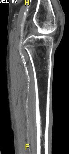
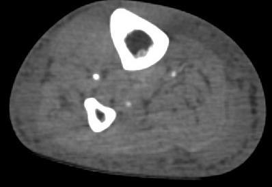
25 REVOLUTION GSI GSI MAR - metal artifact reduction in arthrodesis Metal artifact reduction arthrodesis GSI at 140 kev w/ MARS Conventional CT axial images GSI at 140 kev w/ MARS Images courtesy of OUH Svendborg, Denmark


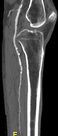
26 Metal artifact reduction, splenic aneurysm embolization Standard 120 kv acquisition GSI at 140 kev w/ MARS Metal artifact reduction with GSI enabled better visualization of splenic aneurysm embolization vs. conventional CT Images courtesy of Dr. JL Sablayrolles, CCN, France 26
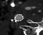
27 Limitations of metal artifact correction algorithms o Location of object (central vs. peripheral) o Orientation of object (transverse vs. oblique) o Motion o Proximity to other high density objects Brook et al. Radiology 2012

28 MARS & artifacts dark star far-field artifact Brook et al. Radiology 2012
29 Conclusions o Metal artifacts is a complex phenomenon o Different strategies for artifact correction have different pros & cons o DECT with VMI is a powerful tool for metal artifacts reduction o Workflow optimization is necessary o Correction algorithms can correct for both beam hardening and photon starvation effects
30 Thank You!
Applications of Low KeV Imaging in Abdomen
 Applications of Low KeV Imaging in Abdomen Dushyant Sahani, M.D Director of CT Associate Professor of Radiology Massachusetts General Hospital Harvard Medical School Email-dsahani@partners.org Disclosure
Metal Artifact Reduction and Dose Efficiency Improvement on Photon Counting Detector CT using an Additional Tin Filter
 Metal Artifact Reduction and Dose Efficiency Improvement on Photon Counting Detector CT using an Additional Tin Filter Wei Zhou, Dilbar Abdurakhimova, Kishore Rajendran, Cynthia McCollough, Shuai Leng
Multi-Energy CT: Principles, Processing
 Multi-Energy CT: Principles, Processing and Clinical Applications Shuai Leng, PhD Associate Professor Department of Radiology Mayo Clinic, Rochester, MN Clinical Motivation CT number depends on x-ray attenuation
Data Acquisition and Image Formation Methods for Multi Energy CT
 Data Acquisition and Image Formation Methods for Multi Energy CT Cynthia H. McCollough, PhD, DABR, FAIMBE, FAAPM, FACR Professor of Medical Physics and Biomedical Engineering Director, CT Clinical Innovation
X. Allen Li. Disclosure. DECT: What, how and Why Why dual-energy CT (DECT)? 7/30/2018. Improving delineation and response assessment using DECT in RT
? 7/30/2018. Improving delineation and response assessment using DECT in RT") Improving delineation and response assessment using DECT in RT X. Allen Li Medical College of Wisconsin MO-A-DBRA-1, AAPM, July 30 th, 2018 Disclosure Research funding support: Siemens Healthineers Elekta
Improving delineation and response assessment using DECT in RT X. Allen Li Medical College of Wisconsin MO-A-DBRA-1, AAPM, July 30 th, 2018 Disclosure Research funding support: Siemens Healthineers Elekta
DUAL ENERGY of the Pancreas
 DUAL ENERGY of the Pancreas Desiree E. Morgan, MD Professor and Vice Chair Clinical Research Director Human Imaging Shared Facility UAB CCC University of Alabama at Birmingham Opportunities to improve?
Carlo N. De Cecco, MD, PhD
 New Contrast Injection Strategies in Low kv and kev Imaging Carlo N. De Cecco, MD, PhD FSCBTMR - FSCCT - FESGAR Consultant for / Research support from: Siemens Bayer Guerbet Low kv and kev imaging Rationale
The relationship between image noise and spatial resolution of CT scanners
 The relationship between image noise and spatial resolution of CT scanners Sue Edyvean, Nicholas Keat, Maria Lewis, Julia Barrett, Salem Sassi, David Platten ImPACT*, St George s Hospital, London *An MDA
Electron density and effective atomic number images generated by dual energy imaging with a 320-detector CT system: A feasibility study
 Electron density and effective atomic number images generated by dual energy imaging with a 320-detector CT system: A feasibility study Poster No.: C-0403 Congress: ECR 2014 Type: Scientific Exhibit Authors:
Two-Material Decomposition From a Single CT Scan Using Statistical Image Reconstruction
 / 5 Two-Material Decomposition From a Single CT Scan Using Statistical Image Reconstruction Yong Long and Jeffrey A. Fessler EECS Department James M. Balter Radiation Oncology Department The University
Abdominal DECT: How to Integrate Into Your Practice
 Abdominal DECT: How to Integrate Into Your Practice Eric Tamm, M.D. Department of Diagnostic Radiology Division of Diagnostic Imaging MD Anderson Cancer Center Houston, TX Disclosure I have no relationships
A practical approach to the introduction of spectral imaging into a large UK acute care teaching hospital
 A practical approach to the introduction of spectral imaging into a large UK acute care teaching hospital Robert Loader Clinical Scientist Clinical & Radiation Physics Directorate of Healthcare Science
Potentials for Dual-energy kv/mv On-board Imaging and Therapeutic Applications
 Potentials for Dual-energy kv/mv On-board Imaging and Therapeutic Applications Fang-Fang Yin Department of Radiation Oncology Duke University Medical Center Acknowledgement Dr Hao Li for his excellent
Detector. * All clinical images are courtesy of. University, Jerusalem. Ami Altman, Ph.D., and Raz Carmi Ph.D., CT BU, PHILIPS Healthcare
 AD Dual-Energy alenerg CT Based on A Double Layer Detector * All clinical images are courtesy of Hadassah Medical Center, The Hebrew University, Jerusalem Ami Altman, Ph.D., and Raz Carmi Ph.D., CT BU,
1-D Fourier Transform Pairs
 1-D Fourier Transform Pairs The concept of the PSF is most easily explained by considering a very small point source being placed in the imaging field-of-view The relationship between the image, I, and
Midterm Review. Yao Wang Polytechnic University, Brooklyn, NY 11201
 Midterm Review Yao Wang Polytechnic University, Brooklyn, NY 11201 Based on J. L. Prince and J. M. Links, Medical maging Signals and Systems, and lecture notes by Prince. Figures are from the textbook.
Reducing metal artefacts and radiation dose in musculoskeletal CT imaging Wellenberg, R.H.H.
 UvA-DARE (Digital Academic Repository) Reducing metal artefacts and radiation dose in musculoskeletal CT imaging Wellenberg, R.H.H. Link to publication Citation for published version (APA): Wellenberg,
ESTIMATION OF 90 SCATTERING COEFFICIENT IN THE SHIELDING CALCULATION OF DIAGNOSTIC X-RAY EQUIPMENT
 Proceedings of the Eleventh EGS4 Users' Meeting in Japan, KEK Proceedings 2003-15, p.107-113 ESTIMATION OF 90 SCATTERING COEFFICIENT IN THE SHIELDING CALCULATION OF DIAGNOSTIC X-RAY EQUIPMENT K. Noto and
Radionuclide Imaging MII Positron Emission Tomography (PET)
") Radionuclide Imaging MII 3073 Positron Emission Tomography (PET) Positron (β + ) emission Positron is an electron with positive charge. Positron-emitting radionuclides are most commonly produced in cyclotron
Radionuclide Imaging MII 3073 Positron Emission Tomography (PET) Positron (β + ) emission Positron is an electron with positive charge. Positron-emitting radionuclides are most commonly produced in cyclotron
Multi-energy CT: Future Directions. Acknowledgements. Overview 7/23/2014. Taly Gilat Schmidt, PhD. Kevin Zimmerman Franco Rupcich Steven Haworth
 Multi-energy CT: Future Directions Taly Gilat Schmidt, PhD Department of Biomedical Engineering Marquette University Acknowledgements Kevin Zimmerman Franco Rupcich Steven Haworth Results in this talk:
Technical University of Denmark
 Technical University of Denmark Page 1 of 10 pages Written test, 12 December 2012 Course name: Introduction to medical imaging Course no. 31540 Aids allowed: None. Pocket calculator not allowed "Weighting":
Differential Absorption Analysis of Nonmagnetic Material in the Phantom using Dual CT
 Journal of Magnetics 21(2), 286-292 (2016) ISSN (Print) 1226-1750 ISSN (Online) 2233-6656 http://dx.doi.org/10.4283/jmag.2016.21.2.286 Differential Absorption Analysis of Nonmagnetic Material in the Phantom
Overview and Status of the Austrian Particle Therapy Facility MedAustron. Peter Urschütz
 Overview and Status of the Austrian Particle Therapy Facility MedAustron Peter Urschütz MedAustron Centre for ion beam therapy and non-clinical research Treatment of 1200 patients/year in full operation
Technical University of Denmark
 Technical University of Denmark Page 1 of 11 pages Written test, 9 December 2010 Course name: Introduction to medical imaging Course no. 31540 Aids allowed: none. "Weighting": All problems weight equally.
Proposed Room Requirements for CT System
 Siemens Proposed Room Requirements for CT System Semarang, 4-5 May 2017 Restricted Siemens Healthcare GmbH, 2016 Page 1 Roles of Medical Physicist CT Image Quality Radiation Protection Optimization Medical
Production of X-rays. Radiation Safety Training for Analytical X-Ray Devices Module 9
 Module 9 This module presents information on what X-rays are and how they are produced. Introduction Module 9, Page 2 X-rays are a type of electromagnetic radiation. Other types of electromagnetic radiation
What is scintigraphy? The process of obtaining an image or series of sequential images of the distribution of a radionuclide in tissues, organs, or
 Let's remind... What is nuclear medicine? Nuclear medicine can be broadly divided into two branches "in vitro" and "in vivo" procedures. There are numerous radioisotopic "in vitro" procedures for genotyping
Ke Li and Guang-Hong Chen
 Ke Li and Guang-Hong Chen Brief introduction of x-ray differential phase contrast (DPC) imaging Intrinsic noise relationship between DPC imaging and absorption imaging Task-based model observer studies
X-ray Interaction with Matter
 X-ray Interaction with Matter 10-526-197 Rhodes Module 2 Interaction with Matter kv & mas Peak kilovoltage (kvp) controls Quality, or penetrating power, Limited effects on quantity or number of photons
Biomedical Imaging. X ray imaging. Patrícia Figueiredo IST
 Biomedical Imaging X ray imaging Patrícia Figueiredo IST 2013-2014 Overview Production of X rays Interaction of electrons with matter X ray spectrum X ray tube Interaction of X rays with matter Photoelectric
PERFORMANCE EVALUATION OF MATERIAL DECOMPOSITION USING RAPID KVP-SWITCHING DUAL-ENERGY CT FOR ASSESSING BONE MINERAL DENSITY
 Texas Medical Center Library DigitalCommons@TMC UT GSBS Dissertations and Theses (Open Access) Graduate School of Biomedical Sciences 5-2014 PERFORMANCE EVALUATION OF MATERIAL DECOMPOSITION USING RAPID
USE OF DLP FOR ESTABLISHING THE SHIELDING OF MULTI- DETECTOR COMPUTED TOMOGRAPHY ROOMS
 USE OF DLP FOR ESTABLISHING THE SHIELDING OF MULTI- DETECTOR COMPUTED TOMOGRAPHY ROOMS F.R. Verdun 1, A. Aroua 1, P.R. Trueb 2, F.O. Bochud 1* 1 University Institute for Radiation Physics, Switzerland
11/19/2014. Chapter 3: Interaction of Radiation with Matter in Radiology and Nuclear Medicine. Nuclide Families. Family Nuclides with Same: Example
 2014-2015 Residents' Core Physics Lectures Mondays 7:00-8:00 am in VA Radiology and UCSDMC Lasser Conference Rooms Topic Chapters Date Faculty 1 Introduction and Basic Physics 1, 2 M 11/17 Andre 2 Interaction
Introduction to SPECT & PET TBMI02 - Medical Image Analysis 2017
 Introduction to SPECT & PET TBMI02 - Medical Image Analysis 2017 Marcus Ressner, PhD, Medical Radiation Physicist, Linköping University Hospital Content What is Nuclear medicine? Basic principles of Functional
CT-PET calibration : physical principles and operating procedures F.Bonutti. Faustino Bonutti Ph.D. Medical Physics, Udine University Hospital.
 CT-PET calibration : physical principles and operating procedures Faustino Bonutti Ph.D. Medical Physics, Udine University Hospital Topics Introduction to PET physics F-18 production β + decay and annichilation
Energy Dependence of Biological Systems Under Radiation Exposure
 Energy Dependence of Biological Systems Under Radiation Exposure Rachel Black Paper G29.00006 Energy Dependence of Cancer Cell Irradiation 09:24 AM 09:36 AM Ariano Munden Paper G29.00007 Calibration Of
The physics of medical imaging US, CT, MRI. Prof. Peter Bogner
 The physics of medical imaging US, CT, MRI Prof. Peter Bogner Clinical radiology curriculum blocks of lectures and clinical practice (7x2) Physics of medical imaging Neuroradiology Head and neck I. Head
AQA Physics /7408
 AQA Physics - 7407/7408 Module 10: Medical physics You should be able to demonstrate and show your understanding of: 10.1 Physics of the eye 10.1.1 Physics of vision The eye as an optical refracting system,
Experimental validation of two dual-energy CT methods for proton therapy using heterogeneous tissue samples
 Experimental validation of two dual-energy CT methods for proton therapy using heterogeneous tissue samples Esther B ar a) Acoustics and Ionising Radiation Team, National Physical Laboratory, Hampton Road,
1. Which of the following statements is true about Bremsstrahlung and Characteristic Radiation?
 BioE 1330 - Review Chapters 4, 5, and 6 (X-ray and CT) 9/27/2018 Instructions: On the Answer Sheet, enter your 2-digit ID number (with a leading 0 if needed) in the boxes of the ID section. Fill in the
A Brief Introduction to Medical Imaging. Outline
 A Brief Introduction to Medical Imaging Outline General Goals Linear Imaging Systems An Example, The Pin Hole Camera Radiations and Their Interactions with Matter Coherent vs. Incoherent Imaging Length
Rad T 290 Worksheet 2
 Class: Date: Rad T 290 Worksheet 2 1. Projectile electrons travel from a. anode to cathode. c. target to patient. b. cathode to anode. d. inner shell to outer shell. 2. At the target, the projectile electrons
M R I Physics Course. Jerry Allison Ph.D., Chris Wright B.S., Tom Lavin B.S., Nathan Yanasak Ph.D. Department of Radiology Medical College of Georgia
 M R I Physics Course Jerry Allison Ph.D., Chris Wright B.S., Tom Lavin B.S., Nathan Yanasak Ph.D. Department of Radiology Medical College of Georgia M R I Physics Course Spin Echo Imaging Hahn Spin Echo
MAPPING FRACTURE APERTURES USING MICRO COMPUTED TOMOGRAPHY
 MAPPING FRACTURE APERTURES USING MICRO COMPUTED TOMOGRAPHY Z. Karpyn, A. Alajmi, C. Parada, A. S. Grader, P.M. Halleck, and O. Karacan. The Pennsylvania State University ABSTRACT Multi-phase flow in fractures
Derivation of factors for estimating the scatter of diagnostic x-rays from walls and ceiling slabs
 Journal of Radiological Protection PAPER Derivation of factors for estimating the scatter of diagnostic x-rays from walls and ceiling slabs To cite this article: C J Martin et al 2012 J. Radiol. Prot.
Evaluation of influencing factors in Dual Energy X-ray imaging
 11th European Conference on Non-Destructive Testing (ECNDT 2014), October 6-10, 2014, Prague, Czech Republic Evaluation of influencing factors in Dual Energy X-ray imaging More Info at Open Access Database
Radhakrishnan B*, Kurup P G G**, Ramakrishnan G***, Chandralingam S****
 Photon Interaction Cross Section of Materials in Heterogeneous Energy Spectrum of Medical Diagnostic X-Ray Beam Radhakrishnan B*, Kurup P G G**, Ramakrishnan G***, Chandralingam S**** *Senior Medical Physicist,
Initial Studies in Proton Computed Tomography
 SCIPP Initial Studies in Proton Computed Tomography L. R. Johnson, B. Keeney, G. Ross, H. F.-W. Sadrozinski, A. Seiden, D.C. Williams, L. Zhang Santa Cruz Institute for Particle Physics, UC Santa Cruz,
FXA UNIT G485 Module X-Rays. Candidates should be able to : I = I 0 e -μx
 1 Candidates should be able to : HISTORY Describe the nature of X-rays. Describe in simple terms how X-rays are produced. X-rays were discovered by Wilhelm Röntgen in 1865, when he found that a fluorescent
Coconuts, grapes, and peppers: Home-made models in the learning process of post-processing softwares used in computed-tomography (CT) imaging
 imaging") Coconuts, grapes, and peppers: Home-made models in the learning process of post-processing softwares used in computed-tomography (CT) imaging Poster No.: C-3019 Congress: ECR 2010 Type: Educational Exhibit
Coconuts, grapes, and peppers: Home-made models in the learning process of post-processing softwares used in computed-tomography (CT) imaging Poster No.: C-3019 Congress: ECR 2010 Type: Educational Exhibit
Proposed Room Requirements for CT System
 Siemens Proposed Room Requirements for CT System Semarang, 4-5 May 2017 Restricted Siemens Healthcare GmbH, 2016 Page 1 Roles of Medical Physicist CT Image Quality Radiation Protection Optimization Medical
Physics of Novel Radiation Modalities Particles and Isotopes. Todd Pawlicki, Ph.D. UC San Diego
 Physics of Novel Radiation Modalities Particles and Isotopes Todd Pawlicki, Ph.D. UC San Diego Disclosure I have no conflicts of interest to disclose. Learning Objectives Understand the physics of proton
Ion- and proton-beams: Experience with Monte Carlo Simulation
 Ion- and proton-beams: Experience with Monte Carlo Simulation Katia Parodi, Ph.D. Heidelberg Ion Therapy Centre, Heidelberg, Germany (Previously: Massachusetts General Hospital, Boston, USA) Workshop on
PARTICLE BEAMS, TOOLS FOR MODERN SCIENCE AND MEDICINE Hans-H. Braun, CERN
 5 th Particle Physics Workshop National Centre for Physics Quaid-i-Azam University Campus, Islamabad PARTICLE BEAMS, TOOLS FOR MODERN SCIENCE AND MEDICINE Hans-H. Braun, CERN 2 nd Lecture Examples of Modern
Spectral Filtering for Improving Quality of Material Discrimination Using Dual Energy X-rays
 Spectral Filtering for Improving Quality of Material Discrimination Using Dual X-rays Y. M. Gil, Y. S. Lee, M. H. Cho, and W. Namgung POSTECH, PAL POSTECH Abstract The well-known dual energy method of
Comparison of Polychromatic and Monochromatic X-rays for Imaging
 Comparison of Polychromatic and Monochromatic X-rays for Imaging M. Hoheisel 1, P. Bernhardt 1, R. Lawaczeck 2, and H. Pietsch 2 1 Siemens AG Medical Solutions, Forchheim, Germany 2 Schering AG Imaging
Evaluation of Phantom Equivalent Materials in Polychromatic Diagnostic X-Ray Beam
 Evaluation of Phantom Equivalent Materials in Polychromatic Diagnostic X-Ray Beam Radhakrishnan B Nair 1*, Ramakrishnan G 2, Chandralingam S 3 and Kurup PGG 1 1 Apollo Speciality Hospital, Chennai, India
Latest developments in PET verification of proton therapy
 Latest developments in PET verification of proton therapy Katia Parodi, Ph.D. Heidelberg Ion Therapy Centre, Heidelberg, Germany Previously: Massachusetts General Hospital and Harvard Medical School, Boston,
Non-invasive Measurement of Pressure Gradients in Pulsatile Flow using Ultrasound
 Paper presented at the IEEE International Ultrasonics Symposium, Prague, Czech Republic, 213: Non-invasive Measurement of Pressure Gradients in Pulsatile Flow using Ultrasound Jacob Bjerring Olesen 1,
An experimental study of dual-energy CT imaging using synchrotron radiation
 Nuclear Science and Techniques 24 (2013) 020102 An experimental study of dual-energy CT imaging using synchrotron radiation HAO Jia 1,2 ZHANG Li 1,2 XING Yuxiang 1,2 KANG Kejun 1,2 1 Department of Engineering
Chapter 2 PET Imaging Basics
 Chapter 2 PET Imaging Basics Timothy G. Turkington PET Radiotracers Positron emission tomography (PET) imaging is the injection (or inhalation) of a substance containing a positron emitter, the subsequent
A GLOBAL LEADER IN METAL AM QUALITY ASSURANCE
 A GLOBAL LEADER IN METAL AM QUALITY ASSURANCE Is Data From In-situ Monitoring Similar to a CT Scan? 0 0 Is Data From In-situ Monitoring Similar to a CT Scan? QM Meltpool objective: Quality assurance Identify
Lecture 5: Tomographic nuclear systems: SPECT
 Lecture 5: Tomographic nuclear systems: SPECT Field trip this saturday at 11 AM at UWMC meet in main hospital lobby at 11 AM if you miss the 'boat', page me at 540-4950 should take ~1 to 1.5 hours, depending
Rad Tech 4912 MRI Registry Review. Outline of the Registry Exam: Certification Fees
 Rad Tech 4912 MRI Registry Review Outline of the Registry Exam: Category: # of questions: A. Patient Care 30 B. Imaging Procedures 62 C. Data Acquisition and Processing 65 D. Physical Principles of Image
Measurement of bone mineral in vivo: An improved method*
 Cl á s i c o Cameron JR, Sorenson J Measurement of bone mineral in vivo: An improved method* John R. Cameron, (1 James Sorenson. (1 Abstract The mineral content of bone can be determined by measuring the
Towards Proton Computed Tomography
 SCIPP Towards Proton Computed Tomography L. R. Johnson, B. Keeney, G. Ross, H. F.-W. Sadrozinski, A. Seiden, D.C. Williams, L. Zhang Santa Cruz Institute for Particle Physics, UC Santa Cruz, CA 95064 V.
Acknowledgements. PET Fundamentals: Ideal Case. Why Are We Excited About PET/CT? PET Fundamentals: Real Case
 PET/CT and Fusion Issues Jon A. Anderson Department of Radiology The University of Texas Southwestern Medical Center at Dallas American Associate of Physicists in Medicine 2003 Annual Meeting Acknowledgements
INTERACTIONS OF RADIATION WITH MATTER
 INTERACTIONS OF RADIATION WITH MATTER Renée Dickinson, MS, DABR Medical Physicist University of Washington Medical Center Department of Radiology Diagnostic Physics Section Outline Describe the various
I. Image Acquisition Physics and Hardware
 CT System Course (SAM) I. Image Acquisition Physics and Hardware Baojun Li, PhD Department of Radiology Medical Center 1 Acknowledgement No conflict of interest to disclose Rolf Behling, Philips Healthcare
Fat to Muscle Ratio Measurements with Dual Energy X Ray Absorbtiometry
 Fat to Muscle Ratio Measurements with Dual Energy X Ray Absorbtiometry A. Chen a,,a. Wang b, C. Broadbent b, J. Zhong b, A. Dilmanian c, F. Zafonte c, and Z. Zhong c* a. Shenzhen College of International
ELEG 479 Lecture #6. Mark Mirotznik, Ph.D. Associate Professor The University of Delaware
 ELEG 479 Lecture #6 Mark Mirotznik, Ph.D. Associate Professor The University of Delaware Summary of Last Lecture X-ray Physics What are X-rays and when are they useful for medical imaging? How are X-rays
Neural Network Approach for Photon-counting Detection The First Step: PPE Correction
 Neural Network Approach for Photon-counting Detection The First Step: PPE Correction Ruibin Feng, Ph.D. Biomedical Imaging Center, CBIS/BME, RPI fengr@rpi.edu David Rundle JairiNovus Technologies Ltd.
arxiv: v1 [physics.med-ph] 21 Nov 2013
![arxiv: v1 [physics.med-ph] 21 Nov 2013](/thumbs/85/92453194.jpg "arxiv: v1 [physics.med-ph] 21 Nov 2013") Preprint typeset in JINST style - HYPER VERSION Reducing beam hardening effects and metal artefacts using Medipix3RX: With applications from biomaterial science arxiv:1311.5303v1 [physics.med-ph] 21 Nov
Preprint typeset in JINST style - HYPER VERSION Reducing beam hardening effects and metal artefacts using Medipix3RX: With applications from biomaterial science arxiv:1311.5303v1 [physics.med-ph] 21 Nov
DEVIL PHYSICS THE BADDEST CLASS ON CAMPUS IB PHYSICS
 DEVIL PHYSICS THE BADDEST CLASS ON CAMPUS IB PHYSICS TSOKOS OPTION I-2 MEDICAL IMAGING Reading Activity Answers IB Assessment Statements Option I-2, Medical Imaging: X-Rays I.2.1. I.2.2. I.2.3. Define
A. I, II, and III B. I C. I and II D. II and III E. I and III
 BioE 1330 - Review Chapters 7, 8, and 9 (Nuclear Medicine) 9/27/2018 Instructions: On the Answer Sheet, enter your 2-digit ID number (with a leading 0 if needed) in the boxes of the ID section. Fill in
WM 07 Conference, February 25 March 01, 2007, Tucson, AZ
 Design and Construction of a High Energy X-Ray R&D Facility, and the Development and Optimization of Real Time Radioisotopic Characterization of Remote Handled Waste at MeV Energies. S. Halliwell, V.J.Technologies
CHIPP Plenary Meeting University of Geneva, June 12, 2008 W. Lustermann on behalf of the AX PET Collaboration
 CHIPP Plenary Meeting University of Geneva, June 12, 2008 W. Lustermann on behalf of the AX PET Collaboration INFN Bari, Ohio State University, CERN, University of Michigan, University of Oslo, INFN Roma,
Principles of MRI EE225E / BIO265. Instructor: Miki Lustig UC Berkeley, EECS
 Principles of MRI EE225E / BIO265 Instructor: Miki Lustig UC Berkeley, EECS Today... Administration http://inst.eecs.berkeley.edu/~ee225e/sp16/ Intro to Medical Imaging and MRI Medical Imaging (Before
Slide 1. Slide 2. Slide 3. Take the Terror Out of Physics. Active and Interactive Games and Activities for Teaching Radiographic Physics
 Slide 1 Active and Interactive Games and Activities for Teaching Radiographic Physics Jennifer Yates, MS, RT(R)(M)(BD) AEIRS 2010 Slide 2 Take the Terror Out of Physics X-Ray Tube Bingo Game Immediate
Investigation of the relationship between linear attenuation coefficients and CT Hounsfield units using radionuclides for SPECT
 Applied Radiation and Isotopes 66 (2008) 1206 1212 www.elsevier.com/locate/apradiso Investigation of the relationship between linear attenuation coefficients and CT Hounsfield units using radionuclides
2015 Ph.D. Comprehensive Examination III. Radiological Sciences - Medical Physics
 January 2015 2015 Ph.D. Comprehensive Examination III Radiological Sciences - Medical Physics In this three-hour exam, you are required to answer all of the questions in Part A and any two (2) out of the
Prompt gamma measurements for the verification of dose deposition in proton therapy. Contents. Two Proton Beam Facilities for Therapy and Research
 Prompt gamma measurements for the verification of dose deposition in proton therapy Two Proton Beam Facilities for Therapy and Research Ion Beam Facilities in Korea 1. Proton therapy facility at National
ELG7173 Topics in signal Processing II Computational Techniques in Medical Imaging
 ELG7173 Topics in signal Processing II Computational Techniques in Medical Imaging Topic #1: Intro to medical imaging Medical Imaging Classifications n Measurement physics Send Energy into body Send stuff
Radiation Dose, Biology & Risk
 ENGG 167 MEDICAL IMAGING Lecture 2: Sept. 27 Radiation Dosimetry & Risk References: The Essential Physics of Medical Imaging, Bushberg et al, 2 nd ed. Radiation Detection and Measurement, Knoll, 2 nd Ed.
EE 4372 Tomography. Carlos E. Davila, Dept. of Electrical Engineering Southern Methodist University
 EE 4372 Tomography Carlos E. Davila, Dept. of Electrical Engineering Southern Methodist University EE 4372, SMU Department of Electrical Engineering 86 Tomography: Background 1-D Fourier Transform: F(
1. Motivation & Detector concept 2. Performance 3. Applications 4. Summary
 A. Takada, T. Tanimori, H. Kubo, K. Miuchi, J. D. Parker, T. Mizumoto, Y. Mizumura, T. Sawano, Y. Matsuoka, S. Komura, S. Nakamura, M. Oda, S. Iwaki, K. Nakamura, S. Sonoda, D. Tomono (Kyoto Univ.) 1.
Current and Recent ICRU Activities in Radiation Protection Dosimetry and Measurements
 Current and Recent ICRU Activities in Radiation Protection Dosimetry and Measurements Hans-Georg Menzel International Commission on Radiation Units and Measurements (ICRU) The principal objective of ICRU
PET. Technical aspects
 PET Technical aspects 15 N 15 O Detector 1 β+ Detector 2 e- Evolution of PET Detectors CTI/Siemens 15 N 15 O Detector block 1 β+ Detector block 2 x e- x y y location line of response Constant fraction
Basic physics Questions
 Chapter1 Basic physics Questions S. Ilyas 1. Which of the following statements regarding protons are correct? a. They have a negative charge b. They are equal to the number of electrons in a non-ionized
Generation of high-brightness electron beams from a needle cathode and their application to make channeling xrays
 Generation of high-brightness electron beams from a needle cathode and their application to make channeling xrays Bill Gabella, Department of Physics and Astronomy, Vanderbilt University, Nashville, Tennessee,
EL-GY 6813/BE-GY 6203 Medical Imaging, Fall 2016 Final Exam
 EL-GY 6813/BE-GY 6203 Medical Imaging, Fall 2016 Final Exam (closed book, 1 sheets of notes double sided allowed, no calculator or other electronic devices allowed) 1. Ultrasound Physics (15 pt) A) (9
Jan Harwin Pachon. Graduate Program In Medical Physics Duke University. Date: Approved: Martin Tornai, Supervisor. Joseph Lo.
 Characterization of Image Quality for 3D Scatter Corrected Breast CT Images by Jan Harwin Pachon Graduate Program In Medical Physics Duke University Date: Approved: Martin Tornai, Supervisor Joseph Lo
Study of Monte Carlo Simulator for Estimation of Anti-Scatter Grid Physical Characteristics on IEC 60627:2013-Based
 American Journal of Physics and Applications 2018; 6(2): 35-42 http://www.sciencepublishinggroup.com/j/ajpa doi: 10.11648/j.ajpa.20180602.12 ISSN: 2330-4286 (Print); ISSN: 2330-4308 (Online) Study of Monte
Projection Radiography
 Projection Radiography Yao Wang Polytechnic University, Brooklyn, NY 11201 Based on J. L. Prince and J. M. Links, Medical Imaging Signals and Systems, and lecture notes by Prince. Figures are from the
This page intentionally left blank
 This page intentionally left blank MCQs for the FRCR Part 1 MCQs for the FRCR Part 1 by Monica Khanna BSc (Hons) MBBS MRCS Department of Clinical Radiology Guy s and St Thomas Hospitals London Leon Menezes
The Physics of PET/CT scanners
 The Physics of PET/CT scanners Ruth E. Schmitz, Adam M. Alessio, and Paul E. Kinahan Imaging Research Laboratory Department of Radiology University of Washington What Makes PET Useful? Positron emission
PHY138Y Nuclear and Radiation Section
 PHY138Y Supplementary Notes II: X rays. A.W. Key Page 1 of 13 PHY138Y Nuclear and Radiation Section Supplementary Notes II X-rays - Production, Characteristics, & Use Contents. 2.1 Introduction 2.2 Production
Procesamiento de Imágenes y Bioseñales
 Procesamiento de Imágenes y Bioseñales Dr. Víctor Castañeda Agenda Physical basis of X-ray- CT, NMR, Ultrasound, Nuclear Medicine Sensors (cameras, gamma probes, microphone) Computational Tomography (CT)
HEAVY ion therapy is an advantageous modality over the
 Measurement of electron density and effective atomic number using dual-energy x-ray CT T. Tsunoo, M. Torikoshi, Y. Ohno, M. Endo, M. Natsuhori, T. Kakizaki, N. Yamada, N. Ito, N. Yagi, and K. Uesugi Abstract
11/10/2014. Chapter 1: Introduction to Medical Imaging. Projection (Transmission) vs. Emission Imaging. Emission Imaging
 vs. Emission Imaging. Emission Imaging") Chapter 1: Introduction to Medical Imaging Overview of Modalities Properties of an Image: Limitations on Information Content Contrast (both object & image): Brightness difference Sharpness (blur): Smallest
Chapter 1: Introduction to Medical Imaging Overview of Modalities Properties of an Image: Limitations on Information Content Contrast (both object & image): Brightness difference Sharpness (blur): Smallest