Carlo N. De Cecco, MD, PhD
|
|
|
- Muriel Hutchinson
- 6 years ago
- Views:
Transcription


1 New Contrast Injection Strategies in Low kv and kev Imaging Carlo N. De Cecco, MD, PhD FSCBTMR - FSCCT - FESGAR
2 Consultant for / Research support from: Siemens Bayer Guerbet
3 Low kv and kev imaging Rationale for Contrast Medium Reduction New Protocols and Future Perspectives

4 Bae KT Radiology 2010
5 Firstly described in 2004 for Chest CT Weight-adapted low-kv contrast-enhanced chest CT can be routinely performed at 80 kv. Good image quality Possible reduction of CM by more than 50% Sigal-Cinqualbre Radiology 2004

6 Lower kvp yields higher iodine contrast Higher iodine contrast compensates for higher image noise at lower kvp Yu L Radiographics 2011

7 Relative change in iodine dose associated with lowering or increasing kv for constant iodine enhancement [phantom experiment] Canstein, White paper, Siemens Healthcare 2015

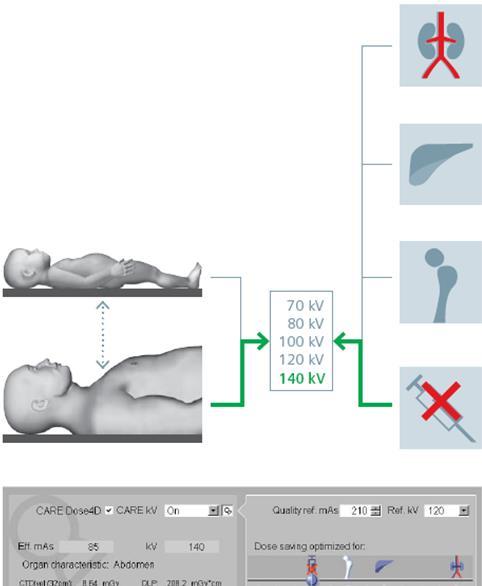
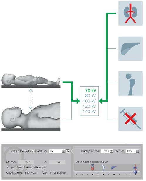
8 On the basis of the attenuation profile the algorithm automatically computes various combinations of tube voltage and tube current that result in the CNR that has been specified. The algorithm selects the most dose-efficient combination.
9
10 Due to current limitations in tube power, larger patients will still require higher kvp to avoid photon starvation Therefore, optimal kvp should be chosen individually for every patient Right dose may mean even higher kv


11 Sperman J, De Cecco C Radiology 2015

12 Virtual Unenhanced Sn 150 kvp 90 kvp Mixed 120 kvp Iodine Map Virtual Monoenergetic

13 Iodine K-edge 33.2 kev 120 kvp 40 kev Stolzmann P Insight Radiol 2011


14 Grant KL Invest Radiol 2014 MONOENERGETIC BASIC 90 kev 110 kev 120 kev 150 kev 190 kev 40 kev 50 kev 60 kev 70 kev 80 kev 90 kev 100 kev 120 kev 150 kev 190 kev MONOENERGETIC PLUS 40 kev 50 kev 60 kev 70 kev 80 kev


15 MONOENERGETIC PLUS MONOENERGETIC BASIC 40 kev 50 kev 40 kev 50 kev 60 kev 70 kev 60 kev 70 kev 90 kev 150 kev 90 kev 150 kev
16

17 80 kvp - 30 ml 120 kvp - 60 ml Ni QQ, De Cecco AJNR 2016


18 80 kvp - 30 ml Ni QQ, De Cecco AJNR 2016


19 70 kvp 30 ml 80 kvp 60 ml 100 kvp 60 ml Zhang LJ, De Cecco CN Medicine 2014


20 BMI BMI BMI >40 Mangold S, De Cecco CN Eur Radiol 2016

21 50 ml CM


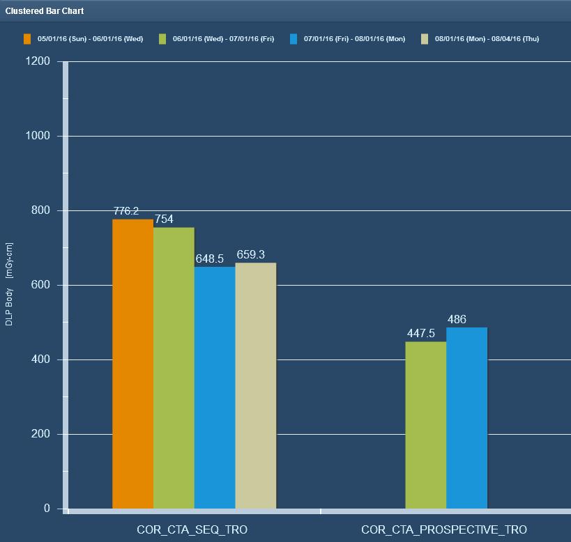
22 20 msv 110 ml CM 3.9 msv 60 ml CM Superior Objective Image Quality Fixed 120 kv - FBP 70 kv ATVS - IR Mangold S, De Cecco CN EJR 2016


23 100 kvp - 60 ml 70 kvp - 40 ml Geyer LL, De Cecco CN Acad Radiol 2015

24 80 kv 50 ml CM


25 Mixed 120 kvp Mono B 40 kev Mono + 40 kev CM 70 ml Wichmann JL, De Cecco Invest Radiol 2015


26 Noda Y Eur J Radiol 2014 Iodine load can be reduced by 33% in CT of the liver with 80 kvp and ASIR technique

27 Combining 80 kvp with IR allows at least a 47% contrast agent dose reduction and 16 % radiation dose reduction for images of comparable quality. Buls N Eur Radiol 2014


28 90 kv 39 M, BMI 21.2 Crohn CM: 100 ml 350 mgi/ml msv 4
29 Low kv/kev protocols can significantly decreased the amount of CM What is the minimum IDR/Contrast Volume we can achieve in clinical practice?
30 120 kv 70 kv 300 mgi/ml 300 mgi/ kg 5 ml/s IDR 1.5 g/s 300 mgi/ml 150 mgi/ kg 2.5 ml/s IDR 0.75 g/s Identical TAC 50% CM reduction in pigs Lell MM Invest Radiol 2015
. Flow rate was adapted to maintain 19 sec of bolus injection.")
31 1 st patients cohort Relative difference of Iodine attenuation at 120 kvp to each tube voltage was used to calculate the amount of CM reduction to achieve equal vascular attenuation ( HU set as target). Flow rate was adapted to maintain 19 sec of bolus injection. Higashigaito K Radiology 2016
32 Results Higashigaito K Radiology 2016


33 Contrast Reduction at Low kvp and kev
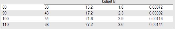
34 Which is the minimum IDR and CM volume required to obtain 350 HU in the aorta at different kv and kev? Two-phases study: 1. Fixed CM different IDRs 2. Fixed IDR lowering CM amount Caruso D, De Cecco CN Submitted

35 All IDRs reached 350 HU (p 0.18)
36 Adequate Time-To-Peak is mandatory
37
38 2 nd approach: Constant IDR and lowering CM amount
39 A combined approach reducing the CM volume for tube voltages <120 kv and increasing the IDR for higher kv settings seems the most effective approach.
40

41 Force Flash Definiton Per-scanner dose monitoring
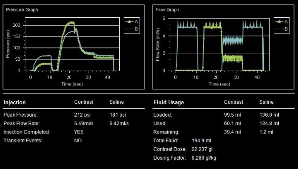
42 Per-Patient Dose Monitoring Protocol Optimization

43 Body Morphology and Weight Right kv/mas Tailored Contrast Medium Dose
")
44 Low kv and kev studies increase Iodine Contrast Enhancement allowing for a significant reduction in contrast media volume New IDR-based injection strategies could further improve contrast medium tailoring Patient-tailored Radiation and Contrast Dose (Kg >> kv/kev >> IDR)
45
Applications of Low KeV Imaging in Abdomen
 Applications of Low KeV Imaging in Abdomen Dushyant Sahani, M.D Director of CT Associate Professor of Radiology Massachusetts General Hospital Harvard Medical School Email-dsahani@partners.org Disclosure
X. Allen Li. Disclosure. DECT: What, how and Why Why dual-energy CT (DECT)? 7/30/2018. Improving delineation and response assessment using DECT in RT
? 7/30/2018. Improving delineation and response assessment using DECT in RT") Improving delineation and response assessment using DECT in RT X. Allen Li Medical College of Wisconsin MO-A-DBRA-1, AAPM, July 30 th, 2018 Disclosure Research funding support: Siemens Healthineers Elekta
Improving delineation and response assessment using DECT in RT X. Allen Li Medical College of Wisconsin MO-A-DBRA-1, AAPM, July 30 th, 2018 Disclosure Research funding support: Siemens Healthineers Elekta
Multi-Energy CT: Principles, Processing
 Multi-Energy CT: Principles, Processing and Clinical Applications Shuai Leng, PhD Associate Professor Department of Radiology Mayo Clinic, Rochester, MN Clinical Motivation CT number depends on x-ray attenuation
Metal Artifact Reduction with DECT
 Metal Artifact Reduction with DECT Daniele Marin, MD Duke University Medical Center Metal artifacts Common clinical problem ( 20%) Boas EF et al. Radiology 2011 Beam Hardening Edge Effects Scatter Metal
Data Acquisition and Image Formation Methods for Multi Energy CT
 Data Acquisition and Image Formation Methods for Multi Energy CT Cynthia H. McCollough, PhD, DABR, FAIMBE, FAAPM, FACR Professor of Medical Physics and Biomedical Engineering Director, CT Clinical Innovation
Metal Artifact Reduction and Dose Efficiency Improvement on Photon Counting Detector CT using an Additional Tin Filter
 Metal Artifact Reduction and Dose Efficiency Improvement on Photon Counting Detector CT using an Additional Tin Filter Wei Zhou, Dilbar Abdurakhimova, Kishore Rajendran, Cynthia McCollough, Shuai Leng
Rad T 290 Worksheet 2
 Class: Date: Rad T 290 Worksheet 2 1. Projectile electrons travel from a. anode to cathode. c. target to patient. b. cathode to anode. d. inner shell to outer shell. 2. At the target, the projectile electrons
Abdominal DECT: How to Integrate Into Your Practice
 Abdominal DECT: How to Integrate Into Your Practice Eric Tamm, M.D. Department of Diagnostic Radiology Division of Diagnostic Imaging MD Anderson Cancer Center Houston, TX Disclosure I have no relationships
Detector. * All clinical images are courtesy of. University, Jerusalem. Ami Altman, Ph.D., and Raz Carmi Ph.D., CT BU, PHILIPS Healthcare
 AD Dual-Energy alenerg CT Based on A Double Layer Detector * All clinical images are courtesy of Hadassah Medical Center, The Hebrew University, Jerusalem Ami Altman, Ph.D., and Raz Carmi Ph.D., CT BU,
A practical approach to the introduction of spectral imaging into a large UK acute care teaching hospital
 A practical approach to the introduction of spectral imaging into a large UK acute care teaching hospital Robert Loader Clinical Scientist Clinical & Radiation Physics Directorate of Healthcare Science
Electron density and effective atomic number images generated by dual energy imaging with a 320-detector CT system: A feasibility study
 Electron density and effective atomic number images generated by dual energy imaging with a 320-detector CT system: A feasibility study Poster No.: C-0403 Congress: ECR 2014 Type: Scientific Exhibit Authors:
Two-Material Decomposition From a Single CT Scan Using Statistical Image Reconstruction
 / 5 Two-Material Decomposition From a Single CT Scan Using Statistical Image Reconstruction Yong Long and Jeffrey A. Fessler EECS Department James M. Balter Radiation Oncology Department The University
ESTIMATION OF 90 SCATTERING COEFFICIENT IN THE SHIELDING CALCULATION OF DIAGNOSTIC X-RAY EQUIPMENT
 Proceedings of the Eleventh EGS4 Users' Meeting in Japan, KEK Proceedings 2003-15, p.107-113 ESTIMATION OF 90 SCATTERING COEFFICIENT IN THE SHIELDING CALCULATION OF DIAGNOSTIC X-RAY EQUIPMENT K. Noto and
DUAL ENERGY of the Pancreas
 DUAL ENERGY of the Pancreas Desiree E. Morgan, MD Professor and Vice Chair Clinical Research Director Human Imaging Shared Facility UAB CCC University of Alabama at Birmingham Opportunities to improve?
X-ray Interaction with Matter
 X-ray Interaction with Matter 10-526-197 Rhodes Module 2 Interaction with Matter kv & mas Peak kilovoltage (kvp) controls Quality, or penetrating power, Limited effects on quantity or number of photons
Assessment of an Advanced Monoenergetic Reconstruction Technique in Dual-Energy Computed Tomography of Head and Neck Cancer
 DOI 10.1007/s00330-015-3627-1 HEAD AND NECK Assessment of an Advanced Monoenergetic Reconstruction Technique in Dual-Energy Computed Tomography of Head and Neck Cancer Moritz H. Albrecht & Jan-Erik Scholtz
Differential Absorption Analysis of Nonmagnetic Material in the Phantom using Dual CT
 Journal of Magnetics 21(2), 286-292 (2016) ISSN (Print) 1226-1750 ISSN (Online) 2233-6656 http://dx.doi.org/10.4283/jmag.2016.21.2.286 Differential Absorption Analysis of Nonmagnetic Material in the Phantom
Midterm Radiation Physics
 Name: Student ID: CHIR 204 Radiographic Physics and Protection Midterm Radiation Physics Friday 12 September, 2003 DO NOT TURN OVER THIS PAGE UNTIL INSTRUCTED TO DO SO Meanwhile, fill in your name and
Comparison of Polychromatic and Monochromatic X-rays for Imaging
 Comparison of Polychromatic and Monochromatic X-rays for Imaging M. Hoheisel 1, P. Bernhardt 1, R. Lawaczeck 2, and H. Pietsch 2 1 Siemens AG Medical Solutions, Forchheim, Germany 2 Schering AG Imaging
1. Which of the following statements is true about Bremsstrahlung and Characteristic Radiation?
 BioE 1330 - Review Chapters 4, 5, and 6 (X-ray and CT) 9/27/2018 Instructions: On the Answer Sheet, enter your 2-digit ID number (with a leading 0 if needed) in the boxes of the ID section. Fill in the
CT-PET calibration : physical principles and operating procedures F.Bonutti. Faustino Bonutti Ph.D. Medical Physics, Udine University Hospital.
 CT-PET calibration : physical principles and operating procedures Faustino Bonutti Ph.D. Medical Physics, Udine University Hospital Topics Introduction to PET physics F-18 production β + decay and annichilation
11/19/2014. Chapter 3: Interaction of Radiation with Matter in Radiology and Nuclear Medicine. Nuclide Families. Family Nuclides with Same: Example
 2014-2015 Residents' Core Physics Lectures Mondays 7:00-8:00 am in VA Radiology and UCSDMC Lasser Conference Rooms Topic Chapters Date Faculty 1 Introduction and Basic Physics 1, 2 M 11/17 Andre 2 Interaction
Multi-energy CT: Future Directions. Acknowledgements. Overview 7/23/2014. Taly Gilat Schmidt, PhD. Kevin Zimmerman Franco Rupcich Steven Haworth
 Multi-energy CT: Future Directions Taly Gilat Schmidt, PhD Department of Biomedical Engineering Marquette University Acknowledgements Kevin Zimmerman Franco Rupcich Steven Haworth Results in this talk:
Computation of energy imparted in diagnostic radiology
 Computation of energy imparted in diagnostic radiology Nikolaos A. Gkanatsios and Walter Huda a) Department of Radiology, University of Florida College of Medicine, P.O. Box 100374, Gainesville, Florida
Theoretical analysis of comparative patient skin dose and exposure technique approaches in planar radiography
 Exposure technique Australian Institute of Radiography The Radiographer 2009; 56 (1): 21 26 Theoretical analysis of comparative patient skin dose exposure technique approaches in planar radiography Faculty
Design of a virtual model of a hand-held Germanium detector and a voxelized ICRP whole body phantom: A Monte Carlo study
 Design of a virtual model of a hand-held Germanium detector and a voxelized ICRP whole body phantom: A Monte Carlo study ASM SABBIR AHMED 1, Gary H Kramer 2, Kurt Ungar 2 1 University of Saskatchewan,
Document downloaded from: This paper must be cited as:
 Document downloaded from: http://hdl.handle.net/10251/47496 This paper must be cited as: Querol Vives, A.; Gallardo Bermell, S.; Ródenas Diago, J.; Verdú Martín, GJ. (2011). Parametric study of the X-ray
Dosimetry. Sanja Dolanski Babić May, 2018.
 Dosimetry Sanja Dolanski Babić May, 2018. What s the difference between radiation and radioactivity? Radiation - the process of emitting energy as waves or particles, and the radiated energy Radioactivity
Radionuclide Imaging MII Positron Emission Tomography (PET)
") Radionuclide Imaging MII 3073 Positron Emission Tomography (PET) Positron (β + ) emission Positron is an electron with positive charge. Positron-emitting radionuclides are most commonly produced in cyclotron
Radionuclide Imaging MII 3073 Positron Emission Tomography (PET) Positron (β + ) emission Positron is an electron with positive charge. Positron-emitting radionuclides are most commonly produced in cyclotron
EXPERIMENTAL DETERMINATION OF SHIELDING REQUIREMENTS FOR PET MEDICAL FACILITIES BRADLEY S. BRINKLEY
 EXPERIMENTAL DETERMINATION OF SHIELDING REQUIREMENTS FOR PET MEDICAL FACILITIES by BRADLEY S. BRINKLEY CLAUDIU T. LUNGU, COMMITTEE CHAIR ALFRED A. BARTOLUCCI STEVEN M. BECKER RIEDAR K. OESTENSTAD SHARON
Geometric series and effective medicine dosage
 Geometric series and effective medicine dosage Introduction This lab concerns a model for a drug being given to a patient at regular intervals. As the drug is broken down by the body, its concentration
This is the last of our four introductory lectures. We still have some loose ends, and in today s lecture, we will try to tie up some of these loose
 This is the last of our four introductory lectures. We still have some loose ends, and in today s lecture, we will try to tie up some of these loose ends. 1 We re going to cover a variety of topics today.
Development of Radioactivity Standards for Quantitative Positron Emission Tomography
 Development of Radioactivity Standards for Quantitative Positron Emission Tomography Brian E. Zimmerman, PhD Radiation Physics Division National Institute of Standards and Technology Gaithersburg, MD 20899-8462
H e a l t h P h y s i c s S e r v i c e s L t d
 1 Title: Adept Medical X-Ray Shield: Scatter Radiation Shielding Grid Format Methodology 2 Background: Adept Medical X-Ray Shield (X-Ray Shield) is embedded with 0.5mm Lead (pb), offering protection from
Radiation Safety and Protection for I-125 Seeding localization: A Day in the Life of a Seed 2013 Update
 Radiation Safety and Protection for I-125 Seeding localization: A Day in the Life of a Seed 2013 Update Department of Education and Clinical Practice High Impact Learning for Remarkable, Compassionate
Evaluation of Phantom Equivalent Materials in Polychromatic Diagnostic X-Ray Beam
 Evaluation of Phantom Equivalent Materials in Polychromatic Diagnostic X-Ray Beam Radhakrishnan B Nair 1*, Ramakrishnan G 2, Chandralingam S 3 and Kurup PGG 1 1 Apollo Speciality Hospital, Chennai, India
1-D Fourier Transform Pairs
 1-D Fourier Transform Pairs The concept of the PSF is most easily explained by considering a very small point source being placed in the imaging field-of-view The relationship between the image, I, and
Physics of Radiography
 EL-GY 6813 / BE-GY 6203 / G16.4426 Medical Imaging Physics of Radiography Jonathan Mamou and Yao Wang Polytechnic School of Engineering New York University, Brooklyn, NY 11201 Based on Prince and Links,
Introduction to SPECT & PET TBMI02 - Medical Image Analysis 2017
 Introduction to SPECT & PET TBMI02 - Medical Image Analysis 2017 Marcus Ressner, PhD, Medical Radiation Physicist, Linköping University Hospital Content What is Nuclear medicine? Basic principles of Functional
Konzepte zur Charakterisierung klinischer CT-Systeme unter Einbeziehung von Bildqualität und Dosis
 Konzepte zur Charakterisierung klinischer CT-Systeme unter Einbeziehung von Bildqualität und Dosis Characterization of clinical CT systems using a dose efficiency index (DEI) Aktenzeichen / FKZ : BfS AG-F
Radiation Detection and Measurement
 Radiation Detection and Measurement June 2008 Tom Lewellen Tkldog@u.washington.edu Types of radiation relevant to Nuclear Medicine Particle Symbol Mass (MeV/c 2 ) Charge Electron e-,! - 0.511-1 Positron
Heuijin Lim, Manwoo Lee, Jungyu Yi, Sang Koo Kang, Me Young Kim, Dong Hyeok Jeong
 Original Article PMP Progress in Medical Physics 28(2), June 2017 https://doi.org/10.14316/pmp.2017.28.2.49 pissn 2508-4445, eissn 2508-4453 Electron Energy Distribution for a Research Electron LINAC Heuijin
October 4th, Bioengineering 508: Physical Aspects of Medical Imaging Some Elementary Particles
 Bioengineering 508: Physical Aspects of Medical Imaging http://courses.washington.edu/bioen508/ For questions, remarks, discussions, errors in the book: Class Discussion Board (link from class website)
Radhakrishnan B*, Kurup P G G**, Ramakrishnan G***, Chandralingam S****
 Photon Interaction Cross Section of Materials in Heterogeneous Energy Spectrum of Medical Diagnostic X-Ray Beam Radhakrishnan B*, Kurup P G G**, Ramakrishnan G***, Chandralingam S**** *Senior Medical Physicist,
Health Physics Services Ltd
 1 Title: Adept Medical STARTable: Scatter Radiation Shielding Grid Format Methodology 2 Background: Adept Medical STARTable Shield is embedded with 0.5mm Lead (Pb), offering protection from scatter radiation
Technical University of Denmark
 Technical University of Denmark Page 1 of 11 pages Written test, 9 December 2010 Course name: Introduction to medical imaging Course no. 31540 Aids allowed: none. "Weighting": All problems weight equally.
1Meherun Nahar, Sazzad, 3Abdus Sattar Mollah and 4Mir Mohammad Akramuzzaman Correspondence Address INTRODUCTION MATERIALS AND METHODS Mathematical
 ORIGINAL ARTICLE Development of MGDA Software for Calculation of Patient Specific Mean Glandular Dose during Mammography 1Meherun Nahar, 2 M Sazzad, 3 Abdus Sattar Mollah and 4 Mir Mohammad Akramuzzaman
Determination of the mass attenuation coefficient for the contrast agent Iohexol using 662 kev photons from a Cesium-137 source.
 Determination of the mass attenuation coefficient for the contrast agent Iohexol using 662 kev photons from a Cesium-137 source. Elizabeth Cañipa *1, 2, Eric Farías 1, 2, Oscar Hernández 1, 3, Echevarria
Physics of Radiography
 Physics of Radiography Yao Wang Polytechnic Institute of NYU Brooklyn, NY 11201 Based on J L Prince and J M Links Medical Imaging Signals and Based on J. L. Prince and J. M. Links, Medical Imaging Signals
Measurement of backscatter factor for kilovoltage x-ray beam using ionization chamber and Gafchromic XR-QA2 film
 IOP Conference Series: Materials Science and Engineering PAPER OPEN ACCESS Measurement of backscatter factor for kilovoltage x-ray beam using ionization chamber and Gafchromic XR-QA2 film To cite this
DEVIL PHYSICS THE BADDEST CLASS ON CAMPUS IB PHYSICS
 DEVIL PHYSICS THE BADDEST CLASS ON CAMPUS IB PHYSICS TSOKOS OPTION I-2 MEDICAL IMAGING Reading Activity Answers IB Assessment Statements Option I-2, Medical Imaging: X-Rays I.2.1. I.2.2. I.2.3. Define
Measurement of bone mineral in vivo: An improved method*
 Cl á s i c o Cameron JR, Sorenson J Measurement of bone mineral in vivo: An improved method* John R. Cameron, (1 James Sorenson. (1 Abstract The mineral content of bone can be determined by measuring the
LECTURE 4 PRINCIPLE OF IMAGE FORMATION KAMARUL AMIN BIN ABDULLAH
 LECTURE 4 PRINCIPLE OF IMAGE FORMATION KAMARUL AMIN BIN ABDULLAH Lesson Objectives At the end of the lesson, student should able to: Define attenuation Explain interactions between x-rays and matter in
Biomedical Imaging. X ray imaging. Patrícia Figueiredo IST
 Biomedical Imaging X ray imaging Patrícia Figueiredo IST 2013-2014 Overview Production of X rays Interaction of electrons with matter X ray spectrum X ray tube Interaction of X rays with matter Photoelectric
We have seen how the Brems and Characteristic interactions work when electrons are accelerated by kilovolts and the electrons impact on the target
 We have seen how the Brems and Characteristic interactions work when electrons are accelerated by kilovolts and the electrons impact on the target focal spot. This discussion will center over how x-ray
Coconuts, grapes, and peppers: Home-made models in the learning process of post-processing softwares used in computed-tomography (CT) imaging
 imaging") Coconuts, grapes, and peppers: Home-made models in the learning process of post-processing softwares used in computed-tomography (CT) imaging Poster No.: C-3019 Congress: ECR 2010 Type: Educational Exhibit
Coconuts, grapes, and peppers: Home-made models in the learning process of post-processing softwares used in computed-tomography (CT) imaging Poster No.: C-3019 Congress: ECR 2010 Type: Educational Exhibit
What is scintigraphy? The process of obtaining an image or series of sequential images of the distribution of a radionuclide in tissues, organs, or
 Let's remind... What is nuclear medicine? Nuclear medicine can be broadly divided into two branches "in vitro" and "in vivo" procedures. There are numerous radioisotopic "in vitro" procedures for genotyping
Radiation protection in the endosuite and the importance of correct use of shields
 Radiation protection in the endosuite and the importance of correct use of shields Anders Wanhainen Professor of Surgery Chief dep. of Vascular Surgery Uppsala University Hospital Sweden High radiation
Initial Certification
 Initial Certification Medical Physics Part 1 Content Guide Part 1 Content Guides and Sample Questions PLEASE NOTE: List of Constants and Physical Values for Use on the Part 1 Physics Exam The ABR provides
FXA UNIT G485 Module X-Rays. Candidates should be able to : I = I 0 e -μx
 1 Candidates should be able to : HISTORY Describe the nature of X-rays. Describe in simple terms how X-rays are produced. X-rays were discovered by Wilhelm Röntgen in 1865, when he found that a fluorescent
Matter, energy, power and heat Units and prefixes used in radiography Radiological units Useful mathematics Proportions and the inverse square law
 Chapter 1 Essential Mathematics and Physics Introduction Chapter contents Matter, energy, power and heat Units and prefixes used in radiography Radiological units Useful mathematics Proportions and the
Comparison of Primary Doses Obtained in Three 6 MV Photon Beams Using a Small Attenuator
 Comparison of Primary Doses Obtained in Three 6 MV Photon Beams Using a Small Attenuator Christoph Trauernicht Groote Schuur Hospital & University of Cape Town Method is based on: Background A method of
11/10/2014. Chapter 1: Introduction to Medical Imaging. Projection (Transmission) vs. Emission Imaging. Emission Imaging
 vs. Emission Imaging. Emission Imaging") Chapter 1: Introduction to Medical Imaging Overview of Modalities Properties of an Image: Limitations on Information Content Contrast (both object & image): Brightness difference Sharpness (blur): Smallest
Chapter 1: Introduction to Medical Imaging Overview of Modalities Properties of an Image: Limitations on Information Content Contrast (both object & image): Brightness difference Sharpness (blur): Smallest
7/30/2018. CT Hardware Basics. X-ray Tube Physics and Dose Reduction
 J. Webster Stayman Advanced Imaging Algorithms and Instrumentation Lab (aiai.jhu.edu) Johns Hopkins University August 30, 018 CT Hardware Basics X-ray Tube Detector (image Credit) https://www.youtube.com/watch?v=bg0inhwarw
Quality-Assurance Check of Collimator and Phantom- Scatter Factors
 Quality-Assurance Check of Collimator and Phantom- Scatter Factors Ramesh C. Tailor, David S. Followill, Nadia Hernandez, Timothy S. Zhu, and Geoffrey S. Ibbott. UT MD Anderson Cancer Center, Houston TX.
Bases of radioisotope diagnostic methods
 Medical, pharmaceutical applications of radioisotopes Bases of radioisotope diagnostic methods Dr. István Voszka Basis of application: radioisotopes have identical behavior in the organism to corresponding
STANDARD WATER PHANTOM BACKSCATTER FACTORS FOR MEDIUM ENERGY X-RAYS
 STANDARD WATER PHANTOM BACKSCATTER FACTORS FOR MEDIUM ENERGY X-RAYS M.A. HASSAN*, M.H. GABER**, E. ESMAT*, H.I. FARAG***, H.M. EISSA* *National Institute for Standards (NIS), Giza, Egypt **Biophysics Department,
Radioactive Decedents What is the risk?
 Radioactive Decedents What is the risk? Glenn M. Sturchio, PhD, CHP Radiation Safety Officer Alan Crutchfield Clinical Research Intern ICCFA Annual Convention & Expo Nashville, TN 08 April 2017 2017 MFMER
Absorption spectra variations of EBT radiochromic film from radiation exposure
 INSTITUTE OF PHYSICS PUBLISHING Phys. Med. Biol. 5 (25) N35 N4 PHYSICS IN MEDICINE AND BIOLOGY doi:.88/3-955/5/3/n2 NOTE Absorption spectra variations of EBT radiochromic film from radiation exposure M
Procesamiento de Imágenes y Bioseñales
 Procesamiento de Imágenes y Bioseñales Dr. Víctor Castañeda Agenda Physical basis of X-ray- CT, NMR, Ultrasound, Nuclear Medicine Sensors (cameras, gamma probes, microphone) Computational Tomography (CT)
Sensitivity of the IRD whole-body counter for in vivo measurements in the case of accidental intakes
 Sensitivity of the IRD whole-body counter for in vivo measurements in the case of accidental intakes B.M. Dantas, E.A. Lucena and A.L.A. Dantas Laboratório de Monitoração In Vivo Divisão de Dosimetria
Reducing metal artefacts and radiation dose in musculoskeletal CT imaging Wellenberg, R.H.H.
 UvA-DARE (Digital Academic Repository) Reducing metal artefacts and radiation dose in musculoskeletal CT imaging Wellenberg, R.H.H. Link to publication Citation for published version (APA): Wellenberg,
USE OF DLP FOR ESTABLISHING THE SHIELDING OF MULTI- DETECTOR COMPUTED TOMOGRAPHY ROOMS
 USE OF DLP FOR ESTABLISHING THE SHIELDING OF MULTI- DETECTOR COMPUTED TOMOGRAPHY ROOMS F.R. Verdun 1, A. Aroua 1, P.R. Trueb 2, F.O. Bochud 1* 1 University Institute for Radiation Physics, Switzerland
Outline. Indrin J. Chetty, AAPM 2006 Monte Carlo CE course. Indrin J. Chetty Henry Ford Hospital. David W. O. Rogers Carleton University
 AAPM Task Group Report No. 105: Issues associated with clinical implementation of Monte Carlo-based photon and electron external beam treatment planning Indrin J. Chetty Henry Ford Hospital David W. O.
Solid State LightBurst New PET Technology GE PET/CT and PET/MR
 Solid State LightBurst New PET Technology GE PET/CT and PET/MR Osama Mawlawi PhD. Dept. of Imaging Physics MD Anderson Cancer Center Disclosures SIEMENS Research grant GE research grant Discovery MI LYSO
The potential of dual-energy computed tomography for quantitative decomposition of soft tissues to water, protein and lipid in brachytherapy
 The potential of dual-energy computed tomography for quantitative decomposition of soft tissues to water, protein and lipid in brachytherapy Alexandr Malusek, M Karlsson, Maria Magnusson and Gudrun Alm
MEDICAL EQUIPMENT: NUCLEAR MEDICINE. Prof. Yasser Mostafa Kadah
 MEDICAL EQUIPMENT: NUCLEAR MEDICINE Prof. Yasser Mostafa Kadah www.k-space.org Recommended Textbook Introduction to Medical Imaging: Physics, Engineering and Clinical Applications, by Nadine Barrie Smith
METHODS OF ASSESSING PAST EXTERNAL EXPOSURES FROM ENVIRONMENTAL CONTAMINATION OF THE TECHA RIVER IN THE FORMER U.S.S.R.
 METHODS OF ASSESSING PAST EXTERNAL EXPOSURES FROM ENVIRONMENTAL CONTAMINATION OF THE TECHA RIVER IN THE FORMER U.S.S.R. April 10 th, 2015 BC Schwarz, M.S. Advanced Laboratory for Radiation Dosimetry Studies
Prompt gamma measurements for the verification of dose deposition in proton therapy. Contents. Two Proton Beam Facilities for Therapy and Research
 Prompt gamma measurements for the verification of dose deposition in proton therapy Two Proton Beam Facilities for Therapy and Research Ion Beam Facilities in Korea 1. Proton therapy facility at National
Henry Ford NERS/BIOE 481. Lecture 05 Radiographic Image Formation
 NERS/BIOE 481 Lecture 05 Radiographic Image Formation Michael Flynn, Adjunct Prof Nuclear Engr & Rad. Science mikef@umich.edu mikef@rad.hfh.edu Henry Ford Health System RADIOLOGY RESEARCH IV - General
CHAPTER 4 RADIATION ATTENUATION
 HDR202 PHYSICS FOR RADIOGRAPHERS 2 CHAPTER 4 RADIATION ATTENUATION PREPARED BY: MR KAMARUL AMIN BIN ABDULLAH SCHOOL OF MEDICAL IMAGING FACULTY OF HEALTH SCIENCES Learning Objectives At the end of the lesson,
Nuclear Instruments and Methods in Physics Research A 417 (1998) 86 94
 86 94") Nuclear Instruments and Methods in Physics Research A 417 (1998) 86 94 Experimental determination of detector gain, zero frequency detective quantum efficiency, and spectral compatibility of phosphor screens:
Nuclear Instruments and Methods in Physics Research A 417 (1998) 86 94 Experimental determination of detector gain, zero frequency detective quantum efficiency, and spectral compatibility of phosphor screens:
Basic principles of x-ray production
 Production of X-Rays part 1 George Starkschall, Ph.D. Lecture Objectives Identify what is needed to produce x-rays Describe how a diagnostic x-ray tube produces x-rays Describe the types of interactions
Radiation protection issues in proton therapy
 Protons IMRT Tony Lomax, Centre for Proton Radiotherapy, Paul Scherrer Institute, Switzerland Overview of presentation 1. Proton therapy: An overview 2. Radiation protection issues: Staff 3. Radiation
COMPARISON OF PERSONNEL RADIATION MONITORING DOSIMETERS DESIGNED FOR MEDICAL FIELD
 COMPARISON OF PERSONNEL RADIATION MONITORING DOSIMETERS DESIGNED FOR MEDICAL FIELD Kirill SKOVORODKO, Birute GRICIENE, Milda PETKELYTE Radiation Protection Division, Vilnius University Hospital Santaros
CALCULATION OF SHIELDING AND RADIATION DOSES FOR PET/CT NUCLEAR MEDICINE FACILITY
 International Conference on Mathematics and Computational Methods Applied to Nuclear Science and Engineering (M&C 2011) Rio de Janeiro, RJ, Brazil, May 8-12, 2011, on CD-ROM, Latin American Section (LAS)
Radiation Safety In-Service House-Keeping and Security Departments. Petrone Associates LLC Specialists in Applied Medical Physics
 Radiation Safety In-Service House-Keeping and Security Departments 1 MEDICAL RADIATION SOURCES Radiation Producing Machines Radioactive Materials 2 Nuclear Medicine Radiation Therapy Research Laboratories
Neural Network Approach for Photon-counting Detection The First Step: PPE Correction
 Neural Network Approach for Photon-counting Detection The First Step: PPE Correction Ruibin Feng, Ph.D. Biomedical Imaging Center, CBIS/BME, RPI fengr@rpi.edu David Rundle JairiNovus Technologies Ltd.
Potentials for Dual-energy kv/mv On-board Imaging and Therapeutic Applications
 Potentials for Dual-energy kv/mv On-board Imaging and Therapeutic Applications Fang-Fang Yin Department of Radiation Oncology Duke University Medical Center Acknowledgement Dr Hao Li for his excellent
Reconstruction for Proton Computed Tomography: A Monte Carlo Study
 Reconstruction for Proton Computed Tomography: A Monte Carlo Study T Li, Z. Liang, K. Mueller, J. Heimann, L. Johnson, H. Sadrozinski, A. Seiden, D. Williams, L. Zhang, S. Peggs, T. Satogata, V. Bashkirov,
Expand the probe idea functionality (above points) to larger imaging systems. First Applications, Probes, Isotopes
 to larger imaging systems. First Applications, Probes, Isotopes") Gamma Ray and Beta Ray Probes Larry MacDonald Imaging Research Laboratory University of Washington Radiology http://depts.washington.edu/nucmed/irl/ 23 Oct. 2007 Oct. 2007 1 Gamma Ray and Beta Ray Probes
Gamma Ray and Beta Ray Probes Larry MacDonald Imaging Research Laboratory University of Washington Radiology http://depts.washington.edu/nucmed/irl/ 23 Oct. 2007 Oct. 2007 1 Gamma Ray and Beta Ray Probes
Outline. Absorbed Dose in Radioactive Media. Introduction. Radiation equilibrium. Charged-particle equilibrium
 Absorbed Dose in Radioactive Media Chapter F.A. Attix, Introduction to Radiological Physics and Radiation Dosimetry Outline General dose calculation considerations, absorbed fraction Radioactive disintegration
A. I, II, and III B. I C. I and II D. II and III E. I and III
 BioE 1330 - Review Chapters 7, 8, and 9 (Nuclear Medicine) 9/27/2018 Instructions: On the Answer Sheet, enter your 2-digit ID number (with a leading 0 if needed) in the boxes of the ID section. Fill in
Part 01 Introduction to X-ray imaging. Overview Module 07 Part 1 X-ray
 1 Introduction to Medical Image Processing (5XSA0), Module 07 Part 01 Introduction to X-ray imaging Peter H.N. de With (p.h.n.de.with@tue.nl ) With Contributions from D. Ruijters of Philips Healthcare
Chapter 2 PET Imaging Basics
 Chapter 2 PET Imaging Basics Timothy G. Turkington PET Radiotracers Positron emission tomography (PET) imaging is the injection (or inhalation) of a substance containing a positron emitter, the subsequent
Proposed Room Requirements for CT System
 Siemens Proposed Room Requirements for CT System Semarang, 4-5 May 2017 Restricted Siemens Healthcare GmbH, 2016 Page 1 Roles of Medical Physicist CT Image Quality Radiation Protection Optimization Medical
Gy can be used for any type of radiation. Gy does not describe the biological effects of the different radiations.
 Absorbed Dose Dose is a measure of the amount of energy from an ionizing radiation deposited in a mass of some material. SI unit used to measure absorbed dose is the gray (Gy). 1J 1 Gy kg Gy can be used
ATTENUATION STUDIES ON DRY AND HYDRATED CROSS-LINKED HYDROPHILIC COPOLYMER MATERIALS AT 8.02 TO kev USING X-RAY FLUORESCENT SOURCES
 Journal of Physical Science, Vol. 18(1), 23 32, 27 23 ATTENUATION STUDIES ON DRY AND HYDRATED CROSS-LINKED HYDROPHILIC COPOLYMER MATERIALS AT 8.2 TO 28.43 kev USING X-RAY FLUORESCENT SOURCES Sabar Bauk
Mayneord-Phillips Summer School St Edmund Hall, University of Oxford July Proton decays to n, e +, ν
 Positron Emission Tomography Physics & Instrumentation Dimitra G. Darambara, Ph.D Multimodality Molecular Imaging Joint Department of Physics RMH/ICR Outline Introduction PET Physics overview Types of
A new neutron monitor for pulsed fields at high-energy accelerators
 A new neutron monitor for pulsed fields at high-energy accelerators Marlies Luszik-Bhadra *, Eike Hohmann Physikalisch-Technische Bundesanstalt, Bundesallee 100, D-38116, Braunschweig, Germany. Abstract.