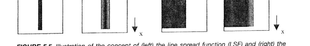
1-D Fourier Transform Pairs
|
|
|
- Christian Jones
- 5 years ago
- Views:
Transcription

1 1-D Fourier Transform Pairs
2
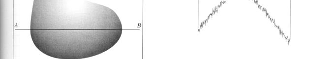
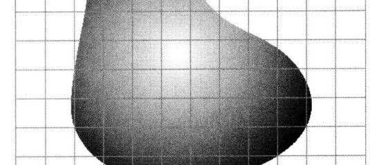
3 The concept of the PSF is most easily explained by considering a very small point source being placed in the imaging field-of-view The relationship between the image, I, and the object, O, can be represented by: I x,y,z = O x,y,z *h x,y,z ( ) ( ) ( ) where * represents a convolution, and h(x,y,z) is the three-dimensional PSF. Ideally, the PSF would be a delta function in all three dimensions, and the image and object would be identical. Object y z x Image (left) the object to be imaged consists of a small sphere. (right) the image obtained is larger than the actual object, and may be blurred asymmetrically in the x, y and z dimensions. In this particular case the PSF is shown as being broad in the x direction and relatively narrow in the z and y directions.
4
5
6


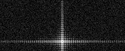

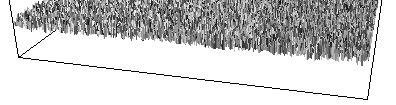
7 Object with noise 2-D Fourier Transform


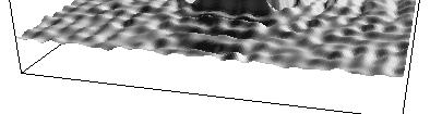
8 Low-Pass Filter Inverse Fourier Transform


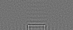
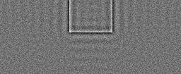
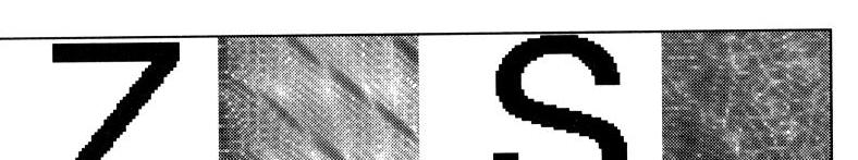
9 High-Pass Filter Inverse Fourier Transform


10 Bandpass Filter Inverse Fourier Transform
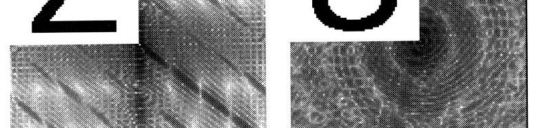
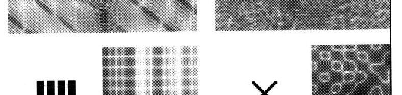
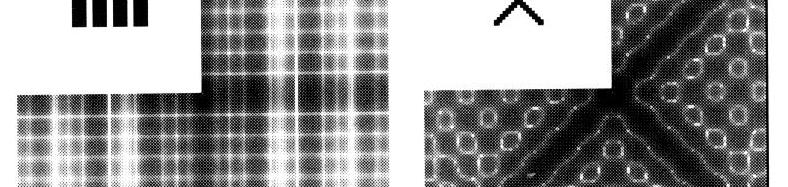
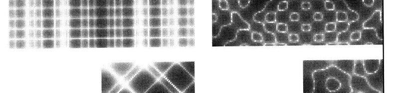
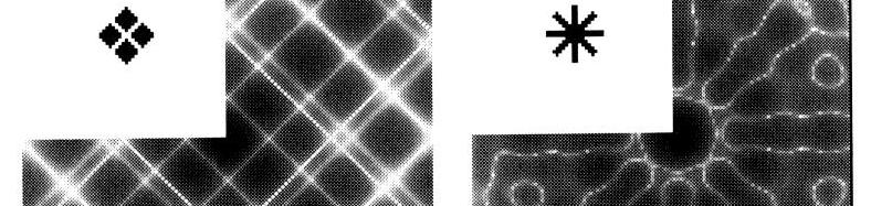
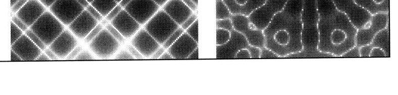
11 Two-Dimensional o Fourier Transform Pairs
12 2-D Fourier Transform Pairs


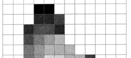
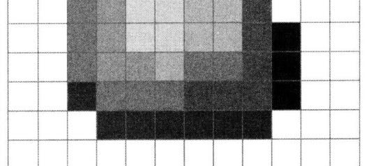
13 Sampling and Quantization

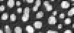
14 Sampling and Quantization


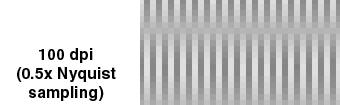


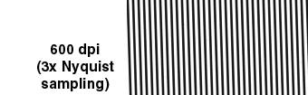
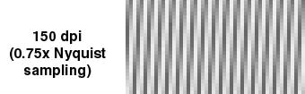
15 Sampling and Quantization

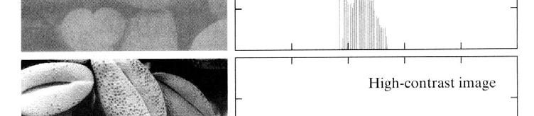
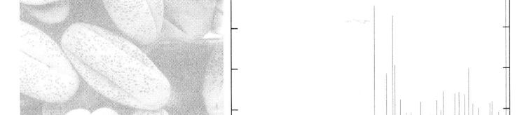
16 Sampling and Quantization Histograms
17 Image Filtering Original Original + Noise
18 Image Filtering Noisy Image Low-Pass Filtered
19 Image Filtering Original Low-Pass Filtered
20 Image Filtering Blurred Image High-Pass Filtered
21 Image Filtering Original Edge-Detection
22 Chapter 1: Medical Imaging: X-rays, CT Note: The class will be taught using the black board instead of using multiple power point slides. However, there is often the time where pictures are just too difficult to draw (e.g. images) and therefore power point will be used. These slides are taken from information directly from your text and will not take the place of taking notes in class. Therefore, it is NOT necessary to print these slides for the lecture.
23 X-ray and Computed Tomography (CT) Conventional x-ray: chest x-ray, dental x- ray Fluoroscopy, Angiography, Mammography Conventional and computed tomography Spiral or helical CT


24 X-rays were discovered by a German physicist, W. K. Roentgen, in 1895 and received the Nobel Prize in University of Wuerzburg One of his earliest photographic plate from his experiments was a film of his wife, Bertha's hand with a ring, was produced on Friday, November 8, 1895.

25 x-ray source low attenuation, low ρ collimator high attenuation, high ρ anti-scatter grid x-ray film (left) Basic set-up for x-ray imaging. The collimator restricts the beam of x-rays, and an anti-scatter grid increases image contrast by reducing the contribution from x-rays that have been scattered, rather than absorbed, by tissue. (right) Contrast: A typical chest x-ray radiograph in which the highly attenuating regions of bone appear white.
26 X-ray wave properties Wavelength, propagation speed, and frequency f λ = c f = frequency Hz (or ν) Particle: E = hf, hν (or photon) λ = wavelength, m c = light speed =3x10 8 m/s 18 h = Plank constant = kev s 19 1 ev = ( J) Joule
 is smaller than the electron")
27 Major components of an x-ray tube. The tube is typically surrounded by an oil bath and lead housing. The magnified view of the target illustrates the line focus principle, whereby the focal spot size (F) is smaller than the electron beam (L) because of the anode angle.
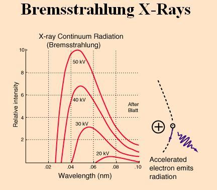
28 X-ray Production -Diagnostic x-rays are produced when electrons with energies of 20 to 150 kev are stopped in matter. -The kinetic energy of the electron is transformed into heat and x-rays when the electrons strike the anode (tungsten). -Electrons rapidly lose their energy by ionization (loose e - ) and excitation (add energy, low to hi nrg system) of electrons in the anode material. -X-rays are generated by two different processes known as bremsstrahlung and characteristic x-ray production.
29
30
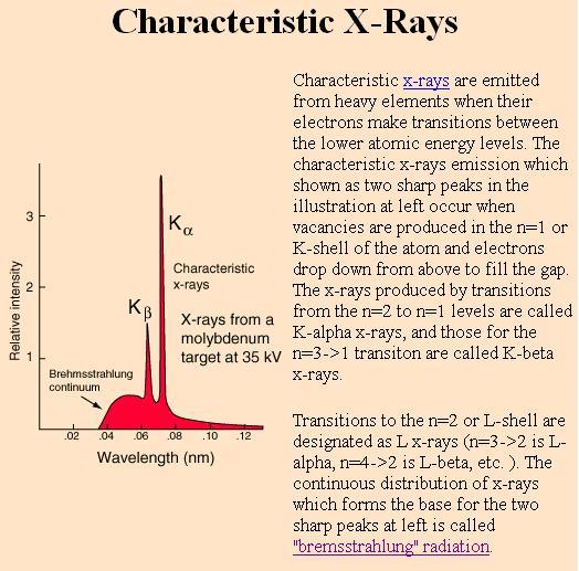
31 Bremsstrahlung (breaking, auf Deutsch, or general) x-rays are produced when incident electrons interact with electric fields, which slow them down and change their direction. -Bremsstrahlung x-rays produce a continuous spectrum of radiation, up to a maximum energy determined by the maximum kinetic energy of the incident electron. -Bremsstrahlung x-ray production increases with the accelerating voltage (kv) and the atomic number (Z) of the anode. tungsten Bremsstrahlung radiation is produced when an energetic electron (1) (with initial energy E1,) passes close to an atomic nucleus. The attractive force of the positively charged nucleus causes the electron to change direction and lose energy. The electron (2) now has a lower energy (E2). The energy difference (E1 -E2) is released as an x-ray photon (3).
32 -Characteristic radiation is produced when inner shell electrons of the anode target are ejected by the incident electrons. -The resultant vacancies are filled by outer shell electrons, and the energy difference is emitted as characteristic radiation as shown below. -Each anode material emits characteristic x-rays of a given energy. -K-shell electrons are ejected only if incident electrons have energies greater than the K-shell binding energy. M L K nucleus Atomic structure showing the maximum number of electrons that can occupy the K (2), L (8) and M (18) shells.
33 Characteristic radiation (cont) -L-shell radiation also normally accompanies K-shell radiation. L-shell characteristic x-rays have very low energies and are absorbed by the glass of the x-ray tube. Only K-shell characteristic x-rays are important in diagnostic radiology. -Most incident electrons interact with outer shell electrons and produce heat but not x-rays. Characteristic radiation is produced when an incoming electron (1) interacts with an inner shell electron (2) and both are ejected (3). When one of the outer shell electrons moves to fill the inner shell vacancy, the excess energy is emitted as characteristic radiation (4).
34
35
36 Interaction of x-ray and matter Scattering and absorption X-ray photon E Nucleus Electrons X-ray photon E E < E Absorption = Photoelectric effect Scattered = Compton scatter

37 X-ray interactions When passing through matter photons may: pass through (i.e. penetrate) absorbed (and x-fer nrg) scattered (change direction and loose nrg) Compton scatter and photoelectric (PE) effect are the important interaction in diagnostic radiology. Others are coherent scatter pair production photodisintegration
 is totally absorbed by an inner shell electron, which is ejected as a photoelectron (2).")
38 X-ray interactions Photoelectric (PE) effect occurs between tightly bound (inner shell) electrons and the incident x-ray photons. The PE effect occurs when an incident x-ray (1) is totally absorbed by an inner shell electron, which is ejected as a photoelectron (2). The vacancy (3) is filled by an outer shell electron, and the energy difference is emitted as characteristic radiation (4) or as an Auger electron (5).
39 X-ray interactions In Compton scatter, incident photons interact with loosely bound (outer shell) electrons. -A Compton interaction results in a scattered photon that has less energy than the incident photon and travels in a new direction. - A scattered electron carries the energy lost by the incident photon. (4) -This electron loses energy by ionizing other atoms in the tissue, thereby contributing to the patient dose. incoming x-ray photon (1) interacts with outer shell electron (2) x-ray photon loses energy and changes direction (3) Compton electron (4) carries away energy lost by scattered photon
40 X-ray interactions -As a result of the Compton interaction, a positive atomic ion, which has lost an outer shell electron, remains. -Compton interactions occur most commonly with electrons with a low binding energy. -Outer shell electrons have binding energies of only a few electron volts, which is negligible compared to the high energy (30 kev) of a typical diagnostic energy x-ray photon. -Compton interactions account for most scattered radiation encountered in diagnostic radiology.
41 Intensity of x-ray beam Intensity of the power per unit area of the beam. Intensity ~ energy/time/area ~ function of number of photons (E) /time/area Two Units associated with X-rays 1) Roentgen - R: Internationally accepted unit of measurement of exposure to x- and gamma radiation. One roentgen is the photon exposure that produces (under standard temperature and pressure) a total positive or negative charge of 2.58x10⁻⁴ coulomb/kg in 1 ml of air. -ortotal number of ion pairs produced due to the radiation in 1 ml of air under standard conditions equivalent to 2.58x10⁻⁴ coulomb/kg in 1 ml of air 2) Radiation absorbed dose (rad): 0.01 joule of radiation absorbed by a 1 kg of material (1 Gy (gray) = 100 rad) 1 rad / R for soft tissues, 4 rad / R in bone at 30 kev
42 Attenuation of the x-ray beam I = I o e -µx where µ is defined as the linear attenuation coefficient with a unit of 1/cm (or np/cm, db/cm). Linear attenuation coefficient is the fraction of photons "lost" from the beam when traveling a unit distance. Two sources for attenuation: Compton, coherent scattering and absorption (photoelectric effect) attenuation = scattering + absorption
43 Attenuation of the x-ray beam -The linear attenuation coefficient normally depends on the density of the absorbing material. -For any absorbing medium, however, the attenuation is the same with only half the thickness but double the density. The propagation length required to reduce the intensity of the beam by 1/2 is given by Half value layer thickness (HVL) = 0.693/µ=ln2/ µ
44 Mass attenuation coefficient (cm 2g -1) 10 1 Bone Muscle 0.1 Fat X-ray energy (kev)

45 Subject contrast decreases with increasing photon energy. As energy increases, so does the ability of the x-ray to penetrate, resulting in less difference in the x-ray attenuation between the air and bone at high energies.
 from the beam by absorbing them and permits higher energy photons to pass through. This reduces the amount of radiation received by a patient.")
46 Hard and soft x-ray soft x-ray = Low energy x-ray, long λ hard x-ray = High energy x-ray, shorter λ Filtration removes low-energy photons (long-wavelength or "soft" x-rays) from the beam by absorbing them and permits higher energy photons to pass through. This reduces the amount of radiation received by a patient.
47 X-ray detectors Photographic film, digital or solid state detectors Conventional radiography Digital or computed radiography Intensifying screen and fluorescent screen For film For human eye Purpose of an intensifying screen is to maximize for light photons at λ's that are optimal for photographic film. Process of converting X-ray photons into visible photons is called fluorescence.
48 Intensifying screen - emits light photons when struck with X-ray photons X-ray photons Substrate, 0.5 mm Reflecting layer, mm Phosphor, 0.2 mm Protective layer, 0.02 mm Phosphor's: CaWO 4, (calcium tungstate) emits light at 430 nm blue ( range) or Gd 2 O 2 S, (gadolinium lanthanum) 410 nm blue Speed of a screen (ability of photons to escape from screen) ~ thickness Exposure ~ energy, same amount of energy produces more visible photons with a thicker screen which can darken the film in a shorter period of time
49 Image intensifier for fluoroscopy Increase brightness for human visualization Film Optical density (OD) = log 10 (I i /I t ) I i I t
50 Film fog, characteristic curve, film gamma γ, measure of the film contrast Characteristic curve between film density and exposure Optical density OD 2 OD 1 loge 1 loge 2 Log exposure Film gamma = ( OD 2 OD 1 ) - loge 2 - loge 1 / ( )
51 X-ray diagnostic methods Conventional x-ray is a map of the intensity distribution of the x-ray beam that has traversed the medium being interrogated. Higher intensity results in a darker image. The 3-D information is compressed into a 2-D image. No depth info in the image. Lesion density smaller lighter Darker, high intensity
52 Penumbra (shadow) and Geometric Unsharpness (object size distortion) S o f S 1 P f = effective focal spot size, t =distance from object to film, S = distance from F to film P = f (S 1 -S o )/ S o P is the geometric penumbra or geometric unsharpness
53 X-ray is most useful for objects which differ greatly in density from surrounding structures e.g. lung and bone. It is not good for soft tissue differentiation. Fluoroscopy, angiography, mammography In fluoroscopy, a contrast agent is taken by the patient and the movement of the contrast agent in the body is followed via a fluorescent screen or an image intensifier tube. Radiation exposure is high (50 R/min)
54
55 Angiography is an x-ray method for imaging the lumen of a blood vessel where a contrast agent is injected either intravenously or arterially. Venography, arteriography, multi-view angiography Lesion Vessel wall X-ray Vessel lumen
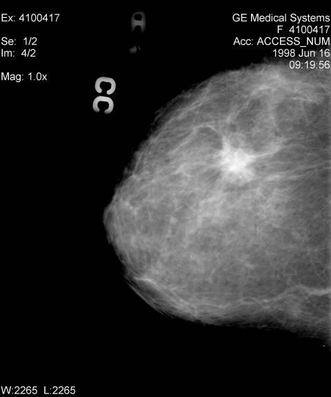
56 Mammography Low energy x-ray photons for better soft tissue differentiation and high resolution (0.1 mm) for diagnosing microcalcifications are needed. Breast compression, short exposure time, single emulsion film, Mo target yielding energy levels between 20 and 30 kev, 0.1 to 0.4 R radiation Alternatives: MR and ultrasound
57
58 Disadvantage The major disadvantage of both x-ray and CT imaging is the fact that the technique uses ionizing radiation, in the energy range kev. Since this ionizing radiation can cause tissue damage, there is a limit on the number of x- ray examinations per year that can be performed on a patient.
59 Computed tomography (CT) (def) tomography (to-mòg re-fê) noun tomo - Greek, to cut Any of several techniques for making detailed x-rays of a predetermined plane section of a solid object while blurring out the images of other planes.
60 CT reconstruction Parallel beam projections Fan beam projections Cone beam projections (Multi-slice Spiral CT) Spatial coordinate system transformation must be implemented for fan beam and cone beam reconstruction

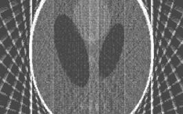
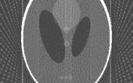
61 x-ray source x-ray detectors Principle of computed tomography with the x-ray source and detector unit rotating synchronously around the patient. (right) An example of a brain CT.
62 CT Scanners: hardware GE: Prospeed, LightSpeed, Synergy Philips: Tomoscan Siemens: Somatom Patient Fan beam Ring of detectors X-ray source
63 Computed tomography (CT) I 0 I 0 x y µ 1 µ 2 µ 3 µ 4 I 1 =I 0 e -(µ 1+µ 2 ) x I 2 =I 0 e -(µ 3+µ 4 ) x I 3 =I 0 e -(µ 1+µ 3 ) y I 4 =I 0 e -(µ 2+µ 4 ) y 4 equations solved for 4 unknown µ s
64 (a) (b) (c) (d) P 4 P 3 P 1 P 2
65
 (d)")
66 (a) (b) (c) (d) (e)
67 H(k) k
68
69 φ p(r,φ)


70


71
Rad T 290 Worksheet 2
 Class: Date: Rad T 290 Worksheet 2 1. Projectile electrons travel from a. anode to cathode. c. target to patient. b. cathode to anode. d. inner shell to outer shell. 2. At the target, the projectile electrons
Shell Atomic Model and Energy Levels
 Shell Atomic Model and Energy Levels (higher energy, deeper excitation) - Radio waves: Not absorbed and pass through tissue un-attenuated - Microwaves : Energies of Photos enough to cause molecular rotation
FXA UNIT G485 Module X-Rays. Candidates should be able to : I = I 0 e -μx
 1 Candidates should be able to : HISTORY Describe the nature of X-rays. Describe in simple terms how X-rays are produced. X-rays were discovered by Wilhelm Röntgen in 1865, when he found that a fluorescent
X-ray Interaction with Matter
 X-ray Interaction with Matter 10-526-197 Rhodes Module 2 Interaction with Matter kv & mas Peak kilovoltage (kvp) controls Quality, or penetrating power, Limited effects on quantity or number of photons
INTERACTIONS OF RADIATION WITH MATTER
 INTERACTIONS OF RADIATION WITH MATTER Renée Dickinson, MS, DABR Medical Physicist University of Washington Medical Center Department of Radiology Diagnostic Physics Section Outline Describe the various
Basic physics Questions
 Chapter1 Basic physics Questions S. Ilyas 1. Which of the following statements regarding protons are correct? a. They have a negative charge b. They are equal to the number of electrons in a non-ionized
1. Which of the following statements is true about Bremsstrahlung and Characteristic Radiation?
 BioE 1330 - Review Chapters 4, 5, and 6 (X-ray and CT) 9/27/2018 Instructions: On the Answer Sheet, enter your 2-digit ID number (with a leading 0 if needed) in the boxes of the ID section. Fill in the
Physics of Radiography
 EL-GY 6813 / BE-GY 6203 / G16.4426 Medical Imaging Physics of Radiography Jonathan Mamou and Yao Wang Polytechnic School of Engineering New York University, Brooklyn, NY 11201 Based on Prince and Links,
Warm-up Which of the following is NOT made up of photons?
 Warm-up Which of the following is NOT made up of photons? 1. Laser light 2. Radio waves 3. Ultrasonic waves 4. X-rays Radio µwaves fir IR uv x-ray gamma-ray... Xrays Discovered by Wm. Roentgen in 1895
Ba (Z = 56) W (Z = 74) preferred target Mo (Z = 42) Pb (Z = 82) Pd (Z = 64)
 W (Z = 74) preferred target Mo (Z = 42) Pb (Z = 82) Pd (Z = 64)") Produced by accelerating electrons with high voltage and allowing them to collide with metal target (anode), e.g, Tungsten. Three Events (Two types of x-ray) a) Heat X-Ray Tube b) bremsstrahlung (braking
Produced by accelerating electrons with high voltage and allowing them to collide with metal target (anode), e.g, Tungsten. Three Events (Two types of x-ray) a) Heat X-Ray Tube b) bremsstrahlung (braking
CHAPTER 4 RADIATION ATTENUATION
 HDR202 PHYSICS FOR RADIOGRAPHERS 2 CHAPTER 4 RADIATION ATTENUATION PREPARED BY: MR KAMARUL AMIN BIN ABDULLAH SCHOOL OF MEDICAL IMAGING FACULTY OF HEALTH SCIENCES Learning Objectives At the end of the lesson,
We have seen how the Brems and Characteristic interactions work when electrons are accelerated by kilovolts and the electrons impact on the target
 We have seen how the Brems and Characteristic interactions work when electrons are accelerated by kilovolts and the electrons impact on the target focal spot. This discussion will center over how x-ray
For the next several lectures, we will be looking at specific photon interactions with matter. In today s lecture, we begin with the photoelectric
 For the next several lectures, we will be looking at specific photon interactions with matter. In today s lecture, we begin with the photoelectric effect. 1 The objectives of today s lecture are to identify
Physics of Radiotherapy. Lecture II: Interaction of Ionizing Radiation With Matter
 Physics of Radiotherapy Lecture II: Interaction of Ionizing Radiation With Matter Charge Particle Interaction Energetic charged particles interact with matter by electrical forces and lose kinetic energy
Basic principles of x-ray production
 Production of X-Rays part 1 George Starkschall, Ph.D. Lecture Objectives Identify what is needed to produce x-rays Describe how a diagnostic x-ray tube produces x-rays Describe the types of interactions
Physics of Radiography
 Physics of Radiography Yao Wang Polytechnic Institute of NYU Brooklyn, NY 11201 Based on J L Prince and J M Links Medical Imaging Signals and Based on J. L. Prince and J. M. Links, Medical Imaging Signals
Chapter Six: X-Rays. 6.1 Discovery of X-rays
 Chapter Six: X-Rays 6.1 Discovery of X-rays In late 1895, a German physicist, W. C. Roentgen was working with a cathode ray tube in his laboratory. He was working with tubes similar to our fluorescent
Biomedical Imaging. X ray imaging. Patrícia Figueiredo IST
 Biomedical Imaging X ray imaging Patrícia Figueiredo IST 2013-2014 Overview Production of X rays Interaction of electrons with matter X ray spectrum X ray tube Interaction of X rays with matter Photoelectric
October 4th, Bioengineering 508: Physical Aspects of Medical Imaging Some Elementary Particles
 Bioengineering 508: Physical Aspects of Medical Imaging http://courses.washington.edu/bioen508/ For questions, remarks, discussions, errors in the book: Class Discussion Board (link from class website)
Sound wave bends as it hits an interface at an oblique angle. 4. Reflection. Sound wave bounces back to probe
 : Ultrasound imaging and x-rays 1. How does ultrasound imaging work?. What is ionizing electromagnetic radiation? Definition of ionizing radiation 3. How are x-rays produced? Bremsstrahlung Auger electron
CHAPTER 2 RADIATION INTERACTIONS WITH MATTER HDR 112 RADIATION BIOLOGY AND RADIATION PROTECTION MR KAMARUL AMIN BIN ABDULLAH
 HDR 112 RADIATION BIOLOGY AND RADIATION PROTECTION CHAPTER 2 RADIATION INTERACTIONS WITH MATTER PREPARED BY: MR KAMARUL AMIN BIN ABDULLAH SCHOOL OF MEDICAL IMAGING FACULTY OF HEALTH SCIENCE Interactions
In today s lecture, we want to see what happens when we hit the target.
 In the previous lecture, we identified three requirements for the production of x- rays. We need a source of electrons, we need something to accelerate electrons, and we need something to slow the electrons
DEVIL PHYSICS THE BADDEST CLASS ON CAMPUS IB PHYSICS
 DEVIL PHYSICS THE BADDEST CLASS ON CAMPUS IB PHYSICS TSOKOS OPTION I-2 MEDICAL IMAGING Reading Activity Answers IB Assessment Statements Option I-2, Medical Imaging: X-Rays I.2.1. I.2.2. I.2.3. Define
LECTURE 4 PRINCIPLE OF IMAGE FORMATION KAMARUL AMIN BIN ABDULLAH
 LECTURE 4 PRINCIPLE OF IMAGE FORMATION KAMARUL AMIN BIN ABDULLAH Lesson Objectives At the end of the lesson, student should able to: Define attenuation Explain interactions between x-rays and matter in
11/10/2014. Chapter 1: Introduction to Medical Imaging. Projection (Transmission) vs. Emission Imaging. Emission Imaging
 vs. Emission Imaging. Emission Imaging") Chapter 1: Introduction to Medical Imaging Overview of Modalities Properties of an Image: Limitations on Information Content Contrast (both object & image): Brightness difference Sharpness (blur): Smallest
Chapter 1: Introduction to Medical Imaging Overview of Modalities Properties of an Image: Limitations on Information Content Contrast (both object & image): Brightness difference Sharpness (blur): Smallest
Chapter 30 X Rays GOALS. When you have mastered the material in this chapter, you will be able to:
 Chapter 30 X Rays GOALS When you have mastered the material in this chapter, you will be able to: Definitions Define each of the following terms, and use it in an operational definition: hard and soft
Interactions with Matter Photons, Electrons and Neutrons
 Interactions with Matter Photons, Electrons and Neutrons Ionizing Interactions Jason Matney, MS, PhD Interactions of Ionizing Radiation 1. Photon Interactions Indirectly Ionizing 2. Charge Particle Interactions
11/19/2014. Chapter 3: Interaction of Radiation with Matter in Radiology and Nuclear Medicine. Nuclide Families. Family Nuclides with Same: Example
 2014-2015 Residents' Core Physics Lectures Mondays 7:00-8:00 am in VA Radiology and UCSDMC Lasser Conference Rooms Topic Chapters Date Faculty 1 Introduction and Basic Physics 1, 2 M 11/17 Andre 2 Interaction
Outline. Chapter 6 The Basic Interactions between Photons and Charged Particles with Matter. Photon interactions. Photoelectric effect
 Chapter 6 The Basic Interactions between Photons and Charged Particles with Matter Radiation Dosimetry I Text: H.E Johns and J.R. Cunningham, The physics of radiology, 4 th ed. http://www.utoledo.edu/med/depts/radther
CHAPTER 2 INTERACTION OF RADIATION WITH MATTER
 CHAPTER 2 INTERACTION OF RADIATION WITH MATTER 2.1 Introduction When gamma radiation interacts with material, some of the radiation will be absorbed by the material. There are five mechanisms involve in
Midterm Review. Yao Wang Polytechnic University, Brooklyn, NY 11201
 Midterm Review Yao Wang Polytechnic University, Brooklyn, NY 11201 Based on J. L. Prince and J. M. Links, Medical maging Signals and Systems, and lecture notes by Prince. Figures are from the textbook.
X-RAY PRODUCTION. Prepared by:- EN KAMARUL AMIN BIN ABDULLAH
 X-RAY PRODUCTION Prepared by:- EN KAMARUL AMIN BIN ABDULLAH OBJECTIVES Discuss the process of x-ray being produced (conditions) Explain the principles of energy conversion in x-ray production (how energy
Chapter 38. Photons Light Waves Behaving as Particles
 Chapter 38 Photons Light Waves Behaving as Particles 38.1 The Photoelectric Effect The photoelectric effect was first discovered by Hertz in 1887, and was explained by Einstein in 1905. The photoelectric
Atomic Physics. Chapter 6 X ray. Jinniu Hu 24/12/ /20/13
 Atomic Physics Chapter 6 X ray 11/20/13 24/12/2018 Jinniu Hu 1!1 6.1 The discovery of X ray X-rays were discovered in 1895 by the German physicist Wilhelm Roentgen. He found that a beam of high-speed electrons
Outline. Radiation Interactions. Spurs, Blobs and Short Tracks. Introduction. Radiation Interactions 1
 Outline Radiation Interactions Introduction Interaction of Heavy Charged Particles Interaction of Fast Electrons Interaction of Gamma Rays Interactions of Neutrons Radiation Exposure & Dose Sources of
A Brief Introduction to Medical Imaging. Outline
 A Brief Introduction to Medical Imaging Outline General Goals Linear Imaging Systems An Example, The Pin Hole Camera Radiations and Their Interactions with Matter Coherent vs. Incoherent Imaging Length
PHY138Y Nuclear and Radiation Section
 PHY138Y Supplementary Notes II: X rays. A.W. Key Page 1 of 13 PHY138Y Nuclear and Radiation Section Supplementary Notes II X-rays - Production, Characteristics, & Use Contents. 2.1 Introduction 2.2 Production
Introduction to Medical Imaging Chapter 1 Radiation and the Atom Chapter 2 Interaction of Radiation and Matter Chapter 3
 Introduction to Medical Imaging Chapter 1 Radiation and the Atom Chapter 2 Interaction of Radiation and Matter Chapter 3 Professor, Radiology and Medical Education Director, Diagnostic Physics a copy of
Medical biophysics II. X-ray. X-ray. Generation, Spectral features Interaction with matter
 Medical biophysics II Medical biophysics II X-ray - generation and properties X-ray - diagnostic foundations Medical use of electronics Thermodynamics - equilibrium, change, laws Diffusion, Brown-motion,
Slide 1. Slide 2. Slide 3. Take the Terror Out of Physics. Active and Interactive Games and Activities for Teaching Radiographic Physics
 Slide 1 Active and Interactive Games and Activities for Teaching Radiographic Physics Jennifer Yates, MS, RT(R)(M)(BD) AEIRS 2010 Slide 2 Take the Terror Out of Physics X-Ray Tube Bingo Game Immediate
Production of X-rays. Radiation Safety Training for Analytical X-Ray Devices Module 9
 Module 9 This module presents information on what X-rays are and how they are produced. Introduction Module 9, Page 2 X-rays are a type of electromagnetic radiation. Other types of electromagnetic radiation
Interaction of charged particles and photons with matter
 Interaction of charged particles and photons with matter Robert Miyaoka, Ph.D. Old Fisheries Center, Room 200 rmiyaoka@u.washington.edu Passage of radiation through matter depends on Type of radiation
Possible Interactions. Possible Interactions. X-ray Interaction (Part I) Possible Interactions. Possible Interactions. section
 Possible Interactions. Possible Interactions. section") Possible Interactions X-ray Interaction (Part I) Three types of interaction 1. Scattering Interaction with an atom Deflected May or may not loss of energy 1 Possible Interactions Three types of interaction
Possible Interactions X-ray Interaction (Part I) Three types of interaction 1. Scattering Interaction with an atom Deflected May or may not loss of energy 1 Possible Interactions Three types of interaction
Topics. EM spectrum. X-Rays Computed Tomography Direct Inverse and Iterative Inverse Backprojection Projection Theorem Filtered Backprojection
 Bioengineering 28A Principles of Biomedical Imaging Fall Quarter 24 X-Rays/CT Lecture Topics X-Rays Computed Tomography Direct Inverse and Iterative Inverse Backprojection Projection Theorem Filtered Backprojection
MEDICAL EQUIPMENT: NUCLEAR MEDICINE. Prof. Yasser Mostafa Kadah
 MEDICAL EQUIPMENT: NUCLEAR MEDICINE Prof. Yasser Mostafa Kadah www.k-space.org Recommended Textbook Introduction to Medical Imaging: Physics, Engineering and Clinical Applications, by Nadine Barrie Smith
CHAPTER 3 Prelude to Quantum Theory. Observation of X Rays. Thomson s Cathode-Ray Experiment. Röntgen s X-Ray Tube
 CHAPTER Prelude to Quantum Theory.1 Discovery of the X Ray and the Electron. Determination of Electron Charge. Line Spectra.4 Quantization.5 Blackbody Radiation.6 Photoelectric Effect.7 X-Ray Production.8
hν' Φ e - Gamma spectroscopy - Prelab questions 1. What characteristics distinguish x-rays from gamma rays? Is either more intrinsically dangerous?
 Gamma spectroscopy - Prelab questions 1. What characteristics distinguish x-rays from gamma rays? Is either more intrinsically dangerous? 2. Briefly discuss dead time in a detector. What factors are important
ELEG 479 Lecture #6. Mark Mirotznik, Ph.D. Associate Professor The University of Delaware
 ELEG 479 Lecture #6 Mark Mirotznik, Ph.D. Associate Professor The University of Delaware Summary of Last Lecture X-ray Physics What are X-rays and when are they useful for medical imaging? How are X-rays
X-Rays and Scattering from Filters Used in Diagnostic Radiology
 International Journal of Scientific and Research Publications, Volume 3, Issue 7, July 2013 1 X-Rays and Scattering from Filters Used in Diagnostic Radiology Odeh Daniel, G Ogbanje, Prof. S. A. Jonah Department
Topics. EM spectrum. X-Rays Computed Tomography Direct Inverse and Iterative Inverse Backprojection Projection Theorem Filtered Backprojection
 Bioengineering 28A Principles of Biomedical Imaging Fall Quarter 25 X-Rays/CT Lecture Topics X-Rays Computed Tomography Direct Inverse and Iterative Inverse Backprojection Projection Theorem Filtered Backprojection
RED. BLUE Light. Light-Matter
 1 Light-Matter This experiment demonstrated that light behaves as a wave. Essentially Thomas Young passed a light of a single frequency ( colour) through a pair of closely spaced narrow slits and on the
Bioengineering 280A Principles of Biomedical Imaging. Fall Quarter 2005 X-Rays/CT Lecture 1. Topics
 Bioengineering 28A Principles of Biomedical Imaging Fall Quarter 25 X-Rays/CT Lecture Topics X-Rays Computed Tomography Direct Inverse and Iterative Inverse Backprojection Projection Theorem Filtered Backprojection
Basic physics of nuclear medicine
 Basic physics of nuclear medicine Nuclear structure Atomic number (Z): the number of protons in a nucleus; defines the position of an element in the periodic table. Mass number (A) is the number of nucleons
CHAPTER 3 The Experimental Basis of Quantum Theory
 CHAPTER 3 The Experimental Basis of Quantum Theory 3.1 3.2 3.3 3.4 3.5 3.6 3.7 3.8 3.9 Discovery of the X Ray and the Electron Determination of Electron Charge Line Spectra Quantization As far as I can
MEDICAL IMAGING. METHODS OF MODERN IMAGING, BASED ON ELECTRO-MAGNETIC RADIATION (radiowaves, infrared radiation, X-rays, γ-rays ) AND ULTRASOUND
 AND ULTRASOUND") MEDICAL IMAGING MEDICAL IMAGING METHODS OF MODERN IMAGING, BASED ON ELECTRO-MAGNETIC RADIATION (radiowaves, infrared radiation, X-rays, γ-rays ) AND ULTRASOUND MEDICAL IMAGING RADIOLOGY NUCLEAR MEDICINE
MEDICAL IMAGING MEDICAL IMAGING METHODS OF MODERN IMAGING, BASED ON ELECTRO-MAGNETIC RADIATION (radiowaves, infrared radiation, X-rays, γ-rays ) AND ULTRASOUND MEDICAL IMAGING RADIOLOGY NUCLEAR MEDICINE
An Introduction to Diffraction and Scattering. School of Chemistry The University of Sydney
 An Introduction to Diffraction and Scattering Brendan J. Kennedy School of Chemistry The University of Sydney 1) Strong forces 2) Weak forces Types of Forces 3) Electromagnetic forces 4) Gravity Types
Emphasis on what happens to emitted particle (if no nuclear reaction and MEDIUM (i.e., atomic effects)
") LECTURE 5: INTERACTION OF RADIATION WITH MATTER All radiation is detected through its interaction with matter! INTRODUCTION: What happens when radiation passes through matter? Emphasis on what happens
LECTURE 5: INTERACTION OF RADIATION WITH MATTER All radiation is detected through its interaction with matter! INTRODUCTION: What happens when radiation passes through matter? Emphasis on what happens
Radionuclide Imaging MII Positron Emission Tomography (PET)
") Radionuclide Imaging MII 3073 Positron Emission Tomography (PET) Positron (β + ) emission Positron is an electron with positive charge. Positron-emitting radionuclides are most commonly produced in cyclotron
Radionuclide Imaging MII 3073 Positron Emission Tomography (PET) Positron (β + ) emission Positron is an electron with positive charge. Positron-emitting radionuclides are most commonly produced in cyclotron
X-ray Absorption Spectroscopy
 X-ray Absorption Spectroscopy Nikki Truss November 26, 2012 Abstract In these experiments, some aspects of x-ray absorption spectroscopy were investigated. The x-ray spectrum of molybdenum was recorded
GLOSSARY OF BASIC RADIATION PROTECTION TERMINOLOGY
 GLOSSARY OF BASIC RADIATION PROTECTION TERMINOLOGY ABSORBED DOSE: The amount of energy absorbed, as a result of radiation passing through a material, per unit mass of material. Measured in rads (1 rad
CHAPTER 3 The Experimental Basis of Quantum
 CHAPTER 3 The Experimental Basis of Quantum 3.1 Discovery of the X Ray and the Electron 3.2 Determination of Electron Charge 3.3 Line Spectra 3.4 Quantization 3.5 Blackbody Radiation 3.6 Photoelectric
Tomography and Reconstruction
 Tomography and Reconstruction Lecture Overview Applications Background/history of tomography Radon Transform Fourier Slice Theorem Filtered Back Projection Algebraic techniques Measurement of Projection
Modern physics ideas are strange! L 36 Modern Physics [2] The Photon Concept. How are x-rays produced? The uncertainty principle
![Modern physics ideas are strange! L 36 Modern Physics [2] The Photon Concept. How are x-rays produced? The uncertainty principle](/thumbs/88/117098787.jpg "Modern physics ideas are strange! L 36 Modern Physics [2] The Photon Concept. How are x-rays produced? The uncertainty principle") L 36 Modern Physics [2] X-rays & gamma rays How lasers work Medical applications of lasers Applications of high power lasers Medical imaging techniques CAT scans MRI s Modern physics ideas are strange!
L 36 Modern Physics [2] X-rays & gamma rays How lasers work Medical applications of lasers Applications of high power lasers Medical imaging techniques CAT scans MRI s Modern physics ideas are strange!
DR KAZI SAZZAD MANIR
 DR KAZI SAZZAD MANIR PHOTON BEAM MATTER ENERGY TRANSFER IONISATION EXCITATION ATTENUATION removal of photons from the beam by the matter. ABSORPTION SCATTERING TRANSMISSION Taking up the energy from the
Chapter Four (Interaction of Radiation with Matter)
") Al-Mustansiriyah University College of Science Physics Department Fourth Grade Nuclear Physics Dr. Ali A. Ridha Chapter Four (Interaction of Radiation with Matter) Different types of radiation interact
Al-Mustansiriyah University College of Science Physics Department Fourth Grade Nuclear Physics Dr. Ali A. Ridha Chapter Four (Interaction of Radiation with Matter) Different types of radiation interact
CHAPTER 27 Quantum Physics
 CHAPTER 27 Quantum Physics Units Discovery and Properties of the Electron Planck s Quantum Hypothesis; Blackbody Radiation Photon Theory of Light and the Photoelectric Effect Energy, Mass, and Momentum
CHARGED PARTICLE INTERACTIONS
 CHARGED PARTICLE INTERACTIONS Background Charged Particles Heavy charged particles Charged particles with Mass > m e α, proton, deuteron, heavy ion (e.g., C +, Fe + ), fission fragment, muon, etc. α is
Explain how Planck resolved the ultraviolet catastrophe in blackbody radiation. Calculate energy of quanta using Planck s equation.
 Objectives Explain how Planck resolved the ultraviolet catastrophe in blackbody radiation. Calculate energy of quanta using Planck s equation. Solve problems involving maximum kinetic energy, work function,
INTRODUCTION TO MEDICAL PHYSICS 1 Quiz #1 Solutions October 6, 2017
 INTRODUCTION TO MEDICAL PHYSICS 1 Quiz #1 Solutions October 6, 2017 This is a closed book examination. Adequate information is provided you to solve all problems. Be sure to show all work, as partial credit
Technical University of Denmark
 Technical University of Denmark Page 1 of 11 pages Written test, 9 December 2010 Course name: Introduction to medical imaging Course no. 31540 Aids allowed: none. "Weighting": All problems weight equally.
Initial Certification
 Initial Certification Medical Physics Part 1 Content Guide Part 1 Content Guides and Sample Questions PLEASE NOTE: List of Constants and Physical Values for Use on the Part 1 Physics Exam The ABR provides
X-RAY SPECTRA. Theory:
 12 Oct 18 X-ray.1 X-RAY SPECTRA In this experiment, a number of measurements involving x-rays will be made. The spectrum of x-rays emitted from a molybdenum target will be measured, and the experimental
EEE4106Z Radiation Interactions & Detection
 EEE4106Z Radiation Interactions & Detection 2. Radiation Detection Dr. Steve Peterson 5.14 RW James Department of Physics University of Cape Town steve.peterson@uct.ac.za May 06, 2015 EEE4106Z :: Radiation
Physics Modern Physics Professor Jodi Cooley. Welcome back. to PHY Arthur Compton
 Welcome back to PHY 3305 Today s Lecture: X-ray Production Compton Scattering Dual Nature of Light Arthur Compton 1892-1962 The Production of xrays X-rays were discovered in 1895 by German physicist Wihelm
Chapter 10: Wave Properties of Particles
 Chapter 10: Wave Properties of Particles Particles such as electrons may demonstrate wave properties under certain conditions. The electron microscope uses these properties to produce magnified images
Introduction. 6.1 Summary Notes The Quantum. D Notes: ! is wavelength (m) c is the speed of light (m/s)
 c is the speed of light (m/s)") Introduction Matter and energy have a dual nature: wave and particle. Understanding the particle nature of light is necessary for learning about modern physics and technology. 6.1 Summary Notes The Quantum
Introduction Matter and energy have a dual nature: wave and particle. Understanding the particle nature of light is necessary for learning about modern physics and technology. 6.1 Summary Notes The Quantum
Radiation Dose, Biology & Risk
 ENGG 167 MEDICAL IMAGING Lecture 2: Sept. 27 Radiation Dosimetry & Risk References: The Essential Physics of Medical Imaging, Bushberg et al, 2 nd ed. Radiation Detection and Measurement, Knoll, 2 nd Ed.
Particle nature of light & Quantization
 Particle nature of light & Quantization A quantity is quantized if its possible values are limited to a discrete set. An example from classical physics is the allowed frequencies of standing waves on a
Technical University of Denmark
 Technical University of Denmark Page 1 of 10 pages Written test, 12 December 2012 Course name: Introduction to medical imaging Course no. 31540 Aids allowed: None. Pocket calculator not allowed "Weighting":
ELG7173 Topics in signal Processing II Computational Techniques in Medical Imaging
 ELG7173 Topics in signal Processing II Computational Techniques in Medical Imaging Topic #1: Intro to medical imaging Medical Imaging Classifications n Measurement physics Send Energy into body Send stuff
Quantum Model Einstein s Hypothesis: Photoelectric Effect
 VISUAL PHYSICS ONLINE MODULE 7 NATURE OF LIGHT Quantum Model Einstein s Hypothesis: Photoelectric Effect The photoelectric effect was discovered by Hertz in 1887 as he confirmed Maxwell s electromagnetic
Physics 6C. Photons. Prepared by Vince Zaccone For Campus Learning Assistance Services at UCSB
 Physics 6C Photons Photoelectric Effect Here is the basic setup for the experiment. Light shines on the metal plate, and the electrons absorb that light energy. metal plate incoming light Photoelectric
Radiation Therapy Study Guide
 Amy Heath Radiation Therapy Study Guide A Radiation Therapist s Review 123 Radiation Therapy Study Guide Amy Heath Radiation Therapy Study Guide A Radiation Therapist s Review Amy Heath, MS, RT(T) University
05/11/2013. Nuclear Fuel Cycle Ionizing radiation. Typical decay energies. Radiation with energy > 100 ev. Ionize an atom < 15eV
 Nuclear Fuel Cycle 2013 Lecture 4: Interaction of Ionizing Radiation with Matter Ionizing radiation Radiation with energy > 100 ev Ionize an atom < 15eV Break a bond 1-5 ev Typical decay energies α: 4-9
EEE4101F / EEE4103F Radiation Interactions & Detection
 EEE4101F / EEE4103F Radiation Interactions & Detection 1. Interaction of Radiation with Matter Dr. Steve Peterson 5.14 RW James Department of Physics University of Cape Town steve.peterson@uct.ac.za March
Nuclear Decays. Alpha Decay
 Nuclear Decays The first evidence of radioactivity was a photographic plate, wrapped in black paper and placed under a piece of uranium salt by Henri Becquerel on February 26, 1896. Like many events in
PHYS 4 CONCEPT PACKET Complete
 PHYS 4 CONCEPT PACKET Complete Written by Jeremy Robinson, Head Instructor Find Out More +Private Instruction +Review Sessions WWW.GRADEPEAK.COM Need Help? Online Private Instruction Anytime, Anywhere
Chemical Engineering 412
 Chemical Engineering 412 Introductory Nuclear Engineering Lecture 26 Radiation Detection & Measurement II Spiritual Thought 2 I would not hold the position in the Church I hold today had I not followed
AQA Physics /7408
 AQA Physics - 7407/7408 Module 10: Medical physics You should be able to demonstrate and show your understanding of: 10.1 Physics of the eye 10.1.1 Physics of vision The eye as an optical refracting system,
Chapter 9: Quantization of Light
 Chapter 9: Quantization of Light Max Planck started the revolution of quantum theory by challenging the classical physics and the classical wave theory of light. He proposed the concept of quantization
PHYS 3313 Section 001 Lecture #7
 PHYS 3313 Section 001 Lecture #7 Photoelectric Effect Compton Effect Pair production/pair annihilation PHYS 3313-001, Fall 1 Reading assignments: CH3.9 Announcements Homework #2 CH3 end of the chapter
Interaction of particles with matter - 2. Silvia Masciocchi, GSI and University of Heidelberg SS2017, Heidelberg May 3, 2017
 Interaction of particles with matter - 2 Silvia Masciocchi, GSI and University of Heidelberg SS2017, Heidelberg May 3, 2017 Energy loss by ionization (by heavy particles) Interaction of electrons with
Interaction of Ionizing Radiation with Matter
 Type of radiation charged particles photonen neutronen Uncharged particles Charged particles electrons (β - ) He 2+ (α), H + (p) D + (d) Recoil nuclides Fission fragments Interaction of ionizing radiation
Classical and Planck picture. Planck s constant. Question. Quantum explanation for the Wein Effect.
 6.1 Quantum Physics. Particle Nature of Light Particle nature of Light Blackbody Radiation Photoelectric Effect Properties of photons Ionizing radiation Radiation damage x-rays Compton effect X-ray diffraction
AP Physics Study Guide Modern Physics I. Atomic Physics and Quantum Effects 1. Who is generally credited with the discovery of the electron?
 AP Physics Study Guide Modern Physics I. Atomic Physics and Quantum Effects 1. Who is generally credited with the discovery of the electron? 2. What was it that J. J. Thomson actually measured? 3. Regarding
X-ray Energy Spectroscopy (XES).
.") X-ray Energy Spectroscopy (XES). X-ray fluorescence as an analytical tool for element analysis is based on 3 fundamental parameters: A. Specificity: In determining an x-ray emission energy E certainty
X-ray Energy Spectroscopy (XES). X-ray fluorescence as an analytical tool for element analysis is based on 3 fundamental parameters: A. Specificity: In determining an x-ray emission energy E certainty
Quantitative Assessment of Scattering Contributions in MeV-Industrial X-ray Computed Tomography
 11th European Conference on Non-Destructive Testing (ECNDT 2014), October 6-10, 2014, Prague, Czech Republic More Info at Open Access Database www.ndt.net/?id=16530 Quantitative Assessment of Scattering
LET! (de / dx) 1 Gy= 1 J/kG 1Gy=100 rad. m(kg) dose rate
 1 Gy= 1 J/kG 1Gy=100 rad. m(kg) dose rate") Basics of Radiation Dosimetry for the Physicist http://en.wikipedia.org/wiki/ionizing_radiation I. Ionizing radiation consists of subatomic particles or electromagnetic waves that ionize electrons along
Basics of Radiation Dosimetry for the Physicist http://en.wikipedia.org/wiki/ionizing_radiation I. Ionizing radiation consists of subatomic particles or electromagnetic waves that ionize electrons along
4.1b - Cavity Theory Lecture 2 Peter R Al mond 2011 Overview of Lecture Exposure (W/e)air Exposure Exposure and and and Air Air Kerma
air Exposure Exposure and and and Air Air Kerma") 4.1b - Cavity Theory Lecture 2 Peter R Almond 2011 Overview of Lecture Exposure (W/e) air Exposure and Air Kerma Exposure Exposure is symbolized as X and defined by the ICRU as the quotient of dq by dm,
4.1b - Cavity Theory Lecture 2 Peter R Almond 2011 Overview of Lecture Exposure (W/e) air Exposure and Air Kerma Exposure Exposure is symbolized as X and defined by the ICRU as the quotient of dq by dm,
Chap. 15 Radiation Imaging
 Chap. 15 Radiation Imaging 15.1 INTRODUCTION Modern Medical Imaging Devices Incorporating fundamental concepts in physical science and innovations in computer technology Nobel prize (physics) : 1895 Wilhelm