MEDICAL IMAGING. METHODS OF MODERN IMAGING, BASED ON ELECTRO-MAGNETIC RADIATION (radiowaves, infrared radiation, X-rays, γ-rays ) AND ULTRASOUND
|
|
|
- Garry Tyler
- 5 years ago
- Views:
Transcription
1 MEDICAL IMAGING
2 MEDICAL IMAGING METHODS OF MODERN IMAGING, BASED ON ELECTRO-MAGNETIC RADIATION (radiowaves, infrared radiation, X-rays, γ-rays ) AND ULTRASOUND
3 MEDICAL IMAGING RADIOLOGY NUCLEAR MEDICINE ULTRASONOGRAPHY (ECHOGRAPHY) MRI (MAGNETIC RESONANCE IMAGING) THERMOGRAPHY
4 RADIOLOGY BASED ON ABSORPTION OF X-RAYS BY THE TISSUES: RADIOGRAPHS, CT
5 NUCLEAR MEDICINE BASED ON RADIOACTIVE ISOTOPES CONCENTRATED IN TISSUE, EMITTING PHOTONS (γ-rays)
6 ULTRASONOGRAPHY BASED ON HIGH-FREQUENCY MECHANICAL OSCILLATIONS, EMITTED AND RECEIVED SIMULTANEOUSLY BY A TRANSMITTER-SENSOR (TRANSDUCER)
7 MRI (MAGNETIC RESONANCE IMAGING) BASED ON RADIO-FREQUENCY RADIATION PRODUCED BY THE EXCITATION OF ODD ATOMIC NUCLEI IN A STRONG MAGNETIC FIELD
8 THERMOGRAPHY BASED ON INFRARED RADIATION EMITTED BY LIVING TISSUE
9 RADIOLOGY
10 RADIOLOGY BASED ON ABSORPTION OF X-RAYS BY THE TISSUES X-radiation (composed of X-rays) is a form of electromagnetic radiation shorter in wavelength than UV rays X-rays are a form of ionizing radiation

11 RADIOLOGY Wilhelm Conrad Röntgen (27 March February 1923) was a German physicist, who, on 8 November 1895, produced and detected electromagnetic radiation in a wavelength range today known as x-rays or Röntgen rays, an achievement that earned him the first Nobel Prize in Physics in 1901 "Röntgen" in English is spelled "Roentgen"

12 RADIOLOGY The source of X-rays is an X-RAY TUBE

13 HIGH VOLTAGE ELECTRIC CURRENT PASSES ACROSS A VACUUM TUBE WITH TWO ELECTRODES THE BEAM OF ELECTRONS BOOSTED FROM THE INCANDESCENT-HEATED CATHODE STRIKES A HEAVY METAL ANODE THAT PRODUCES HEAT AND X-RAYS RADIOLOGY
14 RADIOLOGY

15 RADIOLOGY BASED ON ABSORPTION OF X-RAYS BY THE TISSUES ABSORPTION OF X-RAYS DEPENDS ON: Density of the structure Thickness of the structure
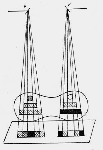
16 RADIOLOGY IMAGING GEOMETRY Distortions arise in an image due to imaging geometry and the characteristics of an object
17 RADIOLOGY Parallax is an apparent exaggeration of the relative position of two objects when viewed along two different lines of sight. Given the two-dimensional nature of radiographs, parallax is an important principle in localizing objects within the body. On the basis of a single frontal view, it is impossible to tell the anteroposterior location of an abnormality. However, a second view from a different perspective can be used to localize the object.
18 RADIOLOGY
19 RADIOLOGY
20 RADIOLOGY RADIOLOGICAL METHODS: Simple (plain) radiography Fluoroscopy Conventional Tomography Computed Tomography (CT)
21 RADIOLOGY Simple (plain) radiography X-ray beam modulated through the patient s body is imprinted on a photographic plate (X-ray film) or received by digital detector (digital radiography)
 radiography")
22 RADIOLOGY Simple (plain) radiography X-RAY FILM


23 RADIOLOGY X-RAY FILM CASSETE DIGITAL DETECTOR


24 RADIOLOGY X-RAY ROOM Radiograph negative image


25 RADIOLOGY FLUOROSCOPY X-ray beam modulated through the patient s body is projected on a fluorescent screen. The image is viewed on the monitor. positive image
26 RADIOLOGY TERMINOLOGY: High dens structures opaque (opacity) bones, calcification, metallic foreign bodies Low dens structures lucent (lucency, translucency, transparency) air


27 RADIOLOGY negative image positive image opacity lucency


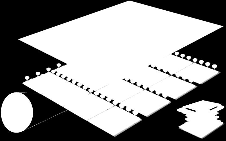
28 RADIOLOGY Conventional Tomography Allows tissue section radiographs. During the exposure, the X-ray tube and the film are moved in opposite directions. The chosen pivot point remains stationary during the whole motion.


29 RADIOLOGY Computed Tomography The X-ray tube emits a sharply collimated fan beam of X-rays which passes the patient and reaches an array of detectors. Tube rotates around the patient.

30 RADIOLOGY Computed Tomography Spiral CT X-ray tube rotates continuously around the patient.
31 RADIOLOGY Computed Tomography TERMINOLOGY: High dens structures hyperdense (hyperdensity) bones, calcification, metallic foreign bodies Low dens structures hypodense (hypodensity) air

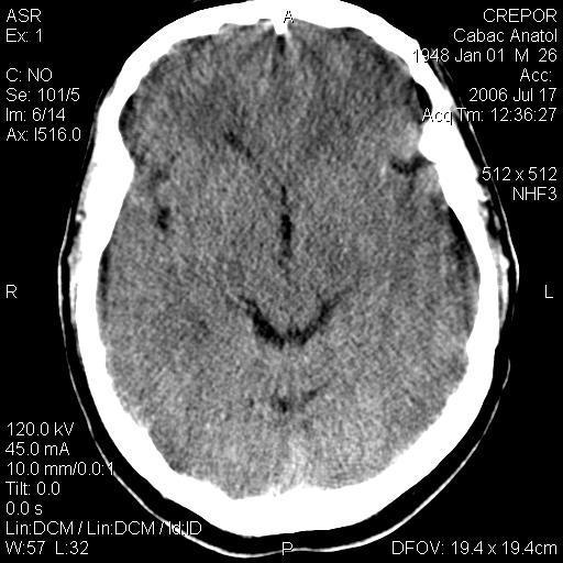
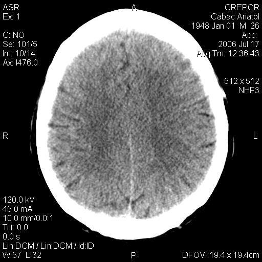
32 RADIOLOGY Computed Tomography
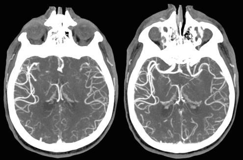
33 RADIOLOGY CT-Angiography

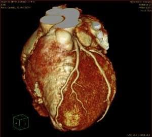
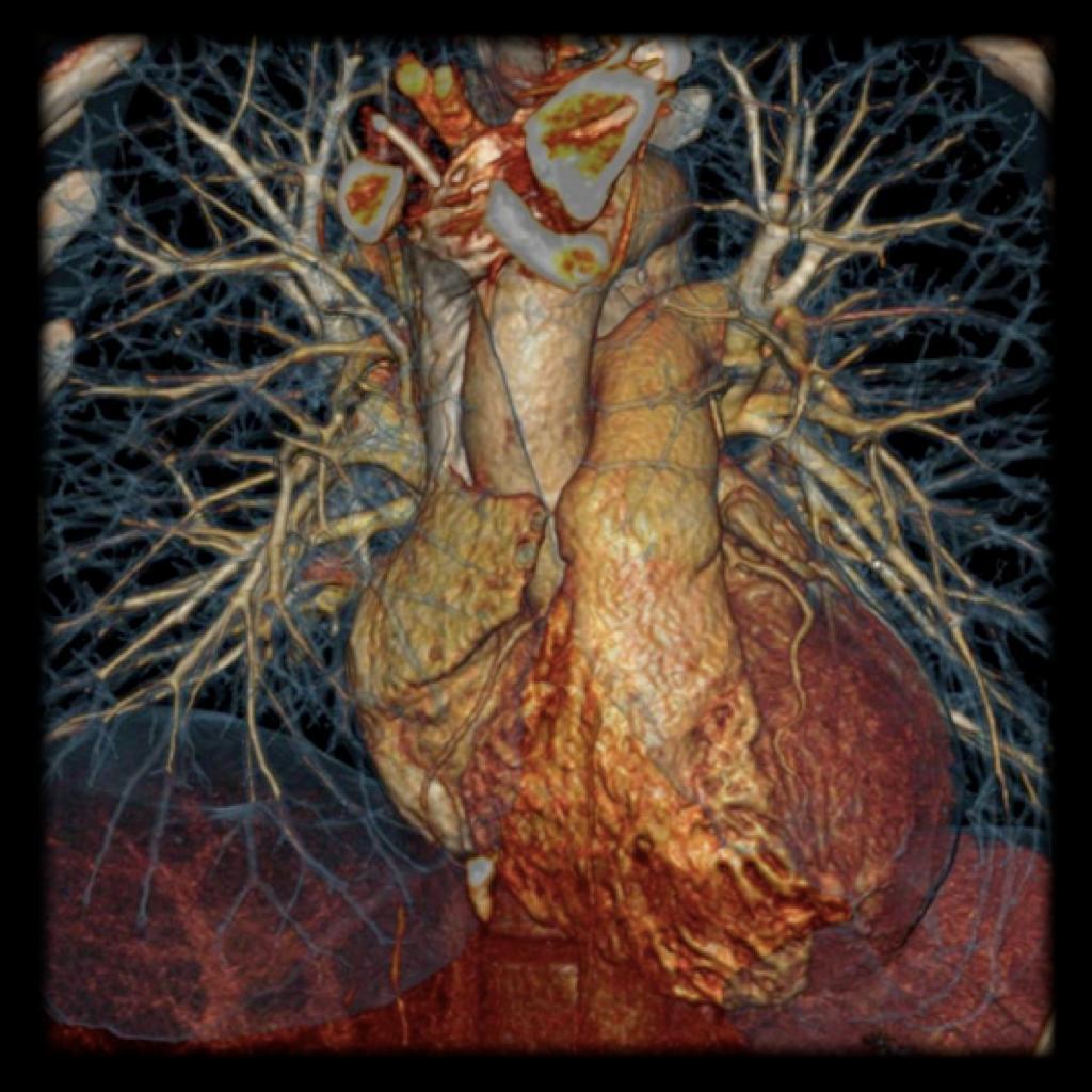
34 RADIOLOGY CT-3D

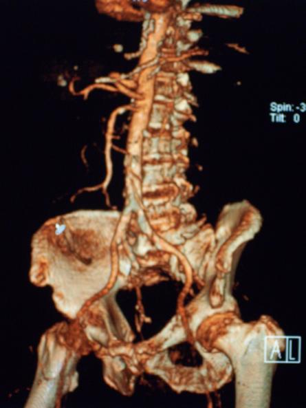
35 RADIOLOGY CT-3D
36 RADIOLOGY CONTRAST MEDIUM (contrast agent) A substance used to enhance the contrast of structures or fluids within the body
37 RADIOLOGY positive media negative media CONTRAST MEDIUM (contrast agent)
38 RADIOLOGY CONTRAST MEDIUM (contrast agent) Positive contrast media and the body's soft tissues contain a similar number of atoms per unit volume. Some atoms in the contrast medium (e.g. iodine or barium) have a much higher atomic number than those of the soft tissues (hydrogen, carbon, nitrogen, oxygen). A higher atomic number is generally associated with an increased ability to attenuate X-rays.
39 Positive contrast media RADIOLOGY CONTRAST MEDIUM (contrast agent) water insoluble contrast media, an aqueous suspension of in soluble crystals of Barium Sulphate water soluble, which in clinical practice today means water solutions of organic compounds with iodine covalently bound to an aromatic structure (Isopaque, Urografin, Angiografin, Gastrografin, Omnipaque, Ultravist..) oily (fat-soluble) contrast medium (Lipiodol)
40 RADIOLOGY CONTRAST MEDIUM (contrast agent) Negative contrast media (air, oxygen, nitric oxide (N 2 O) or carbon dioxide (CO 2 ) and other gases) attenuate X-rays less than the soft tissues of the body, because a gas (the negative contrast medium) contains per unit volume a much lower number of radiation attenuating atoms than the patient's soft tissues.
41 RADIOLOGY RADIOLOGICAL METHODS USING CONTRAST MEDIUM Angiography Bronhography Colecystography, colangiography Oral Barium Sulphate, Barium Enema Limfography Arthrography
42 NUCLEAR MEDICINE

43 NUCLEAR MEDICINE Types of nuclear radiation: Alpha decay Alpha particles Beta decay Beta particles Gamma decay Gamma ray
44 Types of radiation: NUCLEAR MEDICINE α - consist of two protons and two neutrons bound together into a particle identical to a helium nucleus. Electric charge +2. Mass 4 atomic mass units. Low penetration. β - high-energy, high-speed electrons or positrons. Electric charge 1. Mass of electron. Penetration higher than α γ - electromagnetic radiation of high energy. No electic charge. Mass of a photon High penetration
45 NUCLEAR MEDICINE Radionuclide an atom with an unstable nucleus that decays spontaneously with the emission of energy (gamma rays). 99m-Tc, 201-Tl, 131-I, 123-I, 57Co, 133-Xe Positron emitting: 15-O, 13-N, 18F, 11C
46 Radiopharmaceuticals NUCLEAR MEDICINE Substances that contain one or more radioactive atoms (radionuclids), used as tracers in the diagnosis and treatment. The ideal radiopharmaceutical is distributed only to the organs or structures to be imaged.
47 NUCLEAR MEDICINE Methods of investigation - Scintigraphy - SPECT - PET
48 NUCLEAR MEDICINE Scintigraphy a diagnostic procedure consisting of the administration of a radionuclide with an affinity for the organ or tissue of interest, followed by recording the distribution of the radioactivity by a scintillation camera.

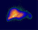
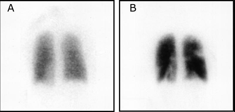
49 NUCLEAR MEDICINE Scintigraphy
 - is able to provide true 3D")

50 NUCLEAR MEDICINE SPECT (Single Photon Emission Computed Tomography) - is able to provide true 3D information - is performed by using a gamma camera to acquire multiple 2-D images from multiple angles.


51 NUCLEAR MEDICINE PET (Positron Emission Tomography) - produces a three-dimensional image of functional processes in the body
52 ULTRASONOGRAPHY (ECHOGRAPHY)
53 ULTRASONOGRAPHY Ultrasound Is an oscillation of pressure transmitted through a solid, liquid, or gas. The sound waves used in ultrasound are between 2 and 10 MHz
54 ULTRASONOGRAPHY Principle Piezoelectric crystals in the transducer convert electricity into high-frequency sound waves, which are sent into tissues. The tissues scatter, reflect, and absorb the sound waves to various degrees. The sound waves that are reflected back (echoes) are converted into electric signals. A computer analyzes the signals and displays the information on a screen.
55 ULTRASONOGRAPHY
56 Modes A-mode: the simplest; ULTRASONOGRAPHY signals are recorded as spikes on a graph; the vertical (Y) axis of the display shows the echo amplitude, and the horizontal (X) axis shows depth or distance into the patient; is used for ophthalmologic scanning.
57 ULTRASONOGRAPHY Modes B-mode (gray-scale): most often used in diagnostic imaging; signals are displayed as a 2-dimensional anatomic image; commonly used to evaluate the developing fetus and to evaluate organs, including the liver, spleen, kidneys, thyroid gland, testes, breasts, and prostate gland; fast enough to show real-time motion, such as the motion of the beating heart or pulsating blood vessels; real-time imaging provides anatomic and functional information.

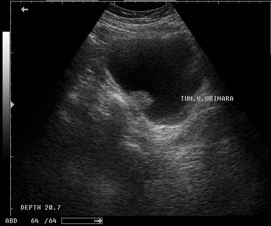
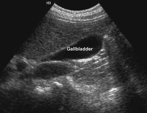
58 Modes B-mode ULTRASONOGRAPHY


59 ULTRASONOGRAPHY Modes M-mode: used to image moving structures; signals reflected by the moving structures are converted into waves that are displayed continuously across a vertical axis; is used primarily for assessment of fetal heartbeat and in cardiac imaging.

; the moving objects are RBCs in")
60 ULTRASONOGRAPHY Modes Doppler ultrasonography : is used to assess blood flow; uses the Doppler effect (alteration of sound frequency by reflection off a moving object); the moving objects are RBCs in blood.
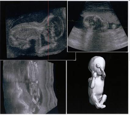
61 Modes 3D ULTRASONOGRAPHY
62 MAGNETIC RESONANCE IMAGING (MRI)

63 MRI Uses magnetic fields and radio waves to produce images of thin slices of tissues (tomographic images).

64 MRI Normally, protons within tissues spin to produce tiny magnetic fields that are randomly aligned. When surrounded by the strong magnetic field of an MRI device, the magnetic axes align along that field.
65 MRI A radiofrequency pulse is then applied, causing the axes of all protons to momentarily align against the field in a high-energy state. After the pulse, some protons relax and resume their baseline alignment within the magnetic field of the MRI device. The magnitude and rate of energy release that occurs as the protons resume this alignment (T1 relaxation) and as they wobble (presses) during the process (T2 relaxation) are recorded as spatially localized signal intensities by a coil (antenna). Computer algorithms analyze these signals and produce anatomic images.


66 Advantages: MRI Does not use ionizing radiation. Produces sectional images in any projection without moving the patient. Requires little patient preparation and is noninvasive. Excellent soft tissue contrast Lack of artifacts from adjacent bones
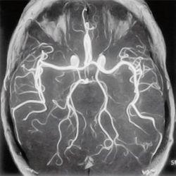
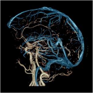
67 MR-Angiography
68 MEDICAL THERMOGRAPHY


69 MEDICAL THERMOGRAPHY Measures body tissue heat energy. Generally "problem areas" show high or low temperatures due to increased or reduced blood flow and metabolic activity, respectively. infrared radiation is emitted by all objects based on their temperatures above -237 С.
70 PACS
71 PACS (Picture Archiving and Communication System)
DEVIL PHYSICS THE BADDEST CLASS ON CAMPUS IB PHYSICS
 DEVIL PHYSICS THE BADDEST CLASS ON CAMPUS IB PHYSICS TSOKOS OPTION I-2 MEDICAL IMAGING Reading Activity Answers IB Assessment Statements Option I-2, Medical Imaging: X-Rays I.2.1. I.2.2. I.2.3. Define
Radioisotopes and PET
 Radioisotopes and PET 1 Radioisotopes Elements are defined by their number of protons, but there is some variation in the number of neutrons. Atoms resulting from this variation are called isotopes. Consider
Radioisotopes in action. Diagnostic application of radioisotopes. Steps of diagnostic procedure. Information from various medical imaging techniques
 Radioisotopes in action Diagnostic application of radioisotopes Steps of diagnostic procedure - Radioactive material introduced into the patient - Distribution and alteration of activity is detected -
Technical University of Denmark
 Technical University of Denmark Page 1 of 11 pages Written test, 9 December 2010 Course name: Introduction to medical imaging Course no. 31540 Aids allowed: none. "Weighting": All problems weight equally.
FXA UNIT G485 Module X-Rays. Candidates should be able to : I = I 0 e -μx
 1 Candidates should be able to : HISTORY Describe the nature of X-rays. Describe in simple terms how X-rays are produced. X-rays were discovered by Wilhelm Röntgen in 1865, when he found that a fluorescent
This Week. 3/23/2017 Physics 214 Summer
 This Week Atoms and nuclei What are we made of? The periodic table Why does it stop? How were the elements made? Radioactive decay Useful but can be toxic Discovery of X Rays: Cathode Rays and TV sets
Structure of Biological Materials
 ELEC ENG 3BA3: Structure of Biological Materials Notes for Lecture #19 Monday, November 22, 2010 6.5 Nuclear medicine imaging Nuclear imaging produces images of the distribution of radiopharmaceuticals
11/10/2014. Chapter 1: Introduction to Medical Imaging. Projection (Transmission) vs. Emission Imaging. Emission Imaging
 vs. Emission Imaging. Emission Imaging") Chapter 1: Introduction to Medical Imaging Overview of Modalities Properties of an Image: Limitations on Information Content Contrast (both object & image): Brightness difference Sharpness (blur): Smallest
Chapter 1: Introduction to Medical Imaging Overview of Modalities Properties of an Image: Limitations on Information Content Contrast (both object & image): Brightness difference Sharpness (blur): Smallest
ELG7173 Topics in signal Processing II Computational Techniques in Medical Imaging
 ELG7173 Topics in signal Processing II Computational Techniques in Medical Imaging Topic #1: Intro to medical imaging Medical Imaging Classifications n Measurement physics Send Energy into body Send stuff
(INCLUDING THIS FRONT PAGE)
") I'IFIITIIBIFI UNIVERSITY OF SCIEI'ICE RITD TECHNOLOGY FACULTY OF HEALTH AND APPLIED SCIENCES DEPARTMENT OF NATURAL AND APPLIED SCIENCES QUALIFICATION: BACHELOR OF SCIENCE (MAJOR AND MINOR) QUALIFICATION
I'IFIITIIBIFI UNIVERSITY OF SCIEI'ICE RITD TECHNOLOGY FACULTY OF HEALTH AND APPLIED SCIENCES DEPARTMENT OF NATURAL AND APPLIED SCIENCES QUALIFICATION: BACHELOR OF SCIENCE (MAJOR AND MINOR) QUALIFICATION
Nuclear Medicine RADIOPHARMACEUTICAL CHEMISTRY
 Nuclear Medicine RADIOPHARMACEUTICAL CHEMISTRY An alpha particle consists of two protons and two neutrons Common alpha-particle emitters Radon-222 gas in the environment Uranium-234 and -238) in the environment
AQA Physics /7408
 AQA Physics - 7407/7408 Module 10: Medical physics You should be able to demonstrate and show your understanding of: 10.1 Physics of the eye 10.1.1 Physics of vision The eye as an optical refracting system,
Modern physics ideas are strange! L 36 Modern Physics [2] The Photon Concept. How are x-rays produced? The uncertainty principle
![Modern physics ideas are strange! L 36 Modern Physics [2] The Photon Concept. How are x-rays produced? The uncertainty principle](/thumbs/88/117098787.jpg "Modern physics ideas are strange! L 36 Modern Physics [2] The Photon Concept. How are x-rays produced? The uncertainty principle") L 36 Modern Physics [2] X-rays & gamma rays How lasers work Medical applications of lasers Applications of high power lasers Medical imaging techniques CAT scans MRI s Modern physics ideas are strange!
L 36 Modern Physics [2] X-rays & gamma rays How lasers work Medical applications of lasers Applications of high power lasers Medical imaging techniques CAT scans MRI s Modern physics ideas are strange!
Technical University of Denmark
 Technical University of Denmark Page 1 of 10 pages Written test, 12 December 2012 Course name: Introduction to medical imaging Course no. 31540 Aids allowed: None. Pocket calculator not allowed "Weighting":
Rad T 290 Worksheet 2
 Class: Date: Rad T 290 Worksheet 2 1. Projectile electrons travel from a. anode to cathode. c. target to patient. b. cathode to anode. d. inner shell to outer shell. 2. At the target, the projectile electrons
Nuclear Radiation. Natural Radioactivity. A person working with radioisotopes wears protective clothing and gloves and stands behind a shield.
 Nuclear Radiation Natural Radioactivity A person working with radioisotopes wears protective clothing and gloves and stands behind a shield. 1 Radioactive Isotopes A radioactive isotope has an unstable
This Week. 7/20/2016 Physics 214 Spring
 This Week Atoms and nuclei What are we made of? The periodic table Why does it stop? How were the elements made? Radioactive decay Useful but can be toxic Discovery of X Rays: Cathode Rays and TV sets
Introduction to Medical Imaging. Medical Imaging
 Introduction to Medical Imaging BME/EECS 516 Douglas C. Noll Medical Imaging Non-invasive visualization of internal organs, tissue, etc. I typically don t include endoscopy as an imaging modality Image
Professor Stuart Bunt 217
 Professor Stuart Bunt 217 Traditional Anatomy Phrenology, the study of bumps on the skull. Measuring brain weights and size (still being done..see the fuss about Einstein s brain). Little link between
The Photon Concept. Modern Physics [2] How are x-rays produced? Gamma rays. X-ray and gamma ray photons. X-rays & gamma rays How lasers work
![The Photon Concept. Modern Physics [2] How are x-rays produced? Gamma rays. X-ray and gamma ray photons. X-rays & gamma rays How lasers work](/thumbs/75/72921848.jpg "The Photon Concept. Modern Physics [2] How are x-rays produced? Gamma rays. X-ray and gamma ray photons. X-rays & gamma rays How lasers work") Modern Physics [2] X-rays & gamma rays How lasers work Medical applications of lasers Applications of high power lasers Medical imaging techniques CAT scans MRI s The Photon Concept a beam of light waves
Modern Physics [2] X-rays & gamma rays How lasers work Medical applications of lasers Applications of high power lasers Medical imaging techniques CAT scans MRI s The Photon Concept a beam of light waves
MEDICAL EQUIPMENT: NUCLEAR MEDICINE. Prof. Yasser Mostafa Kadah
 MEDICAL EQUIPMENT: NUCLEAR MEDICINE Prof. Yasser Mostafa Kadah www.k-space.org Recommended Textbook Introduction to Medical Imaging: Physics, Engineering and Clinical Applications, by Nadine Barrie Smith
Basic physics of nuclear medicine
 Basic physics of nuclear medicine Nuclear structure Atomic number (Z): the number of protons in a nucleus; defines the position of an element in the periodic table. Mass number (A) is the number of nucleons
3. Which of the following statements is (are) TRUE about detector crystals in Anger cameras?
 TRUE about detector crystals in Anger cameras?") BioE 1330 - Exam 2 11/13/2018 Answer Sheet - Correct answer is A for all questions 1. Unlike CT, in nuclear medicine A. Bremsstrahlung is not used to produce high-energy photons. B. signal can be increased
BioE 1330 - Exam 2 11/13/2018 Answer Sheet - Correct answer is A for all questions 1. Unlike CT, in nuclear medicine A. Bremsstrahlung is not used to produce high-energy photons. B. signal can be increased
69 Ga Ga
 Stable isotope Relative atomic mass Mole fraction 69 Ga 68.925 574 0.601 08 71 Ga 70.924 703 0.398 92 Gallium isotopes in medicine 68 Ga is a radioactive isotope that emits positrons, which are used to
The physics of medical imaging US, CT, MRI. Prof. Peter Bogner
 The physics of medical imaging US, CT, MRI Prof. Peter Bogner Clinical radiology curriculum blocks of lectures and clinical practice (7x2) Physics of medical imaging Neuroradiology Head and neck I. Head
Nuclear Physics and Astrophysics
 Nuclear Physics and Astrophysics PHY-302 Dr. E. Rizvi Lecture 24 Medical Imaging Effects of Radiation We now know what radiation is But what does it mean for our bodies? Radioactivity is quantified in
Differentiating Chemical Reactions from Nuclear Reactions
 Differentiating Chemical Reactions from Nuclear Reactions 1 CHEMICAL Occurs when bonds are broken or formed. Atoms remained unchanged, though may be rearranged. Involves valence electrons Small energy
A. I, II, and III B. I C. I and II D. II and III E. I and III
 BioE 1330 - Review Chapters 7, 8, and 9 (Nuclear Medicine) 9/27/2018 Instructions: On the Answer Sheet, enter your 2-digit ID number (with a leading 0 if needed) in the boxes of the ID section. Fill in
Dosimetry. Sanja Dolanski Babić May, 2018.
 Dosimetry Sanja Dolanski Babić May, 2018. What s the difference between radiation and radioactivity? Radiation - the process of emitting energy as waves or particles, and the radiated energy Radioactivity
Medical Biophysics II. Final exam theoretical questions 2013.
 Medical Biophysics II. Final exam theoretical questions 2013. 1. Early atomic models. Rutherford-experiment. Franck-Hertz experiment. Bohr model of atom. 2. Quantum mechanical atomic model. Quantum numbers.
A Brief Introduction to Medical Imaging. Outline
 A Brief Introduction to Medical Imaging Outline General Goals Linear Imaging Systems An Example, The Pin Hole Camera Radiations and Their Interactions with Matter Coherent vs. Incoherent Imaging Length
The physics US and MRI. Prof. Peter Bogner
 The physics US and MRI Prof. Peter Bogner Sound waves mechanical disturbance, a pressure wave moves along longitudinal wave compression rarefaction zones c = nl, (c: velocity, n: frequency, l: wavelength
Chapter 21
 Chapter 21 http://youtu.be/kwasz59f8ga Nuclear reactions involve the nucleus The nucleus opens, and protons and neutrons are rearranged. The opening of the nucleus releases a tremendous amount of energy
Radioisotopes in action. Diagnostic application of radioisotopes. Steps of diagnostic procedure. Information from various medical imaging techniques
 Radioisotopes in action Diagnostic application of radioisotopes Steps of diagnostic procedure - Radioactive material introduced into the patient - Distribution and alteration of activity is detected -Monitoring
Nuclear Medicine Intro & Physics from Medical Imaging Signals and Systems, Chapter 7, by Prince and Links
 Nuclear Medicine Intro & Physics from Medical Imaging Signals and Systems, Chapter 7, by Prince and Links NM - introduction Relies on EMISSION of photons from body (versus transmission of photons through
Name Date Class NUCLEAR RADIATION. alpha particle beta particle gamma ray
 25.1 NUCLEAR RADIATION Section Review Objectives Explain how an unstable nucleus releases energy Describe the three main types of nuclear radiation Vocabulary radioisotopes radioactivity radiation alpha
ENG4BF3 Medical Image Processing
 ENG4BF3 Medical Image Processing Medical Imaging Modalities Imaging in Medical Sciences Imaging is an essential aspect of medical sciences for visualization of anatomical structures and functional or metabolic
Sodium isotopes in biology
 Stable Relative Mole isotope atomic mass fraction 23 Na 22.989 769 28 1 Sodium isotopes in biology Both 22 Na and 24 Na can be used as radioactive tracers to study electrolytes in the human body [102-104].
Magnetic resonance imaging MRI
 Magnetic resonance imaging MRI Introduction What is MRI MRI is an imaging technique used primarily in medical settings that uses a strong magnetic field and radio waves to produce very clear and detailed
Radiopharmaceuticals and Contrast Media. Lec: 7
 Radiopharmaceuticals and Contrast Media Lec: 7 Radiopharmaceuticals Radioisotopes every atom of an element is composed of a nucleus, containing protons and neutrons, surrounded by electrons. In the electrically
Bases of radioisotope diagnostic methods
 Medical, pharmaceutical applications of radioisotopes Bases of radioisotope diagnostic methods Dr. István Voszka Basis of application: radioisotopes have identical behavior in the organism to corresponding
Radionuclide Imaging MII Positron Emission Tomography (PET)
") Radionuclide Imaging MII 3073 Positron Emission Tomography (PET) Positron (β + ) emission Positron is an electron with positive charge. Positron-emitting radionuclides are most commonly produced in cyclotron
Radionuclide Imaging MII 3073 Positron Emission Tomography (PET) Positron (β + ) emission Positron is an electron with positive charge. Positron-emitting radionuclides are most commonly produced in cyclotron
Physics 30 Modern Physics Unit: Atomic Basics
 Physics 30 Modern Physics Unit: Atomic Basics Models of the Atom The Greeks believed that if you kept dividing matter into smaller and smaller pieces, you would eventually come to a bit of matter that
CHAPTER 4 RADIATION ATTENUATION
 HDR202 PHYSICS FOR RADIOGRAPHERS 2 CHAPTER 4 RADIATION ATTENUATION PREPARED BY: MR KAMARUL AMIN BIN ABDULLAH SCHOOL OF MEDICAL IMAGING FACULTY OF HEALTH SCIENCES Learning Objectives At the end of the lesson,
EL-GY 6813/BE-GY 6203 Medical Imaging, Fall 2016 Final Exam
 EL-GY 6813/BE-GY 6203 Medical Imaging, Fall 2016 Final Exam (closed book, 1 sheets of notes double sided allowed, no calculator or other electronic devices allowed) 1. Ultrasound Physics (15 pt) A) (9
Chapter 11 Nuclear Chemistry
 Chapter 11 Nuclear Chemistry 11.1 Nuclear Reactions Nuclear reactions involve the particles located in the nucleus of the atom: The nucleus contains: An atom is characterized by: X A Z - Z the gives the
Year 12 Notes Radioactivity 1/5
 Year Notes Radioactivity /5 Radioactivity Stable and Unstable Nuclei Radioactivity is the spontaneous disintegration of certain nuclei, a random process in which particles and/or high-energy photons are
Name: COMBINED SCIENCE Topics 4, 5 & 6 LEARNING OUTCOMES. Maintain a record of your progress Use the booklet to guide revision
 Name: COMBINED SCIENCE Topics 4, 5 & 6 LEARNING OUTCOMES Maintain a record of your progress Use the booklet to guide revision Close the Gap Contemporary record of the Topics / Learning outcomes that I
Name Date Class NUCLEAR CHEMISTRY
 25 NUCLEAR CHEMISTRY SECTION 25.1 NUCLEAR RADIATION (pages 799 802) This section describes the nature of radioactivity and the process of radioactive decay. It characterizes alpha, beta, and gamma radiation
1-D Fourier Transform Pairs
 1-D Fourier Transform Pairs The concept of the PSF is most easily explained by considering a very small point source being placed in the imaging field-of-view The relationship between the image, I, and
Lecture Presentation. Chapter 21. Nuclear Chemistry. James F. Kirby Quinnipiac University Hamden, CT Pearson Education, Inc.
 Lecture Presentation Chapter 21, Inc. James F. Kirby Quinnipiac University Hamden, CT Energy: Chemical vs. Chemical energy is associated with making and breaking chemical bonds. energy is enormous in comparison.
Waves & Radiation exam questions
 National 5 Physics Waves & Radiation exam questions these questions have been collated from previous Standard Grade (Credit) and Intermediate 2 exams Thurso High School 1. A mountain climber carries a
Application of Nuclear Physics
 Application of Nuclear Physics Frontier of gamma-ray spectroscopy 0.1 IR visible light UV soft X-ray X-ray hard X-ray gamma-ray 1 10 100 1e3 1e4 1e5 1e6 energy [ev] Photoelectric effect e - Compton scattering
MRI Homework. i. (0.5 pt each) Consider the following arrangements of bar magnets in a strong magnetic field.
 Consider the following arrangements of bar magnets in a strong magnetic field.") MRI Homework 1. While x-rays are used to image bones, magnetic resonance imaging (MRI) is used to examine tissues within the body by detecting where hydrogen atoms (H atoms) are and their environment (e.g.
MRI Homework 1. While x-rays are used to image bones, magnetic resonance imaging (MRI) is used to examine tissues within the body by detecting where hydrogen atoms (H atoms) are and their environment (e.g.
Production of X-rays. Radiation Safety Training for Analytical X-Ray Devices Module 9
 Module 9 This module presents information on what X-rays are and how they are produced. Introduction Module 9, Page 2 X-rays are a type of electromagnetic radiation. Other types of electromagnetic radiation
Chapter 20 Nuclear Chemistry. 1. Nuclear Reactions and Their Characteristics
 Chapter 2 Nuclear Chemistry 1. Nuclear Reactions and Their Characteristics Nuclear reactions involve the particles located in the nucleus of the atom: nucleons:. An atom is characterized by its atomic
Doppler echocardiography & Magnetic Resonance Imaging. Doppler echocardiography. History: - Langevin developed sonar.
 1 Doppler echocardiography & Magnetic Resonance Imaging History: - Langevin developed sonar. - 1940s development of pulse-echo. - 1950s development of mode A and B. - 1957 development of continuous wave
1. Which of the following statements is true about Bremsstrahlung and Characteristic Radiation?
 BioE 1330 - Review Chapters 4, 5, and 6 (X-ray and CT) 9/27/2018 Instructions: On the Answer Sheet, enter your 2-digit ID number (with a leading 0 if needed) in the boxes of the ID section. Fill in the
Stable isotope. Relative atomic mass. Mole fraction 203 Tl Tl Thallium isotopes in Earth/planetary science
 Stable isotope Relative atomic mass Mole fraction 203 Tl 202.972 345 0.2952 205 Tl 204.974 428 0.7048 Thallium isotopes in Earth/planetary science Because molecules, atoms, and ions of the stable isotopes
da u g ht er + radiation
 RADIOACTIVITY The discovery of radioactivity can be attributed to several scientists. Wilhelm Roentgen discovered X-rays in 1895 and shortly after that Henri Becquerel observed radioactive behavior while
Chapter. Nuclear Chemistry
 Chapter Nuclear Chemistry Nuclear Reactions 01 Chapter 22 Slide 2 Chapter 22 Slide 3 Alpha Decay: Loss of an α-particle (a helium nucleus) 4 2 He 238 92 U 234 4 U He 90 + 2 Chapter 22 Slide 4 Beta Decay:
Part III Minor Option in Medical Physics 2018 Examples Sheet
 Part III Minor Option in Medical Physics 2018 Examples Sheet Any errors or comments should be addressed sent to: seb53@cam.ac.uk URLs that may be useful: Stanford Event Generation Simulator: http://tinyurl.com/pkg476r
Atomic & Nuclear Physics
 Atomic & Nuclear Physics Life and Atoms Every time you breathe you are taking in atoms. Oxygen atoms to be exact. These atoms react with the blood and are carried to every cell in your body for various
 www.aask24.com www.aask24.com www.aask24.com P=Positron E= Emission T=Tomography Positron emission or beta plus decay (+ ) is a particular type of radioactive decay, in which a proton inside a radionuclide
Procesamiento de Imágenes y Bioseñales
 Procesamiento de Imágenes y Bioseñales Dr. Víctor Castañeda Agenda Physical basis of X-ray- CT, NMR, Ultrasound, Nuclear Medicine Sensors (cameras, gamma probes, microphone) Computational Tomography (CT)
Magnetic Resonance Imaging (MRI)
") Magnetic Resonance Imaging Introduction The Components The Technology (MRI) Physics behind MR Most slides taken from http:// www.slideworld.org/ viewslides.aspx/magnetic- Resonance-Imaging- %28MRI%29-MR-Imaging-
Magnetic Resonance Imaging Introduction The Components The Technology (MRI) Physics behind MR Most slides taken from http:// www.slideworld.org/ viewslides.aspx/magnetic- Resonance-Imaging- %28MRI%29-MR-Imaging-
Chemical Engineering 412
 Chemical Engineering 412 Introductory Nuclear Engineering Lecture 26 Radiation Detection & Measurement II Spiritual Thought 2 I would not hold the position in the Church I hold today had I not followed
12/1/17 OUTLINE KEY POINTS ELEMENTS WITH UNSTABLE NUCLEI Radioisotopes and Nuclear Reactions 16.2 Biological Effects of Nuclear Radiation
 OUTLINE 16.1 Radioisotopes and Nuclear Reactions 16.2 Biological Effects of Nuclear Radiation PET scan X-ray technology CT scan 2009 W.H. Freeman KEY POINTS Radioactivity is the consequence of an unstable
Alpha decay usually occurs in heavy nuclei such as uranium or plutonium, and therefore is a major part of the radioactive fallout from a nuclear
 Radioactive Decay Radioactivity is the spontaneous disintegration of atomic nuclei. This phenomenon was first reported in 1896 by the French physicist Henri Becquerel. Marie Curie and her husband Pierre
Chapter 2. Atomic Structure and Nuclear Chemistry. Atomic Structure & Nuclear Chemistry page 1
 Chapter 2 Atomic Structure and Nuclear Chemistry Atomic Structure & Nuclear Chemistry page 1 Atoms & Elements Part 0: Atomic Structure An Introduction Electrostatics an underlying force throughout chemistry
X-ray Interaction with Matter
 X-ray Interaction with Matter 10-526-197 Rhodes Module 2 Interaction with Matter kv & mas Peak kilovoltage (kvp) controls Quality, or penetrating power, Limited effects on quantity or number of photons
GLOSSARY OF BASIC RADIATION PROTECTION TERMINOLOGY
 GLOSSARY OF BASIC RADIATION PROTECTION TERMINOLOGY ABSORBED DOSE: The amount of energy absorbed, as a result of radiation passing through a material, per unit mass of material. Measured in rads (1 rad
Nuclear Reactions A Z. Radioactivity, Spontaneous Decay: Nuclear Reaction, Induced Process: x + X Y + y + Q Q > 0. Exothermic Endothermic
 Radioactivity, Spontaneous Decay: Nuclear Reactions A Z 4 P D+ He + Q A 4 Z 2 Q > 0 Nuclear Reaction, Induced Process: x + X Y + y + Q Q = ( m + m m m ) c 2 x X Y y Q > 0 Q < 0 Exothermic Endothermic 2
and have low penetrating power) Alpha particles are released through alpha decay. Beta Particles: An electron that comes from a nucleus through
 Alpha particles are released through alpha decay. Beta Particles: An electron that comes from a nucleus through") TOPIC 13: Nuclear Chemistry 1. When the atomic nucleus of one element is changed into the nucleus of a different element, the reaction is called transmutation. Stability of a Nucleus: Any element containing
TOPIC 13: Nuclear Chemistry 1. When the atomic nucleus of one element is changed into the nucleus of a different element, the reaction is called transmutation. Stability of a Nucleus: Any element containing
Name Date Class NUCLEAR CHEMISTRY. Standard Curriculum Core content Extension topics
 28 NUCLEAR CHEMISTRY Conceptual Curriculum Concrete concepts More abstract concepts or math/problem-solving Standard Curriculum Core content Extension topics Honors Curriculum Core honors content Options
Electrical Engineering 3BA3: Structure of Biological Materials
 Electrical Engineering 3BA3: Structure of Biological Materials Day Class Instructor: Dr. I. C. BRUCE Duration of Examination: 3 Hours McMaster University Final Examination December, 2004 This examination
Radioactivity. The Nobel Prize in Physics 1903 for their work on radioactivity. Henri Becquerel Pierre Curie Marie Curie
 Radioactivity Toward the end of the 19 th century, minerals were found that would darken a photographic plate even in the absence of light. This phenomenon is now called radioactivity. Marie and Pierre
1ST SEM MT CHAP 22 REVIEW
 1ST SEM MT CHAP 22 REVIEW Multiple Choice Identify the choice that best completes the statement or answers the question. (CAPITAL LETTERS ONLY PLEASE) 1. Mass defect is the difference between the mass
Lecture 1 Bioradiation
 1 1 Radiation definition: Radiation, when broadly defined, includes the entire spectrum of electromagnetic waves : radiowaves, microwaves, infrared, visible light, ultraviolet, and x-rays and particles.
 Radioactive Decay What is Radioactivity? http://explorecuriocity.org/explore/articleid/3033 http://explorecuriocity.org/explore/articleid/3035 http://explorecuriocity.org/explore/articleid/2160 Quick Review
Chap. 15 Radiation Imaging
 Chap. 15 Radiation Imaging 15.1 INTRODUCTION Modern Medical Imaging Devices Incorporating fundamental concepts in physical science and innovations in computer technology Nobel prize (physics) : 1895 Wilhelm
State the position of protons, neutrons and electrons in the atom
 2.1 The Atom 2.1.1 - State the position of protons, neutrons and electrons in the atom Atoms are made up of a nucleus containing positively charged protons and neutral neutrons, with negatively charged
Number of protons. 2. What is the nuclear symbol for a radioactive isotope of copper with a mass number of 60? A) Cu
 Cu") Chapter 5 Nuclear Chemistry Practice Problems 1. Fill in the missing information in the chart: Medical Use Atomic Mass symbol number Heart imaging 201 Tl 81 Number of protons Number of neutrons Abdominal
Chapter 5 Nuclear Chemistry Practice Problems 1. Fill in the missing information in the chart: Medical Use Atomic Mass symbol number Heart imaging 201 Tl 81 Number of protons Number of neutrons Abdominal
General Physics (PHY 2140)
") General Physics (PHY 2140) Lecture 19 Modern Physics Nuclear Physics Nuclear Reactions Medical Applications Radiation Detectors Chapter 29 http://www.physics.wayne.edu/~alan/2140website/main.htm 1 Lightning
General Physics (PHY 2140) Lecture 19 Modern Physics Nuclear Physics Nuclear Reactions Medical Applications Radiation Detectors Chapter 29 http://www.physics.wayne.edu/~alan/2140website/main.htm 1 Lightning
General Physics (PHY 2140)
") General Physics (PHY 2140) Lightning Review Lecture 19 Modern Physics Nuclear Physics Nuclear Reactions Medical Applications Radiation Detectors Chapter 29 http://www.physics.wayne.edu/~alan/2140website/main.htm
General Physics (PHY 2140) Lightning Review Lecture 19 Modern Physics Nuclear Physics Nuclear Reactions Medical Applications Radiation Detectors Chapter 29 http://www.physics.wayne.edu/~alan/2140website/main.htm
Fundamental MRI Principles Module Two
 Fundamental MRI Principles Module Two 1 Nuclear Magnetic Resonance There are three main subatomic particles: protons neutrons electrons positively charged no significant charge negatively charged Protons
NOTES: 25.2 Nuclear Stability and Radioactive Decay
 NOTES: 25.2 Nuclear Stability and Radioactive Decay Why does the nucleus stay together? STRONG NUCLEAR FORCE Short range, attractive force that acts among nuclear particles Nuclear particles attract one
Sound wave bends as it hits an interface at an oblique angle. 4. Reflection. Sound wave bounces back to probe
 : Ultrasound imaging and x-rays 1. How does ultrasound imaging work?. What is ionizing electromagnetic radiation? Definition of ionizing radiation 3. How are x-rays produced? Bremsstrahlung Auger electron
RADIOACTIVITY & HALF-LIFE Part 2
 RADIOACTIVITY & HALF-LIFE Part 2 Radioactivity Radioactivity: Results from radioactive decay, which is the process whereby unstable atomic nuclei transform and emit radiation. Has existed longer than the
L 36 Modern Physics [3] The atom and the nucleus. Structure of the nucleus. The structure of the nucleus SYMBOL FOR A NUCLEUS FOR A CHEMICAL X
![L 36 Modern Physics [3] The atom and the nucleus. Structure of the nucleus. The structure of the nucleus SYMBOL FOR A NUCLEUS FOR A CHEMICAL X](/thumbs/87/95852290.jpg "L 36 Modern Physics [3] The atom and the nucleus. Structure of the nucleus. The structure of the nucleus SYMBOL FOR A NUCLEUS FOR A CHEMICAL X") L 36 Modern Physics [3] [L36] Nuclear physics what s inside the nucleus and what holds it together what is radioactivity carbon dating [L37] Nuclear energy nuclear fission nuclear fusion nuclear reactors
L 36 Modern Physics [3] [L36] Nuclear physics what s inside the nucleus and what holds it together what is radioactivity carbon dating [L37] Nuclear energy nuclear fission nuclear fusion nuclear reactors
Physics of Radiography
 Physics of Radiography Yao Wang Polytechnic Institute of NYU Brooklyn, NY 11201 Based on J L Prince and J M Links Medical Imaging Signals and Based on J. L. Prince and J. M. Links, Medical Imaging Signals
25.1. Nuclear Radiation
 Nuclear Radiation Marie Curie was a Polish scientist whose research led to many discoveries about radiation and radioactive elements. In 1934 she died from leukemia caused by her long-term exposure to
RADIOACTIVITY. Nature of Radioactive Emissions
 1 RADIOACTIVITY Radioactivity is the spontaneous emissions from the nucleus of certain atoms, of either alpha, beta or gamma radiation. These radiations are emitted when the nuclei of the radioactive substance
11/19/2014. Chapter 3: Interaction of Radiation with Matter in Radiology and Nuclear Medicine. Nuclide Families. Family Nuclides with Same: Example
 2014-2015 Residents' Core Physics Lectures Mondays 7:00-8:00 am in VA Radiology and UCSDMC Lasser Conference Rooms Topic Chapters Date Faculty 1 Introduction and Basic Physics 1, 2 M 11/17 Andre 2 Interaction
U (superscript is mass number, subscript atomic number) - radionuclides nuclei that are radioactive - radioisotopes atoms containing radionuclides
 - radionuclides nuclei that are radioactive - radioisotopes atoms containing radionuclides") Chapter : Nuclear Chemistry. Radioactivity nucleons neutron and proton all atoms of a given element have the same number of protons, atomic number isotopes atoms with the same atomic number but different
Chapter : Nuclear Chemistry. Radioactivity nucleons neutron and proton all atoms of a given element have the same number of protons, atomic number isotopes atoms with the same atomic number but different
EXAMINATION QUESTIONS (6)
") 1. What is a beta-particle? A a helium nucleus B a high-energy electron C four protons D two neutrons EXAMINATION QUESTIONS (6) 2. The diagram shows part of a circuit used to switch street lamps on and
1. What is a beta-particle? A a helium nucleus B a high-energy electron C four protons D two neutrons EXAMINATION QUESTIONS (6) 2. The diagram shows part of a circuit used to switch street lamps on and
Nuclear Chemistry. Background Radiation. Three-fourths of all exposure to radiation comes from background radiation.
 Chapter 11 Nuclear Chemistry Background Radiation Three-fourths of all exposure to radiation comes from background radiation. Most of the remaining one-fourth comes from medical irradiation such as X-rays.
Units and Definition
 RADIATION SOURCES Units and Definition Activity (Radioactivity) Definition Activity: Rate of decay (transformation or disintegration) is described by its activity Activity = number of atoms that decay
2. Which of the following statements help(s) to explain why gas can fill the vessel containing it completely while liquid cannot?
 to explain why gas can fill the vessel containing it completely while liquid cannot?") Name: Class: ( ) There are 30 questions. Time Allowed: 45 min 1. Kinetic theory explains the behaviour of a substance in terms of the behaviour of the molecules in it. Which of the following is/are the
Name: Class: ( ) There are 30 questions. Time Allowed: 45 min 1. Kinetic theory explains the behaviour of a substance in terms of the behaviour of the molecules in it. Which of the following is/are the
Unit 12: Nuclear Chemistry
 Unit 12: Nuclear Chemistry 1. Stability of isotopes is based on the ratio of neutrons and protons in its nucleus. Although most nuclei are stable, some are unstable and spontaneously decay, emitting radiation.