Radionuclide Imaging MII Positron Emission Tomography (PET)
|
|
|
- Andrea Spencer
- 6 years ago
- Views:
Transcription
1 Radionuclide Imaging MII 3073 Positron Emission Tomography (PET)
2 Positron (β + ) emission Positron is an electron with positive charge. Positron-emitting radionuclides are most commonly produced in cyclotron by bombarding a stable element with protons, deuterons or helium nuclei. The produced radionuclides have an excess of protons and decay by the emission of positrons. A proton inside the nucleus is converted into a neutron. The excess energy is emitted as a pair of particles, a positron (β+) and a neutrino.
3 Radioisotope Atomic number (protons) Number of nucleons (protons + neutrons) Approximate half-life (min) Carbon Nitrogen Oxygen Fluorine Common positron emitters for PET and/or PET/CT imaging
4 When a positron is emitted, it travels for a short distance from its site of origin, gradually losing energy to the tissue through which it moves. When most of its kinetic energy has been lost, the positron reacts with a resident electron in an annihilation reaction. This reaction generated two 511 kev gamma photons, which are emitted in opposite direction at about 180 o from each other. In a PET scanner, these photons interact with the detector ring at opposite sites, which defines a line along which the annihilation reaction occurred and permits localization of the reaction. By using many such events, an image can be reconstructed.
5 511 kev β + e kev Positron- electron annihilation reaction
6 PET scanner/camera Dedicated PET camera contains with multiple rings detectors that consist of scintillation crystals coupled with PMTs. The ring design takes advantage of the fact that two photons detected by two opposed detectors in the ring are likely to be from a single annihilation event. Such a simultaneous detection is called a coincidence. The simultaneous detection of two photons provides location information in that the annihilation event can be assumed to occur somewhere on a line between the two detectors (the line of response; LOR).
7 β + e - Coincidence detection
8 The many coincidence events recorded by the PET scanner constitute a raw data set representing projections of the distribution of the positron radiopharmaceutical in the body. These data are then reconstructed by using a filtered back projection algorithm or an iterative algorithm to produce cross-sectional images. Because photons travel at the speed of light, PET cameras require very fast electronics to determine if two detected photons were likely produced by a single annihilation event.
9 In a PET scanner, each annihilation photon reaching a detector generates a single electronic pulse. For this photon to be accepted and used in the PET image, it must be in a specific energy range (ideally approaching 511 kev) and be paired with another photon reaching another detector simultaneously. Coincidence circuitry connecting the many detectors in the rings determines whether two such single pulses fall within a short coincidence time window, typically 6 to 12 nanoseconds. If so, they are deemed to constitute a coincidence event and are recorded in the resultant image.
10 Because of detector ring geometry and photon attenuation through scatter and absorption, many annihilation events result in one of the two 511 kev photons interacting with the PET camera detectors (single event). PET scanners use only photon pairs meeting the coincidence criterion in constructing PET images. Therefore, single events can be identified and discarded. In practice, about 99% of detected photons are rejected by the coincidence circuitry of the PET system. However, this principle of coincidence detection provides a virtual electronic collimation of the events and makes PET scanners inherently more efficient than traditional gamma cameras.
11 Events detected by PET scanners include true, scattered, and random events. All of these may be recorded as coincidences, providing both annihilation photons are actually detected and fall within the coincidence window. True coincidences are those that result when both 511 kev photons from an annihilation reaction are detected within the coincidence time window, neither photon having undergone any form of interaction before reaching the detector. These true coincidence events provide the desired information for constructing accurate images of the distribution of a PET radiopharmaceutical in clinical imaging.
12 Scattered coincidences occur when one or both annihilation photon undergo Compton interaction in body tissues and are deflected away from their expected path but still reach the detectors within the time window and are recorded as a coincidence event. Because the direction of the scattered photon has changed during the Compton interaction, the resulting coincidence event is likely to be assigned as an inaccurate LOR that no longer passes though the point of annihilation, leading to erroneous localization information and decreasing image contrast.
13 Random coincidences arise when two photons, each origination from a different annihilation reaction, reach any detector within the time window and thus appear to represent a true coincidence. Using detectors that allow very precise timing permits the recognition and exclusion of random events with a resultant improvement in image quality. If left uncorrected, both scattered and random coincidences add background to the true coincidence distribution, thereby increasing statistical noise, decreasing contrast, and causing the radioisotope concentrations to be overestimated.
14 a b c
15 There are a number of methods available to reduce the image degrading impact of scattered coincidences. Most scattered photons are not detected because they are absorbed in tissues of the body, are scattered away from the detector rings, or have lost significant energy during Compton scattering. These lower energy scattered events can be rejected by using an energy window designed to exclude photons of certain energies. The success of such rejection depends on the energy resolution characteristics of the detectors being used.
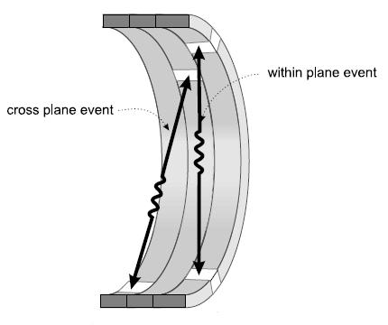
16 Because crystal detectors have only a finite energy resolution, if one were to measure only photons approaching 511 kev and exclude scattered photons of slightly different energies, a large number of true events would also be excluded, thereby either reducing image statistics or increasing image acquisition times unacceptably. Therefore, a rather broad energy window is used that allows some scattered events to be recorded as true events. Another method to reduce scatter from outside the plane of a detector ring is to use thin lead or tungsten septa positioned between the detector elements.
17 Imaging with lead septa is called 2D imaging because most of the photons counted originate in the plane of a single detector ring. 2D imaging improves image quality by reducing image noise. It also minimizes count losses due to system dead time by incidentally reducing the very large numbers of photons reaching the detectors that may occur at high count rates. However, although this reduces the number of scattered events originating outside the field of view (FOV), it also significantly reduces the true counts and increases imaging times.
18 Faster detector crystals and faster electronics in PET instruments have made imaging without septa, so called 3D imaging possible. This allows imaging from the volume defined by the entire FOV of the multiple detector rings of the camera and permits detection of true coincidence events that occur in different detectors on different rings. Compared with 2D imaging, 3D acquisitions increase sensitivity of the system by five folds or more. However, because both true coincidence and scatter rates are increases, better temporal and energy resolutions are needed to accurately eliminate scatter and random events.

19 2D imaging 3D imaging
20 PET scintillation detectors All positron systems use the principle of scintillation whereby the photon interacting with a crystal produces a flash of light, which is then detected and localized by PMTs. The ideal PET crystal detector would have: 1. high stopping power for 511 kev photons providing high efficiency and optimum spatial resolution 2. fast, intense light output with rapid decay of the light for decreased system dead time 3. good energy resolution for accurate scatter rejection 4. Low Compton scatter inside detector crystal 5. Matching of wavelength of fluorescence to response of light detector
21 Stopping power is best for crystalline materials with high density and high effective atomic number (Z value). There are several types of crystalline detector materials used for PET imaging include NaI, BGO, LSO and GSO. The light signal produced by scintillation detectors is not discrete in time but occurs over a short time interval (scintillation decay time, nanoseconds), which includes the period over which the light fades to background. Along with the speed of processing electronics, this decay time is an important determinant of system dead time.
22 Dead time is the brief period during which a crystal- PMT detector is busy producing and recording a scintillation event and having the scintillation light decay so that the next distinct scintillation event can be recognized and recorded. During this time additional arriving events cannot be processed and are lost. High count rate capability of PET instruments is particularly important in 3D acquisitions and in settings requiring high activities of very short-lived radionuclides (e.g., Oxygen-15; physical half-life 124s). Current count rate capabilities are about counts/second.
23 PET detector geometry State-of-the-art PET scanners is multiple full rings detector system that axially surround the patient (360 o ). These cameras have multiple adjacent detector rings that significantly increase the axial FOV of the patient. A larger FOV allows more counts to be detected for a standardized administered radiopharmaceutical dose and a fixed scan time by allowing more time at each table position. The most common detector arrangement consists of rings of individual detector modules of small crystal arrays or cut block scintillation crystal (usually BGO or LSO) coupled with PMTs.
24 In crystal arrays, multiple separate very small scintillation crystals are grouped together in blocks, often arranged in 6 X 6 or 8 X 8 blocks (more economic and cost-effective). These blocks are then assembled to form a crystal ring and coupled to PMTs (about four per block). For multiple rings PET camera, the intrinsic spatial resolution is a function of the crystal size. Thus, the small sizes of the crystal faces allowed by block design permits optimization of intrinsic resolution. Furthermore, a large number of small independent detectors will significantly reduce dead time count losses and allow camera operation at higher count rates.

25 Multiple rings detector Scintillator crystals PMT PMT Block detector unit
26 Sensitivity and resolution The sensitivity is defined as the recorded true coincidence rate (without scatter and random events) divided by the activity concentration (the true emitted events from the source). Sensitivity of a PET camera is determined by multiple factors like scanner geometry, crystal efficiency and photon attenuation in tissue. Spatial resolution in PET scanner is, in large part, a function of detector size, with smaller detectors increasing the resolving capability of the system. The ultimate limit of spatial resolution when using FDG is about 1 mm. However, the practical spatial resolution for clinical imaging is about 4 to 6 mm.
27 Many photons emitted from the patient (about 99%) are not detected because they are emitted in all directions from the patient and the detector rings cover only a fraction of the patient s body surface. When attenuation by absorption or scatter is considered, current systems record substantially less than 0.1% of the true events. However, because state-of-the-art PET scanners typically image in 3D mode, their efficiency for detecting emitted radiation is still considerably greater than that for SPECT imaging.
28 Time-of-flight (TOF) To improve resolution, some systems also measure time-of-flight (TOF) under the assumption that the location of the annihilation can be determined along the LOR of the coincident photons by measuring the time of arrival of each of the photons at the opposing crystals. Except the event that occurs in the exact centre of the detection ring, one of the photons will arrive before the other. The time difference will be proportional to the difference in distances traveled by the two photons and can be used to calculate the position of the event along the LOR.
29 Time-of-flight PET systems
30 Attenuation correction The attenuation in PET imaging, that is, loss of counts due to absorption of photons before they arrive at the detector, is compensated for arithmetically by using data from transmission scans. Depending on the camera design the transmission source can be a positron source, a high energy single photon source or a CT x-ray source. By using this transmission scan data, a patient specific attenuation correction map can be generated. An attenuation-corrected PET image has better image quality as compared with non-corrected one.


31 Non-attenuation corrected PET image Attenuation corrected PET image


32 PET transmission scan PET emission scan + = PET transmission image PET emission image (non-attenuation corrected) Attenuation corrected PET image
33 Standardized uptake value (SUV) Although visual assessment of PET images is often sufficient for image analysis and interpretation, the quantitative image analysis using the standardized uptake value (SUV) is also commonly used to augment the finding by measuring the degree of the tracer uptake on the area of the suspected lesion. SUV is a semiquantitative measurement that normalises the measured radioactivity concentration in a tissue to the body weight and total injected radioactivity, and a unitless ratio.
34
35 Limitations of PET imaging Several limitations of PET imaging are: 1. The attenuation map from the transmission is usually noisy and sensitive to the emission activity in the injected patient. 2. A long acquisition time may limit the throughput capability of the PET scanner. 3. The spatial resolution of PET images is generally poor compared with radiographs, CT or MRI image. 4. The cost of all PET instruments and facilities is relatively high as compared to other conventional imaging modalities. 5. The cost of each PET imaging is also high, which mainly depends on the cost of radiopharmaceuticals used. 6. Patient may suffer to an internal whole body exposure with a non-uniform distribution. 7. The radiation dose largely depends on the amount of injected radioactivity.
Chapter 2 PET Imaging Basics
 Chapter 2 PET Imaging Basics Timothy G. Turkington PET Radiotracers Positron emission tomography (PET) imaging is the injection (or inhalation) of a substance containing a positron emitter, the subsequent
MEDICAL EQUIPMENT: NUCLEAR MEDICINE. Prof. Yasser Mostafa Kadah
 MEDICAL EQUIPMENT: NUCLEAR MEDICINE Prof. Yasser Mostafa Kadah www.k-space.org Recommended Textbook Introduction to Medical Imaging: Physics, Engineering and Clinical Applications, by Nadine Barrie Smith
A. I, II, and III B. I C. I and II D. II and III E. I and III
 BioE 1330 - Review Chapters 7, 8, and 9 (Nuclear Medicine) 9/27/2018 Instructions: On the Answer Sheet, enter your 2-digit ID number (with a leading 0 if needed) in the boxes of the ID section. Fill in
Year 12 Notes Radioactivity 1/5
 Year Notes Radioactivity /5 Radioactivity Stable and Unstable Nuclei Radioactivity is the spontaneous disintegration of certain nuclei, a random process in which particles and/or high-energy photons are
 www.aask24.com www.aask24.com www.aask24.com P=Positron E= Emission T=Tomography Positron emission or beta plus decay (+ ) is a particular type of radioactive decay, in which a proton inside a radionuclide
Radioisotopes and PET
 Radioisotopes and PET 1 Radioisotopes Elements are defined by their number of protons, but there is some variation in the number of neutrons. Atoms resulting from this variation are called isotopes. Consider
Mitigation of External Radiation Exposures
 Mitigation of External Radiation Exposures The three (3) major principles to assist with maintaining doses ALARA are :- 1) Time Minimizing the time of exposure directly reduces radiation dose. 2) Distance
Mayneord-Phillips Summer School St Edmund Hall, University of Oxford July Proton decays to n, e +, ν
 Positron Emission Tomography Physics & Instrumentation Dimitra G. Darambara, Ph.D Multimodality Molecular Imaging Joint Department of Physics RMH/ICR Outline Introduction PET Physics overview Types of
6: Positron Emission Tomography
 6: Positron Emission Tomography. What is the principle of PET imaging? Positron annihilation Electronic collimation coincidence detection. What is really measured by the PET camera? True, scatter and random
The Physics of PET/CT scanners
 The Physics of PET/CT scanners Ruth E. Schmitz, Adam M. Alessio, and Paul E. Kinahan Imaging Research Laboratory Department of Radiology University of Washington What Makes PET Useful? Positron emission
Positron Emission Tomography
 Positron Emission Tomography Presenter: Difei Wang June,2018 Universität Bonn Contents 2 / 24 1 2 3 4 Positron emission Detected events Detectors and configuration Data acquisition Positron emission Positron
Introduction to SPECT & PET TBMI02 - Medical Image Analysis 2017
 Introduction to SPECT & PET TBMI02 - Medical Image Analysis 2017 Marcus Ressner, PhD, Medical Radiation Physicist, Linköping University Hospital Content What is Nuclear medicine? Basic principles of Functional
CT-PET calibration : physical principles and operating procedures F.Bonutti. Faustino Bonutti Ph.D. Medical Physics, Udine University Hospital.
 CT-PET calibration : physical principles and operating procedures Faustino Bonutti Ph.D. Medical Physics, Udine University Hospital Topics Introduction to PET physics F-18 production β + decay and annichilation
PET. Technical aspects
 PET Technical aspects 15 N 15 O Detector 1 β+ Detector 2 e- Evolution of PET Detectors CTI/Siemens 15 N 15 O Detector block 1 β+ Detector block 2 x e- x y y location line of response Constant fraction
Tomography is imaging by sections. 1
 Tomography is imaging by sections. 1 It is a technique used in clinical medicine and biomedical research to create images that show how certain tissues are performing their physiological functions. 1 Conversely,
Nuclear Medicine RADIOPHARMACEUTICAL CHEMISTRY
 Nuclear Medicine RADIOPHARMACEUTICAL CHEMISTRY An alpha particle consists of two protons and two neutrons Common alpha-particle emitters Radon-222 gas in the environment Uranium-234 and -238) in the environment
What is scintigraphy? The process of obtaining an image or series of sequential images of the distribution of a radionuclide in tissues, organs, or
 Let's remind... What is nuclear medicine? Nuclear medicine can be broadly divided into two branches "in vitro" and "in vivo" procedures. There are numerous radioisotopic "in vitro" procedures for genotyping
11/10/2014. Chapter 1: Introduction to Medical Imaging. Projection (Transmission) vs. Emission Imaging. Emission Imaging
 vs. Emission Imaging. Emission Imaging") Chapter 1: Introduction to Medical Imaging Overview of Modalities Properties of an Image: Limitations on Information Content Contrast (both object & image): Brightness difference Sharpness (blur): Smallest
Chapter 1: Introduction to Medical Imaging Overview of Modalities Properties of an Image: Limitations on Information Content Contrast (both object & image): Brightness difference Sharpness (blur): Smallest
Nuclear Medicine Intro & Physics from Medical Imaging Signals and Systems, Chapter 7, by Prince and Links
 Nuclear Medicine Intro & Physics from Medical Imaging Signals and Systems, Chapter 7, by Prince and Links NM - introduction Relies on EMISSION of photons from body (versus transmission of photons through
Study of the feasibility of a compact gamma camera for real-time cancer assessment
 Study of the feasibility of a compact gamma camera for real-time cancer assessment L. Caballero Instituto de Física Corpuscular - CSIC - University of Valencia; C/Catedrático José Beltrán, 2; E-46980;
Detector technology. Aim of this talk. Principle of a radiation detector. Interactions of gamma photons (gas) Gas-filled detectors: examples
 Gas-filled detectors: examples") Aim of this tal Detector technology WMIC Educational Program Nuclear Imaging World Molecular Imaging Congress, Dublin, Ireland, Sep 5-8, 202 You can now the name of a bird in all the languages of the world,
Aim of this tal Detector technology WMIC Educational Program Nuclear Imaging World Molecular Imaging Congress, Dublin, Ireland, Sep 5-8, 202 You can now the name of a bird in all the languages of the world,
Positron Emission Tomography (PET)
") Positron Emission Tomography (PET) A radiological technique for functional imaging Please note that this exercise takes place at the Stockholm Centre for Physics, Astronomy and Biotechniques (Alba Nova).
Positron Emission Tomography (PET) A radiological technique for functional imaging Please note that this exercise takes place at the Stockholm Centre for Physics, Astronomy and Biotechniques (Alba Nova).
3. Which of the following statements is (are) TRUE about detector crystals in Anger cameras?
 TRUE about detector crystals in Anger cameras?") BioE 1330 - Exam 2 11/13/2018 Answer Sheet - Correct answer is A for all questions 1. Unlike CT, in nuclear medicine A. Bremsstrahlung is not used to produce high-energy photons. B. signal can be increased
BioE 1330 - Exam 2 11/13/2018 Answer Sheet - Correct answer is A for all questions 1. Unlike CT, in nuclear medicine A. Bremsstrahlung is not used to produce high-energy photons. B. signal can be increased
Radioisotopes in action. Diagnostic application of radioisotopes. Steps of diagnostic procedure. Information from various medical imaging techniques
 Radioisotopes in action Diagnostic application of radioisotopes Steps of diagnostic procedure - Radioactive material introduced into the patient - Distribution and alteration of activity is detected -
FXA UNIT G485 Module X-Rays. Candidates should be able to : I = I 0 e -μx
 1 Candidates should be able to : HISTORY Describe the nature of X-rays. Describe in simple terms how X-rays are produced. X-rays were discovered by Wilhelm Röntgen in 1865, when he found that a fluorescent
Physics in Nuclear Medicine
 SIMON R. CHERRY, PH.D. Professor Department of Biomedical Engineering University of California-Davis Davis, California JAMES A. SORENSON, PH.D. Emeritus Professor of Medical Physics University of Wisconsin-Madison
Basic physics of nuclear medicine
 Basic physics of nuclear medicine Nuclear structure Atomic number (Z): the number of protons in a nucleus; defines the position of an element in the periodic table. Mass number (A) is the number of nucleons
DEVIL PHYSICS THE BADDEST CLASS ON CAMPUS IB PHYSICS
 DEVIL PHYSICS THE BADDEST CLASS ON CAMPUS IB PHYSICS TSOKOS OPTION I-2 MEDICAL IMAGING Reading Activity Answers IB Assessment Statements Option I-2, Medical Imaging: X-Rays I.2.1. I.2.2. I.2.3. Define
Dana-Farber Cancer Institute, 44 Binney Street, Boston, MA 02115, USA ramsey
 SPECIAL FEATURE: MEDICAL PHYSICS www.iop.org/journals/physed Nuclear medicine Ramsey D Badawi Dana-Farber Cancer Institute, 44 Binney Street, Boston, MA 02115, USA E-mail: ramsey badawi@dfci.harvard.edu
Basic physics Questions
 Chapter1 Basic physics Questions S. Ilyas 1. Which of the following statements regarding protons are correct? a. They have a negative charge b. They are equal to the number of electrons in a non-ionized
Structure of Biological Materials
 ELEC ENG 3BA3: Structure of Biological Materials Notes for Lecture #19 Monday, November 22, 2010 6.5 Nuclear medicine imaging Nuclear imaging produces images of the distribution of radiopharmaceuticals
PET scan simulation. Meysam Dadgar. UMSU, Iran. IFMP, Elbasan, Fig 1: PET camera simulation in gate by cylindrical phantom
 PET scan simulation Meysam Dadgar UMSU, Iran IFMP, Elbasan, 2016 Meysamdadgar10@gmail.com 1 Fig 1: PET camera simulation in gate by cylindrical phantom 2 What is PET? Positron emission tomography (PET),
1st Faculty of Medicine, Charles University in Prague Center for Advanced Preclinical Imaging (CAPI)
") Radioation Resolution and Sensitivity Nuclear Imaging PET + SPECT Radioactive Decay (EC,Ɣ), (β -,Ɣ), (I.T.,Ɣ) β + Projection imaging collimator needed one angular view Projection imaging coincidence imaging,
Radioation Resolution and Sensitivity Nuclear Imaging PET + SPECT Radioactive Decay (EC,Ɣ), (β -,Ɣ), (I.T.,Ɣ) β + Projection imaging collimator needed one angular view Projection imaging coincidence imaging,
QUIZ: Physics of Nuclear Medicine Atomic Structure, Radioactive Decay, Interaction of Ionizing Radiation with Matter
 QUIZ: Physics of Nuclear Medicine Atomic Structure, Radioactive Decay, Interaction of Ionizing Radiation with Matter 1. An atomic nucleus contains 39 protons and 50 neutrons. Its mass number (A) is a)
Wednesday 23 January 2013 Afternoon
 Wednesday 23 January 2013 Afternoon A2 GCE PHYSICS A G485/01 Fields, Particles and Frontiers of Physics *G411600113* Candidates answer on the Question Paper. OCR supplied materials: Data, Formulae and
Gamma ray coincidence and angular correlation
 University of Cape Town Department of Physics Course III laboratory Gamma ray coincidence and angular correlation Introduction Medical imaging based on positron emission tomography (PET) continues to have
A Brief Introduction to Medical Imaging. Outline
 A Brief Introduction to Medical Imaging Outline General Goals Linear Imaging Systems An Example, The Pin Hole Camera Radiations and Their Interactions with Matter Coherent vs. Incoherent Imaging Length
University of Ljubljana Faculty of mathematics and physics Department of physics. Tomography. Mitja Eržen. August 6, Menthor: Dr.
 University of Ljubljana Faculty of mathematics and physics Department of physics Tomography Mitja Eržen August 6, 2009 Menthor: Dr. Matjaž Vencelj Abstract We ll describe some methods for medical imaging.i
CHAPTER 7 TEST REVIEW
 IB PHYSICS Name: Period: Date: # Marks: 94 Raw Score: IB Curve: DEVIL PHYSICS BADDEST CLASS ON CAMPUS CHAPTER 7 TEST REVIEW 1. An alpha particle is accelerated through a potential difference of 10 kv.
Outline Chapter 14 Nuclear Medicine
 Outline Chapter 14 uclear Medicine Radiation Dosimetry I Text: H.E Johns and J.R. Cunningham, The physics of radiology, 4 th ed. http://www.utoledo.edu/med/depts/radther Introduction Detectors for nuclear
CHIPP Plenary Meeting University of Geneva, June 12, 2008 W. Lustermann on behalf of the AX PET Collaboration
 CHIPP Plenary Meeting University of Geneva, June 12, 2008 W. Lustermann on behalf of the AX PET Collaboration INFN Bari, Ohio State University, CERN, University of Michigan, University of Oslo, INFN Roma,
Application of Nuclear Physics
 Application of Nuclear Physics Frontier of gamma-ray spectroscopy 0.1 IR visible light UV soft X-ray X-ray hard X-ray gamma-ray 1 10 100 1e3 1e4 1e5 1e6 energy [ev] Photoelectric effect e - Compton scattering
CHARGED PARTICLE INTERACTIONS
 CHARGED PARTICLE INTERACTIONS Background Charged Particles Heavy charged particles Charged particles with Mass > m e α, proton, deuteron, heavy ion (e.g., C +, Fe + ), fission fragment, muon, etc. α is
GLOSSARY OF BASIC RADIATION PROTECTION TERMINOLOGY
 GLOSSARY OF BASIC RADIATION PROTECTION TERMINOLOGY ABSORBED DOSE: The amount of energy absorbed, as a result of radiation passing through a material, per unit mass of material. Measured in rads (1 rad
Medical Physics. Nuclear Medicine Principles and Applications
 Medical Physics Nuclear Medicine Principles and Applications Dr Roger Fulton Department of PET & Nuclear Medicine Royal Prince Alfred Hospital Sydney Email: rfulton@mail.usyd.edu.au Lectures: http://www-personal.usyd.edu.au/~rfulton/medical_physics
Nuclear Radiation. Natural Radioactivity. A person working with radioisotopes wears protective clothing and gloves and stands behind a shield.
 Nuclear Radiation Natural Radioactivity A person working with radioisotopes wears protective clothing and gloves and stands behind a shield. 1 Radioactive Isotopes A radioactive isotope has an unstable
Radionuclide Imaging MII Detection of Nuclear Emission
 Radionuclide Imaging MII 3073 Detection of Nuclear Emission Nuclear radiation detectors Detectors that are commonly used in nuclear medicine: 1. Gas-filled detectors 2. Scintillation detectors 3. Semiconductor
Radiation Detection and Measurement
 Radiation Detection and Measurement June 2008 Tom Lewellen Tkldog@u.washington.edu Types of radiation relevant to Nuclear Medicine Particle Symbol Mass (MeV/c 2 ) Charge Electron e-,! - 0.511-1 Positron
Development of a High Precision Axial 3-D PET for Brain Imaging
 Development of a High Precision Axial 3-D PET for Brain Imaging On behalf of the AX-PET Collaboration SIENA - IPRD08 October 1st 4th, 2008 1 Outline Basics of Positron Emission Tomography (PET); Principle
12/1/17 OUTLINE KEY POINTS ELEMENTS WITH UNSTABLE NUCLEI Radioisotopes and Nuclear Reactions 16.2 Biological Effects of Nuclear Radiation
 OUTLINE 16.1 Radioisotopes and Nuclear Reactions 16.2 Biological Effects of Nuclear Radiation PET scan X-ray technology CT scan 2009 W.H. Freeman KEY POINTS Radioactivity is the consequence of an unstable
List of Nuclear Medicine Radionuclides. Nuclear Medicine Imaging Systems: The Scintillation Camera. Crystal and light guide
 Nuclear Medicine Imaging Systems: The Scintillation Camera List of Nuclear Medicine Radionuclides Tc99m 140.5 kev 6.03 hours I-131 364, 637 kev 8.06 days I-123 159 kev 13.0 hours I-125 35 kev 60.2 days
Lecture 5: Tomographic nuclear systems: SPECT
 Lecture 5: Tomographic nuclear systems: SPECT Field trip this saturday at 11 AM at UWMC meet in main hospital lobby at 11 AM if you miss the 'boat', page me at 540-4950 should take ~1 to 1.5 hours, depending
EEE4106Z Radiation Interactions & Detection
 EEE4106Z Radiation Interactions & Detection 2. Radiation Detection Dr. Steve Peterson 5.14 RW James Department of Physics University of Cape Town steve.peterson@uct.ac.za May 06, 2015 EEE4106Z :: Radiation
Timing and Energy Response of Six Prototype Scintillators
 Timing and Energy Response of Six Prototype Scintillators CCM Kyba 1, J Glodo 2, EVD van Loef 2, JS Karp 1, KS Shah 2 1 University of Pennsylvania 2 Radiation Monitoring Devices SCINT 2007 June 7, 2007
Atomic & Nuclear Physics
 Atomic & Nuclear Physics Life and Atoms Every time you breathe you are taking in atoms. Oxygen atoms to be exact. These atoms react with the blood and are carried to every cell in your body for various
Nuclear Reactions A Z. Radioactivity, Spontaneous Decay: Nuclear Reaction, Induced Process: x + X Y + y + Q Q > 0. Exothermic Endothermic
 Radioactivity, Spontaneous Decay: Nuclear Reactions A Z 4 P D+ He + Q A 4 Z 2 Q > 0 Nuclear Reaction, Induced Process: x + X Y + y + Q Q = ( m + m m m ) c 2 x X Y y Q > 0 Q < 0 Exothermic Endothermic 2
Properties of the nucleus. 8.2 Nuclear Physics. Isotopes. Stable Nuclei. Size of the nucleus. Size of the nucleus
 Properties of the nucleus 8. Nuclear Physics Properties of nuclei Binding Energy Radioactive decay Natural radioactivity Consists of protons and neutrons Z = no. of protons (Atomic number) N = no. of neutrons
Properties of the nucleus. 9.1 Nuclear Physics. Isotopes. Stable Nuclei. Size of the nucleus. Size of the nucleus
 Properties of the nucleus 9. Nuclear Physics Properties of nuclei Binding Energy Radioactive decay Natural radioactivity Consists of protons and neutrons Z = no. of protons (tomic number) N = no. of neutrons
Technical University of Denmark
 Technical University of Denmark Page 1 of 11 pages Written test, 9 December 2010 Course name: Introduction to medical imaging Course no. 31540 Aids allowed: none. "Weighting": All problems weight equally.
RADIOCHEMICAL METHODS OF ANALYSIS
 RADIOCHEMICAL METHODS OF ANALYSIS 1 Early Pioneers in Radioactivity Rutherfo rd: Discoverer Alpha and Beta rays 1897 Roentge n: Discoverer of X- rays 1895 The Curies: Discoverers of Radium and Polonium
Differentiating Chemical Reactions from Nuclear Reactions
 Differentiating Chemical Reactions from Nuclear Reactions 1 CHEMICAL Occurs when bonds are broken or formed. Atoms remained unchanged, though may be rearranged. Involves valence electrons Small energy
III. Energy Deposition in the Detector and Spectrum Formation
 1 III. Energy Deposition in the Detector and Spectrum Formation a) charged particles Bethe-Bloch formula de 4πq 4 z2 e 2m v = NZ ( ) dx m v ln ln 1 0 2 β β I 0 2 2 2 z, v: atomic number and velocity of
AQA Physics /7408
 AQA Physics - 7407/7408 Module 10: Medical physics You should be able to demonstrate and show your understanding of: 10.1 Physics of the eye 10.1.1 Physics of vision The eye as an optical refracting system,
Optimization of the parameters in the electronics of a PET detector.
 Optimization of the parameters in the electronics of a PET detector. (Optimización de los parámetros de la electrónica de un detector para PET) Ana María Barragán Montero July 4, 2013 Academic project
Compton Camera. Compton Camera
 Diagnostic Imaging II Student Project Compton Camera Ting-Tung Chang Introduction The Compton camera operates by exploiting the Compton Effect. It uses the kinematics of Compton scattering to contract
Radioisotopes in action. Diagnostic application of radioisotopes. Steps of diagnostic procedure. Information from various medical imaging techniques
 Radioisotopes in action Diagnostic application of radioisotopes Steps of diagnostic procedure - Radioactive material introduced into the patient - Distribution and alteration of activity is detected -Monitoring
Sample Spectroscopy System Hardware
 Semiconductor Detectors vs. Scintillator+PMT Detectors Semiconductors are emerging technology - Scint.PMT systems relatively unchanged in 50 years. NaI(Tl) excellent for single-photon, new scintillation
Nuclear Chemistry. Background Radiation. Three-fourths of all exposure to radiation comes from background radiation.
 Chapter 11 Nuclear Chemistry Background Radiation Three-fourths of all exposure to radiation comes from background radiation. Most of the remaining one-fourth comes from medical irradiation such as X-rays.
Some nuclei are unstable Become stable by ejecting excess energy and often a particle in the process Types of radiation particle - particle
 Radioactivity George Starkschall, Ph.D. Lecture Objectives Identify methods for making radioactive isotopes Recognize the various types of radioactive decay Interpret an energy level diagram for radioactive
AEPHY: Nuclear Physics Practise Test
 AEPHY: Nuclear Physics Practise Test Name: OVERALL: Additional 1 mark for units and significant figures. 1. Complete the table below: (2 marks) (63 marks + overall = 64 marks) Element Nuclide Atomic Number
Nuclear Reactions Homework Unit 13 - Topic 4
 Nuclear Reactions Homework Unit 13 - Topic 4 Use the laws of conservation of mass number and charge to determine the identity of X in the equations below. Refer to a periodic table as needed. 222 a. Rn
Chapter 16 Nuclear Chemistry. An Introduction to Chemistry by Mark Bishop
 Chapter 16 Nuclear Chemistry An Introduction to Chemistry by Mark Bishop Chapter Map Nuclides Nuclide = a particular type of nucleus, characterized by a specific atomic number and nucleon number Nucleon
Technical University of Denmark
 Technical University of Denmark Page 1 of 10 pages Written test, 12 December 2012 Course name: Introduction to medical imaging Course no. 31540 Aids allowed: None. Pocket calculator not allowed "Weighting":
Rad T 290 Worksheet 2
 Class: Date: Rad T 290 Worksheet 2 1. Projectile electrons travel from a. anode to cathode. c. target to patient. b. cathode to anode. d. inner shell to outer shell. 2. At the target, the projectile electrons
Travels with a Cyclotron. David Parker University of Birmingham
 Travels with a Cyclotron David Parker University of Birmingham Quick history Current uses of the cyclotron Transfer from Minneapolis 2 History of accelerators at Birmingham 60 Nuffield cyclotron (1948-1999)
Name: COMBINED SCIENCE Topics 4, 5 & 6 LEARNING OUTCOMES. Maintain a record of your progress Use the booklet to guide revision
 Name: COMBINED SCIENCE Topics 4, 5 & 6 LEARNING OUTCOMES Maintain a record of your progress Use the booklet to guide revision Close the Gap Contemporary record of the Topics / Learning outcomes that I
Amorphous selenium (a-se) avalanche photodetector for applications in Positron Emission Tomography (PET)
 avalanche photodetector for applications in Positron Emission Tomography (PET)") Amorphous selenium (a-se) avalanche photodetector for applications in Positron Emission Tomography (PET) By OLEKSANDR BUBON Master of Science in Physics Lakehead University Thunder Bay, Ontario 2011 Amorphous
Amorphous selenium (a-se) avalanche photodetector for applications in Positron Emission Tomography (PET) By OLEKSANDR BUBON Master of Science in Physics Lakehead University Thunder Bay, Ontario 2011 Amorphous
Compton suppression spectrometry
 Compton suppression spectrometry In gamma ray spectrometry performed with High-purity Germanium detectors (HpGe), the detection of low intensity gamma ray lines is complicated by the presence of Compton
Overview of Nuclear Medical Imaging Instrumentation and Techniques*
 Overview of Nuclear Medical Imaging Instrumentation and Techniques* William W. Moses Lawrence Berkeley National Laboratory, University of California, Berkeley, CA 94720 USA Abstract. Nuclear medical imaging
Lecture Presentation. Chapter 21. Nuclear Chemistry. James F. Kirby Quinnipiac University Hamden, CT Pearson Education, Inc.
 Lecture Presentation Chapter 21, Inc. James F. Kirby Quinnipiac University Hamden, CT Energy: Chemical vs. Chemical energy is associated with making and breaking chemical bonds. energy is enormous in comparison.
Physics 3204 UNIT 3 Test Matter Energy Interface
 Physics 3204 UNIT 3 Test Matter Energy Interface 2005 2006 Time: 60 minutes Total Value: 33 Marks Formulae and Constants v = f λ E = hf h f = E k + W 0 E = m c 2 p = h λ 1 A= A T 0 2 t 1 2 E k = ½ mv 2
Chapter 18: Radioactivity And Nuclear Transformation. Presented by Mingxiong Huang, Ph.D.,
 Chapter 18: Radioactivity And Nuclear Transformation Presented by Mingxiong Huang, Ph.D., mxhuang@ucsd.edu 18.1 Radionuclide Decay Terms and Relationships Activity Decay Constant Physical Half-Life Fundamental
Applied Nuclear Physics (Fall 2006) Lecture 21 (11/29/06) Detection of Nuclear Radiation: Pulse Height Spectra
 Lecture 21 (11/29/06) Detection of Nuclear Radiation: Pulse Height Spectra") 22.101 Applied Nuclear Physics (Fall 2006) Lecture 21 (11/29/06) Detection of Nuclear Radiation: Pulse Height Spectra References: W. E. Meyerhof, Elements of Nuclear Physics (McGraw-Hill, New York, 1967),
22.101 Applied Nuclear Physics (Fall 2006) Lecture 21 (11/29/06) Detection of Nuclear Radiation: Pulse Height Spectra References: W. E. Meyerhof, Elements of Nuclear Physics (McGraw-Hill, New York, 1967),
U (superscript is mass number, subscript atomic number) - radionuclides nuclei that are radioactive - radioisotopes atoms containing radionuclides
 - radionuclides nuclei that are radioactive - radioisotopes atoms containing radionuclides") Chapter : Nuclear Chemistry. Radioactivity nucleons neutron and proton all atoms of a given element have the same number of protons, atomic number isotopes atoms with the same atomic number but different
Chapter : Nuclear Chemistry. Radioactivity nucleons neutron and proton all atoms of a given element have the same number of protons, atomic number isotopes atoms with the same atomic number but different
Nuclear Physics. AP Physics B
 Nuclear Physics AP Physics B Nuclear Physics - Radioactivity Before we begin to discuss the specifics of radioactive decay we need to be certain you understand the proper NOTATION that is used. To the
There are three mechanisms by which gamma rays interact with absorber atoms from which two are important for nuclear medicine.
 Measurement of radioactivity. Radioactive decay is a random process and therefore fluctuations are expected in the radioactivity measurement. That is why measurement of radioactivity must be treated by
Positron Emission Tomography
 Positron Emission Tomography CERN Accelerator School Small Accelerators Zeegse, the Netherlands A.M.J. Paans Nuclear Medicine & Molecular Imaging UMC Groningen Elements of Life PET-nuclide Hydrogen Carbon
Comparison of image quality of different iodine isotopes (I-123, I-124 and I-131)
") Comparison of image quality of different iodine isotopes (I-123, I-124 and I-131) Erwann Rault, Stefaan Vandenberghe, Roel Van Holen, Jan De Beenhouwer, Steven Staelens, and Ignace Lemahieu MEDISIP, ELIS
Comparison of image quality of different iodine isotopes (I-123, I-124 and I-131) Erwann Rault, Stefaan Vandenberghe, Roel Van Holen, Jan De Beenhouwer, Steven Staelens, and Ignace Lemahieu MEDISIP, ELIS
Positron Annihilation in Material Research
 Positron Annihilation in Material Research Introduction Positron sources, positron beams Interaction of positrons with matter Annihilation channels: Emission of 1, 2 or 3 γ-quanta Annihilation spectroscopies:
Sodium isotopes in biology
 Stable Relative Mole isotope atomic mass fraction 23 Na 22.989 769 28 1 Sodium isotopes in biology Both 22 Na and 24 Na can be used as radioactive tracers to study electrolytes in the human body [102-104].
Nuclear forces and Radioactivity. Two forces are at work inside the nucleus of an atom
 Nuclear forces and Radioactivity Two forces are at work inside the nucleus of an atom Forces act in opposing directions Electrostatic repulsion: pushes protons apart Strong nuclear force: pulls protons
Advances in PET technology
 Advances in PET technology Jostein Sæterstøl September 15th 2009 Outline Introduction Detectors Time-of-flight PET PET / MRI Conclusions Introduction Fluor-18; FDG β + decay p n + e + + ν Annihilation
Chap. 15 Radiation Imaging
 Chap. 15 Radiation Imaging 15.1 INTRODUCTION Modern Medical Imaging Devices Incorporating fundamental concepts in physical science and innovations in computer technology Nobel prize (physics) : 1895 Wilhelm
Chapter 20 Nuclear Chemistry. 1. Nuclear Reactions and Their Characteristics
 Chapter 2 Nuclear Chemistry 1. Nuclear Reactions and Their Characteristics Nuclear reactions involve the particles located in the nucleus of the atom: nucleons:. An atom is characterized by its atomic
Novel detector systems for the Positron Emission Tomography
 Novel detector systems for the Positron Emission Tomography P. Moskal, P. Salabura, M. Silarski, J. Smyrski, J. Zdebik, M. Zieliński Institute of Physics, Jagiellonian University, 30-059 Cracow, Poland
Chapter 11 Nuclear Chemistry
 Chapter 11 Nuclear Chemistry 11.1 Nuclear Reactions Nuclear reactions involve the particles located in the nucleus of the atom: The nucleus contains: An atom is characterized by: X A Z - Z the gives the
Recent advances and future perspectives of gamma imagers for scintimammography
 3rd International Conference on Imaging Technologies in Biomedical Sciences: ITBS2005 Innovation in Nuclear and Radiological Imaging: From Basic Research to Clinical Application Milos Conference Center,
Chemical Engineering 412
 Chemical Engineering 412 Introductory Nuclear Engineering Lecture 26 Radiation Detection & Measurement II Spiritual Thought 2 I would not hold the position in the Church I hold today had I not followed
Chapter. Nuclear Chemistry
 Chapter Nuclear Chemistry Nuclear Reactions 01 Chapter 22 Slide 2 Chapter 22 Slide 3 Alpha Decay: Loss of an α-particle (a helium nucleus) 4 2 He 238 92 U 234 4 U He 90 + 2 Chapter 22 Slide 4 Beta Decay:
APPLIED RADIATION PHYSICS
 A PRIMER IN APPLIED RADIATION PHYSICS F A SMITH Queen Mary & Westfield College, London fe World Scientific m Singapore * New Jersey London Hong Kong CONTENTS CHAPTER 1 : SOURCES of RADIATION 1.1 Introduction