The EHD protein Past1 controls postsynaptic membrane elaboration and synaptic function
|
|
|
- Shanon Lawrence
- 6 years ago
- Views:
Transcription
1 MBo ARTILE The EHD protein Past1 controls postsynaptic membrane elaboration and synaptic function Kate Koles a, Emily M. Messelaar a, Zachary Feiger a, rystal J. Yu a,. Andrew Frank b, and Avital A. Rodal a a Rosenstiel Basic Medical Sciences Research enter, Department of Biology, Brandeis University, Waltham, MA 2453; b Department of Anatomy and ell Biology, University of Iowa, Iowa ity, IA ABSTRAT Membranes form elaborate structures that are highly tailored to their specialized cellular functions, yet the mechanisms by which these structures are shaped remain poorly understood. Here, we show that the conserved membrane-remodeling -terminal Eps15 Homology Domain (EHD) protein Past1 is required for the normal assembly of the subsynaptic muscle membrane reticulum (SSR) at the Drosophila melanogaster larval neuromuscular junction (NMJ). past1 mutants exhibit altered NMJ morphology, decreased synaptic transmission, reduced glutamate receptor levels, and a deficit in synaptic homeostasis. The membrane-remodeling proteins Amphiphysin and Syndapin colocalize with Past1 in distinct SSR subdomains and collapse into Amphiphysin-dependent membrane nodules in the SSR of past1 mutants. Our results suggest a mechanism by which the coordinated actions of multiple lipidbinding proteins lead to the elaboration of increasing layers of the SSR and uncover new roles for an EHD protein at synapses. Monitoring Editor Patricía Bassereau Institut urie Received: Feb 18, 215 Revised: Jun 24, 215 Accepted: Jul 14, 215 This article was published online ahead of print in MBo in Press ( on July 22, 215. Address correspondence to: Avital A. Rodal (arodal@brandeis.edu). Abbreviations used: BAR, Bin/Amphiphysin/Rvs167; EHD, Eps15-homology domain; F-BAR, Fes/ip4-homology-Bin/Amphiphysin/Rvs167; FRAP, fluorescence recovery after photobleaching; NA, numerical aperture; NMJ, neuromuscular junction; NPF, Asp-Pro-Phe; SSR, subsynaptic reticulum; TEM, transmission electron microscopy. 215 Koles et al. This article is distributed by The American Society for ell Biology under license from the author(s). Two months after publication it is available to the public under an Attribution Noncommercial Share Alike 3. Unported reative ommons License ( ASB, The American Society for ell Biology, and Molecular Biology of the ell are registered trademarks of The American Society for ell Biology. INTRODUTION Dozens of lipid-binding proteins dynamically remodel membranes, generating diverse cell shapes, sculpting organelles, and promoting traffic between subcellular compartments. Although the activities of many of these membrane-remodeling proteins have been studied individually, what is lacking is an understanding of how membraneremodeling factors work together to generate specialized membranes in vivo. -terminal Eps15 Homology Domain (EHD) family proteins encode large membrane-binding ATPases with structural similarity to dynamin and function at a variety of steps of membrane transport (Naslavsky and aplan, 211). These proteins contain an ATPase domain, a helical lipid-binding domain, and a carboxy-terminal EH domain that interacts with Asn-Pro-Phe (NPF) containing binding partners (Naslavsky and aplan, 211). Although their mechanism of action is not fully understood, it is postulated that -terminal EHD proteins bind and oligomerize in an ATP-dependent manner on membrane compartments, where they are involved in the trafficking of cargo (Grant et al., 21; Lin et al., 21; Lee et al., 25; Daumke et al., 27). The mouse and human genomes each contain four highly similar EHD proteins (EHD1 4), which have both unique and overlapping functions (Naslavsky and aplan, 211). EHD proteins interact with several members of the Bin/Amphiphysin/Rvs167 (BAR) and Fes/ip4 homology-bar (F-BAR) protein families, which themselves can remodel membranes via their crescent-shaped dimeric BAR domains (Masuda and Mochizuki, 21). In mammals, EHD proteins associate with the NPF motifs of the F-BAR proteins Syndapin I and II, and these interactions are critical for recycling of cargo from endosomes to the plasma membrane in cultured cells (Xu et al., 24; Braun et al., 25). In aenorhabditis elegans, the sole EHD protein Rme-1 colocalizes and functions with the BAR protein Amphiphysin and the F-BAR protein Syndapin, also via their NPF motifs (Pant et al., 29). Further, EHD1 has been suggested to drive the scission of endosomal recycling tubules generated by the membrane-deforming activities of Syndapin 2 and another NPF-containing protein, MI- AL-L1 (Giridharan et al., 213). However, the combined membraneremodeling activities that might arise in vivo from the shared functions of -terminal EHD and NPF-containing proteins remain unclear. The Drosophila neuromuscular junction (NMJ) is a powerful system in which to study membrane remodeling. On the postsynaptic Volume 26 September 15,
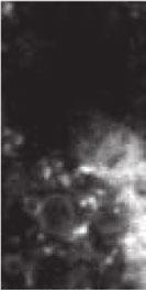
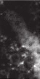
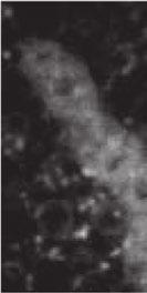
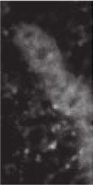
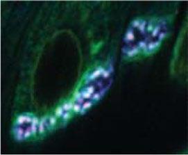
2 side of the NMJ, a highly convoluted array of muscle membrane infoldings called the subsynaptic reticulum (SSR) incorporates neurotransmitter receptors, ion channels, and cell adhesion molecules. Assembly of the SSR during larval growth involves activity-dependent targeted exocytosis mediated by the small GTPase Ral and its effector, the exocyst complex (Teodoro et al., 213), as well as the t-snare (target soluble N-ethylmaleimide sensitive factor attachment protein receptor) receptor gtaxin/syx18 (Gorczyca et al., 27) and scaffolding proteins such as Discs Large (Dlg; Lahey et al., 1994). Many proteins with predicted membrane-remodeling activities, including Drosophila homologues of Syndapin (Synd) and Amphiphysin (Amph), localize extensively to SSR membranes, making them prime candidates to facilitate SSR elaboration (Leventis et al., 21; Razzaq et al., 21; Zelhof et al., 21; Kumar et al., 29b). Amph regulates the postsynaptic turnover of the trans-synaptic cell adhesion molecule FasII (Mathew et al., 23), but its role in organizing the SSR is unknown. The Drosophila melanogaster genome encodes a single -terminal EHD protein called Putative achaete/scute target (Past1). Past1 mutants exhibit defects in endocytic recycling in larval nephrocytes, sterility and aberrant development of the germline, and short lifespan (Olswang-Kutz et al., 29), but the functions of Past1 at the NMJ have not been explored. Mammalian EHD1 localizes to the mouse NMJ, but its function there has been difficult to ascertain, perhaps due to redundancy with other EHD proteins (Mate et al., 212). Here we take advantage of the fact that Past1 encodes the only Drosophila -terminal EHD protein and define its role at the NMJ. RESULTS Past1 localizes to the SSR and is required for proper SSR assembly We first investigated a potential role for Past1 in the neuromuscular system by examining its localization at the larval NMJ relative to the neuronal membrane marker anti horseradish peroxidase (α-hrp) and the presynaptic and postsynaptic scaffolding protein Dlg. Larval muscles are innervated by glutamatergic type Ib NMJs (which are surrounded by extensive SSR and Dlg), glutamatergic type Is NMJs (which are surrounded by a much thinner layer of SSR and Dlg), and peptidergic type II and III NMJs (which lack both SSR and Dlg; Prokop, 26). Polyclonal α-past1 antibodies (Olswang-Kutz et al., 29) revealed endogenous Past1 localization at the NMJ to type Ib and Is arbors but not to type II or type III arbors (Figure 1A), suggesting that Past1 localizes specifically to SSR-surrounded NMJs. Past1 localized to a postsynaptic domain larger than that defined by Dlg and decorated frequent tubule-like extensions into the muscle (Figure 1B, arrows). This Past1 immunolabeling disappeared in past1-null mutants and was lost after postsynaptic depletion of Past1 by RNA interference (RNAi), confirming the specificity of the labeling (Figure 1). Localization of Past1 to the SSR was recapitulated using a Past1 enhanced green fluorescent protein (EGFP) transgene driven by the muscle driver BG487-GAL4 (Figure 1D). By contrast, the mutant Past1 G62E -EGFP protein (analogous to mutations in mammalian and. elegans EHD1/rme-1, which abolish ATP binding and membrane association; Lee et al., 25) was diffusely localized in the muscle, with only a small degree of enrichment at the NMJ (Figure 1D), indicating that ATP binding and membrane binding by Past1 are required for its localization. Past1 also localized to tubulovesicular structures in the muscle cortex (Figure 1B and Supplemental Figure S1A) and to Dlg-labeled muscle muscle junctions. Of interest, however, it did not localize to t-tubules, which are prominent deep invaginations of the muscle plasma membrane that couple muscle membrane depolarization to calcium release from the sarcoplasmic reticulum and also contain Dlg (Razzaq et al., 21; Supplemental Figure S1A). Finally, whereas muscle-specific RNAi revealed negligible expression of Past1 in the motor neuron (Figure 1), ectopic presynaptic expression of Past1- EGFP induced the formation of extensive tubules emanating from the presynaptic arbor, consistent with Past1 having a robust membrane deformation activity. These tubules contained presynaptic membrane marker HRP but were not enriched for the presynaptic proteins we tested (Supplemental Figure S1B), suggesting that they exclude presynaptic cytoplasm and ultrastructures. Given the localization of Past1 to the NMJ and its expected role in membrane remodeling, we next examined the morphology of this synapse in -null mutants (Olswang-Kutz et al., 29). Whereas total bouton number was similar to that for wildtype animals (Figure 1E and Supplemental Table S1), past1 mutants exhibited a marked defect in synaptic bouton shape. Boutons were less round than wild-type boutons, often with ragged edges (Figure 1E). We also found a significant increase in the frequency of ghost boutons (Figure 1, F and G), defined by the dramatic reduction or absence of a postsynaptic marker (Dlg) at α-hrp labeled type Ib and type Is boutons on muscles 6 and 7, which are particularly sensitive to this phenotype (Ataman et al., 28). However, the amount of postsynaptically localized α-hrp labeling (representing shed neuronal membrane debris; Fuentes-Medel et al., 29) was similar in and past1 mutants, suggesting that ghost boutons did not arise from excessive presynaptic membrane shedding (Figure 1H). The NMJ morphology and ghost bouton phenotype were rescued by reexpression of Past1-EGFP in muscles (Figure 1, E G). Therefore Past1 is required postsynaptically for normal synaptic morphogenesis. Past1 is required for synaptic transmission and homeostasis To test the effects of Past1 on NMJ function, we next examined synaptic transmission in past1 mutants. The larval NMJs to which Past1 localizes are glutamatergic synapses and depend on a glutamate receptor (GluR) tetramer consisting of the invariant subunits GluRII, GluRIID, and GlurIIE and the variable subunits GluRIIA and GluRIIB, which determine the electrophysiological properties of the receptor (DiAntonio et al., 1999; Marrus et al., 24). At the larval NMJ, GluRIIA-containing complexes are believed to represent nascent synapses, whereas GluRIIB complexes are believed to represent more mature, stable synapses (Thomas and Sigrist, 212). In addition, this NMJ exhibits robust homeostasis in response to reduced activity of glutamate receptors, responding with an increase in presynaptic release dependent on a retrograde signal and a 2+ influx (Petersen et al., 1997; Frank, 214). To determine the role of Past1 in glutamatergic signaling at the NMJ, we immunostained past1 mutants with glutamate receptor specific antibodies and found a specific reduction in the levels of GluRIIA but not of GluRIIB or GluRII (Figure 2, A and B). Despite reduced GluRIIA levels, Glu- RII clusters in past1 mutants were apposed normally to presynaptic active zones marked by Bruchpilot (BRP) (Figure 2), suggesting that synapse formation was not affected. We then used electrophysiology to examine the functional consequences of loss of Past1. ompared to controls, past1-mutant NMJs exhibited strongly reduced spontaneous miniature excitatory postsynaptic potentials (mepsps; Figure 2, D and E), as well as dampened evoked postsynaptic potentials (EPSPs; Figure 2, D and F). The frequency of mepsps was also depressed (Figure 2G), perhaps because reduced mepsp amplitude precludes detection of some events. Strikingly, quantal content was not increased to compensate for mepsp amplitude reduction (Figure 2H), suggesting a 3276 K. Koles et al. Molecular Biology of the ell
3 defect in synaptic homeostasis. To test this possibility, we used the null mutation GluRIIA SP16, representing a challenge to postsynaptic function that produces a robust homeostatic response (Petersen et al., 1997). ompared to past1 single mutants, we found that past1; GluRIIA SP16 double mutants exhibited a further reduction in mepsp amplitude (Figure 2, D and E). These mepsps in past1; GluRIIA SP16 mutants were very similar in amplitude to GluRIIA SP16 single mutants (Figure 2E), suggesting that the reduction in mepsp amplitude in past1 mutants is predominantly due to diminished Glu- RIIA levels. Remarkably, although we observed robust homeostatic compensation in GluRIIA SP16 single mutants (as evidenced by increased quantal content), GluRIIA SP16 ; past1 double mutants had the same diminished quantal content as past1 single mutants (Figure 2H). These results indicate that past1 mutants have a specific and strong defect in synaptic homeostasis. Past1 organizes the subsynaptic reticulum Past1 is a membrane-remodeling protein, and the defects in synaptic morphology, transmission, and homeostasis that we observed may be due to a direct role in organizing postsynaptic membranes. To test the role of Past1 in the SSR, we examined the organization of postsynaptic membranes using a muscle-expressed myristoylated monomeric red fluorescent protein (myr-mrfp, targeted to membranes via fatty acid modification) or md8-gfp (targeted to membranes using the transmembrane domain of mouse D8). Both reporters localized strongly to the SSR in control and past1- mutant NMJs but were found partly concentrated in nodules at past1-mutant NMJs (Figure 3A, arrows). Myr-mRFP is predicted to diffuse readily within membranes, so we used it as a reporter for mobility within membranes in the SSR, using fluorescence recovery after photobleaching (FRAP) analysis. At the resolution of light microscopy, myr-mrfp intensity and volume were unchanged between wild-type and past1-mutant NMJs (Figure 3B). On photobleaching a string of terminal boutons in either wild-type or past1 mutants, myr-mrfp recovered first in the proximal bouton and last in the distal bouton (Figure 3, arrows), indicating that membrane diffusion occurs primarily between the SSR of adjacent boutons rather than between the NMJ and the muscle cortex. In past1 mutants, myr-mrfp exhibited a similar recovery rate but a significantly higher mobile fraction than in wildtype animals, suggesting that the SSR reticulum may have reduced complexity, allowing more extensive membrane exchange (Figure 3 and Supplemental Movies S1 and S2). To examine further the ultrastructure of the mutant NMJ, we used transmission electron microscopy (TEM). The past1-mutant boutons exhibited normal presynaptic structures, including synaptic vesicles and active zones (Figure 3D). TEM of wild-type type 1b boutons revealed a highly elaborated postsynaptic reticulum (Figure 3D). In striking contrast, Past1- mutant type 1b boutons exhibited aberrant SSR membranes, often separated into distinct nodules. Further, a fraction of the nodules (5 of 12 nodules; n = 5 boutons) exhibited a central core (yellow arrow) surrounded by long membrane extensions 2 nm wide in cross section (Figure 3D, red arrow). Nodules were not seen in wild-type samples (n = 5 boutons). Because thin tubules would be unlikely to extend so frequently and for such long distances in the plane of the thin section, it is likely that these extensions represent flat membrane sheets. We tested this possibility by conducting serial section electron microscopy on NMJs and indeed found that the long membrane extensions in nodules continued through multiple 7-nm sections (Figure 3E), consistent with the hypothesis that they represent membrane sheets. These membrane nodules are likely to correspond to the nodules seen by confocal microscopy of the membrane markers md8-gfp and myr-mrfp (Figure 3A). Thus Past1 is required for normal development and morphology of the SSR. Past1 functionally interacts with Synd and colocalizes with Amph Synd and Amph are two NPF motif containing membrane-remodeling proteins that localize to SSR at the Drosophila larval NMJ (Leventis et al., 21; Razzaq et al., 21; Zelhof et al., 21; Kumar et al., 29b) and interact with EHD proteins in other systems (Xu et al., 24; Braun et al., 25; Pant et al., 29). Amph-A is the only one of three Drosophila Amphiphysin splice isoforms that contains an NPF motif (Zelhof et al., 21), whereas all predicted Drosophila Synd isoforms contain a single NPF motif (Figure 4A). To test whether Synd and Amph might function with Drosophila Past1, we expressed them in cultured Drosophila S2 cells. Past1-EGFP localized to tubular and punctate structures in these cells (Figure 4B). By contrast, the ATP-binding mutant Past1 G62E -GFP localized primarily to the cytoplasm and infrequent aggregates, whereas Past1 lacking its NPF-interacting EH domain (Past1 EH -EGFP) localized primarily to the cytoplasm (Figure 4B). Thus Past1 requires its ATP/membranebinding capacity as well as its EH domain for localization in S2 cells. Amph-A localized to small puncta, as did an Amph NPF motif mutant in which the conserved EH domain interacting phenylalanine was replaced with alanine (Figure 4; Pant et al. 29). The isolated Synd F-BAR domain localized to short tubules and puncta, often emanating in a radial pattern from the center of the cell, whereas full-length Synd localized mainly to the cytoplasm and only weakly to puncta, consistent with previous results demonstrating autoinhibition of the Synd F-BAR by its SH3 domain (Kumar et al., 29b; Rao et al., 21; Figure 4D). We mutated the conserved phenylalanine in the NPF motif of Synd to alanine (Synd NPFmut ) and found a similar localization to wild-type Synd (Figure 4D). We first tested the effects of coexpression of Amph and Past1- EGFP in S2 cells. Past1-EGFP colocalized with a fraction of Amphpositive puncta, and this localization was retained when the Amph NPF motif was mutated (Figure 4). These results suggest that colocalization does not depend on Past1 EH domain Amph NPF motif interactions, although Past1 and Amph can be recruited to the same cellular structures. We next tested the effects of Past1 on Synd localization. Strikingly, when coexpressed with Past1-EGFP, Syndmherry partially relocalized from the cytoplasm to a radial pattern, reminiscent of its isolated F-BAR, and Past1-EGFP colocalized with these structures (Figure 4). By contrast, Past1-EGFP colocalized significantly less with structures formed by the isolated Synd F-BAR, which is missing the Past1-interacting NPF motif (Figure 4D). Further, the Synd NPFmut mutant exhibited reduced colocalization with Past1 compared with wild-type Synd. These results suggest that in S2 cells, Past1 (either directly or indirectly) interacts with and releases Synd from autoinhibition, revealing a membrane-binding activity similar to its isolated F-BAR. Taken together, these data suggest that functional interactions between Past1 and Synd are likely to be conserved for the Drosophila homologues of these proteins. Finally, we found that Past1-EGFP, Synd-mherry, and Amph-myc colocalized in puncta when expressed together in S2 cells (Supplemental Figure S2), indicating that they can associate with the same structures in vivo. Past1 organizes distinct SSR domains defined by Amph and Synd To understand how Past1 sculpts the SSR, we examined its effects on the localization of Amph and Synd at the NMJ. In wild-type Volume 26 September 15, 215 Function of Past1 at the Drosophila NMJ 3277


.")


 G w E st")


 Localization of Past1")


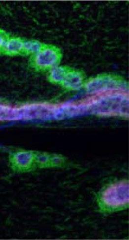
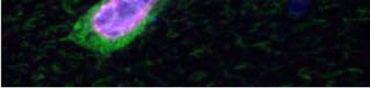
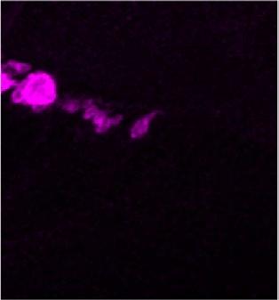
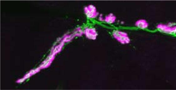
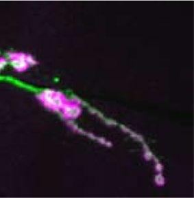
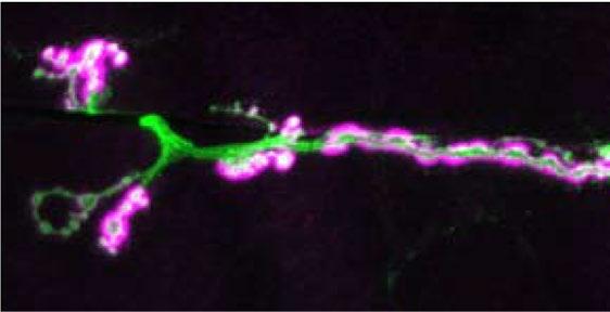

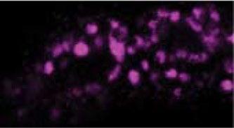
4 A Past1 Dlg HRP muscle 12/13 Ib Past1 HRP II Is III B BG57 > Past1RNAi Past1 Past1 Dlg HRP past111-1 muscle 6/7 HRP yz yz F HRP Dlg past16-4/11-1 Past1-EGFP rescue past16-4/ ty ild 1 6 pe /1 1-1 normalized postsynaptic HRP debris intensity (muscle 4, segment A3).8 1- pe E re GF sc P ue ty pa st 1 6 /1 1-4 pe st ty pa ild 1 6 / w 1-4 / H ild 1. ** 3 w ** Pa 4 pa st 1.5 muscle 4 muscle 6/7 w ild pa typ e st 1 6 Past1-EGFP rescue boutons/1 mm2 muscle area (segment A3) G w E st BG487>Past1G62E-EGFP Past1-EGFP Dlg HRP BG487>Past1-EGFP ghost boutons/nmj (muscle 6/7, segment A3) D Dlg FIGURE 1: Past1 localizes to a postsynaptic tubular-reticular domain at the larval NMJ and is required for postsynaptic membrane assembly. (A) Localization of Past1 to type Ib and Is but not type II or III NMJs. Large image shows 2D projections of 6 spinning-disk confocal stack from an NMJ on muscles 12 and 13. Scale bar, 5 μm. Right, magnified single confocal slices from the area indicated by the dashed rectangle. (B) Large image shows 2D projection of 1 spinning-disk confocal stack from an NMJ on muscles 6 and 7. Right, magnified single confocal slices from the area indicated with the dashed lines; arrows indicate Past1-labeled tubules. () NMJ α-past1 antibody staining is lost in past1 mutants and upon RNAi of Past1 using the muscle-specific driver BG57-GAL4. Images show 2D projections of 6 spinning-disk confocal stacks from muscle 4. (D) Postsynaptically expressed Past1-EGFP exhibits similar NMJ 3278 K. Koles et al. Molecular Biology of the ell
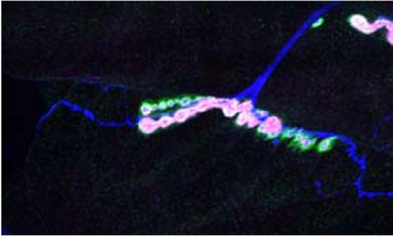
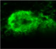
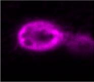
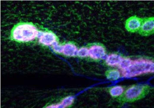
5 animals, Synd and Amph were found at the NMJ in uniformly distributed small postsynaptic puncta (Figure 5A), consistent with previous reports that they colocalize with postsynaptic Dlg (Kumar et al., 29a,b). However, in past1-mutant NMJs, both Synd and Amph were strikingly mislocalized to nodules close to the presynaptic membrane (Figure 5A). Nodules appeared in 82% of past1-mutant NMJs (but no wild-type NMJs; Supplemental Figure S3A) and were distinct from the presynaptic active zone marker BRP, which labels synapses (Supplemental Figure S3B). These nodules coincided with indentations of the presynaptic membrane (Figure 5B, arrows) and colocalized strongly with the postsynaptically expressed membrane marker md8-gfp (Figure 5), suggesting that Synd and Amph nodules represent the membrane structures seen by TEM. Further, Synd nodules were recapitulated by RNAi knockdown of Past1 in muscles, indicating that they arise due to lack of postsynaptic Past1 function (Figure 5D). We next tested whether Synd and Amph levels were altered in past1-mutant NMJs and found that whereas total levels were unchanged at the NMJ (Figure 5E), there was a higher fraction of Synd- and Amph-positive pixels at higher fluorescence intensities relative to wild-type NMJs, consistent with the observed increased clustering of both Synd and Amph in the nodules (Figure 5F). We next tested the localization of a number of additional postsynaptic components in past1 mutants by confocal microscopy. We found that the BAR domain containing protein drich (Nahm et al., 21b) and the F-BAR protein dip4 (Nahm et al., 21a) were localized to structures very similar to Synd nodules (Supplemental Figure S3). The postsynaptic scaffolding molecules Dlg and spectrin, which are not believed to bind to EHD proteins like Past1, were also mislocalized, forming a honeycomb pattern around the NMJ (Supplemental Figure S3D). Thus loss of Past1 causes significant reorganization of postsynaptic membrane-associated and scaffolding proteins. To understand further the organization of SSR components in past1 mutants, we used structured illumination microscopy (SIM), with which we could image their organization beyond the diffraction limit of conventional fluorescence microscopy. In wild-type animals, SIM revealed Synd localization in a punctate pattern, along tubules that extended beyond the Dlg domain (Figure 5G), similar to Past1 localization (Figure 1B) and providing increased resolution over previous studies (Kumar et al., 29a,b). In wild-type animals, Amph localized in small puncta that closely overlapped with the Dlg domain of the SSR (Figure 5G). Thus, in wild-type animals, Synd localizes to a more extensive SSR domain than Amph. By contrast, intensity profiles averaged across multiple past1-mutant nodules revealed that Synd localized to the center of the nodule, whereas Amph labeled a region larger than the central domain and Dlg occupied a further domain on the muscle side of the nodule (Figure 5, G and H). Thus the relative positions of Synd and Amph in the SSR are reversed in past1-mutant NMJs. These results suggest that the aberrant membrane structures visualized by TEM and md8-gfp or myr-mrfp labeling represent a core containing Synd, a larger domain containing Amph (potentially in the membrane sheets we observed by TEM), and a surrounding concentric region containing Dlg. Finally, we tested the role of Past1 ATP binding/membrane binding in the consolidation of the Synd-labeled membrane domain into nodules. First, we confirmed that postsynaptic reexpression of Past1-EGFP (using the muscle driver BG57-GAL4) in the past1 mutant rescued the SSR distribution of Synd (Supplemental Figure S4). By contrast, control larvae expressing md8-gfp in the past1-mutant background exhibited Synd nodules, with which md8-gfp colocalized. The ATP/membrane-binding mutant Past1 G62E -EGFP failed to rescue the Synd nodule phenotype and localized to the nodules (Supplemental Figure S4). Taken together, our results indicate that ATP/membrane binding by Past1 is required to fully elaborate the SSR and that, in its absence, a membrane domain defined by Amph aberrantly surrounds a domain defined by Synd. Amph is required to consolidate Synd into nodules To test the order of action of Amph and Synd microdomains in postsynaptic membrane elaboration, we examined the localization of Synd in amph; past1 double mutants by confocal microscopy. Strikingly, in these double mutants, Synd was no longer localized to nodules but instead was strongly depleted from the NMJ, and its overall levels in the muscle were slightly but significantly reduced (Figure 6, A and B). By contrast, amph single mutants had Synd levels similar to those of wild-type NMJs (Figure 6, A and B). This result suggests that Amph is required to pack Synd into the nodules that form in past1 mutants and that in the absence of NMJ localization, Synd may be destabilized. We next used confocal microscopy to assess the localization of Dlg as a proxy for overall SSR organization. Dlg was localized to a slightly but significantly larger domain in amph single mutants than in past1 single mutants (Figure 6, A and ). In contrast to the dramatic mislocalization of Synd, amph; past1 mutant NMJs did not have significantly different Dlg volume or intensity than wild-type or past1-mutant NMJs. This suggests that unpacking of Synd nodules and mislocalization of Synd from the SSR in amph; past1 mutants are specific to the Synd structure rather than due to a general loss of SSR. Of importance, GluRIIA levels were similar in amph; past1 double mutants compared with past1 single mutants (Figure 6D), suggesting that loss of GluRIIA is a general property of past1 mutants and not an indirect effect of Synd nodule formation. To examine further the phenotype of amph; past1 double mutants, we examined their SSR organization by TEM. The amphmutant SSR had a similar appearance to wild-type SSR (Figure 6E). Further, in contrast to past1 single-mutant SSR, we did not observe any nodules or concentric membrane sheets in the amph; past1 double-mutant SSR (Figure 6E). Thus we conclude that in the localization to endogenous Past1, whereas an ATP-binding mutant (contrast enhanced) is largely cytoplasmic. Large images show 2D projection of 6 spinning-disk confocal stack from an NMJ on muscles 6 and 7. Bottom, magnified single confocal slices from the area indicated by the dashed rectangle. (E) α-hrp staining showing aberrant bouton morphology in past1 mutants that is rescued by postsynaptic reexpression of Past1-EGFP. Overall bouton number per muscle area is normal in past1 mutants (muscles 4 and 6/7, segment A3). Image shows 2D projection of 6 spinningdisk confocal stacks from muscle 4. (F) past1-mutant NMJs exhibit ghost boutons. Images shows 2D projection of 6 spinning-disk confocal stacks from muscle 6/7, segment A3. (G) Quantification of ghost boutons. (H) Quantification of postsynaptic α-hrp debris from three-dimensional (3D) volumes surrounding presynaptic terminals. Number in bar graphs indicates number of NMJs measured; scale bars, 2 μm (B F). Past1-EGFP rescue represents the genotype UAS-Past1-EGFP/+; BG57, past /past Volume 26 September 15, 215 Function of Past1 at the Drosophila NMJ 3279

 1.5 1.")


 Past1 mutants have")


 Quantification of GluR")

")


6 A α-hrp α-gluriia α-hrp α-gluriib α-hrp α-glurii B GluR sum intensity/hrp volume (normalized to ) GluRIIA GluRIIB GluRII α-glurii α-brp α-hrp D mepsp amplitude (mv) 1. ** GluRIIA SP16 ; E F G H ** GluRIIA SP16 GluRIIA SP16 ; EPSP amplitude (mv) FIGURE 2: Past1 mutants have reduced postsynaptic responses and defective homeostatic compensation. (A) Past1 mutants have reduced GluRIIA levels but normal GluRIIB and GluRII levels. Maximum intensity projections of 6 spinning-disk confocal stacks from a representative muscle 4. Scale bar, 2 μm. (B) Quantification of GluR levels from 3D volumes surrounding HRP staining. () Normal presynaptic (α-brp) and postsynaptic (α-glurii) apposition in past1 mutants. Maximum intensity projections of 1 spinning-disk confocal stacks from a representative muscle 4. Scale bar, GluRIIA SP16 GluRIIA SP16 ; mepsp frequency (Hz) 5 * GluRIIA SP16 GluRIIA SP16 ; NLS quantal content ** ** GluRIIA SP16 GluRIIA SP16 ; 328 K. Koles et al. Molecular Biology of the ell
7 absence of Past1, Amph-dependent membrane sheets are required to consolidate or pack a Synd domain, and these events lead to dysfunctional elaboration of the SSR. DISUSSION Roles of Past1, Synd, and Amph in membrane elaboration Although there is great diversity in the morphologies of subcellular membranes and organelles, we have relatively little understanding of the mechanisms by which these complex shapes are generated. Here we uncovered novel functional roles in membrane remodeling at the NMJ for Past1, the sole -terminal EHD protein in Drosophila. Putting together our observations at the NMJ and in S2 cells with previous results from other groups, we propose a new working model for how Past1 functions in synaptic membrane elaboration (Figure 7). Our first key observation is that Past1 is required for normal elaboration of the SSR and that this function depends on its ATP-binding and thus membrane-remodeling activity. Next we found that in wild-type SSR, Amph localizes to a domain proximal to the bouton, whereas Past1 and Synd localize to a more extended tubulovesicular domain. By contrast, in the absence of Past1, the SSR rearranges into highly organized subdomains, with a core of Synd surrounded by a shell of Amph (likely corresponding to membrane sheets seen in Figure 3D by transmission electron microscopy [TEM]). We found that Amph is required for the formation of the sheets (perhaps by regulating the tight curvature at the tips of these membrane structures) and for consolidation of Synd into nodules. Further, our FRAP data indicate that the nodules result in significantly increased membrane flow within the SSR relative to wild-type SSR, suggesting reduced complexity. Finally, our S2 cell data indicate that Past1 may activate the membrane- binding/remodeling activity of Synd. These results suggest a novel mechanism for SSR elaboration at the wild-type NMJ involving sequential steps of membrane remodeling (Figure 7). In this model, Amph localizes and generates membrane tubules proximal to the bouton, and Past1 and Synd work together to further elaborate the tubules distal to the bouton. Successive rounds of these events could lead to the growth and expansion of layers of reticulum. In past1 mutants, this process is severely compromised, resulting in nodules containing a core of inactive Synd packed by Amph-dependent membrane sheets. One issue that remains to be resolved is whether direct physical interactions among Past1, Amph, and Synd (within the SSR subdomains to which they colocalize) contribute to Past1-dependent membrane remodeling at the NMJ, as they do in other systems (Xu et al., 24; Braun et al., 25; Pant et al., 29). Our S2 cell data (Figure 4) suggest that Past1 and Synd functionally interact in vivo. However, we were unable to biochemically detect Past1-Amph or Past1-Synd complexes using coprecipitation experiments in extracts from Drosophila larvae or S2 cells or with purified proteins (unpublished results), suggesting that either they do not directly interact or their interactions are not preserved in solution under the conditions tested. Genetic experiments at the NMJ using mutations that disrupt putative Past1-Synd and Past1-Amph interactions are unlikely to be informative because synd and amph single mutants exhibit no dramatic phenotype in SSR organization (Leventis et al., 21; Razzaq et al., 21; Zelhof et al., 21; Kumar et al., 29b), perhaps due to redundancy with other membrane-remodeling proteins. In fact, in addition to Amph and Synd, we found that the BAR proteins ip4 and drich are localized to nodules in past1 mutants (Supplemental Figure S3), suggesting that multiple membrane-remodeling proteins are available to function in the Past1-dependent pathway. In the future, it will be important to build into our working model the additional roles of these and other SSR-localized membrane-remodeling proteins, as well as the timing of exocyst-dependent membrane addition (Teodoro et al., 213). Role of Past1 in NMJ development and function Our results demonstrate that postsynaptic Past1 plays critical roles in the structure and function of the Drosophila NMJ. Past1 mutant NMJs exhibit aberrant morphology and excess ghost boutons. These ghost boutons are unlikely to be due to defective clearance of excess neuronal membrane as previously described (Fuentes- Medel et al., 29), since we did not observe large amounts of neuronal debris (Figure 1H). They are also unlikely to be related to excess ghost boutons seen in Wingless (Wg) signaling pathway mutants (Ataman et al., 26, 28), since past1 mutants do not phenocopy many other aspects of reduced Wg signaling, including increased GluR levels, disrupted presynaptic function, and reduction in bouton number (Packard et al., 22; Speese et al., 212). The likeliest interpretation is that Past1 functions directly in SSR membrane elaboration, consistent with our EM observations, and ghost boutons may arise when membrane nodules become too severe to allow SSR assembly around boutons that form toward the end of larval development. Another prominent synaptic phenotype that we found in past1 mutants is a strong and specific reduction in localization of GluRIIA to postsynaptic specializations, resulting in decreased mepsp amplitude (Figure 2). This decrease in GluRIIA could potentially arise by many mechanisms, including altered transcriptional or translational regulation or GluR traffic to or from the synapse. Indeed, expression of a dominant-negative EHD1 suppresses AMPA (a-amino-3- hydroxy-5-methyl-4-isoxazolepropionic acid type) glutamate receptor recycling in hippocampal dendritic spines (Park et al., 24). Although there has been little evidence that Drosophila GluRs are regulated by membrane traffic, our data implicating the membraneremodeling protein Past1 indicate that this may be the case. Finally, unlike the great majority of perturbations that reduce GluRIIA levels (reviewed in Frank, 214), past1 mutants surprisingly fail to compensate for this loss by homeostatic up-regulation of presynaptic release, suggesting that Past1 could be involved in relaying an as-yet-unidentified retrograde signal for synaptic homeostasis. Further work exploring mechanisms of GluRIIA regulation and retrograde signaling will be required to understand the role of Past1 in these events. Our present data cannot distinguish whether the function of Past1 in GluR traffic or homeostasis is directly related to its role in SSR elaboration, and it is possible that membrane compartments independent of the SSR are required for these functions and are disrupted in the mutant. Our finding that GluRIIA levels are still reduced in amph; past1 double mutants although SSR nodules are suppressed (Figure 6D) supports the conclusion that GluR localization defects are independent of aberrant SSR morphogenesis. Of note, 1 μm. (D) Representative traces from muscle recordings. The x-axis scale bar, 5 ms (EPSPs), 1, ms (mepsps); y-axis scale bar, 5 mv (EPSPs), 1 mv (mepsps). (E H) Quantification of electrophysiological phenotypes. (E) mepsp amplitude, (F) EPSP amplitude, (G) mepsp frequency, and (H) quantal content, adjusted for nonlinear summation (NLS). Genotypes include white (), n = 31;, n = 13; GluRIIA SP16, n = 44; and GluRIIA SP16 ;, n = 16. Volume 26 September 15, 215 Function of Past1 at the Drosophila NMJ 3281


")


.5.4.3.2.")

 6 4 2")

 Third-instar larval")


 Time-lapse of")


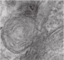
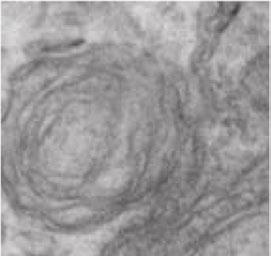
8 A BG57>myr-mRFP + / past1 6-4 BG57>mD8-GFP B myr-mrfp volume / HRP volume myrmrfp sum intensity/ HRP volume (normalized to ) D BG57>myr-mRFP pre-bleach post-bleach +3 sec +6 sec +12 sec +18 sec +24 sec +3 sec fraction fluorescence recovery fraction fluorescence recovery (t = 6 sec) time (sec) ** E t 1/2 recovery (sec) FIGURE 3: Postsynaptic membrane organization is altered in past1 mutants. (A) Third-instar larval NMJ expressing myr-mrfp or md8-gfp under the control of BG57-GAL4. Images and insets show a single 1 spinning-disk confocal slice from muscle 4, segment A3. Arrows indicate postsynaptic foci. Scale bar, 1 μm. (B) Quantifications of myr-mrfp sum intensity and volume relative to HRP-labeled neuronal arbor volume. () Time-lapse of representative FRAP analysis of myr-mrfp turnover in wild-type and mutant larvae. A single spinning-disk 6 confocal slice from muscle 4, segment A3/A4, is shown. Arrows indicate progressive recovery of fluorescence from proximal to distal boutons. Scale bar, 5 μm. Quantification of FRAP recovery. (D) TEM of NMJs from wild-type and past1-mutant larvae. SSR is highlighted in yellow; red arrow indicates membrane sheets, and yellow arrow indicates membrane core. Scale bar, 1 μm. (E) past1-mutant NMJs exhibit membrane sheets. Three 7-nm serial sections from a past1-mutant NMJ. Scale bar, 25 nm K. Koles et al. Molecular Biology of the ell


 Domain structure of")
 Expression of")


9 A B Past1 helical ATP-binding helical EH G domain Past1-EGFP Past1G62E-EGFP Past1 EH-EGFP Synd-mherry SyndFBAR-mherry SyndNPFmut-mherry Synd-mherry SyndF-BAR-mherry SyndNPFmut-mherry NPF Synd F-BAR SH3 NPF Amph-A SH3 AmphNPFmut-myc Amph-myc AmphNPFmut-myc D Past1-EGFP Amph-myc rry m AR ut -m -m he he he -m Sy nd FB nd Sy Sy rry yc ut -m -m Fm ph ph N P Am Am 13.4 PF N.2 rry.4.8 nd Pearson s orrelation oefficient vs Past1-EGFP.6 yc Pearson s orrelation oefficient vs Past1-EGFP Amph-myc Synd-mherry Past1-EGFP BAR FIGURE 4: Past1, Amph, and Synd colocalization in heterologous cells. (A) Domain structure of Past1, Amph-A, and Synd. (B D) Expression of Past1-EGFP, Synd-mherry, and Amph-A in S2 cells spread on concanavalin A. Single 1 confocal slices 1 μm from the cell coverslip interface from representative cells. Bar graphs show mean Pearson s r; number in bar indicates number of cells measured. Scale bars, 1 μm. Volume 26 September 15, 215 Function of Past1 at the Drosophila NMJ 3283


 Maximum intensity projections")

 Single 63 laser scanning")

. Scale bar, 5 μm.")


 Quantification of Amph and")

 Quantification of Synd and")


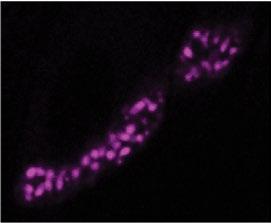
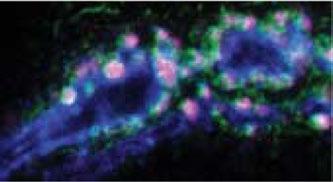
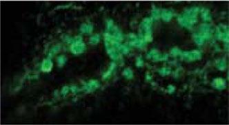
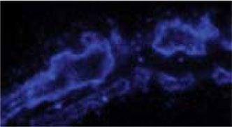
10 A α-amph α-syndy α-hrp B α-amph α-synd α-hrp BG57-GAL4 + md8-gfp α-synd D BG57-GAL4 α-past1 α-synd α-hrp α-amph BG57-GAL4 > Past1-RNAi E Normalized sum intensity/ HRP volume α-synd α-amph F % pixels in intensity range % 1-2% 2-3% 3-4% percent of mean Synd intensity 4-5% % pixels in intensity range ** -1% 1-2% 2-3% * 3-4% percent of mean Amph intensity 4-5% G α-hrp α-amph α-synd α-dlg motor neuron 1. FIGURE 5: Past1 is required for postsynaptic subdomain organization of Synd and Amph. (A) Maximum intensity projections of 6 spinning-disk confocal stacks from representative muscle 4 NMJs. Scale bar, 1 μm. (B) Single 63 laser scanning confocal slice (muscle 4) showing membrane nodules adjacent to neuronal membrane indentations (arrows). Scale bar, 5 μm. () Localization of BG57-GAL4 driven md8-gfp in past1 NMJs. A single 6 confocal slice from a representative muscle 4 is shown. Scale bar, 1 μm. (D) Nodules are recapitulated by postsynaptic past1 RNAi. Maximum intensity projections of 6 spinning-disk confocal stacks from representative muscle 4 NMJs. Scale bar, 1 μm. (E) Quantification of Amph and Synd intensity in 3D volumes surrounding HRP-positive motor neuron. (F) Quantification of Synd and Amph nodules in past1 NMJs. Histograms depict fraction of pixels at indicated intensities in 1.5-μm dilated area surrounding α-hrp positive motor neuron terminal. (G) Single structured illumination slices of wild-type and past1-mutant NMJs. Scale bar, 2 μm. (H) Mean intensity profiles of nodules in past1 mutants along a line traced perpendicular to the neuronal membrane (as indicated in image) and normalized to the width of the Synd peak. H fraction maximum fluorescence intensity muscle.5 1. normalized width muscle motor neuron HRP Amph Synd Dlg 3284 K. Koles et al. Molecular Biology of the ell

 1.5 1.")
 normalized sum")


 Synd nodules are lost in amph; past1")


 Quantification of Synd at the NMJ and")


 Dlg volume and intensity are similar in")


 GluRIIA intensity is")


11 A α-synd α-dlg α-hrp amph 26 ; amph 26 B normalized Synd intensity E NMJ Synd (sum intensity/hrp volume) Muscle Synd (mean intensity/ voxel) amph 26 amph 26 amph 26 ; amph 26 amph 26 ; volume ratio (Dlg/HRP) normalized sum Dlg intenisty/hrp volume ** * ** amph 26 ; normalized sum GluRIIA intensity/ HRP volume amph 26 amph 26 ; amph 26 D amph 26 amph 26 ; FIGURE 6: Amph is required for Synd nodule formation. (A) Synd nodules are lost in amph; past1 double mutants. Maximum intensity projections of 1 confocal stacks from representative muscle 4 NMJs labeled with the indicated antibodies. Scale bar, 2 μm. (B) Quantification of Synd at the NMJ and in the muscle. NMJ Synd is more drastically depleted than muscle Synd. Statistical analyses represent t tests comparing experiments done in parallel. () Dlg volume and intensity are similar in past1 and past1; amph mutants. Top, quantification of Dlg volume surrounding muscle 4, segment A3, presynaptic terminals. Bottom, quantification of Dlg intensity surrounding presynaptic terminals. (D) GluRIIA intensity is similarly reduced in past1 and past1; amph mutants compared with. Quantification of GluRIIA intensity on muscle 4, segment A3. (E) TEM of NMJs from amph and amph; past1 mutant larvae. past1 membrane nodules are suppressed by loss of amph. SSR is highlighted in yellow; scale bar, 5 nm. Volume 26 September 15, 215 Function of Past1 at the Drosophila NMJ 3285
12 Synd Amph Past1 GluR (IIA) 1. SSR elaboration 3. synaptic homeostasis 2. GluR traffic past1 mutant FIGURE 7: Model depicting the role of Past1 in postsynaptic membrane organization and function. In wild-type animals, sequential steps of membrane remodeling by components of the Amph membrane domain (generating sheets) and the Synd membrane domain (forming tubules from these sheets) lead to the elaboration of increasing layers of reticulum. In past1 mutants, the Synd domain collapses into nodules that are packed in by Amph-dependent sheets. Antibodies α-past1 (1:1; Olswang-Kutz et al., 29), α-amph (1:1; Zelhof et al., 21), α- Synd (1:1; Kumar et al., 29b), α-drich (1:1; Nahm et al., 21b), and GlurIIB (1:2) and α-glurii (1:3; Marrus et al., 24) antibodies have been described previously. hicken α-ip4 antibodies were obtained from N. Harden (Simon Fraser University, Burnaby, B, anada. (#4698, 1:1). α-dlg (4F3, 1:1), α- Futsch (22c1, 1:5), α-sp, α-spectrin-α (3A9, 1:5), α-gluriia (8B4D2, 1:1), and α-brp (nc82, 1:1) antibodies were obtained from the Developmental Studies Hybridoma Bank (Iowa ity, IA). For double labeling of BRP and GluRII, antibodies were directly conjugated to Alexa 488 (α- BRP) and Alexa 546 (α-glurii) (Jorquera et al., 212). Otherwise, α-hrp antibodies and secondary antibodies for imaging were conjugated to Dylight 45, Dylight 488, Rhodamine Red-X, or Alexa 647 (Jackson ImmunoResearch, West Grove, PA). many mutants with severely defective SSR and/or reduced GluR levels exhibit normal homeostasis (e.g., GluRIIA, which also has reduced SSR (Petersen et al., 1997; Schmid et al., 26); Dlg (Budnik et al., 1996); Gtaxin, (Gorczyca et al., 27); and Pak1 (Albin and Davis, 24), suggesting that homeostasis is a specific function of Past1 rather than a general SSR- or GluR-related defect. onservation of -terminal EHD protein function at the NMJ Past1 represents the sole EHD homologue in Drosophila, whereas mammals express four EHD proteins with distinct functions (Naslavsky and aplan, 211). Of importance, many of the roles we defined for EHD proteins at the NMJ and in muscle are likely to be conserved. Past1 localizes to the NMJ, the muscle cortex, and myotendinous junctions (Supplemental Figure S1). However, unlike EHD1 (Posey et al., 214), Past1 does not significantly localize to t- tubules. The activities we identified for Past1 at the Drosophila NMJ may inform mechanisms by which EHD2 participates in sarcolemmal repair at the muscle cortex (Marg et al., 212), EHD3 functions in cardiac muscle physiology (urran et al., 214), and EHD1 and EHD4 act at the mouse NMJ (Mate et al., 212). Our findings set the stage for uncovering how neuromuscular synapses are formed and elaborated and illustrate how cooperation between lipid-remodeling proteins can create highly complex membrane structures. MATERIALS AND METHODS Fly stocks Flies were cultured using standard media and techniques. UAS- Past1-EGFP and UAS-Past1 G62E -EGFP lines were constructed in pbi- UAS-Gateway (Wang et al., 211) using the PAST1-RB transcript and inserted into the Attp4 locus (Markstein et al., 28) on Drosophila chromosome II at Genetic Services (ambridge, MA). P{TRiP.HMS557}attP2 was used for RNAi. The past1 6-4 and past (Olswang-Kutz et al., 29), amph 26 (Razzaq et al., 21; Zelhof et al., 21), synd ex22 and synd 1d (Kumar et al., 29a), GluRI- IA SP16 (Petersen et al., 1997), and BG57-GAL4 and BG487-GAL4 (Budnik et al., 1996) lines have been described previously. Immunohistochemistry, imaging, and analysis of NMJ morphology For analysis of NMJ morphology and protein localization at the NMJ, flies were cultured at controlled density at 25º. Wandering third-instar larvae were dissected in calcium-free HL3.1 saline (Feng et al., 24) and fixed in HL3.1 containing 4% formaldehyde (or Bouin s fix for α-glurriia, α-spectrin, and ip4 staining) before antibody staining. For analysis of NMJ morphology, NMJs on muscle 6/7 and muscle 4, segment A3, were selected. Spinning-disk confocal Z-stacks (.3 μm) were collected at room temperature on an Andor spinning-disk confocal system consisting of a Nikon Ni-E upright microscope equipped with 6 (numerical aperture [NA] 1.4) and 1 (NA 1.45) oil immersion objectives, a Yokogawa SU-W1 spinning-disk head, and an Andor ixon 897U electron-multiplying charge-coupled device camera (Andor, Belfast, Northern Ireland). Images were collected using NIS Elements AR software (Nikon, Melville, NY). Laser scanning confocal images were acquired at room temperature on a Leica SP5 confocal microscope equipped with an HX PL APO lambda blue 63. (NA 1.4) oil immersion objective and Leica software (Leica, Wetzlar, Germany). SIM images were collected at room temperature on a Nikon N-SIM instrument equipped with an Apo TIRF 1 (NA 1.4) objective. Images were acquired using a violet-to-red diffraction grating at three angles and five phases of illumination, producing 15 raw images for SIM analysis using NIS Elements software. For TEM, samples were fixed, embedded, and sectioned as previously described (Karnovsky, 1965). Sections were imaged on a JEOL 12EX 8-kV electron microscope at 65, 8, or 12, magnification (JEOL, Peabody, MA). Data were collected from type Ib boutons, defined by their extensive SSR. Live imaging and fluorescence recovery after photobleaching Wandering third-instar larvae were dissected in HL3.1 saline, and the NS was removed. Type Ib NMJs on muscle 4 were imaged on a spinning-disk confocal microscope (see earlier description) with a 6 water-dipping objective (NA 1.). From three to 1 confocal stacks 3286 K. Koles et al. Molecular Biology of the ell
13 were collected for the prebleach intensity before two to three regions/ NMJ were bleached using a Mosaic 3 photoillumination device equipped with a 45-mW, 45-nm laser (Andor). Fluorescence recovery was measured by collecting confocal stacks at 1-s intervals for 45 s. Z-drift was corrected by manually selecting an in-focus range of confocal slices, and XY drift was corrected automatically using the Image registration function in NIS Elements software. Fluorescence recovery in two-dimensional (2D) projections of these confocal stacks were normalized to prebleach (1) and postbleach () fluorescence intensities and analyzed with GraphPad Prism (GraphPad, La Jolla, A). S2 ell culture and imaging S2 cells (herbas and herbas, 1998) were cultured according to standard protocols in Schneider s medium supplemented with 1% fetal bovine serum and.1 mg/ml penicillin/streptomycin. Past1- EGFP and Past1 G62E -EGFP were described earlier. Past1 EH -EGFP was generated by deleting sequences coding amino acids of Past1. Synd (isoform A)-mherry and Synd F-BAR -mherry (amino acids 1 3, as previously described in Becalska et al. 213) were generated in pbi-uasc (Wang et al., 211). Amph-A was constructed in puast and tagged with five copies of the myc epitope. Site-directed mutagenesis was used to generate Amph-A F516A (Amph-A NPFmut ) and Syndapin F418A (Syndapin NPFmut ). onstructs were cotransfected with Actin-GAL4 using Effectene reagent (Qiagen, Valencia, A) and incubated for 2 3 d at 25. ells were spread for 1 h on coverslips coated with concanavalin A (Rogers et al., 23; Sigma-Aldrich, St. Louis, MO), fixed for 1 min in 4% formaldehyde in phosphate-buffered saline (PBS), and then, where indicated, permeabilized, stained with primary and secondary antibodies, and washed with PBS plus.1% Triton X-1. Samples were mounted in Mowiol with DABO (Sigma-Aldrich), and cells were imaged by spinning-disk confocal microscopy as described earlier. Electrophysiology and analysis Wandering third-instar larvae were chosen for electrophysiology. Larvae were dissected in a modified HL3 saline: Nal (7 mm), Kl (5 mm), Mgl 2 (1 mm), NaHO 3 (1 mm), sucrose (115 mm = 3.9%), trehalose (4.2 mm =.16%), HEPES (4-(2-hydroxyethyl)-1- piperazineethanesulfonic acid; 5. mm =.12%), and al 2 (.5 mm). Sharp electrode recordings were taken from muscle 6 of abdominal segment A2 or A3, as previously described (Davis et al., 1998; Frank et al., 26). Data were collected using an Axopatch 2B amplifier (Molecular Devices, Sunnyvale, A), digitized using a Digidata 144A data acquisition system (Molecular Devices), and recorded with plamp 1 acquisition software (Molecular Devices). For presynaptic nerve stimulation, a Master-8 pulse stimulator (A.M.P. Instruments, Jerusalem, Israel) and an ISO-Flex isolation unit (A.M.P. Instruments) were used to deliver 1-ms suprathreshold stimuli to the appropriate segmental nerve. The average spontaneous miniature EPSP (mepsp) amplitude was quantified by measuring the amplitude of 1 2 individual spontaneous release events per NMJ. The average evoked EPSP amplitude was calculated for each NMJ. Quantal content (Q) was determined for each NMJ by calculating the ratio of average EPSP and average mepsp amplitudes. Q was corrected for nonlinear summation as described (Martin, 1955). Image and statistical analyses Analyses of postsynaptic volumes and volumetric intensities were conducted using the Volocity 5.5 lassification Module (ImproVision, Waltham, MA). Volumes corresponding to presynaptic (as determined by HRP) labeling were determined using intensity thresholds, as previously described (Ramachandran et al., 29). Postsynaptic areas were determined by dilating the HRP signal to include all of the quantified postsynaptic label intensity for each antigen and kept identical between control and experimental samples. Presynaptic, postsynaptic, or total (presynaptic and postsynaptic) immunolabeling intensities were normalized to total presynaptic volumes (i.e., HRP-delineated bouton volumes). olocalization analyses in S2 cells were conducted in single confocal slices μm from the coverslip cell interface. Pearson s r was calculated from background-subtracted images using the ImageJ plug-in coloc2 and ostes randomization test (fiji.sc/oloc_2). Synd and Amph pixel intensity distributions at the NMJ were calculated in ImageJ from sum intensity projections of confocal stacks. The α- HRP positive presynaptic area was dilated by 1.5 μm to define the postsynaptic region. Images were then normalized to the mean Synd/Amph intensity in this area, and pixel intensity distributions were calculated using the Sixteen Bit Histogram plugin in Image-J. Data were binned as indicated and graphed using GraphPad Prism. All errors shown are mean ± SEM. Statistical significance was calculated using GraphPad Prism software using analysis of variance followed by pairwise Tukey s tests or using Student s t tests where only two groups were compared; *p < 5, **p < 1, p < 5;, not significantly different. AKNOWLEDGMENTS Stocks and reagents were generously provided by Aaron DiAntonio, Seungbok Lee, Andrew Zelhof, Mani Ramaswami, Mia Horowitz, Troy Littleton, the Bloomington Drosophila Stock enter (Bloomington, IN; National Institutes of Health P4OD18537), the Drosophila Genomics Resource enter (Bloomington, IN), and the Developmental Studies Hybridoma Bank (University of Iowa). We thank Bruce Goode, Troy Littleton, Daniela Nicastro, Suzanne Paradis, Neil Ritter, and ShiYu Wang for helpful discussions and technical assistance, the Electron Microscopy Facility at Harvard Medical School, and hristopher O onnell (Nikon Instruments) for assistance with the SIM experiments. This work was supported by the National Institutes of Health/National Institute of Neurological Disorders and Stroke (DP2 NS82127 to A.A.R.; NS62738 to.a.f.), a Pew Scholar Award (to A.A.R.), the National Science Foundation (MRI DBI ), and funds from the University of Iowa arver Trust and arver ollege of Medicine (to.a.f.). REFERENES Albin SD, Davis GW (24). oordinating structural and functional synapse development: postsynaptic p21-activated kinase independently specifies glutamate receptor abundance and postsynaptic morphology. J Neurosci 24, Ataman B, Ashley J, Gorczyca D, Gorczyca M, Mathew D, Wichmann, Sigrist SJ, Budnik V (26). Nuclear trafficking of Drosophila Frizzled-2 during synapse development requires the PDZ protein dgrip. Proc Natl Acad Sci USA 13, Ataman B, Ashley J, Gorczyca M, Ramachandran P, Fouquet W, Sigrist SJ, Budnik V (28). Rapid activity-dependent modifications in synaptic structure and function require bidirectional Wnt signaling. Neuron 57, Becalska AN, Kelley F, Berciu, Stanishneva-Konovalova TB, Fu X, Wang S (213). Formation of membrane ridges and scallops by the F-BAR protein Nervous Wreck. Mol Biol ell 24, Braun A, Pinyol R, Dahlhaus R, Koch D, Fonarev P, Grant BD, Kessels MM, Qualmann B (25). EHD proteins associate with syndapin I and II and such interactions play a crucial role in endosomal recycling. Mol Biol ell 16, Budnik V, Koh YH, Guan B, Hartmann B, Hough, Woods D, Gorczyca M (1996). Regulation of synapse structure and function by the Drosophila tumor suppressor gene dlg. Neuron 17, Volume 26 September 15, 215 Function of Past1 at the Drosophila NMJ 3287
The EHD protein Past1 controls postsynaptic membrane elaboration and synaptic function
 The EHD protein Past1 controls postsynaptic membrane elaboration and synaptic function Kate Koles 1, Emily M. Messelaar 1, Zachary Feiger 1, Crystal J. Yu 1, C. Andrew Frank 2, and Avital A. Rodal 1,*
SUPPLEMENTARY INFORMATION
 doi:10.1038/nature10923 Supplementary Figure 1 Ten-a and Ten-m antibody and cell type specificities. a c, Representative single confocal sections of a Drosophila NMJ stained with antibodies to Ten-a (red),
T H E J O U R N A L O F C E L L B I O L O G Y
 Supplemental material Kasprowicz et al., http://www.jcb.org/cgi/content/full/jcb.201310090/dc1 T H E J O U R N A L O F C E L L B I O L O G Y Figure S1. NMJ morphology of shi 12-12B ; shi-4c not treated
SUPPLEMENTARY INFORMATION
 DOI: 10.1038/ncb2215 Figure S1 Number of egfp-vps4a bursts versus cellular expression levels. The total number of egfp-vps4a bursts, counted at the end of each movie (frame 2000, after 1h 28 min) are plotted
Anterograde Activin Signaling Regulates Postsynaptic Membrane Potential and GluRIIA/B Abundance at the Drosophila Neuromuscular Junction
 Anterograde Activin Signaling Regulates Postsynaptic Membrane Potential and GluRIIA/B Abundance at the Drosophila Neuromuscular Junction Myung-Jun Kim, Michael B. O Connor* Department of Genetics, Cell
Synaptic Homeostasis Is Consolidated by the Cell Fate Gene gooseberry,adrosophila pax3/7 Homolog
 The Journal of Neuroscience, June 16, 2010 30(24):8071 8082 8071 Development/Plasticity/Repair Synaptic Homeostasis Is Consolidated by the Cell Fate Gene gooseberry,adrosophila pax3/7 Homolog Bruno Marie,
SUPPLEMENTARY INFORMATION
 DOI: 10.1038/ncb2647 Figure S1 Other Rab GTPases do not co-localize with the ER. a, Cos-7 cells cotransfected with an ER luminal marker (either KDEL-venus or mch-kdel) and mch-tagged human Rab5 (mch-rab5,
Ral mediates activity-dependent growth of postsynaptic membranes via recruitment of the exocyst
 The EMBO Journal (2013) 32, 2039 2055 www.embojournal.org Ral mediates activity-dependent growth of postsynaptic membranes via recruitment of the exocyst THE EMBO JOURNAL Rita O Teodoro 1,2, *, Gulc in
13-3. Synthesis-Secretory pathway: Sort lumenal proteins, Secrete proteins, Sort membrane proteins
 13-3. Synthesis-Secretory pathway: Sort lumenal proteins, Secrete proteins, Sort membrane proteins Molecular sorting: specific budding, vesicular transport, fusion 1. Why is this important? A. Form and
Supplementary Figure 1. Real time in vivo imaging of SG secretion. (a) SGs from Drosophila third instar larvae that express Sgs3-GFP (green) and
 SGs from Drosophila third instar larvae that express Sgs3-GFP (green) and") Supplementary Figure 1. Real time in vivo imaging of SG secretion. (a) SGs from Drosophila third instar larvae that express Sgs3-GFP (green) and Lifeact-Ruby (red) were imaged in vivo to visualize secretion
Supplementary Figure 1. Real time in vivo imaging of SG secretion. (a) SGs from Drosophila third instar larvae that express Sgs3-GFP (green) and Lifeact-Ruby (red) were imaged in vivo to visualize secretion
Neurons and Nervous Systems
 34 Neurons and Nervous Systems Concept 34.1 Nervous Systems Consist of Neurons and Glia Nervous systems have two categories of cells: Neurons, or nerve cells, are excitable they generate and transmit electrical
Nature Neuroscience: doi: /nn.2662
 Supplementary Figure 1 Atlastin phylogeny and homology. (a) Maximum likelihood phylogenetic tree based on 18 Atlastin-1 sequences using the program Quicktree. Numbers at internal nodes correspond to bootstrap
SUPPLEMENTARY INFORMATION
 DOI: 10.1038/ncb3267 Supplementary Figure 1 A group of genes required for formation or orientation of annular F-actin bundles and aecm ridges: RNAi phenotypes and their validation by standard mutations.
Homeostatic Control of Presynaptic Release Is Triggered by Postsynaptic Membrane Depolarization
 Neuron, Vol. 30, 737 749, June, 2001, Copyright 2001 by Cell Press Homeostatic Control of Presynaptic Release Is Triggered by Postsynaptic Membrane Depolarization Suzanne Paradis, Sean T. Sweeney, and
Protein Sorting, Intracellular Trafficking, and Vesicular Transport
 Protein Sorting, Intracellular Trafficking, and Vesicular Transport Noemi Polgar, Ph.D. Department of Anatomy, Biochemistry and Physiology Email: polgar@hawaii.edu Phone: 692-1422 Outline Part 1- Trafficking
Chapter 4 Evaluating a potential interaction between deltex and git in Drosophila: genetic interaction, gene overexpression and cell biology assays.
 Evaluating a potential interaction between deltex and git in Drosophila: genetic interaction, gene overexpression and cell biology assays. The data described in chapter 3 presented evidence that endogenous
The neuron as a secretory cell
 The neuron as a secretory cell EXOCYTOSIS ENDOCYTOSIS The secretory pathway. Transport and sorting of proteins in the secretory pathway occur as they pass through the Golgi complex before reaching the
Supplementary Materials for
 www.sciencesignaling.org/cgi/content/full/6/301/ra98/dc1 Supplementary Materials for Regulation of Epithelial Morphogenesis by the G Protein Coupled Receptor Mist and Its Ligand Fog Alyssa J. Manning,
Open Syntaxin Docks Synaptic Vesicles
 Marc Hammarlund 1,2[, Mark T. Palfreyman 1,2[, Shigeki Watanabe 1,2[, Shawn Olsen 1,2, Erik M. Jorgensen 1,2* PLoS BIOLOGY 1 Department of Biology, University of Utah, Salt Lake City, Utah, United States
Nature Methods: doi: /nmeth Supplementary Figure 1. In vitro screening of recombinant R-CaMP2 variants.
 Supplementary Figure 1 In vitro screening of recombinant R-CaMP2 variants. Baseline fluorescence compared to R-CaMP1.07 at nominally zero calcium plotted versus dynamic range ( F/F) for 150 recombinant
Nervous Systems: Neuron Structure and Function
 Nervous Systems: Neuron Structure and Function Integration An animal needs to function like a coherent organism, not like a loose collection of cells. Integration = refers to processes such as summation
Highwire Regulates Synaptic Growth in Drosophila
 Neuron, Vol. 26, 313 329, May, 2000, Copyright 2000 by Cell Press Highwire Regulates Synaptic Growth in Drosophila Hong I. Wan,* Aaron DiAntonio,* Richard D. Fetter,* Kendra Bergstrom,* Roland Strauss,
The BMP Ligand Gbb Gates the Expression of Synaptic Homeostasis Independent of Synaptic Growth Control
 Article The BMP Ligand Gbb Gates the Expression of Synaptic Homeostasis Independent of Synaptic Growth Control Carleton P. Goold 1 and Graeme W. Davis 1, * 1 Department of Biochemistry and Biophysics,
Monitoring neurite morphology and synapse formation in primary neurons for neurotoxicity assessments and drug screening
 APPLICATION NOTE ArrayScan High Content Platform Monitoring neurite morphology and synapse formation in primary neurons for neurotoxicity assessments and drug screening Suk J. Hong and Richik N. Ghosh
Neurophysiology. Danil Hammoudi.MD
 Neurophysiology Danil Hammoudi.MD ACTION POTENTIAL An action potential is a wave of electrical discharge that travels along the membrane of a cell. Action potentials are an essential feature of animal
Cells. Steven McLoon Department of Neuroscience University of Minnesota
 Cells Steven McLoon Department of Neuroscience University of Minnesota 1 Microscopy Methods of histology: Treat the tissue with a preservative (e.g. formaldehyde). Dissect the region of interest. Embed
Chapter 37 Active Reading Guide Neurons, Synapses, and Signaling
 Name: AP Biology Mr. Croft Section 1 1. What is a neuron? Chapter 37 Active Reading Guide Neurons, Synapses, and Signaling 2. Neurons can be placed into three groups, based on their location and function.
Nervous Tissue. Neurons Electrochemical Gradient Propagation & Transduction Neurotransmitters Temporal & Spatial Summation
 Nervous Tissue Neurons Electrochemical Gradient Propagation & Transduction Neurotransmitters Temporal & Spatial Summation What is the function of nervous tissue? Maintain homeostasis & respond to stimuli
Supplemental Information. The Mitochondrial Fission Receptor MiD51. Requires ADP as a Cofactor
 Structure, Volume 22 Supplemental Information The Mitochondrial Fission Receptor MiD51 Requires ADP as a Cofactor Oliver C. Losón, Raymond Liu, Michael E. Rome, Shuxia Meng, Jens T. Kaiser, Shu-ou Shan,
Supplementary Figures Supplementary Tables 1 and 2. Supplementary References
 The Nuclear Import of a Non-Canonical Frizzled2 Signal by Importins-β11 and α2 Promotes Postsynaptic Development Timothy J. Mosca and Thomas L. Schwarz * The F.M. Kirby Neurobiology Center, Children s
!"#$%&'%()*%+*,,%-&,./*%01%02%/*/3452*%3&.26%&4752*,,*1%%
*%+*,,%-&,./*%01%02%/*/3452*%3&.26%&4752*,,*1%%") !"#$%&'%()*%+*,,%-&,./*%01%02%/*/3452*%3&.26%&4752*,,*1%% !"#$%&'(")*++*%,*'-&'./%/,*#01#%-2)#3&)/% 4'(")*++*% % %5"0)%-2)#3&) %%% %67'2#72'*%%%%%%%%%%%%%%%%%%%%%%%4'(")0/./% % 8$+&'&,+"/7 % %,$&7&/9)7$*/0/%%%%%%%%%%
!"#$%&'%()*%+*,,%-&,./*%01%02%/*/3452*%3&.26%&4752*,,*1%% !"#$%&'(")*++*%,*'-&'./%/,*#01#%-2)#3&)/% 4'(")*++*% % %5"0)%-2)#3&) %%% %67'2#72'*%%%%%%%%%%%%%%%%%%%%%%%4'(")0/./% % 8$+&'&,+"/7 % %,$&7&/9)7$*/0/%%%%%%%%%%
Heather Currinn, Benjamin Guscott, Zita Balklava, Alice Rothnie and Thomas Wassmer*
 Online Resources APP controls the formation of PI(3,5)P 2 vesicles through its binding of the PIKfyve complex. Cellular and Molecular Life Sciences Heather Currinn, Benjamin Guscott, Zita Balklava, Alice
Nervous Tissue. Neurons Neural communication Nervous Systems
 Nervous Tissue Neurons Neural communication Nervous Systems What is the function of nervous tissue? Maintain homeostasis & respond to stimuli Sense & transmit information rapidly, to specific cells and
MEMBRANE POTENTIALS AND ACTION POTENTIALS:
 University of Jordan Faculty of Medicine Department of Physiology & Biochemistry Medical students, 2017/2018 +++++++++++++++++++++++++++++++++++++++++++++++++++++++++++++++++++++++++ Review: Membrane physiology
MEMBRANE STRUCTURE. Lecture 9. Biology Department Concordia University. Dr. S. Azam BIOL 266/
 MEMBRANE STRUCTURE Lecture 9 BIOL 266/4 2014-15 Dr. S. Azam Biology Department Concordia University RED BLOOD CELL MEMBRANE PROTEINS The Dynamic Nature of the Plasma Membrane SEM of human erythrocytes
Nervous System Organization
 The Nervous System Nervous System Organization Receptors respond to stimuli Sensory receptors detect the stimulus Motor effectors respond to stimulus Nervous system divisions Central nervous system Command
SUPPLEMENTARY INFORMATION
 SUPPLEMENTARY INFORMATION doi:10.1038/nature12791 Supplementary Figure 1 (1/3) WWW.NATURE.COM/NATURE 1 RESEARCH SUPPLEMENTARY INFORMATION Supplementary Figure 1 (2/3) 2 WWW.NATURE.COM/NATURE SUPPLEMENTARY
Nervous System Organization
 The Nervous System Chapter 44 Nervous System Organization All animals must be able to respond to environmental stimuli -Sensory receptors = Detect stimulus -Motor effectors = Respond to it -The nervous
Practical applications of TIRF microscopy
 Practical applications of TIRF microscopy Evgeny Pryazhnikov University of Helsinki, Neuroscience Center Functional and Morphological Plasticity of the Tripartite Synapse Vesicular release ATP Perisynaptic
PROPERTY OF ELSEVIER SAMPLE CONTENT - NOT FINAL. The Nervous System and Muscle
 The Nervous System and Muscle SECTION 2 2-1 Nernst Potential 2-2 Resting Membrane Potential 2-3 Axonal Action Potential 2-4 Neurons 2-5 Axonal Conduction 2-6 Morphology of Synapses 2-7 Chemical Synaptic
downstream (0.8 kb) homologous sequences to the genomic locus of DIC. A DIC mutant strain (ro- 6
 homologous sequences to the genomic locus of DIC. A DIC mutant strain (ro- 6") A B C D ts Figure S1 Generation of DIC- mcherry expressing N.crassa strain. A. N. crassa colony morphology. When a cot1 (top, left panel) strain is grown at permissive temperature (25 C), it exhibits straight
A B C D ts Figure S1 Generation of DIC- mcherry expressing N.crassa strain. A. N. crassa colony morphology. When a cot1 (top, left panel) strain is grown at permissive temperature (25 C), it exhibits straight
Control and Integration. Nervous System Organization: Bilateral Symmetric Animals. Nervous System Organization: Radial Symmetric Animals
 Control and Integration Neurophysiology Chapters 10-12 Nervous system composed of nervous tissue cells designed to conduct electrical impulses rapid communication to specific cells or groups of cells Endocrine
Role of Mitochondrial Remodeling in Programmed Cell Death in
 Developmental Cell, Vol. 12 Supplementary Data Role of Mitochondrial Remodeling in Programmed Cell Death in Drosophila melanogaster Gaurav Goyal, Brennan Fell, Apurva Sarin, Richard J. Youle, V. Sriram.
7.06 Cell Biology EXAM #3 April 21, 2005
 7.06 Cell Biology EXAM #3 April 21, 2005 This is an open book exam, and you are allowed access to books, a calculator, and notes but not computers or any other types of electronic devices. Please write
Introduction. Gene expression is the combined process of :
 1 To know and explain: Regulation of Bacterial Gene Expression Constitutive ( house keeping) vs. Controllable genes OPERON structure and its role in gene regulation Regulation of Eukaryotic Gene Expression
لجنة الطب البشري رؤية تنير دروب تميزكم
 1) Hyperpolarization phase of the action potential: a. is due to the opening of voltage-gated Cl channels. b. is due to prolonged opening of voltage-gated K + channels. c. is due to closure of the Na +
PHYSIOLOGY CHAPTER 9 MUSCLE TISSUE Fall 2016
 PHYSIOLOGY CHAPTER 9 MUSCLE TISSUE Fall 2016 2 Chapter 9 Muscles and Muscle Tissue Overview of Muscle Tissue types of muscle: are all prefixes for muscle Contractility all muscles cells can Smooth & skeletal
CELB40060 Membrane Trafficking in Animal Cells. Prof. Jeremy C. Simpson. Lecture 2 COPII and export from the ER
 CELB40060 Membrane Trafficking in Animal Cells Prof. Jeremy C. Simpson Lecture 2 COPII and export from the ER Today s lecture... The COPII coat - localisation and subunits Formation of the COPII coat at
Dendrites - receives information from other neuron cells - input receivers.
 The Nerve Tissue Neuron - the nerve cell Dendrites - receives information from other neuron cells - input receivers. Cell body - includes usual parts of the organelles of a cell (nucleus, mitochondria)
According to the diagram, which of the following is NOT true?
 Instructions: Review Chapter 44 on muscular-skeletal systems and locomotion, and then complete the following Blackboard activity. This activity will introduce topics that will be covered in the next few
Visual pigments. Neuroscience, Biochemistry Dr. Mamoun Ahram Third year, 2019
 Visual pigments Neuroscience, Biochemistry Dr. Mamoun Ahram Third year, 2019 References Webvision: The Organization of the Retina and Visual System (http://www.ncbi.nlm.nih.gov/books/nbk11522/#a 127) The
Muscle tissue. Types. Functions. Cardiac, Smooth, and Skeletal
 Types Cardiac, Smooth, and Skeletal Functions movements posture and body position Support soft tissues Guard openings body temperature nutrient reserves Muscle tissue Special Characteristics of Muscle
Neurite formation & neuronal polarization. The cytoskeletal components of neurons have characteristic distributions and associations
 Mechanisms of neuronal migration & Neurite formation & neuronal polarization Paul Letourneau letou001@umn.edu Chapter 16; The Cytoskeleton; Molecular Biology of the Cell, Alberts et al. 1 The cytoskeletal
Dimorphism and Homeostasis of Neuromuscular Synaptic Function
 Dimorphism and Homeostasis of Neuromuscular Synaptic Function - Epp m ε Release + + φ - q Sensitivity The nature of the problem Size, Strength, Resistance.. The Power of Drosophila The nature of the problem
7.06 Spring 2004 PS 6 KEY 1 of 14
 7.06 Spring 2004 PS 6 KEY 1 of 14 Problem Set 6. Question 1. You are working in a lab that studies hormones and hormone receptors. You are tasked with the job of characterizing a potentially new hormone
Our patient for the day...
 Muscles Ch.12 Our patient for the day... Name: Eddy Age: Newborn Whole-body muscle contractions No relaxation Severe difficulty breathing due to inadequate relaxation of breathing muscles Diagnosed with
In order to confirm the contribution of diffusion to the FRAP recovery curves of
 Fonseca et al., Supplementary Information FRAP data analysis ) Contribution of Diffusion to the recovery curves In order to confirm the contribution of diffusion to the FRAP recovery curves of PH::GFP
Postsynaptic Rab GTPases and Exocyst: a screen at the Drosophila neuromuscular junction
 Postsynaptic Rab GTPases and Exocyst: a screen at the Drosophila neuromuscular junction Cátia F Patrício-Rodrigues, Rita O Teodoro CEDOC, Nova Medical School, Universidade Nova de Lisboa Correspondence
Nerve Signal Conduction. Resting Potential Action Potential Conduction of Action Potentials
 Nerve Signal Conduction Resting Potential Action Potential Conduction of Action Potentials Resting Potential Resting neurons are always prepared to send a nerve signal. Neuron possesses potential energy
Lecture 6 - Intracellular compartments and transport I
 01.26.11 Lecture 6 - Intracellular compartments and transport I Intracellular transport and compartments 1. Protein sorting: How proteins get to their appropriate destinations within the cell 2. Vesicular
Physiology Unit 2. MEMBRANE POTENTIALS and SYNAPSES
 Physiology Unit 2 MEMBRANE POTENTIALS and SYNAPSES Neuron Communication Neurons are stimulated by receptors on dendrites and cell bodies (soma) Ligand gated ion channels GPCR s Neurons stimulate cells
A role for PS integrins in morphological growth and synaptic function at the postembryonic neuromuscular junction of Drosophila
 Development 126, 5833-5846 (1999) Printed in Great Britain The Company of Biologists Limited 1999 DEV8655 5833 A role for PS integrins in morphological growth and synaptic function at the postembryonic
Cellular Neuroanatomy I The Prototypical Neuron: Soma. Reading: BCP Chapter 2
 Cellular Neuroanatomy I The Prototypical Neuron: Soma Reading: BCP Chapter 2 Functional Unit of the Nervous System The functional unit of the nervous system is the neuron. Neurons are cells specialized
Neurite initiation. Neurite formation begins with a bud that sprouts from the cell body. One or several neurites can sprout at a time.
 Neurite initiation. Neuronal maturation initiation f-actin polarization and maturation tubulin stage 1: "spherical" neuron stage 2: neurons extend several neurites stage 3: one neurite accelerates its
Chapter 11. Development: Differentiation and Determination
 KAP Biology Dept Kenyon College Differential gene expression and development Mechanisms of cellular determination Induction Pattern formation Chapter 11. Development: Differentiation and Determination
Baz, Par-6 and apkc are not required for axon or dendrite specification in Drosophila
 Baz, Par-6 and apkc are not required for axon or dendrite specification in Drosophila Melissa M. Rolls and Chris Q. Doe, Inst. Neurosci and Inst. Mol. Biol., HHMI, Univ. Oregon, Eugene, Oregon 97403 Correspondence
Synapses. Electrophysiology and Vesicle release
 Synapses Electrophysiology and Vesicle release Major point Cell theory (cells being separated) implies that cells must communicate with each other through extracellular connections most communication is
Neuronal Differentiation: Synapse Formation NMJ
 Neuronal Differentiation: Synapse Formation NMJ Approaches to study synapse formation Rodent NMJ (cholinergic) Drosophila NMJ (glutamatergic) Rodent CNS synapse (glutamatergic or GABAergic) C. elegans
Lecture 2. Excitability and ionic transport
 Lecture 2 Excitability and ionic transport Selective membrane permeability: The lipid barrier of the cell membrane and cell membrane transport proteins Chemical compositions of extracellular and intracellular
Supplementary Figure 1. Biochemical and sequence alignment analyses the
 Supplementary Figure 1. Biochemical and sequence alignment analyses the interaction of OPTN and TBK1. (a) Analytical gel filtration chromatography analysis of the interaction between TBK1 CTD and OPTN(1-119).
Neuroscience 201A Exam Key, October 7, 2014
 Neuroscience 201A Exam Key, October 7, 2014 Question #1 7.5 pts Consider a spherical neuron with a diameter of 20 µm and a resting potential of -70 mv. If the net negativity on the inside of the cell (all
SUPPLEMENTARY INFORMATION
 med!1,2 Wild-type (N2) end!3 elt!2 5 1 15 Time (minutes) 5 1 15 Time (minutes) med!1,2 end!3 5 1 15 Time (minutes) elt!2 5 1 15 Time (minutes) Supplementary Figure 1: Number of med-1,2, end-3, end-1 and
Honors Biology Reading Guide Chapter 11
 Honors Biology Reading Guide Chapter 11 v Promoter a specific nucleotide sequence in DNA located near the start of a gene that is the binding site for RNA polymerase and the place where transcription begins
A Hierarchy of Cell Intrinsic and Target-Derived Homeostatic Signaling
 Article A Hierarchy of Cell Intrinsic and Target-Derived Homeostatic Signaling Sharon Bergquist, 1 Dion K. Dickman, 1 and Graeme W. Davis 1, * 1 Department of Biochemistry and Biophysics, Program in Neuroscience,
Cell Biology Review. The key components of cells that concern us are as follows: 1. Nucleus
 Cell Biology Review Development involves the collective behavior and activities of cells, working together in a coordinated manner to construct an organism. As such, the regulation of development is intimately
Fig. S1. Expression pattern of moody-gal4 in third instar. Maximum projection illustrating a dissected moody-gal4>ngfp L3 larva stained for Repo
 Fig. S1. Expression pattern of moody-gal4 in third instar. Maximum projection illustrating a dissected moody-gal4>ngfp L3 larva stained for Repo (magenta), Fas2 (blue) and GFP (green) in overview (A) and
This script will produce a series of pulses of amplitude 40 na, duration 1ms, recurring every 50 ms.
 9.16 Problem Set #4 In the final problem set you will combine the pieces of knowledge gained in the previous assignments to build a full-blown model of a plastic synapse. You will investigate the effects
Profiling Synaptic Proteins Identifies Regulators of Insulin Secretion and Lifespan
 Profiling Synaptic Proteins Identifies Regulators of Insulin Secretion and Lifespan QueeLim Ch ng. a *, Derek Sieburth. b, Joshua M. Kaplan* Department of Molecular Biology, Massachusetts General Hospital,
Neuronal Activity and CaMKII Regulate Kinesin- Mediated Transport of Synaptic AMPARs
 Article Neuronal Activity and CaMKII Regulate Kinesin- Mediated Transport of Synaptic AMPARs Highlights d AMPAR transport is regulated by synaptic transmission, VGCCs and CaMKII Authors Frédéric J. Hoerndli,
80% of all excitatory synapses - at the dendritic spines.
 Dendritic Modelling Dendrites (from Greek dendron, tree ) are the branched projections of a neuron that act to conduct the electrical stimulation received from other cells to and from the cell body, or
Chapter 48 Neurons, Synapses, and Signaling
 Chapter 48 Neurons, Synapses, and Signaling Concept 48.1 Neuron organization and structure reflect function in information transfer Neurons are nerve cells that transfer information within the body Neurons
Endophilin Functions as a Membrane- Bending Molecule and Is Delivered to Endocytic Zones by Exocytosis
 Endophilin Functions as a Membrane- Bending Molecule and Is Delivered to Endocytic Zones by Exocytosis Jihong Bai, 1,2 Zhitao Hu, 1,2 Jeremy S. Dittman, 3 Edward C.G. Pym, 1,2 and Joshua M. Kaplan 1,2,
Introduction Principles of Signaling and Organization p. 3 Signaling in Simple Neuronal Circuits p. 4 Organization of the Retina p.
 Introduction Principles of Signaling and Organization p. 3 Signaling in Simple Neuronal Circuits p. 4 Organization of the Retina p. 5 Signaling in Nerve Cells p. 9 Cellular and Molecular Biology of Neurons
Nature Neuroscience: doi: /nn.2717
 Supplementary Fig. 1. Dendrite length is not secondary to body length. Dendrite growth proceeds independently of the rate of body growth and decreases in rate in adults. n 20 on dendrite measurement, n
The Diffusion Coefficient for PGK Folding in Eukaryotic Cells
 Biophysical Journal, Volume 99 Supporting Material The Diffusion Coefficient for PGK Folding in Eukaryotic Cells Apratim Dhar, Simon Ebbinghaus, Zhen Shen, Tripta Mishra, and Martin Gruebele 1 Supplementary
Signal Transduction. Dr. Chaidir, Apt
 Signal Transduction Dr. Chaidir, Apt Background Complex unicellular organisms existed on Earth for approximately 2.5 billion years before the first multicellular organisms appeared.this long period for
Segment boundary formation in Drosophila embryos
 Segment boundary formation in Drosophila embryos Development 130, August 2003 Camilla W. Larsen, Elizabeth Hirst, Cyrille Alexandre and Jean Paul Vincent 1. Introduction: - Segment boundary formation:
7.06 Cell Biology EXAM #3 KEY
 7.06 Cell Biology EXAM #3 KEY May 2, 2006 This is an OPEN BOOK exam, and you are allowed access to books, a calculator, and notes BUT NOT computers or any other types of electronic devices. Please write
Physiology Unit 2. MEMBRANE POTENTIALS and SYNAPSES
 Physiology Unit 2 MEMBRANE POTENTIALS and SYNAPSES In Physiology Today Ohm s Law I = V/R Ohm s law: the current through a conductor between two points is directly proportional to the voltage across the
Biology September 2015 Exam One FORM G KEY
 Biology 251 17 September 2015 Exam One FORM G KEY PRINT YOUR NAME AND ID NUMBER in the space that is provided on the answer sheet, and then blacken the letter boxes below the corresponding letters of your
Biology September 2015 Exam One FORM W KEY
 Biology 251 17 September 2015 Exam One FORM W KEY PRINT YOUR NAME AND ID NUMBER in the space that is provided on the answer sheet, and then blacken the letter boxes below the corresponding letters of your
Math in systems neuroscience. Quan Wen
 Math in systems neuroscience Quan Wen Human brain is perhaps the most complex subject in the universe 1 kg brain 10 11 neurons 180,000 km nerve fiber 10 15 synapses 10 18 synaptic proteins Multiscale
Chapter 9. Nerve Signals and Homeostasis
 Chapter 9 Nerve Signals and Homeostasis A neuron is a specialized nerve cell that is the functional unit of the nervous system. Neural signaling communication by neurons is the process by which an animal
Neurite formation & neuronal polarization
 Neurite formation & neuronal polarization Paul Letourneau letou001@umn.edu Chapter 16; The Cytoskeleton; Molecular Biology of the Cell, Alberts et al. 1 An immature neuron in cell culture first sprouts
Microsystems for Neuroscience and Medicine. Lecture 9
 1 Microsystems for Neuroscience and Medicine Lecture 9 2 Neural Microsystems Neurons - Structure and behaviour Measuring neural activity Interfacing with neurons Medical applications - DBS, Retinal Implants
Neurons, Synapses, and Signaling
 Chapter 48 Neurons, Synapses, and Signaling PowerPoint Lecture Presentations for Biology Eighth Edition Neil Campbell and Jane Reece Lectures by Chris Romero, updated by Erin Barley with contributions
R7.3 Receptor Kinetics
 Chapter 7 9/30/04 R7.3 Receptor Kinetics Professional Reference Shelf Just as enzymes are fundamental to life, so is the living cell s ability to receive and process signals from beyond the cell membrane.
CELL BIOLOGY - CLUTCH CH. 9 - TRANSPORT ACROSS MEMBRANES.
 !! www.clutchprep.com K + K + K + K + CELL BIOLOGY - CLUTCH CONCEPT: PRINCIPLES OF TRANSMEMBRANE TRANSPORT Membranes and Gradients Cells must be able to communicate across their membrane barriers to materials
Modulation of central pattern generator output by peripheral sensory cells in Drosophila larvae. BioNB4910 Cornell University.
 Modulation of central pattern generator output by peripheral sensory cells in Drosophila larvae BioNB4910 Cornell University Goals 1) Observe the behavioral effects of remotely activating different populations
SUPPLEMENTARY INFORMATION
 GP2 Type I-piliated bacteria FAE M cell M cell pocket idc T cell mdc Generation of antigenspecific T cells Induction of antigen-specific mucosal immune response Supplementary Figure 1 Schematic diagram