Neuronal Differentiation: Synapse Formation NMJ
|
|
|
- August Moore
- 5 years ago
- Views:
Transcription
1 Neuronal Differentiation: Synapse Formation NMJ

")
 Rodent")

2 Approaches to study synapse formation Rodent NMJ (cholinergic) Drosophila NMJ (glutamatergic) Rodent CNS synapse (glutamatergic or GABAergic) C. elegans NMJ (cholinergic and GABAergic)
3 Approaches to study synapse formation and elimination (1) Rodent NMJ (cholinergic) accessibility in vivo in transgenic mice allows for elegant imaging experiments: field used to be dominated by Jeff Lichtman and Josh Sanes (2) Genetic screens: Drosophila NMJ (glutamatergic) Broadie and Davis C. Elegans Jin, Bargmann, and Nonet ** early screens had only limited success perhaps due to late onset of synaptogenesis ** recent development of GFP fusion reporter genes has changed this by allowing visualization of synapses in living animals (3) Rodent CNS synapses in culture recent advances in cell culture, GFP fusions, and live imaging techniques
 alpha bungarotoxin Sanes and Lichtman 2001")
4 Comparison of the NMJ and Central Synapses Advantages of using the NMJ: (1) Large size (2) Relative simplicity (3) Unparalleled accessibility **owing to these features, the concept of the postsynaptic receptor was proposed by Langley over a century ago (4) alpha bungarotoxin Sanes and Lichtman 2001
5 The Adult NMJ Motor neurons in the ventral horn of the spinal cord send axons through peripheral nerves to innervate muscles Axons branch intramuscularly to innervate many muscle fibers, but each fiber receives only one synapse The axonal branch loses its myelin sheath to terminate in a spray of boutons on the muscle fiber surface 3 cells that constitute the synapse: (1) the motor neuron (nerve terminal) (2) the muscle fiber (3) the Schwann cell **at the adult NMJ, there are > 10,000 AChRs/um 2 Hall and Sanes 1993
 **AChRs are concentrated at the synaptic")
 in which clusters of synaptic vesicles accumulate in the nerve")
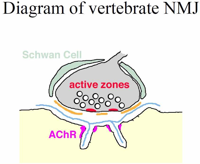
6 Pre and postsynaptic membranes at the NMJ are highly specialized Electron micrograph of a NMJ shows the nerve terminal capped by a Schwann cell situated in a shallow depression of a muscle cell membrane, which is invaginated further into deep postjunctional folds (arrows) **AChRs are concentrated at the synaptic site Burden et al 1979 Higher mag view shows that AChRs are concentrated at the crests and along the sides of the postjunctional folds (white arrow). **postjunctional folds are situated directly across from presynaptic active zones (long arrow) in which clusters of synaptic vesicles accumulate in the nerve terminal
7 Molecular Components of the NMJ the nerve terminal occupies a shallow gutter in the muscle fiber and is capped by processes of Schwann cells active zones in the nerve terminal directly appose junctional folds in the postsynaptic membrane some of the proteins in the synapse are shown, with their subcellular localization indicated by arrows those for which knockout mice have been generated are indicated in BOLD **Postsynaptic membrane = highly specialized to respond rapidly to NT released from the overlying nerve terminal with an extremely high conc. of AChRs: >10,000/um 2 vs. <10/um 2 extrasynaptically Sanes and Lichtman 1999
8 Differentiation of the NMJ 3 Steps: (1) Formation of selective connections between the developing axon and its target (2) Differentiation of the axon s growth cone into a nerve terminal (3) Elaboration of a postsynaptic apparatus in the target cell
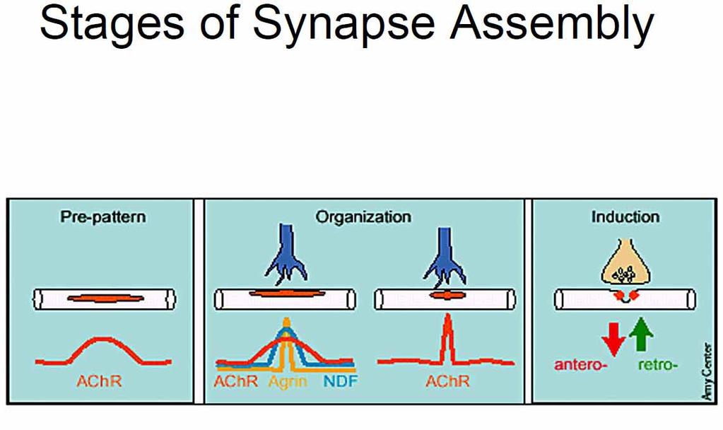
9 Early steps in the formation of the neuromuscular junction Sanes and Lichtman, 1999
10 Differentiation of the NMJ A growth cone approaches a myotube (a) and forms an unspecialized but functional contact on its surface (b). The terminal then differentiates and basal lamina appears in a widened cleft (c). As basal lamina appears extrasynaptically, multiple axons converge on the synaptic site (d). Finally, all axons are eliminated except one. The survivor expands, each terminal bouton is ensheathed by a Schwann cell process, the preterminal axon acquires a myelin sheath, and folds form in the subterminal postsynaptic membrane (e). Hall and Sanes 1993
11 NMJ Formation (1) Both pre- and postsynaptic cells have the machinery to form synapses BEFORE contact - isolated axonal growth cones release Ach - isolated muscle cells express AChRs (2) Contact serves to cluster presynaptic vesicles and postsynaptic AChRs (3) Synapses attain their mature structure and physiological properties over the next days to weeks
12 Spontaneous Release of ACh from Growth Cones A biological sensor for ACh ( a sniffer ) was created by excising a patch of membrane from a muscle cell with a recording pipet. The membrane contained AChRs that were facing outward. Thus, when the pipet was advanced near a growth cone, a current could be recorded when Ach bound to the receptors. Recording of ACh evoked currents (down ward deflections) when the sniffer patch was distant from the growth cone (left) and when it was within a few microns of the growth cone (right). The increased activity indicates that the growth cone was releasing ACh. Young and Poo 1983; Hume et al 1983
 during the first 20min after contact.")
13 Presynaptic Differentiation: Contact with a Muscle Cell RAPIDLY Stimulates ACh Release Cultures of Xenopus spinal neurons were grown and pipets were used to record from round muscle cells and to manipulate them into contact with the neuron A continuous recording from a muscle cell shows spontaneous transmission (downward deflections) during the first 20min after contact. Nerve evoked postsynaptic currents increased in amplitude from the moment of contact to 5min later. **** Increase in synaptic transmission WITHIN MINUTES!!!!!!! Xie and Poo 1986
14 Muscle Signals Presynaptic Differentiation Rudimentary synaptic transmission begins immediately, but the efficacy = low: transmission is weak and subthreshold After about 1 week, fully functional, but immature synapse forms: synaptic vesicles accumulate and quantal content increases. Transmission is still prone to failures and the action potential is broad. After several weeks, mature NMJ is formed: preterminal region is myelinated and transmission has a high safety factor Sanes and Lichtman 1999

 detachment of growth cone from substrate. After 1.")
15 Rapid adhesion between growth cone and postsynaptic muscle cell A muscle cell was manipulated into contact with a growth cone in a dissociated culture of Xenopus spinal cord. After 1.5 or 15 minutes, the muscle cell was withdrawn, and the degree of adhesion was graded: (0) no attachment, (1) filamentous attachment, (2) deformation of growth cone, and (3) detachment of growth cone from substrate. After 1.5 minutes of contact, most pairs exhibited only grade 0 1 adhesion. However, after 15 minutes of contact, the level of adhesion shifted to grade 1 3. (Adapted from Evers et al., 1989)
, and the growth cone was imaged while either a muscle cell or a neuron was brought into contact (middle).")
16 Contact with target increases free calcium in the growth cone. Dissociated neurons were filled with a Ca 2+ sensitive dye (top), and the growth cone was imaged while either a muscle cell or a neuron was brought into contact (middle). Intracellular free calcium increased only during contact with the muscle (red). The muscleevoked rise in Ca 2+ did not occur when the cells were bathed in a Ca 2+ free medium (bottom), indicating the involvement of calcium channels. (Adapted from Dai and Peng, 1993)
17
18 What are the retrograde signals that affect presynaptic differentiation?
19
20
21
22
23
24
25
26
27
28

29 Wnts activate a retrograde signal
, the ACh evoked a similar response across the entire muscle surface (sites 1, 2, and 3).")
30 Postsynaptic Differentiation: AChR clusters form during development ACh was applied to different areas of a muscle cell while the evoked response was monitored with an intracellular pipet. At embryonic day 17 (E17), the ACh evoked a similar response across the entire muscle surface (sites 1, 2, and 3). In adult muscle, an AChevoked response can only be obtained close to the synapse (site 2). AChreceptors were bound with radiolabeled α bungarotoxin, and the distribution was assessed autoradiographically.at E15, the label is spread uniformly across the muscle surface, but by P16 the label is restricted to the synaptic region. Diamond and Miledi 1962; Bevan and Steinbach 1977
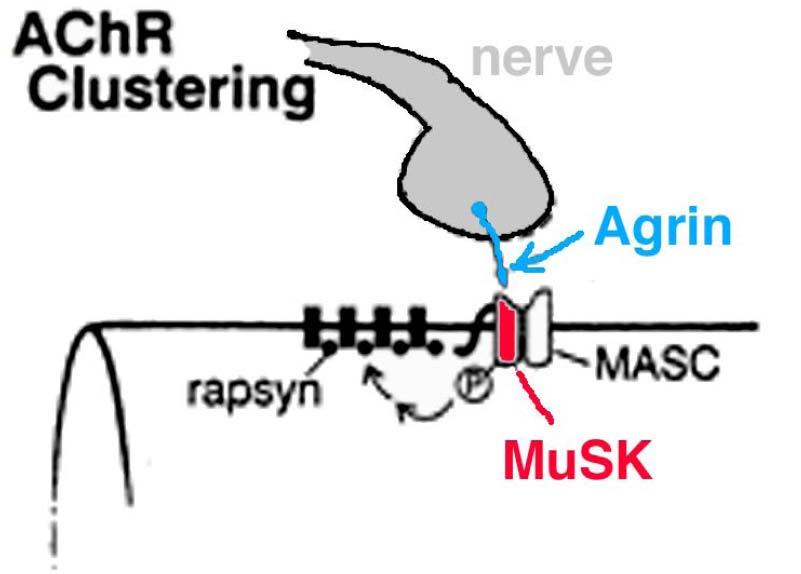
31 Nerve Induces Postsynaptic Differentiation Accumulation of AChRs in the Postsynaptic Membrane nuclei throughout newly formed myotubes express AChR subunit genes, and AChRs are diffusely distributed on the myotube surface (~1000AChRs/um 2 ) the nerve then sends 3 signals to the muscle to affect AChR distribution: (1) AGRIN interacts with MuSK to organize rapsyn mediated AChR clustering (2) NEUREGULIN (ARIA) induces selective expression of AChR subunit genes by synaptic nuclei (3) ACETYLCHOLINE (ACh) activates AChRs to generate a voltageand calcium dependent signal that represses AChR subunit gene expression in extrasynaptic nuclei **Together, these signals lead to selective synthesis of AChRs in synaptic areas and precise accumulation of AChRs in the postsynaptic membrane Sanes and Lichtman 1999
 are spread diffusely over the surface of the myotube.")
32 Postsynaptic Differentiation: (1) Agrin Induces Synaptic Clustering of AChRs Motor neurons synthesize and secrete agrin into the synaptic basal lamina. Before innervation, AChRs (green) are spread diffusely over the surface of the myotube. The release of agrin by the motor neuron results in redistribution of previously unclustered AChRs to synaptic sites, immediately adjacent to the nerve terminal.
33
34
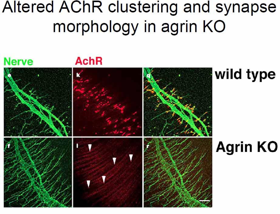
35 Agrin Induces Synaptic Clustering of AChRs Development of NMJs in agrin deficient mice Diaphragm muscles from control (left) and agrin / mice (right) at E15, 16, and 18 were double stained for AChRs and axon, and then were drawn. The developing muscle fibers run vertically. In both control and mutant muscles, an intramuscular nerve (black) and aggregates of AChRs (red) are present by E15. In controls, axonal branches and AChR clusters are confined to a band at the central end plate at all stages. Gautam et al 1996 Mutant AChR aggregates are smaller, less dense, and less numerous; axons form fewer branches and their synaptic relationships are disorganized.
36
37

38
39
40
41
42
43

44 Agrin binds to a receptor complex, and MuSK is required for clustering. Agrin activates a receptor complex that includes muscle specific kinase (MuSK) and an unidentified accessory protein (MASC). The intracellular peripheral membrane protein, Rapsyn, is required for the Agrin mediated MuSK activation to produce AChR phosphorylation, and it participates in receptor clustering. There are other Agrin binding sites, including laminin and α dystroglycan. Laminin has been shown to induce AChR clusters independent of MuSK signaling.


45 Agrin released by nerve terminals acts through MuSK and rapsyn to aggregate AChRs at synaptic sites in the muscle fiber Muscles from a wild type neonatal mouse and from mutants lacking agrin, MuSK, or rapsyn Axons=brown AChRs=green ** Few AChR clusters are present in the agrin mutant, and none are present in the MuSK or rapsyn mutants. All 3 mutants also have nerve abnormalities, reflecting the inability of the muscle to supply proper retrograde factors. Kandel, Schwartz, and Jessell 2000
46
47
48


49 Genetic analysis of early events in AChR clustering Sanes and Lichtman 2001
. B.")
50 Postsynaptic Differentiation: (2) Increase in newly synthesized AChRs Insertion of new ACh receptors occurs within hours of innervation A. Cultures of muscle cells were prelabeled with rhodamine conjugated bungarotoxin (red). B. In one set of cultures, motor neurons were added (left), while a second set of cultures remained without neurons (right). C. After eight hours, both cultures were labeled with a fluorescein conjuctated antibody against AChRs (yellow). The cultures with motor neurons contained many AChRs that were labeled with only antibody (yellow), indicating that they had been newly inserted after the addition of motor neurons. The muscle cell only cultures had AChRs that were primarily labeled by both Rhod Btx (red) and Fluor MAb (yellow). (Adapted from Role et al., 1985)

51 Neuregulin regulates synapse specific AChR expression Expression of Ach receptor genes is modulated as the myotube matures: initially, AChR genes are expressed throughout the myotube innervation leads to upregulation of AChR gene expression in the nuclei that lie directly beneath the nerve terminal and downregulation in nuclei in extrasynaptic regions Kandel, Schwartz, and Jessell 2000

52 Neuregulin stimulates expression of the genes encoding ACh receptors Neuregulins are synthesized and secreted by motor axons Binding of neuregulin to erbb kinases in the postsynaptic membrane activates transcription of Ach receptor genes via a cascade of protein kinases A critical role for neuregulin in vivo was demonstrated in heterozygous neuregulin mutant mice: both the density of the ACh receptors and the amplitude of the muscle response (miniature end plate potential, MEPP) are decreased **suggests that neuregulin is a ratelimiting factor for AChR expression Kandel, Schwartz, and Jessell 2000
.")
, extrajunctional ACh receptors are distributed over the entire muscle surface.")
53 Postsynaptic Differentiation: (3) ACh decreases transcription of extrasynaptic AChRs Extrasynaptic ACh receptors accumulate when the nerve is inactive. A. At the control nerve muscle junction, the electrically active terminal releases ACh and the receptors are clustered at the postsynaptic membrane. The activity dependent signal that suppresses extrajunctional receptors involves calcium influx and activation of the calcium calmodulin dependent protein kinase II (CamKII). A transcription factor found in muscle (myogenin) is phosphorylated and blocks transcription in extrasynatpic nuclei. B. When motor axon activity is blocked with the sodium channel blocker, tetrodotoxin (TTX), extrajunctional ACh receptors are distributed over the entire muscle surface. (Adapted from Lømo and Rosenthal, 1972)


54 Revised View of NMJ Formation 1990 Model Revised Model
55
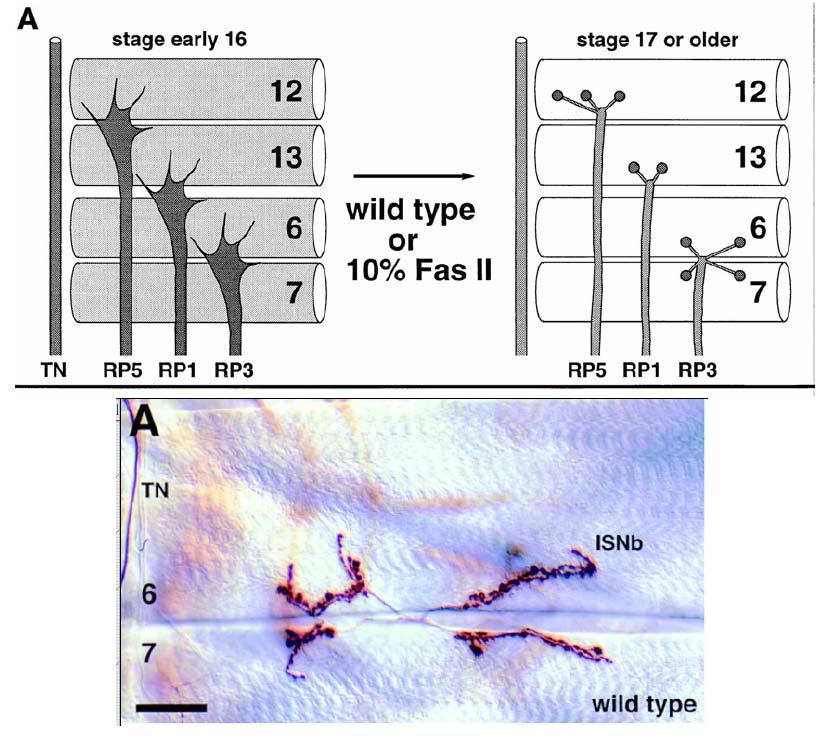
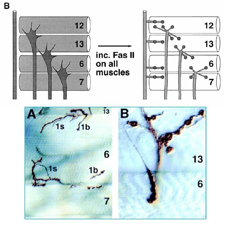
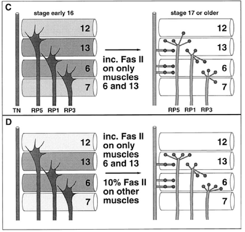
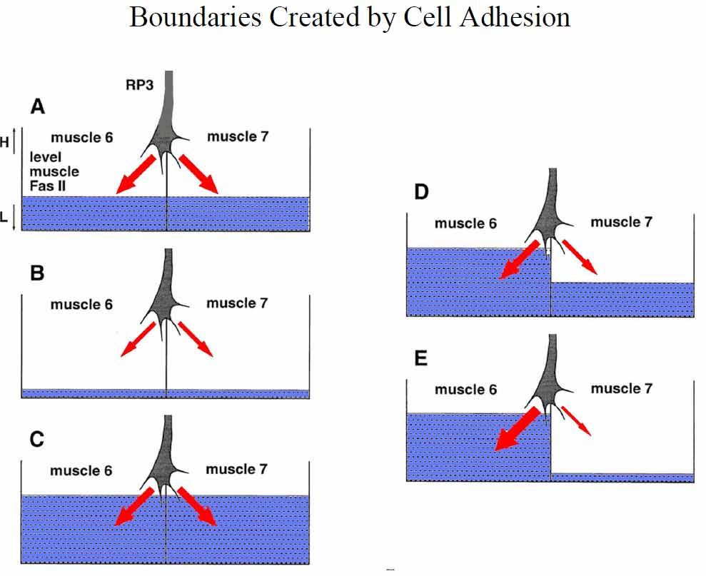
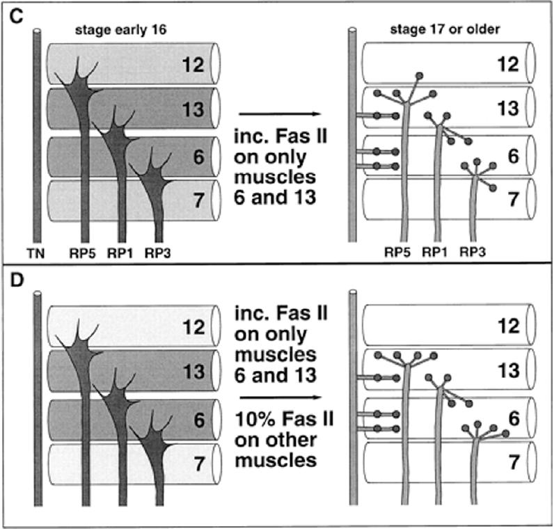
56 Cell Adhesion Molecules and Synapse Specificity Drosophila NMJ
57
58
59
60
61
62
63
64
65 Synaptic Competition during synapse formation
66
67

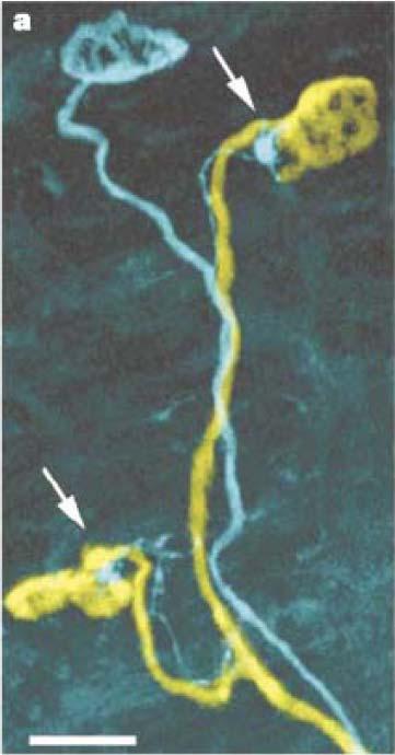
68 Presynaptic terminals fight over the same postsynaptic space

69 The losing axon retracts
70
71
72
73
74
75

76
77
78
79 Glia are also involved in synapse elimination at the NMJ
 activity is in Schwann cells sheathing")
80 Figure 4. LysoTracker (LysoT) activity is in Schwann cells sheathing retreating axons Song, J. W. et al. J. Neurosci. 2008;28:
81
Neurons and Nervous Systems
 34 Neurons and Nervous Systems Concept 34.1 Nervous Systems Consist of Neurons and Glia Nervous systems have two categories of cells: Neurons, or nerve cells, are excitable they generate and transmit electrical
Nervous System Organization
 The Nervous System Nervous System Organization Receptors respond to stimuli Sensory receptors detect the stimulus Motor effectors respond to stimulus Nervous system divisions Central nervous system Command
INDUCTION, ASSEMBLY, MATURATION AND MAINTENANCE OF A POSTSYNAPTIC APPARATUS
 INDUCTION, ASSEMBLY, MATURATION AND MAINTENANCE OF A POSTSYNAPTIC APPARATUS Joshua R. Sanes and Jeff W. Lichtman The postsynaptic apparatus of the skeletal neuromuscular junction, like that of other synapses,
Nervous Tissue. Neurons Neural communication Nervous Systems
 Nervous Tissue Neurons Neural communication Nervous Systems What is the function of nervous tissue? Maintain homeostasis & respond to stimuli Sense & transmit information rapidly, to specific cells and
Nervous Systems: Neuron Structure and Function
 Nervous Systems: Neuron Structure and Function Integration An animal needs to function like a coherent organism, not like a loose collection of cells. Integration = refers to processes such as summation
Chapter 48 Neurons, Synapses, and Signaling
 Chapter 48 Neurons, Synapses, and Signaling Concept 48.1 Neuron organization and structure reflect function in information transfer Neurons are nerve cells that transfer information within the body Neurons
Nervous Tissue. Neurons Electrochemical Gradient Propagation & Transduction Neurotransmitters Temporal & Spatial Summation
 Nervous Tissue Neurons Electrochemical Gradient Propagation & Transduction Neurotransmitters Temporal & Spatial Summation What is the function of nervous tissue? Maintain homeostasis & respond to stimuli
NEURONS, SENSE ORGANS, AND NERVOUS SYSTEMS CHAPTER 34
 NEURONS, SENSE ORGANS, AND NERVOUS SYSTEMS CHAPTER 34 KEY CONCEPTS 34.1 Nervous Systems Are Composed of Neurons and Glial Cells 34.2 Neurons Generate Electric Signals by Controlling Ion Distributions 34.3
Nervous System Organization
 The Nervous System Chapter 44 Nervous System Organization All animals must be able to respond to environmental stimuli -Sensory receptors = Detect stimulus -Motor effectors = Respond to it -The nervous
Information processing. Divisions of nervous system. Neuron structure and function Synapse. Neurons, synapses, and signaling 11/3/2017
 Neurons, synapses, and signaling Chapter 48 Information processing Divisions of nervous system Central nervous system (CNS) Brain and a nerve cord Integration center Peripheral nervous system (PNS) Nerves
According to the diagram, which of the following is NOT true?
 Instructions: Review Chapter 44 on muscular-skeletal systems and locomotion, and then complete the following Blackboard activity. This activity will introduce topics that will be covered in the next few
BIOLOGY 11/10/2016. Neurons, Synapses, and Signaling. Concept 48.1: Neuron organization and structure reflect function in information transfer
 48 Neurons, Synapses, and Signaling CAMPBELL BIOLOGY TENTH EDITION Reece Urry Cain Wasserman Minorsky Jackson Lecture Presentation by Nicole Tunbridge and Kathleen Fitzpatrick Concept 48.1: Neuron organization
Chapter 37 Active Reading Guide Neurons, Synapses, and Signaling
 Name: AP Biology Mr. Croft Section 1 1. What is a neuron? Chapter 37 Active Reading Guide Neurons, Synapses, and Signaling 2. Neurons can be placed into three groups, based on their location and function.
Neurons, Synapses, and Signaling
 LECTURE PRESENTATIONS For CAMPBELL BIOLOGY, NINTH EDITION Jane B. Reece, Lisa A. Urry, Michael L. Cain, Steven A. Wasserman, Peter V. Minorsky, Robert B. Jackson Chapter 48 Neurons, Synapses, and Signaling
Chapter 9. Nerve Signals and Homeostasis
 Chapter 9 Nerve Signals and Homeostasis A neuron is a specialized nerve cell that is the functional unit of the nervous system. Neural signaling communication by neurons is the process by which an animal
Introduction Principles of Signaling and Organization p. 3 Signaling in Simple Neuronal Circuits p. 4 Organization of the Retina p.
 Introduction Principles of Signaling and Organization p. 3 Signaling in Simple Neuronal Circuits p. 4 Organization of the Retina p. 5 Signaling in Nerve Cells p. 9 Cellular and Molecular Biology of Neurons
PHYSIOLOGY CHAPTER 9 MUSCLE TISSUE Fall 2016
 PHYSIOLOGY CHAPTER 9 MUSCLE TISSUE Fall 2016 2 Chapter 9 Muscles and Muscle Tissue Overview of Muscle Tissue types of muscle: are all prefixes for muscle Contractility all muscles cells can Smooth & skeletal
Neurophysiology. Danil Hammoudi.MD
 Neurophysiology Danil Hammoudi.MD ACTION POTENTIAL An action potential is a wave of electrical discharge that travels along the membrane of a cell. Action potentials are an essential feature of animal
MEMBRANE POTENTIALS AND ACTION POTENTIALS:
 University of Jordan Faculty of Medicine Department of Physiology & Biochemistry Medical students, 2017/2018 +++++++++++++++++++++++++++++++++++++++++++++++++++++++++++++++++++++++++ Review: Membrane physiology
BIOLOGY. 1. Overview of Neurons 11/3/2014. Neurons, Synapses, and Signaling. Communication in Neurons
 CAMPBELL BIOLOGY TENTH EDITION 48 Reece Urry Cain Wasserman Minorsky Jackson Neurons, Synapses, and Signaling Lecture Presentation by Nicole Tunbridge and Kathleen Fitzpatrick 1. Overview of Neurons Communication
Nerve Signal Conduction. Resting Potential Action Potential Conduction of Action Potentials
 Nerve Signal Conduction Resting Potential Action Potential Conduction of Action Potentials Resting Potential Resting neurons are always prepared to send a nerve signal. Neuron possesses potential energy
NOTES: CH 48 Neurons, Synapses, and Signaling
 NOTES: CH 48 Neurons, Synapses, and Signaling A nervous system has three overlapping functions: 1) SENSORY INPUT: signals from sensory receptors to integration centers 2) INTEGRATION: information from
Neurons, Synapses, and Signaling
 Chapter 48 Neurons, Synapses, and Signaling PowerPoint Lecture Presentations for Biology Eighth Edition Neil Campbell and Jane Reece Lectures by Chris Romero, updated by Erin Barley with contributions
Neurons, Synapses, and Signaling
 Chapter 48 Neurons, Synapses, and Signaling PowerPoint Lecture Presentations for Biology Eighth Edition Neil Campbell and Jane Reece Lectures by Chris Romero, updated by Erin Barley with contributions
Cells. Steven McLoon Department of Neuroscience University of Minnesota
 Cells Steven McLoon Department of Neuroscience University of Minnesota 1 Microscopy Methods of histology: Treat the tissue with a preservative (e.g. formaldehyde). Dissect the region of interest. Embed
Neurons, Synapses, and Signaling
 Chapter 48 Neurons, Synapses, and Signaling PowerPoint Lectures for Biology, Eighth Edition Lectures by Chris Romero, updated by Erin Barley with contributions from Joan Sharp and Janette Lewis Copyright
Neurons, Synapses, and Signaling
 CAMPBELL BIOLOGY IN FOCUS URRY CAIN WASSERMAN MINORSKY REECE 37 Neurons, Synapses, and Signaling Lecture Presentations by Kathleen Fitzpatrick and Nicole Tunbridge, Simon Fraser University SECOND EDITION
The Nervous System. Nerve Impulses. Resting Membrane Potential. Overview. Nerve Impulses. Resting Membrane Potential
 The Nervous System Overview Nerve Impulses (completed12/03/04) (completed12/03/04) How do nerve impulses start? (completed 19/03/04) (completed 19/03/04) How Fast are Nerve Impulses? Nerve Impulses Nerve
37 Neurons, Synapses, and Signaling
 CAMPBELL BIOLOGY IN FOCUS Urry Cain Wasserman Minorsky Jackson Reece 37 Neurons, Synapses, and Signaling Lecture Presentations by Kathleen Fitzpatrick and Nicole Tunbridge Overview: Lines of Communication
Physiology Unit 2. MEMBRANE POTENTIALS and SYNAPSES
 Physiology Unit 2 MEMBRANE POTENTIALS and SYNAPSES Neuron Communication Neurons are stimulated by receptors on dendrites and cell bodies (soma) Ligand gated ion channels GPCR s Neurons stimulate cells
Dendrites - receives information from other neuron cells - input receivers.
 The Nerve Tissue Neuron - the nerve cell Dendrites - receives information from other neuron cells - input receivers. Cell body - includes usual parts of the organelles of a cell (nucleus, mitochondria)
Organization of the nervous system. Tortora & Grabowski Principles of Anatomy & Physiology; Page 388, Figure 12.2
 Nervous system Organization of the nervous system Tortora & Grabowski Principles of Anatomy & Physiology; Page 388, Figure 12.2 Autonomic and somatic efferent pathways Reflex arc - a neural pathway that
Neurophysiology. + = Na + - = Cl - Proteins HOW? HOW?
 All animal cells have electric potential differences (voltages) across plasma s only electrically excitable cells can respond with APs Luigi Galvani (1791) Animal electricity Electrical fluid passed through
PROPERTY OF ELSEVIER SAMPLE CONTENT - NOT FINAL. The Nervous System and Muscle
 The Nervous System and Muscle SECTION 2 2-1 Nernst Potential 2-2 Resting Membrane Potential 2-3 Axonal Action Potential 2-4 Neurons 2-5 Axonal Conduction 2-6 Morphology of Synapses 2-7 Chemical Synaptic
Math in systems neuroscience. Quan Wen
 Math in systems neuroscience Quan Wen Human brain is perhaps the most complex subject in the universe 1 kg brain 10 11 neurons 180,000 km nerve fiber 10 15 synapses 10 18 synaptic proteins Multiscale
SUPPLEMENTARY INFORMATION
 doi:10.1038/nature10923 Supplementary Figure 1 Ten-a and Ten-m antibody and cell type specificities. a c, Representative single confocal sections of a Drosophila NMJ stained with antibodies to Ten-a (red),
Membrane Protein Channels
 Membrane Protein Channels Potassium ions queuing up in the potassium channel Pumps: 1000 s -1 Channels: 1000000 s -1 Pumps & Channels The lipid bilayer of biological membranes is intrinsically impermeable
Purpose: Perception, Movement, Learning, Memory, Thinking, Communication Functions:
 Nervous System Purpose: Perception, Movement, Learning, Memory, Thinking, Communication Functions: Sensory Input: Obtaining stimulation from the environment (light, heat, pressure, vibration, chemical,
Structure of Biological Materials
 ELEC ENG 3BA3: Structure of Biological Materials Notes for Lecture #7 Monday, September 24, 2012 3.2 Muscle biomechanics Organization: skeletal muscle is made up of muscle fibers each fiber is a single
Physiology Unit 2. MEMBRANE POTENTIALS and SYNAPSES
 Physiology Unit 2 MEMBRANE POTENTIALS and SYNAPSES In Physiology Today Ohm s Law I = V/R Ohm s law: the current through a conductor between two points is directly proportional to the voltage across the
Propagation& Integration: Passive electrical properties
 Fundamentals of Neuroscience (NSCS 730, Spring 2010) Instructor: Art Riegel; email: Riegel@musc.edu; Room EL 113; time: 9 11 am Office: 416C BSB (792.5444) Propagation& Integration: Passive electrical
MEMBRANE STRUCTURE. Lecture 9. Biology Department Concordia University. Dr. S. Azam BIOL 266/
 MEMBRANE STRUCTURE Lecture 9 BIOL 266/4 2014-15 Dr. S. Azam Biology Department Concordia University RED BLOOD CELL MEMBRANE PROTEINS The Dynamic Nature of the Plasma Membrane SEM of human erythrocytes
Lecture 2. Excitability and ionic transport
 Lecture 2 Excitability and ionic transport Selective membrane permeability: The lipid barrier of the cell membrane and cell membrane transport proteins Chemical compositions of extracellular and intracellular
Synapses. Electrophysiology and Vesicle release
 Synapses Electrophysiology and Vesicle release Major point Cell theory (cells being separated) implies that cells must communicate with each other through extracellular connections most communication is
Neurons: Cellular and Network Properties HUMAN PHYSIOLOGY POWERPOINT
 POWERPOINT LECTURE SLIDE PRESENTATION by LYNN CIALDELLA, MA, MBA, The University of Texas at Austin Additional text by J Padilla exclusively for physiology at ECC UNIT 2 8 Neurons: PART A Cellular and
Overview Organization: Central Nervous System (CNS) Peripheral Nervous System (PNS) innervate Divisions: a. Afferent
 Peripheral Nervous System (PNS) innervate Divisions: a. Afferent") Overview Organization: Central Nervous System (CNS) Brain and spinal cord receives and processes information. Peripheral Nervous System (PNS) Nerve cells that link CNS with organs throughout the body.
Overview Organization: Central Nervous System (CNS) Brain and spinal cord receives and processes information. Peripheral Nervous System (PNS) Nerve cells that link CNS with organs throughout the body.
Control and Integration. Nervous System Organization: Bilateral Symmetric Animals. Nervous System Organization: Radial Symmetric Animals
 Control and Integration Neurophysiology Chapters 10-12 Nervous system composed of nervous tissue cells designed to conduct electrical impulses rapid communication to specific cells or groups of cells Endocrine
Neurochemistry 1. Nervous system is made of neurons & glia, as well as other cells. Santiago Ramon y Cajal Nobel Prize 1906
 Neurochemistry 1 Nervous system is made of neurons & glia, as well as other cells. Santiago Ramon y Cajal Nobel Prize 1906 How Many Neurons Do We Have? The human brain contains ~86 billion neurons and
Cells to Tissues. Peter Takizawa Department of Cell Biology
 Cells to Tissues Peter Takizawa Department of Cell Biology From one cell to ensembles of cells. Multicellular organisms require individual cells to work together in functional groups. This means cells
Ch 33. The nervous system
 Ch 33 The nervous system AP bio schedule Tuesday Wed Thursday Friday Plant test Animal behavior lab Nervous system 25 Review Day (bring computer) 27 Review Day (bring computer) 28 Practice AP bio test
R7.3 Receptor Kinetics
 Chapter 7 9/30/04 R7.3 Receptor Kinetics Professional Reference Shelf Just as enzymes are fundamental to life, so is the living cell s ability to receive and process signals from beyond the cell membrane.
thebiotutor.com A2 Biology Unit 5 Responses, Nervous System & Muscles
 thebiotutor.com A2 Biology Unit 5 Responses, Nervous System & Muscles 1 Response Mechanism tropism Definition A growth movement of part of plant in response to a directional stimulus examples Positive:
NOTE: LOOK ON MY WEBSITE FOR THE MUSCLE LABELING POWER POINT/PDF Part I. Identify the parts of the neuron that are labeled below.
 Anatomy & Physiology Nervous System Part I 2/26/16 NOTE: LOOK ON MY WEBSITE FOR THE MUSCLE LABELING POWER POINT/PDF Part I. Identify the parts of the neuron that are labeled below. 1. 2. 3. 5. 4. 6. Part
BIOLOGY. Neurons, Synapses, and Signaling CAMPBELL. Reece Urry Cain Wasserman Minorsky Jackson
 CAMPBELL BIOLOGY TENTH EDITION Reece Urry Cain Wasserman Minorsky Jackson 48 Neurons, Synapses, and Signaling Lecture Presentation by Nicole Tunbridge and Kathleen Fitzpatrick Lines of Communication The
Fundamentals of Neurosciences. Smooth Muscle. Dr. Kumar Sambamurti 613-SEI; ;
 Fundamentals of Neurosciences Smooth Muscle Dr. Kumar Sambamurti 613-SEI; 792-4315; sambak@musc.edu 1 Smooth Muscle Structure Cells much smaller than skeletal muscle (2-5µM diam, 100-400µM long) Single
Open Syntaxin Docks Synaptic Vesicles
 Marc Hammarlund 1,2[, Mark T. Palfreyman 1,2[, Shigeki Watanabe 1,2[, Shawn Olsen 1,2, Erik M. Jorgensen 1,2* PLoS BIOLOGY 1 Department of Biology, University of Utah, Salt Lake City, Utah, United States
the axons of the nerve meet with the muscle cell.
 Steps to Contraction 1. A nerve impulse travels to the neuromuscular junction on a muscle cell. The neuromuscular junction is the point where the axons of the nerve meet with the muscle cell. 2. Ach is
Intercellular Communication. Department of Physiology School of Medicine University of Sumatera Utara
 Intercellular Communication Department of Physiology School of Medicine University of Sumatera Utara Intercellular Communication and Signal Transduction The ability of cells to communicate with each other
The Nervous System. Nervous System Organization. Nerve Tissue. Two parts to the nervous system 11/27/2016
 The Nervous System Nervous System Organization Animals must be able to respond to environmental stimuli. Three functions of the nervous system: Sensory input conduction of signals from sensory receptors.
Cellular Neuroanatomy II The Prototypical Neuron: Neurites. Reading: BCP Chapter 2
 Cellular Neuroanatomy II The Prototypical Neuron: Neurites Reading: BCP Chapter 2 Major Internal Features of a Neuron The neuron is the functional unit of the nervous system. A typical neuron has a soma
The vertebrate neuromuscular junction (NMJ),
,") Perisynaptic Schwann Cells at the Neuromuscular Synapse: Adaptable, Multitasking Glial Cells Chien-Ping Ko 1 and Richard Robitaille 2,3 1 Section of Neurobiology, Department of Biological Sciences, University
Perisynaptic Schwann Cells at the Neuromuscular Synapse: Adaptable, Multitasking Glial Cells Chien-Ping Ko 1 and Richard Robitaille 2,3 1 Section of Neurobiology, Department of Biological Sciences, University
Separate pathways for synapse-specific and electrical activity-dependent gene expression in skeletal muscle
 Development 120, 1799-1804 (1994) Printed in Great Britain The Company of Biologists Limited 1994 1799 Separate pathways for synapse-specific and electrical activity-dependent gene expression in skeletal
Axon guidance I. Paul Garrity March 15, /9.013
 Axon guidance I Paul Garrity March 15, 2004 7.68/9.013 Neuronal Wiring: Functional Framework of the Nervous System Stretch reflex circuit Early theories of axonogenesis Schwann: many neurons link to form
CELL BIOLOGY - CLUTCH CH. 9 - TRANSPORT ACROSS MEMBRANES.
 !! www.clutchprep.com K + K + K + K + CELL BIOLOGY - CLUTCH CONCEPT: PRINCIPLES OF TRANSMEMBRANE TRANSPORT Membranes and Gradients Cells must be able to communicate across their membrane barriers to materials
Neurons. The Molecular Basis of their Electrical Excitability
 Neurons The Molecular Basis of their Electrical Excitability Viva La Complexity! Consider, The human brain contains >10 11 neurons! Each neuron makes 10 3 (average) synaptic contacts on up to 10 3 other
Neurite formation & neuronal polarization
 Neurite formation & neuronal polarization Paul Letourneau letou001@umn.edu Chapter 16; The Cytoskeleton; Molecular Biology of the Cell, Alberts et al. 1 An immature neuron in cell culture first sprouts
Our patient for the day...
 Muscles Ch.12 Our patient for the day... Name: Eddy Age: Newborn Whole-body muscle contractions No relaxation Severe difficulty breathing due to inadequate relaxation of breathing muscles Diagnosed with
Neurite formation & neuronal polarization. The cytoskeletal components of neurons have characteristic distributions and associations
 Mechanisms of neuronal migration & Neurite formation & neuronal polarization Paul Letourneau letou001@umn.edu Chapter 16; The Cytoskeleton; Molecular Biology of the Cell, Alberts et al. 1 The cytoskeletal
Reading: Chapter 5, pp ; Reference chapter D, pp Problem set F
 Mosaic Analysis Reading: Chapter 5, pp140-141; Reference chapter D, pp820-823 Problem set F Twin spots in Drosophila Although segregation and recombination in mitosis do not occur at the same frequency
Membrane Potentials, Action Potentials, and Synaptic Transmission. Membrane Potential
 Cl Cl - - + K + K+ K + K Cl - 2/2/15 Membrane Potentials, Action Potentials, and Synaptic Transmission Core Curriculum II Spring 2015 Membrane Potential Example 1: K +, Cl - equally permeant no charge
Neurophysiology. Review from 12b. Topics in neurophysiology 7/08/12. Lecture 11b BIOL241
 Neurophysiology Lecture 11b BIOL241 Review from 12b. CNS brain and spinal cord PNS nerves SNS (somatic) ANS (autonomic) Sympathetic NS Parasympathetic NS Afferent vs efferent (SAME) Cells of the nervous
Regulation and signaling. Overview. Control of gene expression. Cells need to regulate the amounts of different proteins they express, depending on
 Regulation and signaling Overview Cells need to regulate the amounts of different proteins they express, depending on cell development (skin vs liver cell) cell stage environmental conditions (food, temperature,
Central synapse and neuromuscular junction: same players, different roles
 Review TRENDS in Genetics Vol.19 No.7 July 2003 395 Central synapse and neuromuscular junction: same players, different roles Kwok-On Lai and Nancy Y. Ip Department of Biochemistry, Molecular Neuroscience
Ch 8: Neurons: Cellular and Network Properties, Part 1
 Developed by John Gallagher, MS, DVM Ch 8: Neurons: Cellular and Network Properties, Part 1 Objectives: Describe the Cells of the NS Explain the creation and propagation of an electrical signal in a nerve
Vertebrate Physiology 437 EXAM I NAME, Section (circle): am pm 23 September Exam is worth 100 points. You have 75 minutes.
: am pm 23 September Exam is worth 100 points. You have 75 minutes.") 1 Vertebrate Physiology 437 EXAM I NAME, Section (circle): am pm 23 September 2004. Exam is worth 100 points. You have 75 minutes. True or False (write true or false ; 10 points total; 1 point each) 1.
1 Vertebrate Physiology 437 EXAM I NAME, Section (circle): am pm 23 September 2004. Exam is worth 100 points. You have 75 minutes. True or False (write true or false ; 10 points total; 1 point each) 1.
QUESTION? Communication between neurons depends on the cell membrane. Why is this so?? Consider the structure of the membrane.
 QUESTION? Communication between neurons depends on the cell membrane Why is this so?? Consider the structure of the membrane. ECF ICF Possible ANSWERS?? Membrane Ion Channels and Receptors: neuron membranes
Cellular Neurobiology BIPN 140 Fall 2016 Problem Set #8
 Cellular Neurobiology BIPN 140 Fall 2016 Problem Set #8 1. Inductive signaling is a hallmark of vertebrate and mammalian development. In early neural development, there are multiple signaling pathways
THE PROBLEMS OF DEVELOPMENT. Cell differentiation. Cell determination
 We emphasize these points from Kandel in Bi/CNS 150 Bi/CNS/NB 150: Neuroscience Read Lecture Lecture Friday, October 2, 2015 Development 1: pp 5-10 Introduction Brains evolved All higher animals have brains
Characterization of the Molecular Mechanisms Regulating the Agrin Signaling Pathway: a Dissertation
 University of Massachusetts Medical School escholarship@umms GSBS Dissertations and Theses Graduate School of Biomedical Sciences 10-4-1999 Characterization of the Molecular Mechanisms Regulating the Agrin
A. Visceral and somatic divisions. B. Sympathetic and parasympathetic divisions. C. Central and peripheral divisions
 Ch 8: Neurons: Cellular and Network Properties, Part 1 Review of the Nervous System Objectives: Describe the Cells of the NS Explain the creation and propagation of an electrical signal in a nerve cell
Animal structure and function
 Animal structure and function The nervous system Parts of the nervous system 43C, 44B, 45D Brain structure and function Eyes Retina Neurons: How neurons communicate: Resting potential: The resting
لجنة الطب البشري رؤية تنير دروب تميزكم
 1) Hyperpolarization phase of the action potential: a. is due to the opening of voltage-gated Cl channels. b. is due to prolonged opening of voltage-gated K + channels. c. is due to closure of the Na +
The Agrin Hypothesis (McMahan, 1990)
") The Agrin Hypothesis (McMahan, 1990) Ectopic Expression of Agrin Reist et al., 1987 Kröger and Schröder, 2002 Agrin is a Component of b-amyloid Plaques in Alzheimer s Brains Donahue et al., (1999) PNAS
The Agrin Hypothesis (McMahan, 1990) Ectopic Expression of Agrin Reist et al., 1987 Kröger and Schröder, 2002 Agrin is a Component of b-amyloid Plaques in Alzheimer s Brains Donahue et al., (1999) PNAS
Cell Death & Trophic Factors II. Steven McLoon Department of Neuroscience University of Minnesota
 Cell Death & Trophic Factors II Steven McLoon Department of Neuroscience University of Minnesota 1 Remember? Neurotrophins are cell survival factors that neurons get from their target cells! There is a
Lecture 13, 05 October 2004 Chapter 10, Muscle. Vertebrate Physiology ECOL 437 University of Arizona Fall instr: Kevin Bonine t.a.
 Lecture 13, 05 October 2004 Chapter 10, Muscle Vertebrate Physiology ECOL 437 University of Arizona Fall 2004 instr: Kevin Bonine t.a.: Nate Swenson Vertebrate Physiology 437 18 1. Muscle A. Sarcomere
Neurite initiation. Neurite formation begins with a bud that sprouts from the cell body. One or several neurites can sprout at a time.
 Neurite initiation. Neuronal maturation initiation f-actin polarization and maturation tubulin stage 1: "spherical" neuron stage 2: neurons extend several neurites stage 3: one neurite accelerates its
The neuron as a secretory cell
 The neuron as a secretory cell EXOCYTOSIS ENDOCYTOSIS The secretory pathway. Transport and sorting of proteins in the secretory pathway occur as they pass through the Golgi complex before reaching the
Biosciences in the 21st century
 Biosciences in the 21st century Lecture 1: Neurons, Synapses, and Signaling Dr. Michael Burger Outline: 1. Why neuroscience? 2. The neuron 3. Action potentials 4. Synapses 5. Organization of the nervous
Nervous & Endocrine System
 3/19 HW Day 1 Read pages 897-900 Complete Vocab. on pg 897 Aim: What is Regulation? Do Now: What 2 organ systems are involved in regulation? Nervous & Endocrine System Regulation: The control and coordination
Introduction to Cellular Communication *
 OpenStax-CNX module: m53235 1 Introduction to Cellular Communication * Steven Telleen This work is produced by OpenStax-CNX and licensed under the Creative Commons Attribution License 4.0 1 Why Cells Communicate
Chapter 11. Development: Differentiation and Determination
 KAP Biology Dept Kenyon College Differential gene expression and development Mechanisms of cellular determination Induction Pattern formation Chapter 11. Development: Differentiation and Determination
Signal Transduction. Dr. Chaidir, Apt
 Signal Transduction Dr. Chaidir, Apt Background Complex unicellular organisms existed on Earth for approximately 2.5 billion years before the first multicellular organisms appeared.this long period for
Building the Vertebrate Neuromuscular Synapse
 Building the Vertebrate Neuromuscular Synapse Steven J. Burden Molecular Neurobiology Program, Skirball Institute, NYU Medical School, 540 First Avenue, New York City, New York 10016 Received 10 June 2002;
The Wnt and BMP Families of Signaling Morphogens at the Vertebrate Neuromuscular Junction
 Int. J. Mol. Sci. 2011, 12, 8924-8946; doi:10.3390/ijms12128924 OPEN ACCESS Review International Journal of Molecular Sciences ISSN 1422-0067 www.mdpi.com/journal/ijms The Wnt and BMP Families of Signaling
Nature Neuroscience: doi: /nn.2662
 Supplementary Figure 1 Atlastin phylogeny and homology. (a) Maximum likelihood phylogenetic tree based on 18 Atlastin-1 sequences using the program Quicktree. Numbers at internal nodes correspond to bootstrap
Neural Tissue. PowerPoint Lecture Presentations prepared by Jason LaPres. Lone Star College North Harris Pearson Education, Inc.
 12 Neural Tissue PowerPoint Lecture Presentations prepared by Jason LaPres Lone Star College North Harris An Introduction to the Nervous System The Nervous System Includes all neural tissue in the body
1. Action Potentials. Housekeeping, 04 February 2008
 Lecture 8, 04 Feb 2008 Vertebrate Physiology ECOL 437 (MCB/VetSci 437) Univ. of Arizona, spring 2008 Kevin Bonine & Kevin Oh 1. Neurons & Synapses (Ch11&12) (finish slides posted for 30 Jan 2008) Housekeeping,
The Neuron - F. Fig. 45.3
 excite.org(anism): Electrical Signaling The Neuron - F. Fig. 45.3 Today s lecture we ll use clickers Review today 11:30-1:00 in 2242 HJ Patterson Electrical signals Dendrites: graded post-synaptic potentials
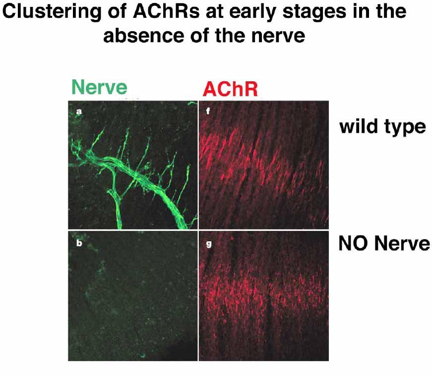
Patterning of Muscle Acetylcholine Receptor Gene Expression in the Absence of Motor Innervation
 Neuron, Vol. 30, 399 410, May, 2001, Copyright 2001 by Cell Press Patterning of Muscle Acetylcholine Receptor Gene Expression in the Absence of Motor Innervation Xia Yang, 1 Silvia Arber, 2,5 Christopher
Formation of the neuromuscular junction
 Eur. J. Biochem. 265, 1±10 (1999) q FEBS 1999 REVIEW ARTICLE Formation of the neuromuscular junction Agrin and its unusual receptors Werner Hoch Max-Planck-Institut fuèr Entwicklungsbiologie, Abteilung