STRUCTURE AND FUNCTION OF HETEROTRIMERIC G PROTEIN-REGULATED RHO GUANINE NUCLEOTIDE EXCHANGE FACTORS
|
|
|
- Martin Blankenship
- 5 years ago
- Views:
Transcription
1 Molecular Pharmacology This article has Fast not been Forward. copyedited Published and formatted. on The October final version 30, may 2009 differ as from doi: /mol this version. STRUCTURE AND FUNCTION OF HETEROTRIMERIC G PROTEIN-REGULATED RHO GUANINE NUCLEOTIDE EXCHANGE FACTORS Mohamed Aittaleb *, Cassandra A. Boguth *, John J. G. Tesmer Life Sciences Institute (M.A., C.A.B., & J. J. G. T.) and the Department of Pharmacology (J. J. G. T.) University of Michigan, Ann Arbor, Michigan 1 Copyright 2009 by the American Society for Pharmacology and Experimental Therapeutics.
2 Running Title: Heterotrimeric G protein-regulated RhoGEFs Corresponding Author: Abbreviations: John J. G. Tesmer Life Sciences Institute 210 Washtenaw Avenue University of Michigan Ann Arbor, Michigan Tel: (734) Fax: (734) johntesmer@umich.edu Number of text pages: 57 Number of tables: 2 Number of Figures: 3 Number of References: 141 Number of words in abstract: 148 Number of words in introduction: 1207 Number of words in discussion (conclusions): 583 DH, Dbl homology; DEP, Disheveled, EGL-10, Pleckstrin; Gα, heterotrimeric G protein α subunit; Gβγ heterotrimeric G protein β and γ heterodimer; GPCR, G protein-coupled receptor; GTPase, guanine nucleotide triphosphatase; IP4P, inositol polyphosphate-4-phosphatase; LARG, leukemia-associated RhoGEF; LPA, lysophosphatidic acid; PDZ, Postsynaptic density 95, Disk large, Zona occludens-1; PH, Pleckstrin homology; PI3K, phosphatidylinositol 3-kinase; PLCβ, phospholipase Cβ; P-Rex, phosphatidylinositol (3,4,5)-triphosphate (PIP 3 )-dependent Rac exchanger; RhoGEF, Rho guanine nucleotide exchange factor; rgrgs, RhoGEF fragment containing both N-terminal GAP motif and RH domain; RGS, regulator of G protein signaling; RH, RGS homology; SH3, Src homology 3; SRE, serum response element; SRF, serum response factor. 2
3 3
4 Abstract Activation of certain classes of G protein-coupled receptors (GPCRs) can lead to alterations in the actin cytoskeleton, gene transcription, cell transformation, and other processes that are known to be regulated by Rho family small molecular weight GTPases. Although these responses can occur indirectly via cross-talk from canonical heterotrimeric G protein cascades, it has recently been demonstrated that Dbl family Rho guanine nucleotide exchange factors (RhoGEFs) can serve as the direct downstream effectors of heterotrimeric G proteins. Heterotrimeric Gα 12/13, Gα q, and Gβγ subunits are each now known to directly bind and regulate RhoGEFs. Atomic structures have recently been determined for several of these RhoGEFs and their G protein complexes, providing fresh insight into the molecular mechanisms of signal transduction between GPCRs and small molecular weight G proteins. This review covers what is currently known about the structure, function, and regulation of these recently recognized effectors of heterotrimeric G proteins. 4
5 I. Introduction Heterotrimeric GTP-binding (G) proteins are master regulators of cell homeostasis. By coordinating signaling between the ~800 G protein-coupled receptors (GPCRs) in the human genome and a relatively small handful of effector enzymes and channels in the cell, they control processes such as muscle contractility, glycogen metabolism, neurotransmission, and the concentration of intracellular ions. Their profound impact on nearly all cellular processes and their therapeutic potential have rendered them one of the most intensely studied signal transduction paradigms at the biochemical and molecular level (Sprang et al., 2007). When heterotrimeric G proteins are in their inactive, GDP-bound state, they exist as an inert complex composed of α, β, and γ subunits (Gαβγ). In this state, they are substrates for activated GPCRs, which catalyze nucleotide exchange on the α subunit (Gα). When bound to GTP, Gα releases the effector binding surface of the β and γ heterodimer (Gβγ) so that both Gα and Gβγ can interact with and modulate the activity of specific downstream enzymes and channels. The Gα subunit has weak guanine nucleotide triphosphatase (GTPase) activity that slowly returns the G protein to its GDP-bound state. Gα GDP then becomes re-sequestered by Gβγ. Beyond serving as conduits for extracellular signals, heterotrimeric G proteins contribute to the fidelity, duration, and amplitude of GPCR signaling. A given class of heterotrimeric G protein can typically recognize only a subset of GPCRs, and can only interact with one or a few downstream effector targets, ensuring the specificity of signaling from receptor to effector. The rate of GTP hydrolysis on Gα dictates the length of time its signal is in play, and this rate can be dramatically accelerated by either the effector target or regulators of G protein signaling (RGS) proteins, which serve as GAPs for some classes of Gα subunits (Ross and Wilkie, 2000). Finally, if the activated receptor is not uncoupled from the G protein, such as via 5
6 phosphorylation by GPCR kinases and binding of arrestins, then Gαβγ can undergo multiple rounds of activation. Thus, regulating the relative rates of G protein activation and deactivation can control the amplitude of signals initiated by GPCRs. Crystallographic studies have uncovered the molecular mechanism by which activated Gα and Gβγ subunits engage their effector targets. In the case of Gα, the binding of GTP induces a conformational change in structural elements that interact with the γ-phosphate of GTP, known as Switch I, II, and III (Fig. 1a). This conformational change lowers the affinity of Gα for Gβγ while increasing its affinity for effectors. Upon binding GTP, a shallow hydrophobic canyon is created between Switch II and the α3 helix of Gα that is used for binding all the effectors that have been structurally characterized thus far (Chen et al., 2008; Chen et al., 2005; Lutz et al., 2007; Slep et al., 2001; Tesmer et al., 1997b; Tesmer et al., 2005). There is sufficient variability in the residues that line the perimeter of this effector docking site to explain the exquisite specificity of Gα for its downstream targets. In the case of Gβγ, its ability to bind effectors depends on the unmasking of a broad, relatively hydrophobic surface that is sequestered in the Gαβγ complex. Conformational changes between the Gα- and effector-bound states of Gβγ have thus far proved relatively minor. Although the binding surface for the effectors of Gβγ are believed to overlap with that of Gα, they are believed to involve distinct regions in each case (Davis et al., 2005; Ford et al., 1998). A role for GPCRs in cell migration and transformation. GPCRs are traditionally thought of as regulators of cellular metabolism or as mediators of sensory perception, such as vision, smell, and taste. The canonical effector enzymes involved in these processes include adenylyl cyclase, cgmp phosphodiesterase, and phospholipase Cβ (PLCβ). The Gα subunits that couple receptors to these effectors represent three of the four subfamilies of human heterotrimeric Gα subunits: 6
7 Gα s, Gα i/t, and Gα q, respectively. However, GPCRs can also serve as potent oncogenes in a manner more typical of tyrosine kinase growth factor receptors (Whitehead et al., 2001). Indeed, constitutively activated Gα subunits, in particular members of the Gα 12/13 subfamily, can lead to strong cell proliferative responses (Dhanasekaran et al., 1998). The mechanism by which this occurs began to be understood by the observation that lysophosphatidic acid (LPA) and bombesin, known ligands for GPCRs, can activate the small molecular weight G protein RhoA and induce the formation of stress fibers and focal adhesions (Ridley and Hall, 1992). In the following years, numerous studies lent additional support to the idea that Rho GTPases were activated downstream of many GPCRs (Sah et al., 2000; Seasholtz et al., 1999). The Rho GTPases are a family of peripheral membrane proteins that regulate essential cellular processes including cell shape, cell migration, cell cycle progression, and gene transcription (Etienne-Manneville and Hall, 2002; Wennerberg and Der, 2004). Like their larger Gα subunit homologs, they cycle between an inactive GDP-bound and an active GTP-bound state that can interact with specific downstream effector targets, depending on the Rho GTPase. In vivo, Rho GTPases require an upstream GEF for activation. The largest and best characterized RhoGEF family is characterized by a catalytic Dbl homology (DH) domain, an extended helical domain of ~200 amino acids that forms the principal binding site for the nucleotide-free GTPase. The DH domain is almost always immediately followed in the primary sequence by a pleckstrin homology (PH) domain (Rossman et al., 2005; Schmidt and Hall, 2002). In some DH-family RhoGEFs, the PH domain inhibits the intrinsic GEF activity of the DH domain (Bellanger et al., 2003; Das et al., 2000; Lutz et al., 2007; Welch et al., 2002), a constraint that is presumably released upon the interaction of the PH domain with phospholipids or other proteins. In other Dbl family members, the PH domain can play a positive signaling role (Liu et al., 1998; Reuther et al., 2001; Rossman et al., 2002). 7
8 GPCRs could regulate Rho GTPase signaling either directly via the interaction of activated heterotrimeric G proteins with a RhoGEF, or indirectly, for example, via activation of PKA or PKC, which can then phosphorylate RhoGEFs, their upstream regulators, or their downstream targets. In 1998, it was shown that Gα 13 subunits bind directly to p115rhogef and stimulate its activity (Hart et al., 1998), thereby revealing a simple, direct path from LPA or thrombin receptors to RhoA (Wang et al., 2004). In 2005, it was shown that Gα q/11 could activate the activity of another RhoA-selective RhoGEF, p63rhogef, providing another direct pathway, this time initiated by bombesin, muscarinic, and angiotensin receptors (Lutz et al., 2005). Gβγ is also now known to directly bind and activate RhoGEFs, such as P-Rex1, which links G i -coupled chemokine receptors in neutrophils to the activation of Rac2 (Welch et al., 2002). In this review, we examine what is currently known about the structure and function of Dbl-family RhoGEFs that are directly regulated by heterotrimeric G proteins (Table 1, 2, Fig. 2). We focus primarily on three aspects relevant to molecular pharmacology: 1) the structures of RhoGEF domains involved in signal transduction, 2) the specific details of how heterotrimeric G proteins are known to interact with these domains, and 3) the potential mechanisms by which these interactions lead to RhoGEF activation. II. Gα 12/13 -regulated RhoGEFs There are currently four RhoGEFs believed to be directly stimulated by the Gα 12/13 subfamily of heterotrimeric G proteins: p115-rhogef, LARG, PDZ-RhoGEF, and Lbc (Siehler, 2009; Suzuki et al., 2009a) (Table 1, Fig. 2a). Although these enzymes all belong to the Lbc subfamily of RhoGEFs, it is not yet clear if their homology correlates with an ability to be regulated by 8
9 heterotrimeric G proteins. The supporting data is weakest for Lbc, which has not yet been shown to bind or be activated by Gα 12/13 subunits using purified proteins in vitro. A) The RH-RhoGEFs Background. Members of the Gα 12/13 subfamily are potent regulators of the actin cytoskeleton (Buhl et al., 1995), and are important for vascular development and for chemotaxis (Offermanns et al., 1997). Given that Rho GTPases were also well-established regulators of these responses, the hunt was on for RhoGEFs that could operate directly downstream of Gα 12/13. One candidate was p115rhogef, a RhoA-selective GEF containing a region with very weak homology to the catalytic RGS homology (RH) domain characteristic of RGS proteins (Tesmer, 2009). Indeed, this proved to be the binding site for activated Gα 12 and Gα 13 subunits, although only Gα 13 could stimulate GEF activity (Hart et al., 1998; Kozasa et al., 1998; Mao et al., 1998). Soon thereafter, PDZ-RhoGEF (Fukuhara et al., 1999), also known as GTRAP48 (Jackson et al., 2001), and leukemia-associated RhoGEF (LARG) (Fukuhara et al., 2000) were identified as RH-domain containing RhoGEFs. These three proteins form the RH-RhoGEF subfamily, a small subfamily of Gα 13 -regulated RhoGEFs. LARG is the only one currently known to be activated by Gα 12, although this appears to require phosphorylation of the RhoGEF by Tec tyrosine kinase (Suzuki et al., 2003). All three RH-RhoGEFs are widely expressed in mammals (Fukuhara et al., 1999; Hart et al., 1996; Kourlas et al., 2000; Kuner et al., 2002), but p115rhogef and PDZ-RhoGEF are most abundantly expressed in hematopoietic cells and the central nervous system, respectively. In resting cells, RH-RhoGEFs are predominantly localized to the cytosol, with some presence at the cell membrane or bound to actin. Upon activation of Gα 12/13 coupled receptors or co-expression of constitutively active mutants of Gα 12 or Gα 13, RH-RhoGEFs translocate to the 9
10 membrane surface (Siehler, 2009; Suzuki et al., 2009a). RH-RhoGEFs also exhibit some receptor selectivity. In HEK293 and PC3 cells, RhoA is activated by PDZ-RhoGEF via LPA receptors, and by LARG via thrombin receptors (Wang et al., 2004). The mechanism by which this selectivity occurs is not known, but it is not apparently due to differences in their PDZ domains, as both PDZ-RhoGEF and LARG can bind to the C-termini of LPA receptors (Yamada et al., 2005). The RH RhoGEFs are complex proteins with multiple signaling domains and functional motifs hidden within the low complexity regions that bracket them. There is considerable evidence that the mechanism of activation is also complex, involving not only recruitment to specific sites at the membrane surface, but also multiple intra- and inter-molecular interactions that mediate inhibition of basal activity as well as allosteric activation (Bhattacharyya et al., 2009; Bhattacharyya and Wedegaertner, 2003; Suzuki et al., 2009b; Zheng et al., 2009). The specific details vary among subfamily members (e.g. p115rhogef lacks a PDZ domain) and context (e.g. LPA versus thrombin receptors). In the following sections, we summarize what is known about the structure and function of the major domains and other functional motifs of RH- RhoGEFs, highlighting their potential roles in regulating the activity of the DH/PH domains. We end by describing recent solution-based studies of LARG and PDZ-RhoGEF that provide new insights into the molecular mechanism of RH-RhoGEF activation. The RhoGEF RH domain. As the principal binding site for Gα 12/13 subunits, structural analysis of the RhoGEF RH domain was anticipated to explain the molecular basis for selectivity and GAP activity, and to shed some light on the mechanism of regulation by heterotrimeric G proteins. The canonical RH domain is a bundle of nine helices that forms a relatively flat domain with two lobes (Tesmer, 2009). In RGS proteins, the α5-α6 loop of the domain interacts 10
11 with catalytic residues in Switch I and II of Gα to accelerate GAP activity (Tesmer et al., 1997a). In contrast, the RH domains of p115rhogef and LARG do not have GAP activity. Instead, a short sequence motif positioned immediately N-terminal to the RH domain is required for stimulating GTP hydrolysis in p115rhogef and LARG (Chen et al., 2003). The region spanning this N-terminal motif and the RH domain has been termed the rgrgs domain (Chen et al., 2001) (Fig. 2a). Crystal structures of the p115rhogef (Chen et al., 2001) and the PDZ-RhoGEF (Longenecker et al., 2001) RH domains confirmed that they indeed retain most of the canonical RH domain fold, albeit with a ~65-residue extension that contributes three extra α helices to the C-terminus (Fig. 1b, c). However, these initial structures did not reveal an obvious mode of interaction with Gα 12/13 subunits, in part because the Gα-interacting residues of RGS proteins are not conserved in the RhoGEF RH domain (Tesmer, 2009). Although the entire rgrgs protein of p115rhogef was crystallized, the N-terminal GAP motif was not ordered. The structure of the p115rhogef rgrgs domain in complex with a chimeric Gα 13/i1 protein (Chen et al., 2005) revealed a surprising bipartite interaction mediated by the N-terminal GAP motif and the RH domain (Fig. 1b). The Gα 13/i1 chimera used in this study was created to circumvent problems in expressing functional Gα 13 in E. coli, and contains the α helical domain and Switch I and II elements from Gα 13, with the remainder from Gα i1. As a consequence, the effector docking site contains residues that are not native to Gα 13, including some that directly contact the RH domain. Notably, this chimera was never shown to activate p115rhogef. The N-terminal motif (residues 22-37) begins with an IIG sequence motif that anchors the peptide on the α-helical domain of Gα 13/i1, and is followed by an acidic sequence in which Glu27 and Phe31 contact Gα 13/i1 -Arg200 in Switch I and Gln226 in Switch II, respectively, stabilizing them in a more transition state-like conformation. The structure thereby provides a molecular explanation 11
12 for the mild GAP activity exhibited by p115rhogef. The RH domain (residues ), as if supplanted from Switch I by the N-terminal GAP motif, binds to the canonical effector docking site of Gα 13/i1 (Fig. 1b). Instead of using the α5-α6 loop to bind Gα, the interaction is mediated by structural elements from the unique C-terminal extension of the RH domain, in particular the α8-α9 loop, which bears hydrophobic residues that pack into the effector docking site. Consequently, the canonical core of the RhoGEF RH domain can be thought of as a scaffold that positions unique functional motifs/extensions at its N- and C-termini that are required for high affinity binding, recognition of the activated conformation of the Gα subunit, and, in p115rhogef and LARG, GAP activity. Recent crystal structures of complexes between a different Gα 13 chimera (all but the N- terminal helix was native to Gα 13 ) and the PDZ-RhoGEF rgrgs domain recapitulate the major findings of the Gα 13/i1 -p115rhogef complex (Chen et al., 2008). As before, the N-terminal motif of the RhoGEF anchors to the α-helical domain of Gα 13 using an IIG sequence motif and interacts with the switch regions (Fig. 1c). However, in this case Glu312 and Tyr315 (analogous to Glu27 and Phe32 in p115rhogef) do not make productive interactions with residues in the active site of Gα 13, explaining the lack of GAP activity as well as the observation that PDZ-RhoGEF has similar affinity for the GTPγS- and GDP AlF - 4 -bound states of Gα 13 (Chen et al., 2008). Interestingly, residues in the RH domain of p115rhogef that bind to the effector docking site of Gα 13 are not conserved in PDZ-RhoGEF (Fig. 1b,c). Despite this difference, a similar complex is formed, with the effector docking site expanding to accommodate the larger αe helix that occupies the position analogous to the α8-α9 loop of p115rhogef. However, it is not clear if the differences in the structure of the effector docking site are a consequence of differences in the chimeric Gα 13 subunits used, of differences in the RH domains, or of both. The structure of the PDZ-RhoGEF rgrgs domain in complex with 12
13 deactivated Gα 13 GDP was also determined. Interestingly, Switch II retains its active conformation despite the presence of GDP. Thus, the duration of signaling via RH-RhoGEFs could be regulated not only by the rate of GTP hydrolysis, but also by the rate that Gα 13 GDP is sequestered by Gβγ (Chen et al., 2008). The PDZ domain. PDZ-RhoGEF and LARG can also be activated by receptors other than GPCRs via interactions mediated by the postsynaptic density 95, disk large, zona occludens-1 (PDZ) domain found at their N-terminus. PDZ domains are common structural domains that typically bind the C-termini of proteins found at the cell membrane (Nourry et al., 2003). Compared to other PDZ domains, the RH-RhoGEF PDZ domain binds to surprisingly diverse biological targets (for a review see (Smietana et al., 2008)). Perhaps the best characterized is plexin B1, a semaphorin 4D receptor, which mediates RhoA activation and axonal growth cone collapse (Aurandt et al., 2002; Hirotani et al., 2002; Perrot et al., 2002; Swiercz et al., 2002). Both RhoGEFs also bind to LPA-1 and LPA-2 receptors, which couple primarily with Gα 12/13 subunits. Interaction between the C-termini of these receptors and the PDZ domain was required for RhoA activation (Yamada et al., 2005). NMR structures of the LARG PDZ domain alone and in complex with a plexin-b1 C- terminal octapeptide have been reported (Liu et al., 2008) (Fig. 1d). A solution structure of the PDZ-RhoGEF PDZ domain is also available (PDB entry 2DLS). They retain the typical PDZ domain fold, which consists of six anti-parallel strands (βa-βf) and two helices (αa and αb). Peptide ligands dock in an extended conformation into a groove formed between βb and αb, where they form a β-sheet like interaction with the βb strand. The LARG PDZ domain undergoes an atypical conformational change upon ligand binding relative to other characterized PDZ domains (Fig. 1d), and the apo form exhibits a high degree of conformational flexibility 13
14 (Liu et al., 2008). These characteristics may facilitate the binding of these PDZ domains to wide variety of targets. DH/PH domain structures. The canonical DH domain is composed of ten or more helices that assemble into an oblong shape reminiscent of a chaise lounge (Worthylake et al., 2000). The last helix (called α6) transverses the longest dimension of the domain, and often forms a continuous helix with αn, the first helix of the PH domain (Fig. 1e). The PH domain consists of a flattened 7-stranded anti-parallel β-barrel, capped at one end by αc, the hallmark C-terminal helix found in all PH domains. In some PH domains, loops on the opposite end of the barrel from αc are used to bind specific phospholipid head groups. The αn helix is characteristic of PH domains found in DH/PH tandems. Structures have been determined for the LARG DH/PH domains, both alone and in complex with RhoA (Kristelly et al., 2004) and for the PDZ-RhoGEF DH/PH-RhoA complex (Derewenda et al., 2004). Both RhoA complexes exhibit similar domain organization and intersubunit contacts (Fig. 3f). As in Dbs, another RhoGEF whose PH domain contributes positively to GEF activity (Rossman et al., 2003; Rossman et al., 2002), direct contacts are formed between RhoA and the PH domain. Although this interface accounts for only ~8% of the total buried surface area in the LARG RhoA-DH/PH complex, mutation of residues in the PH domain-rhoa interface diminished GEF activity to the level of the DH domain alone (Kristelly et al., 2004). Mutation of residues in the PDZ-RhoGEF interface did not lead to significant differences in vitro (Oleksy et al., 2006), and hence the functional importance of the RhoA-PH domain interface appears to vary among RH-RhoGEFs. Examination of the DH/PH structures reveals several structural elements that could play a role in regulation. The first was a flexible, helical extension at the N-terminus of the DH domain 14
15 (Fig. 1e). Either truncation of the extension or mutation of a tryptophan buried between the extension and the DH domain (LARG-Trp769) reduced GEF activity (Kristelly et al., 2004). However, the LARG-W769D mutation only exhibited a minor defect in SRE transcription assays in cells (Aittaleb et al., 2009). Sequence analysis predicts that the extension exists in all Lbc subfamily RhoGEFs, suggesting that it is not involved in regulation specific to the RH- RhoGEFs. Instead, recent data suggests that the N-terminal extension could be more important for autoinhibition of basal activity (see below). The second notable feature is a relatively long helical linker between the DH and PH domains that allows for considerable conformational flexibility between the two domains. For example, there is a ~30 difference in orientation of the PH domain relative to the DH domain between the LARG DH/PH and DH/PH-RhoA structures, with the hinge occurring at the junction of the α6 and αn helices. The four unique DH/PH domains in the LARG DH/PH- RhoA structure also exhibit conformational differences in the α6-αn linker (Kristelly et al., 2004). Because mutation of residues in the PH domain that directly interact with RhoA inhibit GEF activity (Kristelly et al., 2004), these contacts were proposed to alter the conformation of the α6-αn linker region in a manner that facilitates the binding of RhoA. Indeed, residues at the end of α6 make critical interactions with Switch II of the bound GTPase (Aghazadeh et al., 1998). Interactions with other proteins or phospholipids that likewise influence the relative orientation of the DH and PH domains could likewise affect LARG activity. Solution studies of the PDZ-RhoGEF DH/PH domains did not find evidence for an analogous large conformational change in the linker region (Cierpicki et al., 2009), suggesting that in PDZ-RhoGEF the PH domain may simply serve to stabilize the α6 helix of the DH domain. Lastly, in all the DH/PH structures a solvent-exposed patch of conserved hydrophobic residues is observed in the PH domain (Fig. 1e,f). Depending on the crystal structure, the patch 15
16 forms either a 2-fold crystallographic or a quasi-2-fold molecular interface that buries ~800 Å 2 of surface area. Located on the side of the PH domain β barrel, the patch would be in position to bind adjacent peripheral membrane proteins or domains at the cell membrane. Mutation of residues in the hydrophobic patch of LARG had no effect on nucleotide exchange activity in vitro, but abrogated the ability of LARG to induce RhoA activation and stress fiber formation in cells (Aittaleb et al., 2009). Because the activity of these mutants could be rescued by fusion with nonspecific membrane targeting motifs, the hydrophobic patch appears to contribute to the proper localization of LARG by interacting with unknown target(s) at the cell membrane. However, membrane targeting by constitutively activated Gα 13 did not rescue activity, suggesting that the hydrophobic patch also plays an essential role in GPCR-mediated regulation, although it does not yet appear to involve direct interactions with activated Gα 13 (Aittaleb et al., 2009). The hydrophobic patch is conserved among all Lbc subfamily members, once again indicating a common functional role. Indeed, mutation of the hydrophobic patch of Lbc likewise abolished its ability to promote RhoA-mediated transcriptional activation in cells (Aittaleb et al., 2009). Other functional motifs. p115rhogef, LARG, and PDZ-RhoGEF undergo homo- and heterodimerization mediated by their C-terminal tails, and specific coiled-coil motifs have been identified in p115rhogef and LARG (Chikumi et al., 2004; Eisenhaure et al., 2003; Grabocka and Wedegaertner, 2007) (Fig. 2a). Deletion of the C-terminus augments the ability of RH- RhoGEFs to induce RhoA activation in cells, suggesting a negative regulatory role for dimerization, presumably via the recruitment of unknown inhibitory factors (Chikumi et al., 2004; Eisenhaure et al., 2003). A proline-rich motif positioned immediately C-terminal to the PDZ-RhoGEF PH domain, (residues ) has been shown to be essential for plasma 16
17 membrane localization of PDZ-RhoGEF, and is required for PDZ-RhoGEF induced cortical actin re-organization and cell rounding (Togashi et al., 2000). The RH-DH/PH linker region also contains functional elements. In addition to an acidic autoinhibitory motif (Zheng et al., 2009), the RH-DH/PH linker of PDZ-RhoGEF contains a unique actin-binding motif (Banerjee et al., 2009; Banerjee and Wedegaertner, 2004). Solution studies. Two recent solution-based studies have provided new insights into RH- RhoGEF regulation (Fig. 3a). Using surface plasmon resonance, the first study showed that Gα 13 could interact with not only the RH but also the DH/PH and C-terminal regions of LARG (Suzuki et al., 2009b), consistent with earlier observations using co-immunoprecipitation (Bhattacharyya and Wedegaertner, 2003; Wells et al., 2002). The presence of all three domains (RH, DH, and PH) was required for formation of the highest affinity complex, and the K204A mutation of Gα 13, which abrogates binding to the RH domain (Grabocka and Wedegaertner, 2005; Nakamura et al., 2004), did not eliminate binding to the DH/PH domain. Thus, Gα 13 appears to exhibit at least two LARG binding surfaces: one for the rgrgs domain that is dependent on the activation status of Gα 13, and another for the DH/PH domains that uses activation-independent structural elements, potentially the C-terminal region of the Ras-like domain (Kreutz et al., 2007). Truncation studies of PDZ-RhoGEF identified a conserved acidic motif (residues ) a few amino acids upstream of the N-terminal helical extension of the DH domain that serves an autoinhibitory role (Zheng et al., 2009). NMR spectroscopy comparing the chemical shifts in DH/PH fragments with or without this acidic motif suggests that it interacts with basic residues on the DH domain, in particular Arg867 and Arg868. Because these residues also interact with the bound GTPase, their interaction with the acidic motif would form an 17
18 autoinhibited state reminiscent of that of Vav (Aghazadeh et al., 2000). Indeed, charge-reversing mutations of four of the acidic residues in the motif (the activation box, Fig. 2a) enhanced GEF activity ~20-fold in a DH/PH fragment of PDZ-RhoGEF. Curiously, the enhancement was only ~2 fold for the same mutants in the context of a larger fragment that also contained the PDZ and RH domain. This may indicate that the acidic motif and the PDZ and RH domains act synergistically to autoinhibit activity. Acidic motifs also occur in the RH-DH linker regions of p115rhogef and LARG, although it remains to be seen if they function similarly. If the activation box is used in all Lbc subfamily members to regulate basal activity, it may provide a functional role for the N-terminal extension of the DH domain, which folds in such a manner that it would juxtapose the acidic motif of PDZ-RhoGEF with Arg867 and Arg868 on the surface of the DH domain. B) Lbc-RhoGEF. Lymphoid blast crisis (Lbc) RhoGEF was originally isolated as a transforming oncogene (onco-lbc) from myeloid leukemia patients (Toksoz and Williams, 1994) and is the founding member of the Lbc subfamily of RhoGEFs. No atomic structures are currently available for this enzyme. Beyond the DH/PH domains, proto-lbc contains a predicted α-helical domain and a proline rich motif in its C-terminus that have been shown to act as negative regulators of proto-lbc transforming activity and localize the enzyme to the membrane fraction of cells (Sterpetti et al., 1999). Onco-Lbc lacks this C-terminal region. The Lbc- RhoGEF splice variant AKAP-Lbc, or AKAP13, is highly expressed in the heart and functions both as a camp-dependent protein kinase (PKA)-scaffolding protein and a guanine nucleotide exchange factor (GEF) for RhoA (Diviani et al., 2001). Similar to the RH-RhoGEFs, there is evidence that AKAP-Lbc can undergo homo-oligomerization through a leucine zipper motif in 18
19 the C-terminal region. Deletion of this C-terminal region in AKAP-Lbc enhances its GEF activity, suggesting a negative regulatory role for oligomerization (Baisamy et al., 2005). Proto-Lbc and AKAP-Lbc have been shown to be specifically stimulated by Gα 12 as well as via activation of the LPA receptor (Diviani et al., 2001; Dutt et al., 2004; Majumdar et al., 1999). The activation appears to be direct, because Lbc can be co-immunoprecipitated from HEK293 cells with constitutively active Gα 12 (Diviani et al., 2001; Dutt et al., 2004). However, it should be noted that Gα q can also be precipitated with Lbc, although with no obvious effect on RhoA activation (Pi et al., 2002; Sagi et al., 2001). The binding site for Gα 12 is not known, but a region C-terminal to the PH domain with debatable homology to RH domains has been identified (Dutt et al., 2004). This region overlaps with the leucine zipper motif shown to mediate homodimerization (Baisamy et al., 2005). III) Gα q -regulated RhoGEFs Background. The involvement of Gα q in Rho-mediated signaling has been recognized for over a decade. Dominant negative RhoA and C3 endotoxin were used to demonstrate the existence of a unique Rho-dependent pathway in response to the activation of Gq-coupled receptors in cardiac myocytes (Sah et al., 1996). Later, the development of cell-based, RhoA pulldown assays enabled a direct demonstration of an increase in RhoA GTP in response to the activation of Gα q -coupled receptors in HEK293 cells, establishing that Gα q operated upstream of RhoA, at least in some signaling contexts (Chikumi et al., 2002). Studies of mouse embryonic fibroblasts deficient in either Gα 12 /Gα 13 or Gα q /Gα 11 subunits confirmed that these G protein families activate RhoA via independent pathways, although RhoA activation by Gα 12 /Gα 13 subunits was more robust (Vogt et al., 2003). 19
20 A number of RhoGEFs have been reported to interact with, and thus potentially be regulated by Gα q. These include LARG (Booden et al., 2002; Chikumi et al., 2002; Vogt et al., 2003) and Lbc (Pi et al., 2002; Sagi et al., 2001). In neither case has a direct interaction between the RhoGEF and Gα q been demonstrated with purified components, nor has Gα q -mediated stimulation of GEF activity been reconstituted in vitro. Furthermore, reports conflict on whether or not the RH domain of LARG is responsible for binding Gα q. A) p63rhogef. In 2005, p63rhogef, which is expressed predominantly in the brain and heart, was shown to be activated by G q -coupled receptors in a manner that directly competes with the activation of PLCβ, the canonical effector of Gα q (Lutz et al., 2005). A splice variant lacking the first 106 amino acids, known as GEFT, was reported to activate Cdc42 and Rac1 in cell-based assays and with purified proteins in vitro (Guo et al., 2003). Although there is the possibility of cross-talk in cells, GEF activity towards Cdc42 and Rac1 has not been demonstrated by other groups (Lutz et al., 2004; Rojas et al., 2007; Souchet et al., 2002). Primary sequence analysis also strongly suggests that p63rhogef and its splice variants are selective for RhoA. The primary sequence of p63rhogef lacked an obvious RH domain that could serve as a binding site for Gα q. Initial truncation studies instead localized the binding site to a fragment of p63rhogef that includes the PH domain (Lutz et al., 2005). Subsequently, more detailed truncation studies, site-directed mutagenesis, and structure determination revealed that the C- terminal helix of the PH domain formed the principal binding site for activated Gα q (Lutz et al., 2007; Rojas et al., 2007). The closely related, C-terminal set of DH/PH domains in Trio and Kalirin were also shown to bind and be activated by Gα q in vitro (Lutz et al., 2007; Rojas et al., 2007) and in cells (Lutz et al., 2007). Although the physiological role of p63rhogef in 20
21 mammals likely involves smooth muscle function (S. Lutz, personal communication), the p63rhogef homolog in C. elegans, UNC-73E, is known to serve as a bona fide downstream effector of Gα q and works in conjunction with PLCβ to regulate the release of acetylcholine vesicles at neuromuscular synapses (Williams et al., 2007). Its knockout, in combination with a PLCβ null allele, generates the same phenotype as knockouts of Gα q, confirming the necessary involvement of both pathways downstream of Gα q (Williams et al., 2007). Finally, the kinase domain of mixed lineage kinase 3 (MLK3) was reported to bind to p63rhogef and inhibit its interaction with Gα q, thereby regulating directed cell migration (Swenson-Fields et al., 2008). Although it is not yet known what moiety of p63rhogef could be involved in binding MLK3, the PH domain would be an obvious candidate. Interestingly, Gα 16, a divergent member of the Gα q subfamily, binds but fails to activate p63rhogef, suggesting that Gα 16 serves as a general inhibitor of Gα q/11 signaling through p63rhogef, just as Gα 12 may inhibit Gα 13 signaling through p115rhogef (Yeung and Wong, 2009). The idea of Gα 16 serving as a p63rhogef trap is also supported by at least one other study (Moepps et al., 2008) Structural studies of p63rhogef. After delineation of the minimal fragment of p63rhogef required for high affinity binding to Gα q, the Gα q -DH/PH-RhoA complex was isolated and crystallized. Its structure determination revealed that the C-terminal helix of the p63rhogef PH domain was unusually long (Fig. 1g), with hydrophobic residues on the extension of this helix binding directly into the effector docking site of Gα q (Lutz et al., 2007). Mutation of these hydrophobic residues abrogates binding (Lutz et al., 2007; Rojas et al., 2007). Other residues, principally from the β1-β4 sheet of the PH domain, form specific contacts that help to dictate specificity for the Gα q/11 subfamily. The interface is highly reminiscent of the interface between 21
22 the PH domain of GRK2 and Gβγ (Lodowski et al., 2003), establishing a theme for how PH domains serve as protein-protein interaction modules. Notably, the manner in which p63rhogef engages Gα q is distinct from how the rgrgs domain engages Gα 13 in that the GAP binding site on Switch I is left freely accessible to RGS proteins. p63rhogef lacks significant GAP activity on its own, but can allosterically regulate the binding of RGS proteins (Shankaranarayanan et al., 2008). Although the principal interaction is via the PH domain, the C-terminal region of the Gα protein, including its C-terminal α5 helix, forms direct interactions with the DH domain and residues in the DH/PH domain interface. Mutation of some of these residues in p63rhogef leads to a loss of Gα q activation in vitro, but had only minor effects on binding, indicating that these contacts are most important for regulation of activity (Lutz et al., 2007). More recently, mutation of Tyr356 in the C-terminus of Gα q has also been shown to abrogate activation of p63rhogef (Shankaranarayanan et al., 2009). Because the equivalent residue in Gα 16 is isoleucine, this specific contact may help explain why Gα 16 binds yet fails to activate p63rhogef. Compared to structures of other characterized DH/PH tandem domains, the PH domain of p63rhogef in complex with Gα q is held in an unusual orientation relative to the DH domain, being rotated ~50 around the axis of the α6 helix relative to those of Dbs and TrioN, the closest homologs of known structure (Chhatriwala et al., 2007; Snyder et al., 2002). The PH domain makes no direct contacts with the substrate RhoA. Although the structure of p63rhogef in the absence of Gα q is not yet available, it is anticipated that the domain-bridging interactions of Gα q help to constrain the DH and PH domains in this conformation, and that the DH/PH domains adopt a more Dbs-like conformation in the basal state. 22
23 Mechanism of activation. Unlike the RH-RhoGEFs, p63rhogef is predominantly found localized to actin-rich structures (Souchet et al., 2002) and overexpressed p63rhogef is localized to the cell surface (Shankaranarayanan et al., 2009). Therefore, membrane translocation is not expected to play a major role in activation. Also unlike RH-RhoGEFs and Dbs, the PH domain of p63rhogef is autoinhibitory, with the isolated DH/PH fragment having far less activity than the isolated DH domain (Lutz et al., 2004; Lutz et al., 2007; Rojas et al., 2007). Thus, the PH domain must somehow interfere with the binding of RhoA. Indeed, the binding of Gα q greatly enhances the affinity of RhoA for the DH/PH domains (Shankaranarayanan et al., 2009). Because the PH domain does not inhibit GEF activity when added in trans (Shankaranarayanan et al., 2009), the inhibition mediated by the PH domain appears to require a covalent linkage between the DH and PH domains, once again implicating the α6-αn region as a structure of regulatory importance. Inspection of residues in the α6-αn linker region indicate that Arg341 of p63rhogef might sterically and electrostatically repel Arg68 in switch II of RhoA if the PH domain were in a more Dbs-like orientation relative to the DH domain. An R341A mutation indeed enhances the basal activity of p63rhogef, but, surprisingly, does not eliminate activation by Gα q (Shankaranarayanan et al., 2009). In fact, the R341A mutation allows Gα q to enhance activity to levels greater than the DH domain alone, indicating that Gα q can activate p63rhogef not only by removing an autoinhibitory restraint, but also by activating the DH domain allosterically. This is presumably carried out via the interactions of the C-terminal region of Gα q with the DH domain, as observed in the crystal structure, which in turn alters the conformation of the α6-αn linker (Shankaranarayanan et al., 2009). A schematic model for Gα q -mediated activation of p63rhogef and related DH/PH domains is shown in Fig. 3b. 23
24 B) Trio and Kalirin. Trio and Kalirin are closely related proteins that contain two sets of DH/PH domains and a much more complex domain structure than p63rhogef (Fig. 2b). Both proteins play critical roles in neurite growth and neuronal morphology (Table 1). The second set of DH/PH domains in Trio (TrioC) is closely related to that of p63rhogef, and is likewise selective for RhoA. The first set, TrioN, which is selective for RhoG and Rac, has been structurally characterized (Chhatriwala et al., 2007; Liu et al., 1998; Skowronek et al., 2004). The PH domain of TrioC likewise inhibits GEF activity (Bellanger et al., 2003), and the regulation of the TrioC and KalirinC DH/PH domains by Gα q is expected to be similar to that of p63rhogef (Lutz et al., 2007; Rojas et al., 2007). It is not known how the other domains in these complex proteins will impact regulation by heterotrimeric G proteins, especially in light of the fact that downstream signaling from each set of DH/PH domains has the potential to antagonize each other. There are, however, isoforms of Trio and Kalirin in mammals (Kawai et al., 1999; McPherson et al., 2005) and of UNC-73 in C. elegans (Steven et al., 2005) that contain only the C-terminal DH/PH domains. These splice variants are thus expected to act in an analogous way to p63rhogef in cells. IV) Gβγ regulated RhoGEFs Background. As of yet, there are no reported structures of Gβγ in complex with a RhoGEF, and compared to the Gα 12/13 - and Gα q -regulated RhoGEFs, there is relatively little known about the specific residues involved in binding or the molecular mechanisms involved. In two of the four examples covered here, the physiological roles of the enzyme are unknown (Table 1). The example with the strongest experimental support is P-Rex1, in which Gβγ-mediated regulation has been demonstrated by a number of different groups and a direct interaction was quantitatively measured in vitro using SPR (Urano et al., 2008). Gβγ is also reported to regulate 24
25 directional sensing mediated by the RhoGEF PIXα via complex formation with p21-associated protein kinase (Li et al., 2003). However, because the interaction of Gβγ with PIXα is thought to be indirect, it is not discussed further in this review. A) The P-Rex Subfamily. The P-Rex subfamily of RhoGEFs is made up of phosphatidylinositol (3,4,5)-triphosphate (PIP 3 )-dependent Rac exchanger 1 (P-Rex1) (Welch et al., 2002) and P-Rex2 (Donald et al., 2004; Rosenfeldt et al., 2004). P-Rex1 and P-Rex2 are of similar size (~185 kda) and have high sequence identity (59%) (Donald et al., 2004). Their domain architecture begins with the characteristic DH/PH tandem domains, followed by two DEP domains, two PDZ domains, and a C terminal domain that exhibits closest homology to inositol polyphosphate-4-phosphatase (IP4P) (Welch et al., 2002) (Fig. 2c). The IP4P domain does not appear to have phosphatase activity (Hill et al., 2005). A splice variant of P-Rex2, P- Rex2b, lacks the IP4P domain (Rosenfeldt et al., 2004). P-Rex1, P-Rex2, and P-Rex2b are all directly regulated by Gβγ subunits and PIP 3 (Donald et al., 2004; Li et al., 2005; Welch et al., 2002). P-Rex1 is highly expressed in white blood cells and the brain. In neutrophils, P-Rex1 couples GPCRs to Rac-dependent formation of reactive oxygen species (Welch et al., 2002). Upon binding chemokines, G i -coupled GPCRs release Gβγ subunits, which also increase the concentration of PIP 3 by activating class IB phosphatidyl inositol-3 kinases (PI3Ks). Thus in neutrophils, the activation of Rac can occur independently from activation of tyrosine kinase receptors. P-Rex2 has wide tissue distribution, but is most strongly expressed in skeletal muscle, small intestine, and placenta, whereas the splice variant P-Rex2b is expressed highly in the heart (Donald et al., 2004; Rosenfeldt et al., 2004). Neither isoform of P-Rex2 is expressed in neutrophils. Because the cells in which P-Rex2 and P-Rex2b are expressed generally lack class 25
26 IB PI3Ks, it is believed that P-Rex2 integrates signals generated from both GPCRs and tyrosine kinase receptors, the latter of which activates the more ubiquitously expressed class IA PI3Ks (Donald et al., 2004). Šƒ ˆ ƒ ƒ In cells, P-Rex1 is activated when both Gβγ and PI3K are over expressed, and in vitro, PIP 3 and Gβγ act synergistically to activate the enzyme (Welch et al, 2002). Not all Gβγ subunit isoforms can activate P-Rex1 equally, although the ones with the most activity appear to be the same ones activate PI3K the most (Mayeenuddin et al., 2006). Two of the Gγ subunits that were less effective at activating P-Rex1, Gγ 1 and Gγ 11, are farnesylated instead of geranylgeranylated, suggesting that their ability to stably associate with the cell membrane is important for regulation by Gβγ. Phosphorylation of P-Rex1 by protein kinase A (PKA) inhibits activation by Gβγ by about 50-fold (Mayeenuddin and Garrison, 2006), and thus P-Rex1 can be regulated by GPCRs via both direct (Gβγ) and indirect (PIP 3 and PKA) pathways. To delineate the regions that are important for Gβγ and PIP 3 binding, truncation studies of P-Rex1 were conducted. The isolated DH/PH tandem domain fragment exhibited higher activity than the full-length protein, indicating that other domains of the protein are involved in autoinhibition of basal activity. Of all the domains, the PH domain was found to be most important for PIP 3 activation of P-Rex1, although it lacks obvious sequence signatures for phospholipid binding found in other PIP 3 -binding PH domains, and there was evidence for at least one other PIP 3 binding site in the protein (Hill et al., 2005). Identification of the Gβγ binding-site has not been as clear-cut. The isolated DH/PH tandem domain of P-Rex1 were reportedly activated by Gβγ, as could a variant with the PH domain deleted, suggesting that the DH domain was sufficient for binding and regulation by Gβγ 26
27 (Hill et al., 2005). However, direct activation of the isolated DH domain was not shown. Coimmunoprecipitation assays using various fragments of P-Rex2b suggested instead that the PH domain contained the binding site for Gβγ (Li et al., 2005). A more recent study of P-Rex1, again using co-immunoprecipitation, indicated that the DH domain does not directly bind Gβγ. Rather, an intramolecular complex formed by the second DEP, first PDZ, and IP4P domains creates a binding site for Gβγ. PKA phosphorylation, presumably at consensus sites found in these domains, weakened these interdomain contacts and thus inhibits Gβγ binding (Urano et al., 2008). This latter result, however, is difficult to reconcile with the fact that P-Rex2b appears to be regulated by Gβγ and altogether lacks a IP4P domain (Li et al., 2005), and that the DH/PH tandem domains of P-Rex1 are sufficient to observe PIP 3 and Gβγ-mediated membrane translocation (Barber et al., 2007). Thus, although PIP 3 -mediated membrane recruitment represents one component of the activation mechanism for P-Rex subfamily RhoGEFs, the molecular basis for allosteric control of enzymatic activity by Gβγ remains obscure, except that it likely results in the release of the inhibition mediated by domains C-terminal to the DH domain (Fig. 3c). B) p114rhogef. p114rhogef is another member of the Lbc subfamily of RhoGEFs that can be activated via stimulation of LPA or M 3 -muscarinic receptors. Beyond the characteristic tandem DH/PH module, the protein contains no other recognized structural domains, but does contain a C-terminal proline-rich region (Blomquist et al., 2000). The protein is widely expressed (Blomquist et al., 2000; Niu et al., 2003), and a shorter variant was detected in placenta and smooth muscle (Niu et al., 2003). There are conflicting reports on whether it is selective for RhoA (Blomquist et al., 2000; Nagata and Inagaki, 2005) or both RhoA and Rac1 (Niu et al., 2003). However, it should be noted that its DH/PH domains are closely related in 27
28 sequence to those of other Lbc subfamily RhoGEFs, which are thought to be specific for RhoA. The physiological roles of p114rhogef are unknown, but it has been identified as a binding partner for septin 9b, a protein involved in cytokinesis. This interaction is believed to be mediated by regions C-terminal to the DH/PH domains (Nagata and Inagaki, 2005). Initial studies indicated that p114rhogef activity is directly regulated by Gβγ, and not Gα 12/13 subunits (Niu et al., 2003). A subsequent study, however, found no evidence that the enzyme could be activated by Gβγ (Ueda et al., 2008). The reason for this discrepancy is not clear. Truncations of p114rhogef demonstrated that the DH/PH module has higher basal GEF activity than wild type, suggesting that the C-terminal region is inhibitory (Niu et al., 2003), and hence Gβγ binding would be anticipated to release autoinhibition. To identify the Gβγ binding site, an N-terminal fragment containing the DH/PH domains, and C-terminal fragments (with and without the proline-rich region) were co-expressed in the presence or absence of Gβγ in NIH3T3 cells (Niu et al., 2003). Although the N-terminal catalytic fragment (residues 1-647) was as fully responsive to Gβγ activation as WT in driving SRF-mediated transcription, coimmunoprecipitation from cell lysates suggested that Gβγ could bind to either N or C-terminal fragments. C) Common-site lymphoma/leukemia guanine nucleotide exchange factor (Clg). Clg (also known as PLEKHG2 or FLJ00018) is a widely-expressed GEF that is selective for Cdc42, although some activity for Rac1 has been demonstrated (Himmel et al., 2002; Ueda et al., 2008). The enzyme was singled out in a screen for RhoGEF enzymes whose ability to augment SRFmediated transcription could be enhanced by the co-expression of Gβγ subunits. The existence of such an enzyme could help explain how G i -coupled receptors induce cell-spreading in NIH3T3 fibroblasts (Ueda et al., 2001). In addition to the DH/PH tandem domains, putative 28
29 formin homology and PDZ domains are predicted in the C-terminus of Clg. The importance of these motifs is not known, although the C-terminus of the enzyme clearly plays a role in inhibition of basal GEF activity (Ueda et al., 2008). Constructs of Clg that contain only the DH domain do not activate transcription, indicating that the PH domain is necessary for activity in cells, perhaps via the direct interactions of this domain with the Rho GTPase or the cell membrane. Treatment with wortmannin demonstrated that Clg activity is not dependent on PIP 3, which is known to interact with the PH domain of P-Rex1. To identify the binding site for Gβγ, a series of fragments of Clg with increasing portions of the C-terminus removed were expressed, purified, and tested for their ability to bind Gβγ (Ueda et al., 2008). A DH domain fragment was also tested. All fragments except the DH domain fragment could bind Gβγ in a GST-pulldown assay. The smallest fragment that retained nearly full ability to bind Gβγ spans residues 1-134, which includes a small portion of the DH domain. This fragment could inhibit the ability of Clg to promote SRF-dependent gene transcription and induce cell spreading in a concentration dependent manner, supporting its identification as the Gβγ binding site(ueda et al., 2008). D) Tim/Arhgef5. Tim, also known as Arhgef5, was first identified in a screen for focus forming genes in NIH3T3 cells (Chan et al., 1994), and more recently in an si-rna based screen for enzymes essential for macrophage chemotaxis (Wang et al., 2009). The enzyme is selective for RhoA and related GTPases (Snyder et al., 2002; Wang et al., 2009; Xie et al., 2005). Tim is a relatively simple RhoGEF, having in addition to the DH/PH domains a C-terminal SH3 domain that interacts with an N-terminal proline-rich region of the protein (Yohe et al., 2008). In addition, a helix N-terminal to the DH domain is predicted to bind directly to the DH domain to autoinhibit activity (Yohe et al., 2007). Both release of the SH3 domain (e.g. via competition 29
30 with other proline rich motifs) and tyrosine phosphorylation of the N-terminal helix are postulated to be required for full activation of Tim (Yohe et al., 2008). Because Gβγ subunits are known to be involved in leukocyte chemotaxis, the ability of Gβγ to bind and regulate the activity of Tim was tested. In co-immunoprecipitation assays, Gβγ was shown to bind to fragments that contained the PH domain and the preceding DH/PH linker region. Pull-down assays using purified proteins demonstrated that the interaction between Tim and Gβγ was direct. Gβγ also activated Tim in an SRE response assay as well as in vitro using purified proteins (Wang et al., 2009). It remains to be seen how regulation by Gβγ is integrated into the layers of autoinhibition already mediated by the SH3 domain and N-terminal helix motif, or if it simply provides a mechanism to bypass them. It also remains to be seen if other members of the Tim subfamily of RhoGEFs, which includes ephexin/ngef, neuroblastoma, WGEF, SGEF, and Vsm-RhoGEF (Rossman et al., 2005), are also regulated by Gβγ. V) Conclusions Crystal structures and other biophysical studies of RH-RhoGEFs and p63rhogef have yielded substantial new insights into the molecular mechanisms governing their regulation by heterotrimeric G proteins. The current data suggest that in most cases heterotrimeric G proteins interact directly with the catalytic core of the enzyme to stimulate activity. For example, the Gα 13 -RH domain complex is expected to bind to the DH/PH domain in RH-RhoGEFs, and Gα q forms direct contacts with both DH and PH domains of p63rhogef in the Gα q -p63rhogef- RhoA crystal structure. The binding of heterotrimeric G proteins does not always have to be stimulatory, as it has been proposed that Gα 12 and Gα 16 can antagonize signaling by their closely related paralogs. These proteins bind with high affinity to the same sites as Gα 13 and Gα q, 30
31 respectively, yet somehow fail to induce a conformation change that leads to RhoGEF activation. The molecular basis for this failure to activate is not yet understood. Based on the available structural data from the RH-RhoGEFs and p63rhogef, the conformation of the α6-αn linker, and hence the relative orientation of their DH and PH domains (Fig. 1e,f,g), is expected to play a role in tuning activity. In RH-RhoGEFs, the PH domain augments the activity of the adjacent DH domain, even in the absence of heterotrimeric G proteins, by trapping the linker region in a more optimal conformation. In this case, it is tempting to speculate that the role of heterotrimeric G proteins may primarily be to displace autoinhibitory elements from the DH/PH core domain. In TrioC RhoGEFs, the PH domain and linker adopt a conformation that instead inhibits the binding of RhoA in the basal state. Confronted with this scenario, heterotrimeric G proteins must bind directly to the DH/PH core and constrain the DH and PH domains in a manner that relieves inhibition in a manner that does not block the binding of RhoA and that allosterically activates nucleotide exchange (Fig. 1g). As in other Gα-effector complexes, the effector docking site of Gα forms the principal binding site for binding downstream RhoGEFs. The effector docking site of Gα 13 appears to be conformationally flexible, not only in response to GTP loading and hydrolysis, but also when challenged by the unique docking motifs presented by p115rhogef and PDZ-RhoGEF (Chen et al., 2008) (Fig. 1b,c). Thus, even closely-related effectors need not necessarily have well conserved Gα-binding motifs. Contacts made with Gα outside of the effector docking site mandate specificity, and can also contribute to regulation, as in the case of RH-RhoGEFs and p63rhogef. Unfortunately, the structural data reported thus far are still not definitive with regards to the mechanism of signal transduction for any heterotrimeric G protein-regulated RhoGEF. In no case are structures known for both the inactive and active forms of these enzymes, and future 31
32 efforts will likely focus on obtaining other snapshots of these enzymes along their reaction coordinates. Structures of RH-RhoGEFs that include the RH domain in addition to the DH/PH domains, and of apo-p63rhogef would be especially informative. This is a challenging task, especially when considering domains that are connected by long, unstructured linkers. High resolution spectroscopic studies, such as those employed by (Zheng et al., 2009), hold promise in these situations. Our understanding of the molecular basis for regulation of the Gβγ-regulated RhoGEFs lags even farther behind those that are regulated by Gα subunits. Structural analysis of these enzymes and their Gβγ complexes will be important for rapidly advancing our understanding of the molecular mechanisms that control their activity. ACKNOWLEDGMENTS The authors would like to thank Dr. Thomas Wieland (U. of Heidelberg) and Tomek Cierpicki (U. of Michigan) for critical reading of the manuscript. 32
33 REFERENCES Aghazadeh B, Lowry WE, Huang XY and Rosen MK (2000) Structural basis for relief of autoinhibition of the Dbl homology domain of proto-oncogene Vav by tyrosine phosphorylation. Cell 102(5): Aghazadeh B, Zhu K, Kubiseski TJ, Liu GA, Pawson T, Zheng Y and Rosen MK (1998) Structure and mutagenesis of the Dbl homology domain. Nat Struct Biol 5(12): Aittaleb M, Gao G, Evelyn CR, Neubig RR and Tesmer JJ (2009) A conserved hydrophobic surface of the LARG pleckstrin homology domain is critical for RhoA activation in cells. Cell Signal 21(11): Appert-Collin A, Cotecchia S, Nenniger-Tosato M, Pedrazzini T and Diviani D (2007) The A- kinase anchoring protein (AKAP)-Lbc-signaling complex mediates 1 adrenergic receptor-induced cardiomyocyte hypertrophy. Proc Natl Acad Sci U S A 104(24): Aurandt J, Vikis HG, Gutkind JS, Ahn N and Guan KL (2002) The semaphorin receptor plexin- B1 signals through a direct interaction with the Rho-specific nucleotide exchange factor, LARG. Proc Natl Acad Sci U S A 99(19): Baisamy L, Jurisch N and Diviani D (2005) Leucine zipper-mediated homo-oligomerization regulates the Rho-GEF activity of AKAP-Lbc. J Biol Chem 280(15): Banerjee J, Fischer CC and Wedegaertner PB (2009) The amino acid motif L/IIxxFE defines a novel actin-binding sequence in PDZ-RhoGEF. Biochemistry 48(33): Banerjee J and Wedegaertner PB (2004) Identification of a novel sequence in PDZ-RhoGEF that mediates interaction with the actin cytoskeleton. Mol Biol Cell 15(4):
34 Barber MA, Donald S, Thelen S, Anderson KE, Thelen M and Welch HC (2007) Membrane translocation of P-Rex1 is mediated by G protein subunits and phosphoinositide 3- kinase. J Biol Chem 282(41): Bellanger JM, Estrach S, Schmidt S, Briancon-Marjollet A, Zugasti O, Fromont S and Debant A (2003) Different regulation of the Trio Dbl-Homology domains by their associated PH domains. Biol Cell 95(9): Bhattacharyya R, Banerjee J, Khalili K and Wedegaertner PB (2009) Differences in G 12 - and G 13 -mediated plasma membrane recruitment of p115-rhogef. Cell Signal 21(6): Bhattacharyya R and Wedegaertner PB (2003) Characterization of G 13 -dependent plasma membrane recruitment of p115rhogef. Biochem J 371(Pt 3): Blomquist A, Schworer G, Schablowski H, Psoma A, Lehnen M, Jakobs KH and Rumenapp U (2000) Identification and characterization of a novel Rho-specific guanine nucleotide exchange factor. Biochem J 352 Pt 2: Booden MA, Siderovski DP and Der CJ (2002) Leukemia-associated Rho guanine nucleotide exchange factor promotes Gα q -coupled activation of RhoA. Mol Cell Biol 22(12): Buhl AM, Johnson NL, Dhanasekaran N and Johnson GL (1995) G 12 and G 13 stimulate Rho- dependent stress fiber formation and focal adhesion assembly. J Biol Chem 270(42): Carnegie GK, Soughayer J, Smith FD, Pedroja BS, Zhang F, Diviani D, Bristow MR, Kunkel MT, Newton AC, Langeberg LK and Scott JD (2008) AKAP-Lbc mobilizes a cardiac hypertrophy signaling pathway. Mol Cell 32(2):
35 Chan AM, McGovern ES, Catalano G, Fleming TP and Miki T (1994) Expression cdna cloning of a novel oncogene with sequence similarity to regulators of small GTP-binding proteins. Oncogene 9(4): Chen Z, Singer WD, Danesh SM, Sternweis PC and Sprang SR (2008) Recognition of the activated states of G 13 by the rgrgs domain of PDZRhoGEF. Structure 16(10): Chen Z, Singer WD, Sternweis PC and Sprang SR (2005) Structure of the p115rhogef rgrgs domain-g 13/i1 chimera complex suggests convergent evolution of a GTPase activator. Nat Struct Mol Biol 12(2): Chen Z, Singer WD, Wells CD, Sprang SR and Sternweis PC (2003) Mapping the Ga 13 binding interface of the rgrgs domain of p115rhogef. J Biol Chem 278(11): Chen Z, Wells CD, Sternweis PC and Sprang SR (2001) Structure of the rgrgs domain of p115rhogef. Nat Struct Biol 8(9): Chhatriwala MK, Betts L, Worthylake DK and Sondek J (2007) The DH and PH domains of trio coordinately engage Rho GTPases for their efficient activation. J Mol Biol 368(5): Chikumi H, Barac A, Behbahani B, Gao Y, Teramoto H, Zheng Y and Gutkind JS (2004) Homoand hetero-oligomerization of PDZ-RhoGEF, LARG and p115rhogef by their C- terminal region regulates their in vivo Rho GEF activity and transforming potential. Oncogene 23(1): Chikumi H, Vazquez-Prado J, Servitja JM, Miyazaki H and Gutkind JS (2002) Potent activation of RhoA by G q and Gq-coupled receptors. J Biol Chem 277(30): Cierpicki T, Bielnicki J, Zheng M, Gruszczyk J, Kasterka M, Petoukhov M, Zhang A, Fernandez EJ, Svergun DI, Derewenda U, Bushweller JH and Derewenda ZS (2009) The solution 35
36 structure and dynamics of the DH-PH module of PDZRhoGEF in isolation and in complex with nucleotide-free RhoA. Protein Sci. Das B, Shu X, Day GJ, Han J, Krishna UM, Falck JR and Broek D (2000) Control of intramolecular interactions between the pleckstrin homology and Dbl homology domains of Vav and Sos1 regulates Rac binding. J Biol Chem 275(20): Davis TL, Bonacci TM, Sprang SR and Smrcka AV (2005) Structural and molecular characterization of a preferred protein interaction surface on G protein subunits. Biochemistry 44(31): Derewenda U, Oleksy A, Stevenson AS, Korczynska J, Dauter Z, Somlyo AP, Otlewski J, Somlyo AV and Derewenda ZS (2004) The crystal structure of RhoA in complex with the DH/PH fragment of PDZRhoGEF, an activator of the Ca 2+ sensitization pathway in smooth muscle. Structure 12(11): Dhanasekaran N, Tsim ST, Dermott JM and Onesime D (1998) Regulation of cell proliferation by G proteins. Oncogene 17(11 Reviews): Diviani D, Soderling J and Scott JD (2001) AKAP-Lbc anchors protein kinase A and nucleates G 12 -selective Rho-mediated stress fiber formation. J Biol Chem 276(47): Donald S, Hill K, Lecureuil C, Barnouin R, Krugmann S, John Coadwell W, Andrews SR, Walker SA, Hawkins PT, Stephens LR and Welch HC (2004) P-Rex2, a new guaninenucleotide exchange factor for Rac. FEBS Lett 572(1-3): Donald S, Humby T, Fyfe I, Segonds-Pichon A, Walker SA, Andrews SR, Coadwell WJ, Emson P, Wilkinson LS and Welch HC (2008) P-Rex2 regulates Purkinje cell dendrite morphology and motor coordination. Proc Natl Acad Sci U S A 105(11): Dong X, Mo Z, Bokoch G, Guo C, Li Z and Wu D (2005) P-Rex1 is a primary Rac2 guanine nucleotide exchange factor in mouse neutrophils. Curr Biol 15(20):
37 Dutt P, Nguyen N and Toksoz D (2004) Role of Lbc RhoGEF in G 12/13-induced signals to Rho GTPase. Cell Signal 16(2): Eisenhaure TM, Francis SA, Willison LD, Coughlin SR and Lerner DJ (2003) The Rho guanine nucleotide exchange factor Lsc homo-oligomerizes and is negatively regulated through domains in its carboxyl terminus that are absent in novel splenic isoforms. J Biol Chem 278(33): Etienne-Manneville S and Hall A (2002) Rho GTPases in cell biology. Nature 420(6916): Ford CE, Skiba NP, Bae H, Daaka Y, Reuveny E, Shekter LR, Rosal R, Weng G, Yang CS, Iyengar R, Miller RJ, Jan LY, Lefkowitz RJ and Hamm HE (1998) Molecular basis for interactions of G protein βγ subunits with effectors. Science 280(5367): Francis SA, Shen X, Young JB, Kaul P and Lerner DJ (2006) Rho GEF Lsc is required for normal polarization, migration, and adhesion of formyl-peptide-stimulated neutrophils. Blood 107(4): Fukuhara S, Chikumi H and Gutkind JS (2000) Leukemia-associated Rho guanine nucleotide exchange factor (LARG) links heterotrimeric G proteins of the G 12 family to Rho. FEBS Lett 485(2-3): Fukuhara S, Murga C, Zohar M, Igishi T and Gutkind JS (1999) A novel PDZ domain containing guanine nucleotide exchange factor links heterotrimeric G proteins to Rho. J Biol Chem 274(9): Girkontaite I, Missy K, Sakk V, Harenberg A, Tedford K, Potzel T, Pfeffer K and Fischer KD (2001) Lsc is required for marginal zone B cells, regulation of lymphocyte motility and immune responses. Nat Immunol 2(9):
38 Grabocka E and Wedegaertner PB (2005) Functional consequences of G 13 mutations that disrupt interaction with p115rhogef. Oncogene 24(13): Grabocka E and Wedegaertner PB (2007) Disruption of oligomerization induces nucleocytoplasmic shuttling of leukemia-associated rho Guanine-nucleotide exchange factor. Mol Pharmacol 72(4): Guo X, Stafford LJ, Bryan B, Xia C, Ma W, Wu X, Liu D, Songyang Z and Liu M (2003) A Rac/Cdc42-specific exchange factor, GEFT, induces cell proliferation, transformation, and migration. J Biol Chem 278(15): Harenberg A, Girkontaite I, Giehl K and Fischer KD (2005) The Lsc RhoGEF mediates signaling from thromboxane A2 to actin polymerization and apoptosis in thymocytes. Eur J Immunol 35(6): Hart MJ, Jiang X, Kozasa T, Roscoe W, Singer WD, Gilman AG, Sternweis PC and Bollag G (1998) Direct stimulation of the guanine nucleotide exchange activity of p115 RhoGEF by Gα 13. Science 280(5372): Hart MJ, Sharma S, elmasry N, Qiu RG, McCabe P, Polakis P and Bollag G (1996) Identification of a novel guanine nucleotide exchange factor for the Rho GTPase. J Biol Chem 271(41): Hata K, Kaibuchi K, Inagaki S and Yamashita T (2009) Unc5B associates with LARG to mediate the action of repulsive guidance molecule. J Cell Biol 184(5): Hilgers RH, Todd J, Jr. and Webb RC (2007) Increased PDZ-RhoGEF/RhoA/Rho kinase signaling in small mesenteric arteries of angiotensin II-induced hypertensive rats. J Hypertens 25(8):
39 Hill K, Krugmann S, Andrews SR, Coadwell WJ, Finan P, Welch HC, Hawkins PT and Stephens LR (2005) Regulation of P-Rex1 by phosphatidylinositol (3,4,5)-trisphosphate and Gβγ subunits. J Biol Chem 280(6): Himmel KL, Bi F, Shen H, Jenkins NA, Copeland NG, Zheng Y and Largaespada DA (2002) Activation of clg, a novel dbl family guanine nucleotide exchange factor gene, by proviral insertion at evi24, a common integration site in B cell and myeloid leukemias. J Biol Chem 277(16): Hirotani M, Ohoka Y, Yamamoto T, Nirasawa H, Furuyama T, Kogo M, Matsuya T and Inagaki S (2002) Interaction of plexin-b1 with PDZ domain-containing Rho guanine nucleotide exchange factors. Biochem Biophys Res Commun 297(1): Jackson M, Song W, Liu MY, Jin L, Dykes-Hoberg M, Lin CI, Bowers WJ, Federoff HJ, Sternweis PC and Rothstein JD (2001) Modulation of the neuronal glutamate transporter EAAT4 by two interacting proteins. Nature 410(6824): Kawai T, Sanjo H and Akira S (1999) Duet is a novel serine/threonine kinase with Dbl- Homology (DH) and Pleckstrin-Homology (PH) domains. Gene 227(2): Kourlas PJ, Strout MP, Becknell B, Veronese ML, Croce CM, Theil KS, Krahe R, Ruutu T, Knuutila S, Bloomfield CD and Caligiuri MA (2000) Identification of a gene at 11q23 encoding a guanine nucleotide exchange factor: evidence for its fusion with MLL in acute myeloid leukemia. Proc Natl Acad Sci U S A 97(5): Kozasa T, Jiang X, Hart MJ, Sternweis PM, Singer WD, Gilman AG, Bollag G and Sternweis PC (1998) p115 RhoGEF, a GTPase activating protein for G 12 and G 13. Science 280(5372):
40 Kreutz B, Hajicek N, Yau DM, Nakamura S and Kozasa T (2007) Distinct regions of G 13 participate in its regulatory interactions with RGS homology domain-containing RhoGEFs. Cell Signal 19(8): Kristelly R, Gao G and Tesmer JJ (2004) Structural determinants of RhoA binding and nucleotide exchange in leukemia-associated Rho guanine-nucleotide exchange factor. J Biol Chem 279(45): Kuner R, Swiercz JM, Zywietz A, Tappe A and Offermanns S (2002) Characterization of the expression of PDZ-RhoGEF, LARG and Gα 12 /Gα 13 proteins in the murine nervous system. Eur J Neurosci 16(12): Li Z, Hannigan M, Mo Z, Liu B, Lu W, Wu Y, Smrcka AV, Wu G, Li L, Liu M, Huang CK and Wu D (2003) Directional sensing requires G -mediated PAK1 and PIX -dependent activation of Cdc42. Cell 114(2): Li Z, Paik JH, Wang Z, Hla T and Wu D (2005) Role of guanine nucleotide exchange factor P- Rex-2b in sphingosine 1-phosphate-induced Rac1 activation and cell migration in endothelial cells. Prostaglandins Other Lipid Mediat 76(1-4): Liu J, Zhang J, Yang Y, Huang H, Shen W, Hu Q, Wang X, Wu J and Shi Y (2008) Conformational change upon ligand binding and dynamics of the PDZ domain from leukemia-associated Rho guanine nucleotide exchange factor. Protein Sci 17(6): Liu X, Wang H, Eberstadt M, Schnuchel A, Olejniczak ET, Meadows RP, Schkeryantz JM, Janowick DA, Harlan JE, Harris EA, Staunton DE and Fesik SW (1998) NMR structure and mutagenesis of the N-terminal Dbl homology domain of the nucleotide exchange factor Trio. Cell 95(2):
41 Lodowski DT, Pitcher JA, Capel WD, Lefkowitz RJ and Tesmer JJ (2003) Keeping G proteins at bay: a complex between G protein-coupled receptor kinase 2 and Gβγ. Science 300(5623): Longenecker KL, Lewis ME, Chikumi H, Gutkind JS and Derewenda ZS (2001) Structure of the RGS-like domain from PDZ-RhoGEF: linking heterotrimeric G protein-coupled signaling to Rho GTPases. Structure (Camb) 9(7): Lutz S, Freichel-Blomquist A, Rumenapp U, Schmidt M, Jakobs KH and Wieland T (2004) p63rhogef and GEFT are Rho-specific guanine nucleotide exchange factors encoded by the same gene. Naunyn Schmiedebergs Arch Pharmacol 369(5): Lutz S, Freichel-Blomquist A, Yang Y, Rumenapp U, Jakobs KH, Schmidt M and Wieland T (2005) The guanine nucleotide exchange factor p63rhogef, a specific link between G q/11 -coupled receptor signaling and RhoA. J Biol Chem 280(12): Lutz S, Shankaranarayanan A, Coco C, Ridilla M, Nance MR, Vettel C, Baltus D, Evelyn CR, Neubig RR, Wieland T and Tesmer JJ (2007) Structure of G q -p63rhogef-rhoa complex reveals a pathway for the activation of RhoA by GPCRs. Science 318(5858): Majumdar M, Seasholtz TM, Buckmaster C, Toksoz D and Brown JH (1999) A rho exchange factor mediates thrombin and G 12 -induced cytoskeletal responses. J Biol Chem 274(38): Mao J, Yuan H, Xie W and Wu D (1998) Guanine nucleotide exchange factor GEF115 specifically mediates activation of Rho and serum response factor by the G protein subunit G 13. Proc Natl Acad Sci U S A 95(22):
42 Mayeenuddin LH and Garrison JC (2006) Phosphorylation of P-Rex1 by the cyclic AMPdependent protein kinase inhibits the phosphatidylinositiol (3,4,5)-trisphosphate and Gβγmediated regulation of its activity. J Biol Chem 281(4): Mayeenuddin LH, McIntire WE and Garrison JC (2006) Differential sensitivity of P-Rex1 to isoforms of G protein dimers. J Biol Chem 281(4): McPherson CE, Eipper BA and Mains RE (2005) Multiple novel isoforms of Trio are expressed in the developing rat brain. Gene 347(1): Moepps B, Tulone C, Kern C, Minisini R, Michels G, Vatter P, Wieland T and Gierschik P (2008) Constitutive serum response factor activation by the viral chemokine receptor homologue pus28 is differentially regulated by G q/11 and G 16. Cell Signal 20(8): Nagata K and Inagaki M (2005) Cytoskeletal modification of Rho guanine nucleotide exchange factor activity: identification of a Rho guanine nucleotide exchange factor as a binding partner for Sept9b, a mammalian septin. Oncogene 24(1): Nakamura S, Kreutz B, Tanabe S, Suzuki N and Kozasa T (2004) Critical role of lysine 204 in switch I region of Gα 13 for regulation of p115rhogef and leukemia-associated RhoGEF. Mol Pharmacol 66(4): Niu J, Profirovic J, Pan H, Vaiskunaite R and Voyno-Yasenetskaya T (2003) G Protein βγ subunits stimulate p114rhogef, a guanine nucleotide exchange factor for RhoA and Rac1: regulation of cell shape and reactive oxygen species production. Circ Res 93(9): Nourry C, Grant SG and Borg JP (2003) PDZ domain proteins: plug and play! Sci STKE 2003(179):RE7. 42
43 Offermanns S, Mancino V, Revel JP and Simon MI (1997) Vascular system defects and impaired cell chemokinesis as a result of Gα 13 deficiency. Science 275(5299): Oleksy A, Opalinski L, Derewenda U, Derewenda ZS and Otlewski J (2006) The molecular basis of RhoA specificity in the guanine nucleotide exchange factor PDZ-RhoGEF. J Biol Chem 281(43): Penzes P, Johnson RC, Kambampati V, Mains RE and Eipper BA (2001) Distinct roles for the two Rho GDP/GTP exchange factor domains of kalirin in regulation of neurite growth and neuronal morphology. J Neurosci 21(21): Perrot V, Vazquez-Prado J and Gutkind JS (2002) Plexin B regulates Rho through the guanine nucleotide exchange factors leukemia-associated Rho GEF (LARG) and PDZ-RhoGEF. J Biol Chem 277(45): Pi M, Spurney RF, Tu Q, Hinson T and Quarles LD (2002) Calcium-sensing receptor activation of rho involves filamin and rho-guanine nucleotide exchange factor. Endocrinology 143(10): Reuther GW, Lambert QT, Booden MA, Wennerberg K, Becknell B, Marcucci G, Sondek J, Caligiuri MA and Der CJ (2001) Leukemia-associated Rho guanine nucleotide exchange factor, a Dbl family protein found mutated in leukemia, causes transformation by activation of RhoA. J Biol Chem 276(29): Ridley AJ and Hall A (1992) The small GTP-binding protein rho regulates the assembly of focal adhesions and actin stress fibers in response to growth factors. Cell 70(3): Rojas RJ, Yohe ME, Gershburg S, Kawano T, Kozasa T and Sondek J (2007) G q directly activates p63rhogef and Trio via a conserved extension of the Dbl homologyassociated pleckstrin homology domain. J Biol Chem 282(40):
44 Rosenfeldt H, Vazquez-Prado J and Gutkind JS (2004) P-REX2, a novel PI-3-kinase sensitive Rac exchange factor. FEBS Lett 572(1-3): Ross EM and Wilkie TM (2000) GTPase-activating proteins for heterotrimeric G proteins: regulators of G protein signaling (RGS) and RGS-like proteins. Annu Rev Biochem 69: Rossman KL, Cheng L, Mahon GM, Rojas RJ, Snyder JT, Whitehead IP and Sondek J (2003) Multifunctional Roles for the PH Domain of Dbs in Regulating Rho GTPase Activation. J Biol Chem 278(20): Rossman KL, Der CJ and Sondek J (2005) GEF means go: turning on RHO GTPases with guanine nucleotide-exchange factors. Nat Rev Mol Cell Biol 6(2): Rossman KL, Worthylake DK, Snyder JT, Siderovski DP, Campbell SL and Sondek J (2002) A crystallographic view of interactions between Dbs and Cdc42: PH domain-assisted guanine nucleotide exchange. Embo J 21(6): Sagi SA, Seasholtz TM, Kobiashvili M, Wilson BA, Toksoz D and Brown JH (2001) Physical and functional interactions of G q with Rho and its exchange factors. J Biol Chem 276(18): Sah VP, Hoshijima M, Chien KR and Brown JH (1996) Rho is required for G q and 1- adrenergic receptor signaling in cardiomyocytes. Dissociation of Ras and Rho pathways. J Biol Chem 271(49): Sah VP, Seasholtz TM, Sagi SA and Brown JH (2000) The role of Rho in G protein-coupled receptor signal transduction. Annu Rev Pharmacol Toxicol 40: Schmidt A and Hall A (2002) Guanine nucleotide exchange factors for Rho GTPases: turning on the switch. Genes Dev 16(13):
45 Seasholtz TM, Majumdar M and Brown JH (1999) Rho as a mediator of G protein-coupled receptor signaling. Mol Pharmacol 55(6): Shankaranarayanan A, Boguth CA, Lutz S, Vettel C, Uhleman F, Aittaleb M, Wieland T and Tesmer JJ (2009) Relief of autoinhibition and allosteric activation of p63rhogef by G q. Submitted. Shankaranarayanan A, Thal DM, Tesmer VM, Roman DL, Neubig RR, Kozasa T and Tesmer JJ (2008) Assembly of high order G q -effector complexes with RGS proteins. J Biol Chem 283(50): Siehler S (2009) Regulation of RhoGEF proteins by G 12/13 -coupled receptors. Br J Pharmacol 158(1): Skowronek KR, Guo F, Zheng Y and Nassar N (2004) The C-terminal basic tail of RhoG assists the guanine nucleotide exchange factor trio in binding to phospholipids. J Biol Chem 279(36): Slep KC, Kercher MA, He W, Cowan CW, Wensel TG and Sigler PB (2001) Structural determinants for regulation of phosphodiesterase by a G protein at 2.0 Å. Nature 409(6823): Smietana K, Kasztura M, Paduch M, Derewenda U, Derewenda ZS and Otlewski J (2008) Degenerate specificity of PDZ domains from RhoA-specific nucleotide exchange factors PDZRhoGEF and LARG. Acta Biochim Pol 55(2): Snyder JT, Worthylake DK, Rossman KL, Betts L, Pruitt WM, Siderovski DP, Der CJ and Sondek J (2002) Structural basis for the selective activation of Rho GTPases by Dbl exchange factors. Nat Struct Biol 9(6): Souchet M, Portales-Casamar E, Mazurais D, Schmidt S, Leger I, Javre JL, Robert P, Berrebi- Bertrand I, Bril A, Gout B, Debant A and Calmels TP (2002) Human p63rhogef, a 45
46 novel RhoA-specific guanine nucleotide exchange factor, is localized in cardiac sarcomere. J Cell Sci 115(Pt 3): Sprang SR, Chen Z and Du X (2007) Structural basis of effector regulation and signal termination in heterotrimeric G proteins. Adv Protein Chem 74:1-65. Sterpetti P, Hack AA, Bashar MP, Park B, Cheng SD, Knoll JH, Urano T, Feig LA and Toksoz D (1999) Activation of the Lbc Rho exchange factor proto-oncogene by truncation of an extended C terminus that regulates transformation and targeting. Mol Cell Biol 19(2): Steven R, Zhang L, Culotti J and Pawson T (2005) The UNC-73/Trio RhoGEF-2 domain is required in separate isoforms for the regulation of pharynx pumping and normal neurotransmission in C. elegans. Genes Dev 19(17): Suzuki N, Hajicek N and Kozasa T (2009a) Regulation and physiological functions of G12/13- mediated signaling pathways. Neurosignals 17(1): Suzuki N, Nakamura S, Mano H and Kozasa T (2003) G 12 activates Rho GTPase through tyrosine-phosphorylated leukemia-associated RhoGEF. Proc Natl Acad Sci U S A 100(2): Suzuki N, Tsumoto K, Hajicek N, Daigo K, Tokita R, Minami S, Kodama T, Hamakubo T and Kozasa T (2009b) Activation of leukemia-associated RhoGEF by G 13 with significant conformational rearrangements in the interface. J Biol Chem 284(8): Swenson-Fields KI, Sandquist JC, Rossol-Allison J, Blat IC, Wennerberg K, Burridge K and Means AR (2008) MLK3 limits activated G q signaling to Rho by binding to p63rhogef. Mol Cell 32(1):
47 Swiercz JM, Kuner R, Behrens J and Offermanns S (2002) Plexin-B1 directly interacts with PDZ-RhoGEF/LARG to regulate RhoA and growth cone morphology. Neuron 35(1): Tesmer JJ (2009) Structure and function of regulator of G protein signaling homology domains. Prog Mol Biol Transl Sci 86: Tesmer JJ, Berman DM, Gilman AG and Sprang SR (1997a) Structure of RGS4 bound to AlF activated G i 1: stabilization of the transition state for GTP hydrolysis. Cell 89(2): Tesmer JJ, Sunahara RK, Gilman AG and Sprang SR (1997b) Crystal structure of the catalytic domains of adenylyl cyclase in a complex with G s GTP S. Science 278(5345): Tesmer VM, Kawano T, Shankaranarayanan A, Kozasa T and Tesmer JJ (2005) Snapshot of activated G proteins at the membrane: the Gα q -GRK2-Gβγ complex. Science 310(5754): Togashi H, Nagata K, Takagishi M, Saitoh N and Inagaki M (2000) Functions of a rho-specific guanine nucleotide exchange factor in neurite retraction. Possible role of a proline-rich motif of KIAA0380 in localization. J Biol Chem 275(38): Toksoz D and Williams DA (1994) Novel human oncogene lbc detected by transfection with distinct homology regions to signal transduction products. Oncogene 9(2): Ueda H, Morishita R, Yamauchi J, Itoh H, Kato K and Asano T (2001) Regulation of Rac and Cdc42 pathways by G i during lysophosphatidic acid-induced cell spreading. J Biol Chem 276(9): Ueda H, Nagae R, Kozawa M, Morishita R, Kimura S, Nagase T, Ohara O, Yoshida S and Asano T (2008) Heterotrimeric G protein subunits stimulate FLJ00018, a guanine nucleotide exchange factor for Rac1 and Cdc42. J Biol Chem 283(4):
48 Urano D, Nakata A, Mizuno N, Tago K and Itoh H (2008) Domain-domain interaction of P-Rex1 is essential for the activation and inhibition by G protein βγ subunits and PKA. Cell Signal 20(8): Vogt S, Grosse R, Schultz G and Offermanns S (2003) Receptor-dependent RhoA activation in G 12 /G 13 -deficient cells: genetic evidence for an involvement of G q /G 11. J Biol Chem 278(31): Wang Q, Liu M, Kozasa T, Rothstein JD, Sternweis PC and Neubig RR (2004) Thrombin and lysophosphatidic acid receptors utilize distinct rhogefs in prostate cancer cells. J Biol Chem 279(28): Wang Z, Kumamoto Y, Wang P, Gan X, Lehmann D, Smrcka AV, Cohn L, Iwasaki A, Li L and Wu D (2009) Regulation of immature dendritic cell migration by RhoA guanine nucleotide exchange factor Arhgef5. J Biol Chem 284(42): Welch HC, Coadwell WJ, Ellson CD, Ferguson GJ, Andrews SR, Erdjument-Bromage H, Tempst P, Hawkins PT and Stephens LR (2002) P-Rex1, a PtdIns(3,4,5)P 3 - and G regulated guanine-nucleotide exchange factor for Rac. Cell 108(6): Welch HC, Condliffe AM, Milne LJ, Ferguson GJ, Hill K, Webb LM, Okkenhaug K, Coadwell WJ, Andrews SR, Thelen M, Jones GE, Hawkins PT and Stephens LR (2005) P-Rex1 regulates neutrophil function. Curr Biol 15(20): Wells CD, Liu MY, Jackson M, Gutowski S, Sternweis PM, Rothstein JD, Kozasa T and Sternweis PC (2002) Mechanisms for reversible regulation between G 13 and Rho exchange factors. J Biol Chem 277(2): Wennerberg K and Der CJ (2004) Rho-family GTPases: it's not only Rac and Rho (and I like it). J Cell Sci 117(Pt 8):
49 Whitehead IP, Zohn IE and Der CJ (2001) Rho GTPase-dependent transformation by G proteincoupled receptors. Oncogene 20(13): Williams SL, Lutz S, Charlie NK, Vettel C, Ailion M, Coco C, Tesmer JJ, Jorgensen EM, Wieland T and Miller KG (2007) Trio's Rho-specific GEF domain is the missing G q effector in C. elegans. Genes Dev 21(21): Wirth A, Benyo Z, Lukasova M, Leutgeb B, Wettschureck N, Gorbey S, Orsy P, Horvath B, Maser-Gluth C, Greiner E, Lemmer B, Schutz G, Gutkind JS and Offermanns S (2008) G12-G13-LARG-mediated signaling in vascular smooth muscle is required for saltinduced hypertension. Nat Med 14(1): Wong K, Van Keymeulen A and Bourne HR (2007) PDZRhoGEF and myosin II localize RhoA activity to the back of polarizing neutrophil-like cells. J Cell Biol 179(6): Worthylake DK, Rossman KL and Sondek J (2000) Crystal structure of Rac1 in complex with the guanine nucleotide exchange region of Tiam1. Nature 408(6813): Xie X, Chang SW, Tatsumoto T, Chan AM and Miki T (2005) TIM, a Dbl-related protein, regulates cell shape and cytoskeletal organization in a Rho-dependent manner. Cell Signal 17(4): Yamada T, Ohoka Y, Kogo M and Inagaki S (2005) Physical and functional interactions of the lysophosphatidic acid receptors with PDZ domain-containing Rho guanine nucleotide exchange factors (RhoGEFs). J Biol Chem 280(19): Yeung WW and Wong YH (2009) The RhoA-specific guanine nucleotide exchange factor p63rhogef binds to activated G 16 and inhibits the canonical phospholipase C pathway. Cell Signal 21(8): Ying Z, Giachini FR, Tostes RC and Webb RC (2009) PYK2/PDZ-RhoGEF links Ca2+ signaling to RhoA. Arterioscler Thromb Vasc Biol 29(10):
50 Ying Z, Jin L, Palmer T and Webb RC (2006) Angiotensin II up-regulates the leukemiaassociated Rho guanine nucleotide exchange factor (RhoGEF), a regulator of G protein signaling domain-containing RhoGEF, in vascular smooth muscle cells. Mol Pharmacol 69(3): Yohe ME, Rossman K and Sondek J (2008) Role of the C-terminal SH3 domain and N-terminal tyrosine phosphorylation in regulation of Tim and related Dbl-family proteins. Biochemistry 47(26): Yohe ME, Rossman KL, Gardner OS, Karnoub AE, Snyder JT, Gershburg S, Graves LM, Der CJ and Sondek J (2007) Auto-inhibition of the Dbl family protein Tim by an N-terminal helical motif. J Biol Chem 282(18): Zheng M, Cierpicki T, Momotani K, Artamonov MV, Derewenda U, Bushweller JH, Somlyo AV and Derewenda ZS (2009) On the mechanism of autoinhibition of the RhoA-specific nucleotide exchange factor PDZRhoGEF. BMC Struct Biol 9:36. 50
51 FOOTNOTES *These authors contributed equally. This work was supported by National Institutes of Health [Grants HL086865, HL071818]. 51
52 Figure Legends Fig. 1 Structural studies of heterotrimeric G protein-regulated RhoGEFs (see Table 2). a) Structure of Gα 13 in its active, GDP-AlF - 4 -bound conformation. The Ras-like domain is colored cyan, and the α-helical domain is grey. The three nucleotide-dependent Switch regions (Switch I-III) are red. The canonical effector docking site, a shallow canyon formed between Switch II and the α3 helix, is indicated by the transparent yellow ellipse. b) The p115rhogef rgrgs domain in complex with Gα 13/i1 GDP AlF - 4. The Gα subunit is shown as a molecular surface and is colored as in Fig. 1a. The p115rhogef RH domain (orange) binds to the effector docking site of the Gα 13/i1 chimera with its α8-α9 loop (yellow). Non-canonical helices that extend the C-terminus of the RH domain are colored in gold. The N-terminal motif responsible for GAP activity (green) binds to the α-helical domain with its IIG motif, followed by residues that stabilize catalytic residues in the GTPase active site. c) The rgrgs domain of PDZ- RhoGEF in complex with Gα 13 GDP AlF - 4. A short α helix (αe, colored yellow) binds in the effector docking site, which is expanded relative to the effector docking site in the Gα 13/i1 - p115rhogef complex. The N-terminal motif follows the same path as in the complex with p115rhogef, but there is no productive interaction with catalytic residues, explaining the absence of GAP activity. d) The LARG PDZ domain. The plexin B1 C-terminal octapeptide binds in a cleft formed between the βb strand and the αb helix, with only the last five residues of the peptide being well defined. Ligand binding induces an atypical conformational change in the domain that expands the ligand binding cleft, particularly at the base of the domain (towards bottom of panel). e) The structure of the tandem DH/PH domains of LARG in their RhoAbound conformation. Elements characteristic of the Lbc subfamily (Fig. 2a) include an N- terminal extension (teal) that coalesces around a buried tryptophan side chain (Trp769), and a hydrophobic patch on the surface of the PH domain (light green) that was recently shown to be 52
53 critical for RhoA signaling in cells (Aittaleb et al., 2009). The α6 helix of the DH domain is fused to the αn helix of the PH domain to create a continuous, flexible helix that links the two domains. Residues in the α6-αn linker region form critical contacts with Switch II of RhoA. f) The LARG DH/PH-RhoA complex. The tandem DH/PH domain is shown as a molecular surface and is colored as in panel e. RhoA binds principally to the DH domain, although the PH domain also contributes a small amount of buried surface area. Arg68 from Switch II of RhoA (ball and stick model) interacts with residues in the α6-αn hinge. The complex is oriented such that the expected plane of the membrane is along the top of the panel. g) Atomic structure of the Gα q -p63rhogef-rhoa complex. Gα q is shown as a molecular surface and its domains are colored as for Gα 13 in panel a. The DH (magenta) and PH (purple) domains both contact Gα q, with the extended αc helix of the PH domain binding in the effector docking site. Nucleotidefree RhoA binds on the opposite surface of the DH domain from Gα q. Fig. 2 Domain structure of RhoGEFs reported to be directly regulated by heterotrimeric G proteins. The amino acid numbers shown above each protein correspond to the human ortholog. For domains that have not been structurally characterized, identified domain boundaries were assigned based on sequence alignment with their closest homologs of known structure unless otherwise indicated. a) RhoGEFs regulated by Gα 12/13 subunits. p115rhogef, LARG, and PDZ-RhoGEF constitute the RH-RhoGEF subfamily, and are characterized by an N-terminal RH domain. Short sequence motifs that appear to play functional roles are shown as small bars and are color coded according to the key at the bottom of the panel. b) RhoGEFs regulated by Gα q subunits. All three RhoGEFs have closely related DH/PH domains that constitute the TrioC subfamily. c) RhoGEFs regulated by Gβγ subunits. None of these RhoGEFs are closely reltaed, although p114rhogef belongs to the Lbc subfamily of RhoGEFs and hence its DH/PH domains 53
54 are closely related to those of the RH-RhoGEFs and Lbc. The boundaries for the PDZ domains and the domains shown in grey were assigned based on the GenPept entry for each protein. Fig. 3 Models of RhoGEF activation. a) LARG/PDZ-RhoGEF activation. Membrane recruitment and RhoA activation is mediated by receptors or other proteins that interact with the PDZ domain, such as the plexin B1 receptor, or by activated Gα 13 subunits, which interact with the RH domain. Plexin B1 signaling to RhoA does not appear to require Gα 13 (Perrot et al., 2002). Although membrane recruitment is one aspect of signal transduction to RH-RhoGEFs, additional interactions at the cell membrane involving the various structural domains and sequence motifs of the RH-RhoGEF are likely required for full activation (Bhattacharyya et al., 2009; Bhattacharyya and Wedegaertner, 2003). Indeed, there is evidence that Gα 13 interacts with other regions in addition to the RH domain (indicated by red arrows) to stabilize a high affinity complex and stimulate GEF activity (Suzuki et al., 2009b). Autoinhibition of basal activity is believed to be imposed by the cooperative actions of the N-terminal region and C- terminal tail of the RhoGEF, as well as by an acidic sequence in the RH-DH linker region (Zheng et al., 2009). Not included in this schematic is the hydrophobic patch of the PH domain, which is required for RhoA activation in cells and likely mediates protein-protein interactions at the membrane surface (Aittaleb et al., 2009). b) p63rhogef/trioc subfamily activation. Unlike in RH-RhoGEFs, p63rhogef appears to be membrane associated and the PH domain of p63rhogef autoinhibits basal activity, in part due to unfavorable interactions between Switch II of RhoA and residues in the α6-αn linker region of p63rhogef. The bridging interactions of the Gα subunit are proposed not only to fix the PH domain in a less inhibitory conformation but also to activate the DH domain allosterically. c) Activation of P-Rex1 by Gβγ subunits in neutrophils. In neutrophils, P-Rex1 is a Rac2 specific GEF (Dong et al., 2005; Welch et al., 54
55 2005) that can be fully activated by GPCRs because of the presence of class IB PI3K (Donald et al., 2004), which are likewise activated by Gβγ subunits. The mechanism of activation shown is based on that proposed by Urano et al. (Urano et al., 2008) with Gβγ binding to an intramolecular complex formed by the second DEP, first PDZ, and IP4P domains. Binding to Gβγ is expected to relieve the inhibition imposed by the DEP and PDZ domains (Hill et al., 2005) and recruit the enzyme to the cell surface. 55
56 Table 1 Heterotrimeric G proteins that directly regulate Dbl family RhoGEFs Heterotrimeric G RhoGEF Known Physiological Roles Rho family Protein Family Substrate Gα 12/13 p115rhogef/lsc Neutrophil migration, leukocyte homeostasis (Francis et al., 2006; Girkontaite et al., 2001); Rho regulation of apoptosis in thymocytes (Harenberg et al., 2005) LARG Signal transduction downstream of thrombin receptors (Wang et al., 2004); growth cone collapse (Hata et al., 2009; Swiercz et al., 2002); induction of salt-induced hypertension in vascular smooth muscle (Wirth et al., 2008; Ying et al., 2006) PDZ-RhoGEF Signal transduction downstream of LPA receptors (Wang et al., 2004); Ca 2+ sensitization in smooth muscle (Derewenda et al., 2004); neutrophil polarization (Wong et al., 2007); neurite retraction (Togashi et al., 2000) ; angiotensin II induced contraction in vascular smooth muscle cells (Hilgers et al., 2007; Ying et al., 2009) Lbc Promotion of cardiac hypertrophy (Appert- Collin et al., 2007; Carnegie et al., 2008) Gα q/11 p63rhogef Gα q/11 -dependent activation of RhoA in smooth muscle cells (S. Lutz, personal communication) TrioC In C. elegans, the C-terminal DH/PH domain of UNC-73 promotes acetylcholine vesicle release at neuromuscular junctions (Williams et al., 2007) and regulation of pharynx pumping, speed of locomotion, and egg-laying (Steven et al., 2005) KalirinC Neurite extension and neuronal morphology (Penzes et al., 2001) Gβγ P-Rex1 ROS generation and rate of chemotaxis in neutrophils (Dong et al., 2005; Welch et al., 2005); Purkinje cell morphology and cerebellar function (Donald et al., 2008) P-Rex2 Purkinje cell morphology and cerebellar function (Donald et al., 2008) Rho Rho Rho Rho Rho Rho Rac Rac p114rhogef Unknown Rho/Rac Clg/PLEKHG2 Unknown Rac/Cdc42 Tim/Arhgef5 Dendritic cell chemotaxis, allergenic airway inflammation (Wang et al., 2009) GTPase specificity has not been demonstrated in vitro. Rho 56
57 Table 2 Structural Analysis of Heterotrimeric G protein-regulated RhoGEFs RhoGEF Domain(s) Functional insights PDB entry p115rhogef RH domain Along with 1HTJ, confirmed structural homology of the 1IAP RhoGEF RH domain to those of RGS proteins Gα i/13 -rgrgs domain complex Showed bipartite interaction of the rgrgs domain with an activated chimeric Gα subunit, with the RH domain docking in the effector binding pocket (unlike RGS proteins), and the N- terminal GAP motif interacting primarily with Switch I (like RGS proteins). 1SHZ LARG DH/PH domains Along with 1X86, demonstrated flexibility in the α6-αn linker between the DH and PH domains and, along with 1XCG, revealed unique, potentially regulatory features. PDZ- RhoGEF DH/PH-RhoA complex PDZ domain PDZ domain-plexin B1 C-terminal octapeptide RH domain Gα 13 -rgrgs domain complex DH/PH-RhoA complex PDZ domain Along with 1XCG, showed that both DH and PH domains of RH-RhoGEFs directly engage RhoA, helping to explain the positive signaling role of the PH domain in this subfamily. Along with 2DLS, confirmed expected PDZ domain fold in the RH-RhoGEF subfamily. Described interaction between the PDZ domain and a C- terminal peptide from one of its receptor targets, and the conformational changes induced by ligand binding. Along with 1IAP, confirmed structural homology of the RhoGEF RH domain with those of RGS proteins. Although the Gα binding motifs of PDZ-RhoGEF and p115rhogef are structurally distinct, the effector docking site of Gα 13 changes shape to accommodate either. The GAP motif of PDZ-RhoGEF is shown to interact differently with Gα 13 in its GDP, GDP AlF 4 -, and GTPγS bound states. An explanation for why PDZ-RhoGEF lacks GAP activity was provided. Along with 1X86, showed that both DH and PH domains of RH-RhoGEFs directly engage RhoA, helping to explain the positive signaling role of the PH domain in this subfamily. Along with 2OMJ, confirmed expected PDZ domain fold in the RH-RhoGEF subfamily. 1TXD 1X86 2OMJ (NMR) 2OS6 (NMR) 1HTJ 3CX6 3CX7 3CX8 1XCG 2DLS (NMR) p63rhogef Gα q -DH/PH-RhoA complex Revealed that Gα q interacts with both the DH and PH domains of TrioC subfamily RhoGEFs. The αc helix of the PH domain binds in the effector docking site of Gα q, whereas additional contacts mediated by the DH domain appear required for activation. 2RGN 57
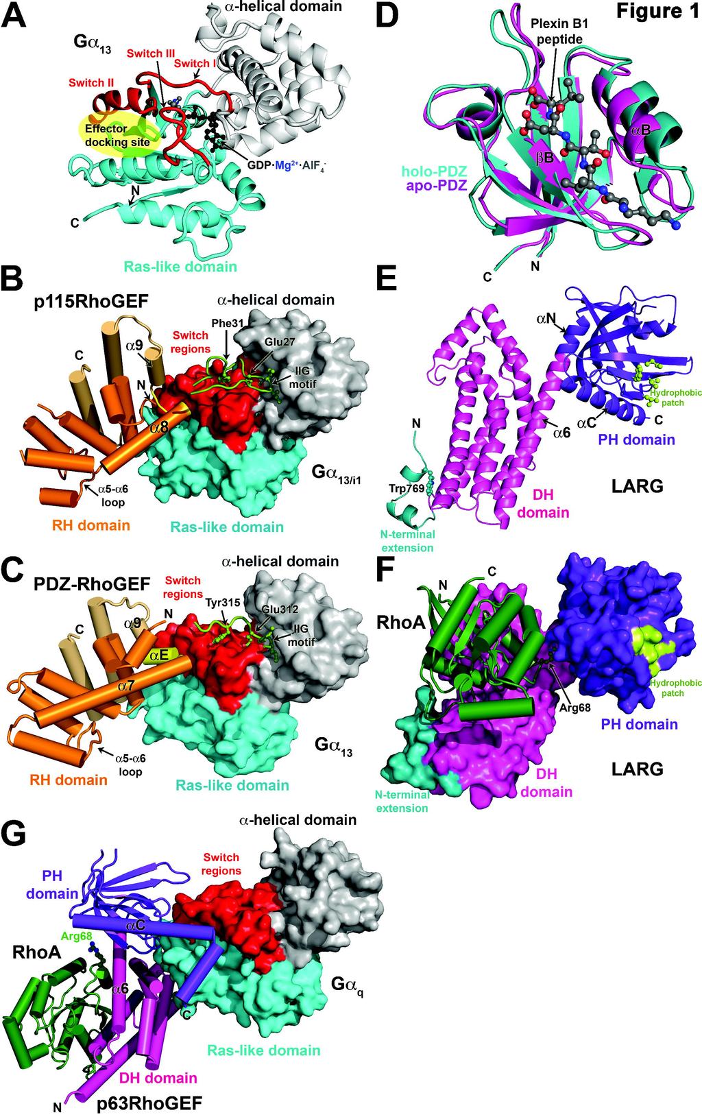
58
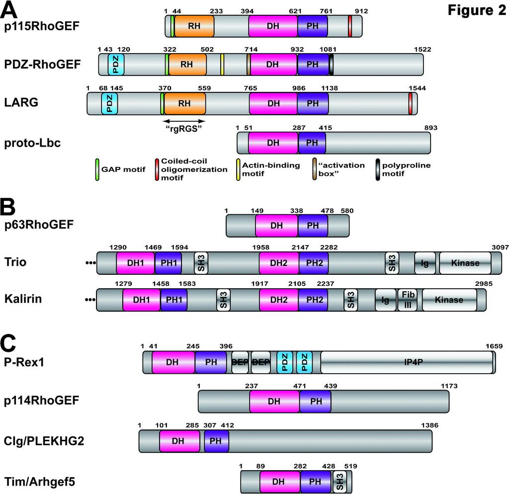
59

60
Signal Transduction Phosphorylation Protein kinases. Misfolding diseases. Protein Engineering Lysozyme variants
 Signal Transduction Phosphorylation Protein kinases Misfolding diseases Protein Engineering Lysozyme variants Cells and Signals Regulation The cell must be able to respond to stimuli Cellular activities
Heterotrimeric G proteins and the role of lipids in signaling. John Sondek, Ph.D. Depts. of Pharmacology and Biochemistry & Biophyscis
 Heterotrimeric G proteins and the role of lipids in signaling John Sondek, Ph.D. Depts. of Pharmacology and Biochemistry & Biophyscis The GTPase cycle molecular switch A GTPases is NOT a kinase Two major
5- Semaphorin-Plexin-Neuropilin
 5- Semaphorin-Plexin-Neuropilin 1 SEMAPHORINS-PLEXINS-NEUROPILINS ligands receptors co-receptors semaphorins and their receptors are known signals for: -axon guidance -cell migration -morphogenesis -immune
MOLECULAR DRUG TARGETS
 MOLECULAR DRUG TARGETS LEARNING OUTCOMES At the end of this session student shall be able to: List different types of druggable targets Describe forces involved in drug-receptor interactions Describe theories
Visual pigments. Neuroscience, Biochemistry Dr. Mamoun Ahram Third year, 2019
 Visual pigments Neuroscience, Biochemistry Dr. Mamoun Ahram Third year, 2019 References Webvision: The Organization of the Retina and Visual System (http://www.ncbi.nlm.nih.gov/books/nbk11522/#a 127) The
The EGF Signaling Pathway! Introduction! Introduction! Chem Lecture 10 Signal Transduction & Sensory Systems Part 3. EGF promotes cell growth
 Chem 452 - Lecture 10 Signal Transduction & Sensory Systems Part 3 Question of the Day: Who is the son of Sevenless? Introduction! Signal transduction involves the changing of a cell s metabolism or gene
Chem Lecture 10 Signal Transduction
 Chem 452 - Lecture 10 Signal Transduction 111202 Here we look at the movement of a signal from the outside of a cell to its inside, where it elicits changes within the cell. These changes are usually mediated
Regulation and signaling. Overview. Control of gene expression. Cells need to regulate the amounts of different proteins they express, depending on
 Regulation and signaling Overview Cells need to regulate the amounts of different proteins they express, depending on cell development (skin vs liver cell) cell stage environmental conditions (food, temperature,
Raghuram et al PNAS 100:9620
 ION CHANNL MACROMOLCULAR COMPLXS A V I R W c g I S L S R d L L LLL I VA S f L LS HP a b D G C S1 S2 S3 S4 S5 I e V N S6 COOH NH 2 PDZ domains; recognition of short peptides with a COOH terminal hydrophobic
Receptors and Ion Channels
 Receptors and Ion Channels Laurie Kellaway Senior Lecturer Department of Human Biology Laurie@curie.uct.ac.za Tel. +27 +21 4066 271 What are the two types of Neurotransmitter receptors Ionotropic receptors
GTPASE-ACTIVATING PROTEINS FOR HETEROTRIMERIC GPROTEINS: Regulators of G Protein Signaling (RGS) and RGS-Like Proteins
 and RGS-Like Proteins") Annu. Rev. Biochem. 2000. 69:795 827 Copyright c 2000 by Annual Reviews. All rights reserved GTPASE-ACTIVATING PROTEINS FOR HETEROTRIMERIC GPROTEINS: Regulators of G Protein Signaling (RGS) and RGS-Like
Annu. Rev. Biochem. 2000. 69:795 827 Copyright c 2000 by Annual Reviews. All rights reserved GTPASE-ACTIVATING PROTEINS FOR HETEROTRIMERIC GPROTEINS: Regulators of G Protein Signaling (RGS) and RGS-Like
Ligand screening system using fusion proteins of G protein coupled receptors with G protein α subunits
 2 Ligand screening system using fusion proteins of G protein coupled receptors with G protein α subunits G protein coupled receptors A key player of signaling transduction. Cell membranes are packed with
Reception The target cell s detection of a signal coming from outside the cell May Occur by: Direct connect Through signal molecules
 Why Do Cells Communicate? Regulation Cells need to control cellular processes In multicellular organism, cells signaling pathways coordinate the activities within individual cells that support the function
Gene regulation I Biochemistry 302. Bob Kelm February 25, 2005
 Gene regulation I Biochemistry 302 Bob Kelm February 25, 2005 Principles of gene regulation (cellular versus molecular level) Extracellular signals Chemical (e.g. hormones, growth factors) Environmental
Identification of Functional Amino Acids in the G Protein Alpha-Subunit
 The University of Maine DigitalCommons@UMaine University of Maine Office of Research and Sponsored Programs: Grant Reports Special Collections 7-31-2001 Identification of Functional Amino Acids in the
References on Kinetics and Mechanisms
 References on Kinetics and Mechanisms Excellent reference for all aspects of enzyme kinetics including important elements of Metabolic Control Analysis of relevance to systems analysis of enzyme function
Signal Transduction. Dr. Chaidir, Apt
 Signal Transduction Dr. Chaidir, Apt Background Complex unicellular organisms existed on Earth for approximately 2.5 billion years before the first multicellular organisms appeared.this long period for
Transmembrane Domains (TMDs) of ABC transporters
 of ABC transporters") Transmembrane Domains (TMDs) of ABC transporters Most ABC transporters contain heterodimeric TMDs (e.g. HisMQ, MalFG) TMDs show only limited sequence homology (high diversity) High degree of conservation
Transmembrane Domains (TMDs) of ABC transporters Most ABC transporters contain heterodimeric TMDs (e.g. HisMQ, MalFG) TMDs show only limited sequence homology (high diversity) High degree of conservation
Activation of a receptor. Assembly of the complex
 Activation of a receptor ligand inactive, monomeric active, dimeric When activated by growth factor binding, the growth factor receptor tyrosine kinase phosphorylates the neighboring receptor. Assembly
Transcription Regulation And Gene Expression in Eukaryotes UPSTREAM TRANSCRIPTION FACTORS
 Transcription Regulation And Gene Expression in Eukaryotes UPSTREAM TRANSCRIPTION FACTORS RG. Clerc March 26. 2008 UPSTREAM TRANSCRIPTION FACTORS Experimental approaches DNA binding domains (DBD) Transcription
Review. Membrane proteins. Membrane transport
 Quiz 1 For problem set 11 Q1, you need the equation for the average lateral distance transversed (s) of a molecule in the membrane with respect to the diffusion constant (D) and time (t). s = (4 D t) 1/2
RNA Synthesis and Processing
 RNA Synthesis and Processing Introduction Regulation of gene expression allows cells to adapt to environmental changes and is responsible for the distinct activities of the differentiated cell types that
Zool 3200: Cell Biology Exam 5 4/27/15
 Name: Trask Zool 3200: Cell Biology Exam 5 4/27/15 Answer each of the following short answer questions in the space provided, giving explanations when asked to do so. Circle the correct answer or answers
Analysis of Rho GTPase Signalling Pathways Regulating Epithelial Morphogenesis. Sean Wallace UCL
 Analysis of Rho GTPase Signalling Pathways Regulating Epithelial Morphogenesis Sean Wallace UCL A thesis submitted for the degree of Doctor of Philosophy (PhD) Declaration I, Sean Wallace, confirm that
Intercellular communication
 Intercellular communication Dewajani Purnomosari Department of Histology and Cell Biology Faculty of Medicine Universitas Gadjah Mada Outline General principle of intercellular communicabon Signal molecules
Visual pigments. Neuroscience, Biochemistry Dr. Mamoun Ahram Third year, 2015
 Visual pigments Neuroscience, Biochemistry Dr. Mamoun Ahram Third year, 2015 References Photoreceptors and visual pigments Webvision: The Organization of the Retina and Visual System (http://www.ncbi.nlm.nih.gov/books/nbk11522/#a127)
Lecture 10: Cyclins, cyclin kinases and cell division
 Chem*3560 Lecture 10: Cyclins, cyclin kinases and cell division The eukaryotic cell cycle Actively growing mammalian cells divide roughly every 24 hours, and follow a precise sequence of events know as
MAPK kinase kinase regulation of SAPK/JNK pathways
 MAPK kinase kinase regulation of SAPK/JNK pathways Lisa Stalheim and Gary L. Johnson Abstract SAPK/JNK members of the MAPK family are regulated by at least fourteen known MAPK kinase kinases (MKKKs). In
Biotechnology of Proteins. The Source of Stability in Proteins (III) Fall 2015
 Fall 2015") Biotechnology of Proteins The Source of Stability in Proteins (III) Fall 2015 Conformational Entropy of Unfolding It is The factor that makes the greatest contribution to stabilization of the unfolded
Biotechnology of Proteins The Source of Stability in Proteins (III) Fall 2015 Conformational Entropy of Unfolding It is The factor that makes the greatest contribution to stabilization of the unfolded
Supplementary Materials for
 advances.sciencemag.org/cgi/content/full/3/4/e1600663/dc1 Supplementary Materials for A dynamic hydrophobic core orchestrates allostery in protein kinases Jonggul Kim, Lalima G. Ahuja, Fa-An Chao, Youlin
Welcome to Class 21!
 Welcome to Class 21! Introductory Biochemistry! Lecture 21: Outline and Objectives l Regulation of Gene Expression in Prokaryotes! l transcriptional regulation! l principles! l lac operon! l trp attenuation!
Protein Structure. W. M. Grogan, Ph.D. OBJECTIVES
 Protein Structure W. M. Grogan, Ph.D. OBJECTIVES 1. Describe the structure and characteristic properties of typical proteins. 2. List and describe the four levels of structure found in proteins. 3. Relate
Analysis of correlated mutations in Ras G-domain
 www.bioinformation.net Volume 13(6) Hypothesis Analysis of correlated mutations in Ras G-domain Ekta Pathak * Bioinformatics Department, MMV, Banaras Hindu University. Ekta Pathak - E-mail: ektavpathak@gmail.com;
targets. clustering show that different complex pathway
 Supplementary Figure 1. CLICR allows clustering and activation of cytoplasmic protein targets. (a, b) Upon light activation, the Cry2 (red) and LRP6c (green) components co-cluster due to the heterodimeric
Biol403 - Receptor Serine/Threonine Kinases
 Biol403 - Receptor Serine/Threonine Kinases The TGFβ (transforming growth factorβ) family of growth factors TGFβ1 was first identified as a transforming factor; however, it is a member of a family of structurally
Chapter 16. Cellular Movement: Motility and Contractility. Lectures by Kathleen Fitzpatrick Simon Fraser University Pearson Education, Inc.
 Chapter 16 Cellular Movement: Motility and Contractility Lectures by Kathleen Fitzpatrick Simon Fraser University Two eukaryotic motility systems 1. Interactions between motor proteins and microtubules
Gene Control Mechanisms at Transcription and Translation Levels
 Gene Control Mechanisms at Transcription and Translation Levels Dr. M. Vijayalakshmi School of Chemical and Biotechnology SASTRA University Joint Initiative of IITs and IISc Funded by MHRD Page 1 of 9
1. Ras-Interactors from the RASOMICS database
 Transformation by ras oncogenes induces the deregulation of intracellular signalling cascades that are critical elements in cell growth control. Ras genes code for small GTPases that act as GDP/ GTP-regulated
7.013 Problem Set
 7.013 Problem Set 5-2013 Question 1 During a summer hike you suddenly spot a huge grizzly bear. This emergency situation triggers a fight or flight response through a signaling pathway as shown below.
JBC Papers in Press. Published on August 13, 2018 as Manuscript RA
 JBC Papers in Press. Published on August 13, 2018 as Manuscript RA118.003290 The latest version is at http://www.jbc.org/cgi/doi/10.1074/jbc.ra118.003290 Bond swapping from a charge cloud allows flexible
Introduction to Comparative Protein Modeling. Chapter 4 Part I
 Introduction to Comparative Protein Modeling Chapter 4 Part I 1 Information on Proteins Each modeling study depends on the quality of the known experimental data. Basis of the model Search in the literature
Nature Structural & Molecular Biology: doi: /nsmb Supplementary Figure 1
 Supplementary Figure 1 Crystallization. a, Crystallization constructs of the ET B receptor are shown, with all of the modifications to the human wild-type the ET B receptor indicated. Residues interacting
SUPPLEMENTARY INFORMATION
 doi:10.1038/nature11085 Supplementary Tables: Supplementary Table 1. Summary of crystallographic and structure refinement data Structure BRIL-NOP receptor Data collection Number of crystals 23 Space group
of the Guanine Nucleotide Exchange Factor FARP2
 Structure, Volume 21 Supplemental Information Structural Basis for Autoinhibition of the Guanine Nucleotide Exchange Factor FARP2 Xiaojing He, Yi-Chun Kuo, Tyler J. Rosche, and Xuewu Zhang Inventory of
SUPPLEMENTARY INFORMATION
 doi:10.1038/nature17991 Supplementary Discussion Structural comparison with E. coli EmrE The DMT superfamily includes a wide variety of transporters with 4-10 TM segments 1. Since the subfamilies of the
Mechanisms of Cell Proliferation
 Mechanisms of Cell Proliferation Cell Cycle G 2 S G 1 Multi-cellular organisms depend on cell division/proliferation; Each organism has a developmental plan that determines its behavior and properties;
Initiation of translation in eukaryotic cells:connecting the head and tail
 Initiation of translation in eukaryotic cells:connecting the head and tail GCCRCCAUGG 1: Multiple initiation factors with distinct biochemical roles (linking, tethering, recruiting, and scanning) 2: 5
RANK. Alternative names. Discovery. Structure. William J. Boyle* SUMMARY BACKGROUND
 RANK William J. Boyle* Department of Cell Biology, Amgen, Inc., One Amgen Center Drive, Thousand Oaks, CA 91320-1799, USA * corresponding author tel: 805-447-4304, fax: 805-447-1982, e-mail: bboyle@amgen.com
1. Introduction. 1. Introduction. 1.1 Signal transduction
 1. Introduction 1.1 Signal transduction During the evolution of multicellular organisms, highly complex and integrative communication networks have evolved to coordinate growth, differentiation and metabolism
Introduction. Gene expression is the combined process of :
 1 To know and explain: Regulation of Bacterial Gene Expression Constitutive ( house keeping) vs. Controllable genes OPERON structure and its role in gene regulation Regulation of Eukaryotic Gene Expression
Drug interactions. (Efficacy is a measure of the size of response produced by receptor activation)
") Drug interactions Receptor A signal transducer, modifying cell function in response to an extracellular signal. - Membrane proteins e.g. AChR, - Cytoplasmic proteins e.g. steroid receptors Drugs usually
Drug interactions Receptor A signal transducer, modifying cell function in response to an extracellular signal. - Membrane proteins e.g. AChR, - Cytoplasmic proteins e.g. steroid receptors Drugs usually
Study Guide 11 & 12 MULTIPLE CHOICE. Choose the one alternative that best completes the statement or answers the question.
 Study Guide 11 & 12 MULTIPLE CHOICE. Choose the one alternative that best completes the statement or answers the question. 1) The receptors for a group of signaling molecules known as growth factors are
Gene regulation II Biochemistry 302. February 27, 2006
 Gene regulation II Biochemistry 302 February 27, 2006 Molecular basis of inhibition of RNAP by Lac repressor 35 promoter site 10 promoter site CRP/DNA complex 60 Lewis, M. et al. (1996) Science 271:1247
1. The plasma membrane of eukaryotic cells is supported by a. actin filaments. b. microtubules. c. lamins. d. intermediate filaments.
 ANALYSIS AND MODELING OF CELL MECHANICS Homework #2 (due 1/30/13) This homework involves comprehension of key biomechanical concepts of the cytoskeleton, cell-matrix adhesions, and cellcell adhesions.
Mechanisms of Cell Proliferation
 Mechanisms of Cell Proliferation Cell Cycle G 2 S G 1 Multi-cellular organisms depend on cell division/proliferation; Each organism has a developmental plan that determines its behavior and properties;
13-3. Synthesis-Secretory pathway: Sort lumenal proteins, Secrete proteins, Sort membrane proteins
 13-3. Synthesis-Secretory pathway: Sort lumenal proteins, Secrete proteins, Sort membrane proteins Molecular sorting: specific budding, vesicular transport, fusion 1. Why is this important? A. Form and
CHAPTER 1 THE STRUCTURAL BIOLOGY OF THE FGF19 SUBFAMILY
 CHAPTER 1 THE STRUCTURAL BIOLOGY OF THE FGF19 SUBFAMILY Andrew Beenken and Moosa Mohammadi* Department of Pharmacology, New York University School of Medicine, New York, New York, USA. *Corresponding Author:
Ras e la Cascata delle Piccole GTPasi
 Roma 26-27 Giugno 2015 Multidisciplinarietà e Biologia Molecolare applicate alla pratica Clinica i Oncologica: un'opportunità ed un vantaggio per tuttitti Ras e la Cascata delle Piccole GTPasi Alvaro Leone
What binds to Hb in addition to O 2?
 Reading: Ch5; 158-169, 162-166, 169-174 Problems: Ch5 (text); 3,7,8,10 Ch5 (study guide-facts); 1,2,3,4,5,8 Ch5 (study guide-apply); 2,3 Remember Today at 5:30 in CAS-522 is the second chance for the MB
Advanced Higher Biology. Unit 1- Cells and Proteins 2c) Membrane Proteins
 Membrane Proteins") Advanced Higher Biology Unit 1- Cells and Proteins 2c) Membrane Proteins Membrane Structure Phospholipid bilayer Transmembrane protein Integral protein Movement of Molecules Across Membranes Phospholipid
Advanced Higher Biology Unit 1- Cells and Proteins 2c) Membrane Proteins Membrane Structure Phospholipid bilayer Transmembrane protein Integral protein Movement of Molecules Across Membranes Phospholipid
Supplemental data for
 Supplemental data for A Real-Time Guanine Nucleotide Exchange Assay using NMR: Activation of RhoA by PDZ- RhoGEF. Geneviève M.C. Gasmi-Seabrook 1,3, Christopher B. Marshall 1,3, Melissa Cheung 1,3, Bryan
Examination paper for Bi3016 Molecular Cell Biology
 Department of Biology Examination paper for Bi3016 Molecular Cell Biology Academic contact during examination: Per Winge Phone: 99369359 Examination date: 20 th December 2017 Examination time (from-to):
Supplementary Figure 1 Schematic overview of ASTNs in neuronal migration. (a) Schematic of roles played by ASTNs 1 and 2. ASTN-1-mediated adhesions
 Schematic of roles played by ASTNs 1 and 2. ASTN-1-mediated adhesions") Supplementary Figure 1 Schematic overview of ASTNs in neuronal migration. (a) Schematic of roles played by ASTNs 1 and 2. ASTN-1-mediated adhesions undergo endocytosis into clathrin-coated vesicles dependent
Supplementary Figure 1 Schematic overview of ASTNs in neuronal migration. (a) Schematic of roles played by ASTNs 1 and 2. ASTN-1-mediated adhesions undergo endocytosis into clathrin-coated vesicles dependent
Abstract. Author Summary. Yougan Cheng 1, Hans Othmer 1,* 1 School of Mathematics, University of Minnesota, Minneapolis, MN, USA. *
 A Model for Direction Sensing in Dictyostelium Discoideum: Ras Activity and Symmetry Breaking Driven by a G βγ - Mediated, G α2 -Ric8 Dependent Signal Transduction Network - Submission to PLOS Computational
Oligomerization of DH Domain Is Essential for Dbl-Induced Transformation
 MOLECULAR AND CELLULAR BIOLOGY, Jan. 2001, p. 425 437 Vol. 21, No. 2 0270-7306/01/$04.00 0 DOI: 10.1128/MCB.21.2.425 437.2001 Copyright 2001, American Society for Microbiology. All Rights Reserved. Oligomerization
Cell-Cell Communication in Development
 Biology 4361 - Developmental Biology Cell-Cell Communication in Development October 2, 2007 Cell-Cell Communication - Topics Induction and competence Paracrine factors inducer molecules Signal transduction
The molecular genetics of chemotaxis: sensing and responding to chemoattractant gradients
 The molecular genetics of chemotaxis: sensing and responding to chemoattractant gradients Richard A. Firtel* and Chang Y. Chung Summary Chemotaxis plays a central role in various biological processes,
Conclusions. The experimental studies presented in this thesis provide the first molecular insights
 C h a p t e r 5 Conclusions 5.1 Summary The experimental studies presented in this thesis provide the first molecular insights into the cellular processes of assembly, and aggregation of neural crest and
Neurite initiation. Neurite formation begins with a bud that sprouts from the cell body. One or several neurites can sprout at a time.
 Neurite initiation. Neuronal maturation initiation f-actin polarization and maturation tubulin stage 1: "spherical" neuron stage 2: neurons extend several neurites stage 3: one neurite accelerates its
ADAM FAMILY. ephrin A INTERAZIONE. Eph ADESIONE? PROTEOLISI ENDOCITOSI B A RISULTATO REPULSIONE. reverse. forward
 ADAM FAMILY - a family of membrane-anchored metalloproteases that are known as A Disintegrin And Metalloprotease proteins and are key components in protein ectodomain shedding Eph A INTERAZIONE B ephrin
Cell Death & Trophic Factors II. Steven McLoon Department of Neuroscience University of Minnesota
 Cell Death & Trophic Factors II Steven McLoon Department of Neuroscience University of Minnesota 1 Remember? Neurotrophins are cell survival factors that neurons get from their target cells! There is a
Molecular Cell Biology 5068 In Class Exam 2 November 8, 2016
 Molecular Cell Biology 5068 In Class Exam 2 November 8, 2016 Exam Number: Please print your name: Instructions: Please write only on these pages, in the spaces allotted and not on the back. Write your
MBLG lecture 5. The EGG! Visualising Molecules. Dr. Dale Hancock Lab 715
 MBLG lecture 5 Dr. Dale Hancock D.Hancock@mmb.usyd.edu.au Lab 715 The EGG! Visualising Molecules In molecular biology and biochemistry it is better to view molecules as killer pythons rather than smarties.
β1 Structure Prediction and Validation
 13 Chapter 2 β1 Structure Prediction and Validation 2.1 Overview Over several years, GPCR prediction methods in the Goddard lab have evolved to keep pace with the changing field of GPCR structure. Despite
Supplementary Figure 1. Aligned sequences of yeast IDH1 (top) and IDH2 (bottom) with isocitrate
 and IDH2 (bottom) with isocitrate") SUPPLEMENTARY FIGURE LEGENDS Supplementary Figure 1. Aligned sequences of yeast IDH1 (top) and IDH2 (bottom) with isocitrate dehydrogenase from Escherichia coli [ICD, pdb 1PB1, Mesecar, A. D., and Koshland,
SUPPLEMENTARY FIGURE LEGENDS Supplementary Figure 1. Aligned sequences of yeast IDH1 (top) and IDH2 (bottom) with isocitrate dehydrogenase from Escherichia coli [ICD, pdb 1PB1, Mesecar, A. D., and Koshland,
CHAPTER 13 PROKARYOTE GENES: E. COLI LAC OPERON
 PROKARYOTE GENES: E. COLI LAC OPERON CHAPTER 13 CHAPTER 13 PROKARYOTE GENES: E. COLI LAC OPERON Figure 1. Electron micrograph of growing E. coli. Some show the constriction at the location where daughter
COMPUTER SIMULATION OF DIFFERENTIAL KINETICS OF MAPK ACTIVATION UPON EGF RECEPTOR OVEREXPRESSION
 COMPUTER SIMULATION OF DIFFERENTIAL KINETICS OF MAPK ACTIVATION UPON EGF RECEPTOR OVEREXPRESSION I. Aksan 1, M. Sen 2, M. K. Araz 3, and M. L. Kurnaz 3 1 School of Biological Sciences, University of Manchester,
Ribosome readthrough
 Ribosome readthrough Starting from the base PROTEIN SYNTHESIS Eukaryotic translation can be divided into four stages: Initiation, Elongation, Termination and Recycling During translation, the ribosome
Interpreting and evaluating biological NMR in the literature. Worksheet 1
 Interpreting and evaluating biological NMR in the literature Worksheet 1 1D NMR spectra Application of RF pulses of specified lengths and frequencies can make certain nuclei detectable We can selectively
Richik N. Ghosh, Linnette Grove, and Oleg Lapets ASSAY and Drug Development Technologies 2004, 2:
 1 3/1/2005 A Quantitative Cell-Based High-Content Screening Assay for the Epidermal Growth Factor Receptor-Specific Activation of Mitogen-Activated Protein Kinase Richik N. Ghosh, Linnette Grove, and Oleg
G protein coupled receptors Structure, function, regulation
 G protein coupled receptors Structure, function, regulation Im menschlichen Genom finden sich Gene für ~ 810 G-Protein-gekoppelte Rezeptoren 350 nicht-geruchbildende Rezeptoren 460 Geruchrezeptoren Vielfalt
Supplemental table S7.
 Supplemental table S7. GO terms significantly enriched in significantly up-regulated genes of the microarray. K: number of genes from the input cluster in the given category. F: number of total genes in
Cellular Neuroanatomy I The Prototypical Neuron: Soma. Reading: BCP Chapter 2
 Cellular Neuroanatomy I The Prototypical Neuron: Soma Reading: BCP Chapter 2 Functional Unit of the Nervous System The functional unit of the nervous system is the neuron. Neurons are cells specialized
Ion Channel Structure and Function (part 1)
") Ion Channel Structure and Function (part 1) The most important properties of an ion channel Intrinsic properties of the channel (Selectivity and Mode of Gating) + Location Physiological Function Types
Ion Channel Structure and Function (part 1) The most important properties of an ion channel Intrinsic properties of the channel (Selectivity and Mode of Gating) + Location Physiological Function Types
arxiv: v1 [q-bio.cb] 31 Dec 2015
![arxiv: v1 [q-bio.cb] 31 Dec 2015](/thumbs/77/76028420.jpg "arxiv: v1 [q-bio.cb] 31 Dec 2015") A Model for Direction Sensing in Dictyostelium Discoideum: Ras Activity and Symmetry Breaking Driven by a G βγ - Mediated, G α2 -Ric8 Dependent Signal Transduction Network arxiv:52.09233v [q-bio.cb] 3
A Model for Direction Sensing in Dictyostelium Discoideum: Ras Activity and Symmetry Breaking Driven by a G βγ - Mediated, G α2 -Ric8 Dependent Signal Transduction Network arxiv:52.09233v [q-bio.cb] 3
Major Types of Association of Proteins with Cell Membranes. From Alberts et al
 Major Types of Association of Proteins with Cell Membranes From Alberts et al Proteins Are Polymers of Amino Acids Peptide Bond Formation Amino Acid central carbon atom to which are attached amino group
Table 1. Kinetic data obtained from SPR analysis of domain 11 mutants interacting with IGF-II. Kinetic parameters K D 1.
 Kinetics and Thermodynamics of the Insulin-like Growth Factor II (IGF-II) Interaction with IGF-II/Mannose 6-phosphate Receptor and the function of CD and AB Loop Solvent-exposed Residues. Research Team:
Signaling to the Nucleus by an L-type Calcium Channel- Calmodulin Complex Through the MAP Kinase Pathway
 Signaling to the Nucleus by an L-type Calcium Channel- Calmodulin Complex Through the MAP Kinase Pathway Ricardo E. Dolmetsch, Urvi Pajvani, Katherine Fife, James M. Spotts, Michael E. Greenberg Science
According to the diagram, which of the following is NOT true?
 Instructions: Review Chapter 44 on muscular-skeletal systems and locomotion, and then complete the following Blackboard activity. This activity will introduce topics that will be covered in the next few
Receptor Based Drug Design (1)
") Induced Fit Model For more than 100 years, the behaviour of enzymes had been explained by the "lock-and-key" mechanism developed by pioneering German chemist Emil Fischer. Fischer thought that the chemicals
Induced Fit Model For more than 100 years, the behaviour of enzymes had been explained by the "lock-and-key" mechanism developed by pioneering German chemist Emil Fischer. Fischer thought that the chemicals
Amneh Auben. Abdulrahman Jabr. Diala Abu-Hassan
 21 Amneh Auben Abdulrahman Jabr Diala Abu-Hassan Matrix polysaccharides Extracellular matrix (ECM): It s a collection of components that fills the spaces outside the cell or between the cells. ---------
with%dr.%van%buskirk%%%
 with%dr.%van%buskirk%%% How$to$do$well?$ Before$class:$read$the$corresponding$chapter$ Come$to$class$ready$to$par9cipate$in$Top$Hat$ Don t$miss$an$exam!!!!!!!!!!!!!!!!!!!!!!!!!!$ But$I m$not$good$with$science
BIRKBECK COLLEGE (University of London)
") BIRKBECK COLLEGE (University of London) SCHOOL OF BIOLOGICAL SCIENCES M.Sc. EXAMINATION FOR INTERNAL STUDENTS ON: Postgraduate Certificate in Principles of Protein Structure MSc Structural Molecular Biology
BIRKBECK COLLEGE (University of London) SCHOOL OF BIOLOGICAL SCIENCES M.Sc. EXAMINATION FOR INTERNAL STUDENTS ON: Postgraduate Certificate in Principles of Protein Structure MSc Structural Molecular Biology
Bypass and interaction suppressors; pathway analysis
 Bypass and interaction suppressors; pathway analysis The isolation of extragenic suppressors is a powerful tool for identifying genes that encode proteins that function in the same process as a gene of
William D. Singer, Ph.D.
 William D. Singer, Ph.D. billsinger@dcccd.edu EDUCATION AND TRAINING University of Texas Southwestern Medical Center; Dallas, Texas 1991-1995 ~ Postdoctoral Studies, Cellular Signal Transduction University
16 The Cell Cycle. Chapter Outline The Eukaryotic Cell Cycle Regulators of Cell Cycle Progression The Events of M Phase Meiosis and Fertilization
 The Cell Cycle 16 The Cell Cycle Chapter Outline The Eukaryotic Cell Cycle Regulators of Cell Cycle Progression The Events of M Phase Meiosis and Fertilization Introduction Self-reproduction is perhaps
Chapter 16 Lecture. Concepts Of Genetics. Tenth Edition. Regulation of Gene Expression in Prokaryotes
 Chapter 16 Lecture Concepts Of Genetics Tenth Edition Regulation of Gene Expression in Prokaryotes Chapter Contents 16.1 Prokaryotes Regulate Gene Expression in Response to Environmental Conditions 16.2
CELB40060 Membrane Trafficking in Animal Cells. Prof. Jeremy C. Simpson. Lecture 2 COPII and export from the ER
 CELB40060 Membrane Trafficking in Animal Cells Prof. Jeremy C. Simpson Lecture 2 COPII and export from the ER Today s lecture... The COPII coat - localisation and subunits Formation of the COPII coat at
Computational modeling of G-Protein Coupled Receptors (GPCRs) has recently become
 has recently become") Homology Modeling and Docking of Melatonin Receptors Andrew Kohlway, UMBC Jeffry D. Madura, Duquesne University 6/18/04 INTRODUCTION Computational modeling of G-Protein Coupled Receptors (GPCRs) has recently
Homology Modeling and Docking of Melatonin Receptors Andrew Kohlway, UMBC Jeffry D. Madura, Duquesne University 6/18/04 INTRODUCTION Computational modeling of G-Protein Coupled Receptors (GPCRs) has recently
Nervous System Organization
 The Nervous System Nervous System Organization Receptors respond to stimuli Sensory receptors detect the stimulus Motor effectors respond to stimulus Nervous system divisions Central nervous system Command