Role of Cadherin-7 in cranial neural crest development and its potential heterotypic interaction with N-cadherin in trigeminal ganglion formation
|
|
|
- Roxanne Scarlett Williams
- 5 years ago
- Views:
Transcription
1 C h a p t e r 4 Role of Cadherin-7 in cranial neural crest development and its potential heterotypic interaction with N-cadherin in trigeminal ganglion formation 4.1 Abstract Cadherins are transmembrane molecules best known for mediating intercellular adhesion through homophilic binding of identical cadherins. A widely held view is that expression of different cadherins drives selective intercellular adhesion that is crucial for tissue segregation and boundary formation. This is largely based on their dynamic spatiotemporal expression patterns during morphogenesis, as well as results from in vitro cell aggregation assays showing segregation of cells expressing different cadherins. However, cadherin interaction is likely more broad. Several recent studies have shown that cadherins of different subtypes can interact with each other in vitro indistinguishably from homophilic binding. This idea remains to be explored in vivo. Here, departing from the conventional model, we test the possibility that interactions between neural crest and ectodermal placodes during trigeminal ganglion formation in the chick may rely on both heterotypic and homotypic interactions of at least two different cadherins. We have shown in Chapter 3 that N-cadherin is expressed by placodal neurons during condensation of cranial ganglia and is critical for proper gangliogenesis. Because of their dual origin, coalescence of trigeminal ganglion may involve crest crest, crest placode, and/or placode placode adhesion. However, the molecular mechanism mediating neural crest placode condensation remains to be explored. To test the intriguing poassibility that interactions between Cadherin-7, which is expressed by migrating neural crest cells (Nakagawa and Takeichi, 1995), and N-cadherin on placodal neurons, may be involved in
2 ganglion formation, we first examined the protein expression and function of Cadherin-7 relative to N-cadherin. The results show that neural crest cells express the Cadherin-7 protein while they are intermixing with placodal neurons expressing N-cadherin in the forming ganglion. Moreover, perturbation of Cadherin-7 affects cell morphology and cell cell interactions during migration and later leads to malformed cranial ganglia. In contrast, the overall migration stream pattern of cranial neural crest appeared unaffected. Misexpression of Cadherin-7 in the placodal ectoderm severely interrupts normal placodal morphogenesis and causes an increase in neuronal cells in the ectoderm. Our results suggest the possibility that both heterophilic and homophilic cadherin mediated adhesion drive coalescence of cranial sensory ganglia, and that there are distinct functions of Cadherin-7 contributing to neural crest cell migration versus cranial ganglia formation. 4.2 Introduction The expression of classical (type I and type II) cadherins during vertebrate development is tightly regulated and coincides with crucial morphogenetic events, such as gastrulation, neurulation, nephrogenesis, neural crest emigration as well as in pathological cases such as cancer (Pla et al., 2001; Taneyhill, 2008; Wheelock et al., 2008). In these examples, the switching of expression of one cadherin subtype to another appears to match changes in tissue adhesive properties. For example, as neural crest cells undergo an epithelial-mesenchymal transition, acquire motility and exit the neural tube, they shift from expressing N-cadherin to Cadherin-6b in the chick dorsal neural tube and then to Cadherin 7 in migrating neural crest cells. Cadherins are thought to primarily mediate selective adhesion between cells expressing the same cadherin subtype. Evidence to this effect
3 comes from experiments showing that distinct cells expressing different cadherin segregate from one another and form distinct aggregates (Nose et al., 1988), suggesting that homophilic binding of cadherins drives tissue segregation and maintains tissue integrity (Takeichi, 1990). More recently, however, in vitro cell binding and aggregation assays have shown that heterophilic interaction can also occur. Cells expressing different classical cadherins either of type I or type II can heterotypically bind and intermix to form aggregates indistinguishable from homotypic interaction (Duguay et al., 2003; Shimoyama et al., 2000). Consistent with this, cadherin bond strengths by force measurements show similar adhesion between heterotypic and homotypic interactions (Prakasam et al., 2006). However, there is little information regarding whether classical type I and type II cadherins interact. A study on the protein structure of the ectodomain of type II suggests that type I and type II are overall very similar but have distinct adhesive interfaces at the EC1 domain type I has one conserved tryptophan residue (W2) versus two conserved tryptophan residues (W2 and W4) and a larger hydrophobic pocket in type II that are thought to preclude their binding (Patel et al., 2006). However, this remains unexplored in vivo. We have previously shown that N-cadherin plays an important role during aggregation of placode-derived neurons into ganglia. However, the mechanism underlying coalescence of their partner cells, the neural crest, is not known. Here, we investigate Cadherin-7 as a possible candidate for mediating neural crest assembly, since it is expressed in the migrating cranial neural crest (Nakagawa and Takeichi, 1995). We find that neural crest cells express the Cadherin-7 protein in a complementary pattern to N- cadherin on placodal neurons, raising the intriguing possibility for interactions between two distinct classes of cadherins (N-cadherin, a type I, and Cadherin-7, a type II) during trigeminal gangliogenesis. Perturbation of Cadherin-7 affects crest crest interactions and morphology, later resulting in disfigurement of the ganglion. The results provide a basis for
4 testing the possibility of a novel heterophilic interaction between classical type I and type II cadherins in vivo. The interaction between neural crest and placodes serves as an excellent new model to examine the dynamic role of cadherins in cell cell interactions during cellular coalescence in vivo. 4.3 Materials and methods Embryos Fertilized chicken (Gallus gallus domesticus) eggs were obtained from local commercial sources and incubated at 37 C to the desired stages according to the criteria of Hamburger and Hamilton (Hamburger and Hamilton, 1992). In situ hybridization Whole mount chick in situ hybridization was performed as described (Shiau et al., 2008). cdna plasmids obtained from BBSRC (ChickEST clones 445e6 and 854n16) were used to transcribe antisense riboprobes against chick Cadherin-7. The plasmids were sequenced and determined to contain the coding sequence of the chick Cadherin-7 gene (NCBI accession number: NM_ ) corresponding to nucleotides (445e6 clone) and (854n16 clone). Both riboprobes gave the same expression patterns. Embryos were imaged and subsequently sectioned at 12 µm.
5 Immunohistochemistry Whole chick embryos were fixed in 4% paraformaldehyde overnight at 4ºC, washed in PBT (PBS + 0.2% tween) and either immunostained as whole embryos and/or processed for 10 µm cryostat sections. Primary antibodies used were anti-n-cadherin (DSHB, MNCD2 clone; 1:1), anti-gfp (Molecular Probes; 1:1000 to 1:2500), anti-hnk-1 (American Type Culture; 1:3 or 1:5), anti-cadherin-7 (DSHB, clone CCD7-1; 1:1), and anti-tuj1 (Covance; 1:250). Secondary antibodies: cyanine 2 or rhodamine red-x conjugated donkey anti-rat IgG (Jackson ImmunoResearch) used at 1:1000 and all others were obtained from Molecular Probes and used at 1:1000 or 1:2000 dilutions (except 1:250 dilution for Alexa Flour 350 conjugated antibodies). Images were taken using the AxioVision software from a Zeiss Axioskop2 plus fluorescence microscope, and processed using Adobe Photoshop CS3. In ovo electroporation of cranial neural crest and the trigeminal ectoderm For the presumptive cranial neural crest, DNA injection was targeted in the neural folds at stage 8 or presumptive neural crest region at gastrula stages 4 6 in ovo. Platinum electrodes were placed horizontally across the neural folds at stage 8 and electrical pulses of 2 25 V in 50 ms at 100 ms intervals were delivered. At stages 4 6, the electrodes were placed vertically across the epiblast and V in 50 ms at 100 ms pulses were delivered. For stages 4 6 electroporation, two points of special care were used: first, eggs were taken out of the incubator individually, manipulated, and immediately placed back into the incubator to minimize the time they sat at room temperature, and second the amount of india ink used to visualize the embryo was minimized (just enough to faintly depict the embryo). For the trigeminal ectoderm, DNA was injected overlying the presumptive trigeminal placodal ectoderm at stages 8 10 by air pressure using a glass micropipette. Platinum electrodes were placed vertically across the chick embryo
6 delivering 5 8 V in 50 ms at 100 ms intervals current pulses. Electroporated eggs were resealed and re-incubated at 37 C to reach the desired stages. Plasmid constructs FL-Cad7-cytopcig expression vector was cloned from excising the full-length coding sequence plus the FLAG epitope at BsiWI (later blunted) and NotI sites from pcmvc7/flag-pa, a vector derived from (Nakagawa and Takeichi, 1998). This fragment was inserted into the pbs II KS cloning vector (Stratagene) at EcoRV and NotI sites. Fulllength Cadherin-7 fragment was then cut out at NotI (later blunted) and also at ClaI; this was inserted into the cyto-pcig vector (Shiau et al., 2008) at ClaI/EcoRV (where the EcorRV site is disrupted). FL-cad7 (clone C1) was determined to be correct by sequencing. EC-Cad7-cytopcig encodes the extracellular Cadherin-7 protein up to the pretransmembrane domain, corresponding to nucleotides of the chick Cadherin-7 mrna (accession: NM_ ). This fragment was cloned by PCR from the original vector pcmv-c7/flag-pa into the PCRII-TOPO vector. Clones were sequenced and the correct clone (TC-4) was used for the subsequent steps. The 5 to 3 sequence of the PCR primers were: (cd7-f) ggaaaaaaaagatgaagttgggc and (dncd7-r) tcatcaattatggttatttgctgc. As an intermediate step, the fragment was cut at EcoRV/BamHI and ligated into the pbs II KS vector at the same sites. Lastly, the EC-Cad7 fragment was excised at BamHI (later blunted) and then at ClaI, and was then inserted into cyto-pcig at ClaI/EcoRV (where the EcorRV site is disrupted). EC-cad7 or tn-cad7 (clone n9) was determined to be correct by sequencing.
7 4.4 Results and discussions Cadherin-7 mrna is expressed in migrating cranial neural crest cells and later restricted to dorsal regions of cranial ganglia We characterized the mrna expression of Cadherin-7 in chick embryos from stages of early cranial neural crest migration to ganglion formation (stages 10 18). By whole mount in situ hybridization, we found that cranial neural crest cells begin to express Cadherin-7 only after they have left the neural tube at all axial levels in the head. By stage 16 when the ganglia have begun coalescing, the mrna expression is mostly restricted to those neural crest cells occupying the dorsal portion of the ganglia (adjacent to the hindbrain) (Fig. 1, A-E). This dorsal expression of Cadherin-7 at exit points of cranial motor axons which correspond to the even-numbered rhombomeres (r2, r4, and r6) has been reported previously; these sites also represent the entry points where sensory axons from the cranial ganglia enter the hindbrain (Nakagawa and Takeichi, 1995; Niederlander and Lumsden, 1996; Osborne et al., 2005). Neural crest cells migrate in well-characterized streams along almost the entire length of the body axis in a rostrocaudal progression. The first migrating neural crest cells emerging from the midbrain region express Cadherin-7 at stage 10, as shown in Fig. 1A. Interestingly, even at stage 10, the expression of Cadherin-7 mrna appears to be downregulated in the most distally located cells that have migrated furthest. Cranial neural crest cells migrate in three streams along the rostrocaudal cranial neuraxis: first (midbrain and r1/r2), second (r4), and third (r6) (Fig. 1, F-M). Cadherin-7 expression appears to be increasingly fainter in migrating neural crest cells as migration progresses. For example, most cells in the first stream express Cadherin-7 at the beginning of migration at stage 10 with the exception of the most ventrally migrating cells (Fig. 1F,G). Toward the end of
8 their migration at stage 13, most neural crest cells have downregulated Cadherin-7 mrna except for those in the dorsal region near the trigeminal motor exit point (Fig. 1, K-M). This expression pattern further refines and becomes smaller after migration and during ganglion coalescence at stages (Fig. 1D,E). Cadherin-7 expression is also detected weakly in the ventrolateral neural tube and in some parts of the surface ectoderm, including the otic placode, but is not in surface ectoderm or placode-derived cells during ganglion formation (Fig. 1G, J). The results suggest that the transcription of Cadherin-7 is dynamically regulated during neural crest migration, such that neural crest cells that have migrated more ventrally downregulate Cadherin-7, whereas late migrating cells that reside near cranial motor exit points retain expression. This contrasts with the idea initially proposed by Nakagawa and Takiechi (1995) that Cadherin-7 is expressed by a distinct subpopulation of neural crest cells that segregate and sequester near the exit points. Our interpretation is supported by in vivo time-lapse imaging that shows that cranial neural crest cells undergo progressive spatially-ordered cell migration. They first leave the dorsal neural tube and then move laterally beneath the surface ectoderm, and thus directionally away from the neural tube. There is no indication of early segregation or clustering of cells near the exit points (Kulesa and Fraser, 2000; Kulesa et al., 2008). Furthermore, previous studies have also shown that late- emigrating neural crest cells remain closer to the dorsal portion of the embryo, whereas early- emigrating cells occupy and give rise to more ventral structures (Baker et al., 1997; Niederlander and Lumsden, 1996).
9 4.4.2 Cadherin-7 protein is similar but broader than the mrna and complementary to N-cadherin expression in the trigeminal ganglion Since mrna expression does not necessarily reflect that of protein which is more directly indicative of function, we examined the expression pattern of Cadherin-7 protein at times of cranial neural crest migration (stage 11) and ganglion condensation (stage 18) (Fig. 2). Surprisingly, Cadherin-7 protein was expressed more broadly than its mrna in the first migratory neural crest stream (Fig. 2A) and during condensation into the trigeminal ganglion (Fig. 2D). During migration, the vast majority of neural crest cells express Cadherin-7 protein, but the cells that have migrated furthest distally appear to have lower expression compared to that of the proximally located cells. This difference may indicate that though the transcript is readily downregulated, Cadherin-7 protein has a slow turnover and is retained on the neural crest cell surface. Cadherin-7 protein expression was found in the most proximal region of the ganglion, which is neural crest derived (Fig. 2B,C) and throughout the forming trigeminal ganglion at stage 18 (Fig. 2D,E). Cadherin-7 protein expression is largely reciprocal to that of N-cadherin on placodal neurons, which can be recognized using the neuronal marker β-neurotubulin (TuJ1) (Fig. 2D,E) with the exception of a few neurons that had weak Cadherin-7 expression, just above background (see example in Fig. 2E, gray arrow). Because the Cadherin-7 protein expression persists longer than mrna, fewer differences were seen in expression levels between dorsal and ventral portions of the trigeminal ganglion. The complementary protein expression of Cadherin-7 on neural crest and N-cadherin on placodal neurons raises an intriguing possibility for heterophilic cadherin interaction in mediating condensation of these highly intermixed cell populations. Similar to the head, distinction between Cadherin-7 mrna and protein also can be noted in the trunk neural crest where mrna is restricted to the proximal dorsal and
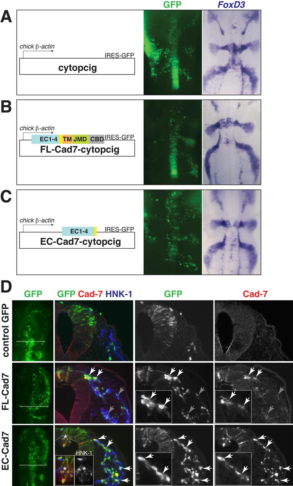
10 ventral roots (Nakagawa and Takeichi, 1995), but protein is expressed by migratory cells forming the dorsal root ganglia (Nakagawa and Takeichi, 1998) In vivo perturbation of Cadherin-7 function in the cranial neural crest affects cell cell interactions and cellular protrusions To investigate the possible role of Cadherin-7 in ganglion formation, we examined the effects of increasing Cadherin-7 levels or blocking its function in the cranial neural crest, which is known to have abundant and dynamic cell cell contacts during migration (Kulesa and Fraser, 2000; Teddy and Kulesa, 2004). For this purpose, we cloned into a bicistronic vector encoding IRES-GFP with a chick β actin promoter (cyto-pcig), a full-length Cadherin-7 (FL-Cad7-cytopcig) as well as a truncated Cadherin- 7 (EC-Cad7-cytopcig), coding sequence. EC-Cad7 was designed to encode the first four cadherin repeats based on a natural splice Cadherin-7 variant previously shown to inhibit full-length Cadheirn-7 (Kawano et al., 2002). Empty vector (cytopcig) was used as a negative control. Constructs were electroporated in ovo into one side of the dorsal neural tube prior to neural crest emigration at stage 8 (3 5 somites stage). Embryos with high levels of GFP expression were analyzed after about hours of incubation (stages 10 14). For controls, we compared the electroporated side of experimental embryos to control GFP embryos as well as to the contralateral side of the same embryos as an internal control. Many embryos that were highly transfected also received construct within the surface ectoderm on the contralateral side (due to leakage of injected DNA outside of the neural tube). Embryos were analyzed for the three characteristic cranial neural crest migration streams using several neural crest markers: HNK-1, FoxD3, and Sox10. No differences were noted in the overall migration pattern between control GFP (n=13), FL-Cad7 (n=7) and EC-Cad7 (n=13) embryos (Fig. 3, A-C and data not shown).
11 However, careful examination of the neural crest cells in transverse sections through these embryos revealed marked differences between experimental and control embryos. Transfection of both FL- and EC- Cad7 vectors into neural crest cells resulted in significant protein overexpression (Fig. 3D). Since endogenous Cadherin-7 levels were generally very weak by immunostaining, most of the antibody staining reflects exogenous expression of FL- or EC- Cad7 expression. The strong Cadherin-7 staining noted in GFP expressing transfected neural crest and neural tube cells on the electroporated compared with the control contralateral side confirms that the expression reflects exogenous proteins. Interestingly, neural crest cells overexpressing FL-Cad7 tend to cluster with each other in a manner that correlates with high expression of FL-Cad7 at the adherens junction (Fig. 3D). Even though these cells interact abnormally, they still manage to leave the neural tube and migrate in separate streams. In contrast, neural crest cells expressing high levels of EC- Cad7 appear to have normal organization, making cell contacts with one another and migrating similarly to control GFP embryos (Fig. 3D). In the future, dynamic analysis of these cells using live imaging will be used to more fully characterize their behaviors. Aberrant protrusion of EC-Cad7 and wild type neural crest cells was observed on the apical side of the dorsal neural tube adjacent to the second rhombomere (r2) level but not elsewhere (Fig. 3D). This suggests that perhaps the EC-Cad7 constructs can disrupt Cadherin-7 or other cadherins in the neural tube (i.e. Cadherin 6b) that are required for emigration in a cell-autonomous and non-autonomous manner. Both transfected and nontransfected (HNK-1+ but GFP-) neural crest cells were found in the protrusion. This is not surprising, since the construct is designed to produce a soluble extracellular form of Cadherin-7 which perhaps may bind to other cadherins at lower affinities and affect other cells. However, this effect may be marginal since most neural crest cells still migrate away from the neural tube normally.
12 Since neural tube electroporation is mosaic and often less than 50% of the neural crest population is transfected, we cannot completely assess effects of exogenous expressions without achieving higher transfection levels. To circumvent this potential limitation, we also introduced these constructs at gastrula stages (stages 4-6) where most if not all of the neural crest cells, as well as some ectodermal cells are transfected. In these embryos, the effect of overexpressing Cadherin-7 on neural crest cell morphology was more severe than that described above at stage 8. Analysis by HNK-1 staining showed that these cells were more clustered and appeared rounded with fewer cellular protrusions (n=4, Fig. 4D). Again the overall pattern of migration streams was normal (Fig. 4, A-B). Taken together, altering the levels of Cadherin-7 appears to have some effects on neural crest cell clustering and morphology but not the formation of the separate migratory streams Subcellular localization and expression pattern of exogeneous full-length Cadherin-7 on migrating neural crest cells suggest dynamic regulation of the Cadherin-7 protein We observed interesting differences in the expressions of full-length (FL) versus extracellular (EC) Cad7 protein in the neural crest. First, the FL-Cad7 was detected mainly at cell boundaries or at junctions between cells that appear more tightly associated and can be either punctate or continuous along the cell membrane (Fig. 3D and Fig. 4C). This is different from the EC-Cad7 protein whose subcellular localization is more widespread, detected both in the cytoplasm and at the membrane but not restricted to the adherens junction (Fig. 3D). The expression of FL-Cad7 is consistent with the idea that FL-Cad7 is mediating cell adhesion and that the protein is dynamically regulated at the cell surface. Second, EC-Cad7 is expressed by most, if not all transfected neural crest cells whereas FL-Cad7 is expressed strongly in only some GFP expressing transfected cells. Other GFP cells express little or no detectable FL-Cad7, at stages (Fig. 3D).
13 The expression of FL-Cad7 often correlates with GFP transfected cells that appear more adherent, usually in a cluster of a few cells but sometimes only two cells along the migration pathway. The regulation of the FL-Cad7 expression pattern may be dependent on the stage of migration and/or levels of the FL-Cad7, as suggested by the observation that GFP transfected cells electroporated at gastrula stages and examined at stages 10 11, appear to all express FL-Cad7 (Fig. 4C). It is intriguing to speculate that there may be some post-translational regulation of the Cadherin-7 protein during neural crest migration. This is unlikely to involve transcriptional control, since the levels of GFP, which is encoded on the same bicistronic mrna with FL-Cad7, were unaffected. One possibility is that Cadherin-7 may undergo similar post-translational modification as that observed for Cadherin-11 by ADAM metalloproteases during Xenopus neural crest migration (McCusker et al., 2009). Regulation of cadherins by ADAMs has also been shown in the trunk neural tube in chick, prior to neural crest emigration (Shoval et al., 2007), as well as in other systems (Maretzky et al., 2005; Reiss et al., 2005). The expression of Cadherin-7 in chick is similar to Cadherin-11 in Xenopus (Vallin et al., 1998), as both are specifically expressed in migrating neural crest cells, whereas in mouse neural crest, Cadherin-6 is expressed in both premigratory and migrating neural crest (Inoue et al., 1997); therefore studies on Xcadherin-11, also a classical type II cadherin, may be relevant to our understanding of Cadherin-7. McCusker et al. (2009) shows that endogenous Cadherin-11 undergoes cleavage during neural crest migration producing a putative extracellular product encompassing the first three cadherin repeats (EC1 3) and thus the remaining cleaved Cadherin-11 no longer has the first extracellular domain (EC1) sited for homophilic binding (Patel et al., 2006). Blocking this cleavage by reducing ADAMs or overexpressing Cadherin-11 inhibits neural crest migration, likely due to an increase in cadherin-mediated adhesion. Increasing ADAMs or the putative extracellular cleavage product can reverse the
14 blockage of migration by Cadherin-11 (McCusker et al., 2009). Thus, cleavage of Cadherin-11 by ADAMs may regulate the levels of full-length and cleaved cadherin proteins which modulates levels of cell cell adhesion between migrating neural crest cells. Since we used an antibody that recognizes the extracellular portion of the Cadherin- 7 to detect FL-Cad7, it is possible that the loss or reduced detection of the extracellular portion of FL-Cad7 on the transfected GFP neural crest cells results from the cleavage of the Cadherin-7 protein by ADAMs during migration. Alternatively, the regulation of catenins by phosphorylation may control the levels of Cadherin-7 protein. For example, the intracellular binding partner p120-catenin of cadherins has been shown to stabilize cadherins and regulate their turnover at the cell surface (Kowalczyk and Reynolds, 2004; Perez-Moreno and Fuchs, 2006) Perturbation of Cadherin-7 in precursors causes malformed cranial ganglia To test the function of Cadherin-7 during ganglion formation, we introduced FL- Cad7 or EC-Cad7 and control GFP into embryos in ovo at early stages 4 6 in order to achieve high transfection efficiency in the neural crest cells. Embryos were analyzed after 48 hours (~ stage 16). Either overexpressing or blocking Cadherin-7 caused defects in ganglia formation, with most dramatic phenotypes in FL-Cad7 embryos (Fig. 5). In FL- Cad7 embryos, trigeminal and epibranchial ganglia appeared reduced in size, were more condensed, and had aberrant placement of placodal neurons (regions of gaps within the ganglia and also aberrant connection between the glossopharyngeal and vagal ganglia at the third and fourth branchial arches) (Fig. 5B). High levels of Cadherin-7 caused dramatic ganglia malformation but normal development of the remaining head. EC-Cad7 overexpressing embryos had much less severe defects, generally with slightly misshapen ganglia that were not reduced in size (Fig. 5C). Control GFP embryos as well as the
15 contralateral unelectroporated side had no apparent effects. The data suggest that overexpression of Cadherin-7 has notable effects on ganglia formation. Whether or not Cadherin-7 is essential for gangliogenesis requires further investigation using a gene knockdown approach (i.e. RNAi or morpholino) Misexpression of Cadherin-7 in the trigeminal ectoderm disrupts placodal morphogenesis and augments the neuronal population in the ectoderm We further examined the phenotype of overexpressing FL-Cad7 on the organization and coalecence of placodal neurons by examining embryos in transverse sections. We observed that misexpression of Cadherin-7 in the placodal ectoderm causes dramatic disruptions (Fig. 6), with aberrant placode-derived neuronal nodules, delamination of sheets of differentiating placodal ectoderm, and widespread neuronal cells in the surface ectoderm, which coexpresses GFP and TuJ1 (Fig. 6, B-D). The most proximal part of the ganglion adjacent to the hindbrain, which is mostly neural crestderived, appears to assemble properly in whole mount (Fig. 5B), probably due to low level transfection of the neural crest (Fig. 6A). Since placodal neurons generally do not express Cadherin-7 but neural crest cells do, we predicted that introducing Cadherin-7 into the placodal cells would cause neural crest cells to be abnormally more adherent to placodal neurons. However, we found that intermixing and association between neural crest and placodal neurons throughout the region of ganglion formation are aberrant. In fact, there are several instances where placodal neurons are devoid of neural crest association (Fig. 6B-D). These are generally placodal cells that appear excessively adherent to each other as they form placodal nodules and delaminate as a sheet of neurogenic ectoderm instead of undergoing proper ingression. Other regions containing placodal neurons that are less adherent and normally associated
16 with each other appear to intermingle with neural crest cells, however there are still spots of segregated placodal cells in the mix (Fig. 6D-D ). This may suggest that cell adhesion between placodes and neural crest may depend less on cadherin subtype identity and more on adhesive properties (i.e. cadherin expression levels) and/or that other factors play a larger role in mediating neural crest placode coalescence than simply expressing the same cadherin (e.g. heterophilic interaction may be required, which perhaps is disrupted by Cadherin-7 overexpression). This is consistent with in vitro cell binding assays that show two cell populations expressing two distinct cadherin subtypes can sort out or intermix depending on their relative cadherin expression levels, independent of subtype specificity. Furthermore two cell lines expressing identical cadherin but at different concentrations also sort out (Duguay et al., 2003). The abnormal, widespread expression of the neuronal marker TuJ1 in the placodal ectoderm may indicate that Cadherin-7 expression has blocked ingression of differentiating placodal neurons and thus has kept many of these cells at the surface and/or increased neuronal differentiation within the placodal ectoderm. The large number of placodal neurons in the surface ectoderm in the distal ganglion, particularly around the first branchial arch, together with the blocked ingression may explain the appearance of the reduced trigeminal ganglion in whole mount (Fig. 5). Taken together, misexpression of Cadherin-7 can severely disrupt normal placodal morphogenesis and perhaps differentiation, likely by causing excessive cell adhesion between placodes.
17 4.5 Conclusion and future work Our data suggest that Cadherin-7 may regulate neural crest cell interactions and adhesion, and later, if overexpressed, can cause severe defects in cranial ganglia formation. However, Cadherin-7 does not appear to play a role in patterning cranial neural crest cell migration into stereotypic streams. This is consistent with results of Cadherin-11 perturbation in Xenopus cranial neural crest (Borchers et al., 2001). In contrast to results observed with Cadherin-11, however, overexpression of Cadherin-7 did not block neural crest migration (Borchers et al., 2001; McCusker et al., 2009). However, the penetrance of this phenotype may depend upon the amount of mrna injected (Borchers et al., 2001). The difference may be due to the levels of transfection, since single blastomere injections (which are not possible in the chick) can achieve higher transfection than electroporation. Cadherin-7 overexpression in the trunk neural tube has previously been reported to block neural crest emigration along the dorsolateral pathway, but not the dorsoventral stream to the dorsal root ganglia (Nakagawa and Takeichi, 1998). However, no such inhibition was observed in the cranial regions, though overexpression of the truncated Cadherin-7 (EC- Cad7) that presumably acts as a dominant-negative can affect proper emigration. During cranial neural crest migration, both mrna and protein expression pattern of Cadherin-7 suggest dynamic regulation not only at transcriptional but also post-translational levels. The largely complementary expression of Cadherin-7 on neural crest and N- cadherin on placodal neurons suggests that coalescence of these precursor cell types may rely on both homophilic and heterophilic interactions between Cadherin-7 and N-cadherin. We propose two testable models for neural crest placode coalescence: 1) that Cadherin- 7 N-cadherin heterotypic binding mediates adhesion between neural crest and placodal cells and a smaller proportion of this interaction may be mediated through Cadherin-7 homotypically; or 2) neural crest placode adhesion is mediated by homotypic Cadherin-7
18 interaction and N-cadherin acts only on placodal neurons, suggesting that at early stages of ganglion formation, there would be far fewer adherens junctions between neural crest placode than between the same precursor type cells. Interestingly, Cadherin-7 misexpression did not increase but instead prevented neural crest placode association. The data suggest that altering cadherin levels is sufficient to block proper placodal morphogenesis and intermixing of neural crest and placodal cells. It remains to be seen whether the difference in cadherin subtype is also crucial for proper neural crest placode coalescence into ganglion. Further investigation using Cadherin-7 gene knockdown approaches (i.e. RNAi or morpholino) will provide deeper insights into the endogenous Cadherin-7 role and its interaction with N-cadherin during gangliogenesis. Co-immunoprecipitation of N-cadherin and Cadherin-7 from cells in the trigeminal ganglion would directly test whether heterophilic interactions occur in vivo. Furthermore, if blocking or depleting N-cadherin whose expression is specific to placodal neurons affects placode neural crest association, this would lend support for heterotypic cadherin interaction. Rescue experiments using one to test whether it can restore the loss of function of the other would establish whether or not these two cadherin members may be functionally equivalent or have distinct biological effects on neural crest and placodal cells. This could also address the significance of subtype specificity on the integration of neural crest and placodal cells. Another pertinent question is whether one cadherin can regulate the other, either cell-autonomously or noncell-autonomously. For example, does misexpression of Cadherin-7 on the placodal ectoderm or reduction of Cadherin-7 on the neural crest, or both, alter N-cadherin expression on placodal cells? Taken together, the results provide exciting new avenues for exploration of cadherin-mediated neural crest placode interactions during cranial gangliogenesis. Future investigation will explore the nature of the functional interactions between these two distinct cadherins and their biological significance.
19 4.6 Acknowledgements I thank my undergraduate students, Emma Broom and Emma Hindley, who were supported by the SURF/Caltech-Cambridge exchange program, for their contribution on the preliminary studies of Cadherin-7 expression and its function during cranial neural crest development. This work was supported by US National Institutes of Health (NIH) National Research Service Award 5T32 GM07616 to C. E. S. and NIH grant DE16459 to M.B.-F.
 first migration stream at stage 10 and subsequently in all three streams as migration progresses at (B)")
20 Figure 1. Figure 1. Cadherin-7 mrna is transiently expressed by migrating cranial neural crest, and later retained in neural crest at the cranial entry/exit points and in the trigeminal ganglion. In situ hybridization with the Cadherin-7 riboprobe shows Cad-7 mrna expression in the (A) first migration stream at stage 10 and subsequently in all three streams as migration progresses at (B) stage 12 and (C) stage 13. Later Cad-7 is expressed in discrete neural crest regions at the cranial motor exit points in the forming ganglia at (D) stage 16 and (E) stage 18 with the expression in the trigeminal ganglion appearing more broad. (F-M) Cad-7 expression overlaps with neural crest marker HNK-1 but is downregulated in the neural crest cells that have migrated the furthest. Downregulation of Cad-7 is apparent early at the beginning of migration (F-G) at stage 10 and (H- M) throughout the time of migration in all three streams. (M, M ) Section through the presumptive trigeminal exit point at the level indicated in K showing high expression in the neural crest cells adjacent to the neural tube at stage 13.
 Cross section through stage 11 showing overlap of Cad-7 with neural crest marker HNK-1.")
21 Figure 2. Figure 2. Cadherin-7 protein expression is broader than the mrna. Schematics of the chick embryos at stage 11 and stage 18 indicate the levels of the sections. (A,A ) Cross section through stage 11 showing overlap of Cad-7 with neural crest marker HNK-1. Cad-7 protein expression is detected throughout the neural crest stream, albeit weaker in the ventrally migrating neural crest cells. (B) Cross section through the trigeminal ganglion at stage 18. (C,C, C ) High magnification of the dotted box in B showing the most dorsal region of the ganglion which is neural crest-derived expressing Cad-7 and not N-cad. The neurotubulin TuJ1 staining outside of the neural tube is probably exiting motor axons rather than placodal neuronal processes. (D) High magnification of the dotted box in B showing a section through the OpV region of the trigeminal ganglion. (E,E,E ) Magnification of the boxed region in D showing a predominantly complementary expression of Cad-7 and N-cad. Placodal neurons as labeled by TuJ1 mostly express N-cad but not Cad-7 (arrows), although some may express weak or background levels of Cad-7 (gray arrow). Cad-7 expression is present at the perimeter of the ganglion lobe as expected for expression in the neural crest which lacks N-cad expression (asterisk).
22 Figure 3.
23 Figure 3. Perturbation of Cadherin-7 does not affect the pattern of cranial neural crest streams but alters cell cell interactions. (A, B, C) Left subpanel showing a schematic of the vector used. Middle subpanel showing the level and region of DNA transfection as indicated by the GFP expression. Right, the same embryo as that shown in the middle subpanel was processed for the neural crest marker FoxD3 in situ hybridization. (A) Control embryo at stage 12. (B) FL-Cad7 embryo and (C) EC-Cad7 embryo at stage 12 also show the typical pattern of the three migration streams. (D) First column from the left shows the neural crest region of transfection by GFP expression in whole mount embryos. Second column shows the color overlay image of GFP (green), Cad-7 protein (red), and HNK-1 (blue) of a cross section through the whole mount embryo at the level indicated by the dotted line. Single channel images of GFP and Cad-7 from the color overlay image are shown in the third and fourth columns. Neural crest cells transfected with FL- Cad7 appear to aberrantly associate with each other and tend to cluster (white arrows), in contrast to the control GFP and EC-Cad7 expressing neural crest cells. Exogenous FL-Cad7 protein is expressed mostly at adherens junctions between neural crest cells (white arrows) and is downregulated in many GFP expressing transfected cells (gray arrows), whereas the EC-Cad7 protein is localized less specifically to the cell membrane and is expressed at high levels in most, if not all, GFP expressing cells (white arrows). Expression of EC-Cad7 can cause disruption to neural crest emigration as shown by the aberrant protrusions of neural crest (HNK-1+) and neural tube cells in the lumen of the neural tube (asterisk).

24 Figure 4.
25 Figure 4. Abundant full-length Cadherin-7 in the cranial neural crest causes abnormal neural crest cell morphology but does not inhibit migration. To increase the percentage of transfected neural crest cells, control GFP or FL-Cad7 expression vectors were introduced into the presumptive neural crest region (neural plate border) at gastrula stages 4 6 by electroporation. Control GFP (A) or FL- Cad7 (B) embryos were electroporated on one side as indicated by the GFP expression, and analyzed at stages Cross section through the midbrain region of the FL-Cad7 embryo at stage 10+ showing the tightly clustered migration stream on the transfected side in contrast to the unelectroporated control side. (C) Higher magnfication of the same section showing the control and electroporated FL-Cad7 sides in the color overlay of GFP (green), Cad-7 protein (red), and HNK-1 (blue) expressions (also shown as individual images). (D) Close examination of the neural crest cell morphology in the control GFP, control side of FL-Cad7, and FL-Cad7 cases shows that the neural crest cells (HNK-1+) expressing high levels of Cad-7 are abnormally more adhered to one another and appear more rounded with fewer cellular protrusions. This contrasts the loosely connecting and mesenchymal appearance of the control neural crest cells.
 indicating area of transfection, neuronal marker TuJ1 (red) on placodal neurons, and HNK-1 (blue) on neural crest cells.")
26 Figure 5. Figure 5. Perturbation of Cadherin-7 in the precursors severely disrupts cranial sensory ganglia formation. Top panels show color overlay of GFP expression (green) indicating area of transfection, neuronal marker TuJ1 (red) on placodal neurons, and HNK-1 (blue) on neural crest cells. To achieve high transfection levels, embryos were electroporated at stages 4 6 and analyzed at stage 16 in the forming ganglia. (A) Control GFP embryo displays typical ganglia formation. (B) By contrast, overexpression of Cadherin-7 resulted in severe ganglia defects, including reduced (or tightly coalesced) trigeminal and epibranchial ganglia (top arrows), with abnormal organization of placodal neurons (regions of gaps within the ganglia [asterisk] and abnormal link between the glossopharyngeal and vagal ganglia [bottom arrow]). These abnormalities correspond to regions of transfection as shown by GFP expression in color overlay image and inset. (C) Blocking Cadherin- 7 by EC-Cad7 showed less severe effects, generally with misshapen ganglia (arrows) that were not reduced in size. Unelectroporated control sides of the FL-Cad7 and EC-Cad7 embryos showed no apparent defects. t, trigeminal; f, facial; g, glossopharyngeal; v,vagal; EP, electroporated.

27 Figure 6.
28 Figure 6. Misexpression of Cadherin-7 leads to aberrant placodal morphogenesis and increased neuronal cells in the ectoderm. Schematic of chick stage 16 embryo showing the levels of the frontal plane sections. FL-Cad7 embryo electroporated in the precursor cell populations (both neural crest and placodal ectoderm) at stages 4 6 showing transfection mostly in the placodal cells by the time of analysis at stage 16. (A) Most proximal part of the trigeminal and facial ganglia adjacent to the hindbrain, which is mostly neural crest-derived, appears to assemble properly. (B) Misexpression of Cadherin-7 in the trigeminal placodal ectoderm causes dramatic disruptions of the placodal ectoderm delaminating sheets of differentiating placodal ectoderm (TuJ1+) that do not associate with neural crest cells (arrows). (C-D ) It also causes aberrant placode-derived neuronal nodules (arrowhead, GFP+/TuJ1+) and widespread neuronal cells in the surface ectoderm (dotted arrow, TuJ1+/GFP+). Placode neural crest interaction appears abnormal. The aberrant nodules and some regions in the ganglion clusters (arrow) are devoid of neural crest (HNK-1+) association. The unelectroporated control side forms the typical OpV and MmV lobes as shown in C. (E-E ) Control embryo electroporated with a GFP expression vector also at stages 4 6 shows normal trigeminal ganglion formation and intermixing of neural crest (HNK-1+) and placodal neurons (TuJ1+). OpV, ophthalmic; MmV, maxillo-mandibular; t, trigeminal; f, facial.
Conclusions. The experimental studies presented in this thesis provide the first molecular insights
 C h a p t e r 5 Conclusions 5.1 Summary The experimental studies presented in this thesis provide the first molecular insights into the cellular processes of assembly, and aggregation of neural crest and
Neural crest placode interactions mediate trigeminal ganglion formation and require Slit1 Robo2 signaling
 C h a p t e r 2 Neural crest placode interactions mediate trigeminal ganglion formation and require Slit1 Robo2 signaling 2.1 Abstract 1 The cellular and molecular interactions allowing generation of complex
In ovo time-lapse analysis after dorsal neural tube ablation shows rerouting of chick hindbrain neural crest
 In ovo time-lapse analysis after dorsal neural tube ablation shows rerouting of chick hindbrain neural crest Paul Kulesa, Marianne Bronner-Fraser and Scott Fraser (2000) Presented by Diandra Lucia Background
SUPPLEMENTARY INFORMATION
 doi:10.1038/nature11589 Supplementary Figure 1 Ciona intestinalis and Petromyzon marinus neural crest expression domain comparison. Cartoon shows dorsal views of Ciona mid gastrula (left) and Petromyzon
N-cadherin acts in concert with Slit1-Robo2 signaling in regulating aggregation of placode-derived cranial sensory neurons
 RESEARCH ARTICLE 4155 Development 137, 4155-4164 (2010) doi:10.1242/dev.034355 N-cadherin acts in concert with Slit1-Robo2 signaling in regulating aggregation of placode-derived cranial sensory neurons
Abstract. Assistant Professor Dr. Lisa Taneyhill, Department of Animal and Avian Sciences
 Abstract Title of Document: THE ROLE OF THE ADHERENS JUNCTION PROTEIN αn-catenin IN NEURAL CREST-DERIVED TRIGEMINAL GANGLIA FORMATION Rachel Hooper, Master of Science 2012 Directed By: Assistant Professor
Dynamic and Differential Regulation of Stem Cell Factor FoxD3 in the Neural Crest Is Encrypted in the Genome
 Dynamic and Differential Regulation of Stem Cell Factor FoxD3 in the Neural Crest Is Encrypted in the Genome Marcos S. Simões-Costa 1., Sonja J. McKeown 1., Joanne Tan-Cabugao 1, Tatjana Sauka-Spengler
Nature Neuroscience: doi: /nn.2662
 Supplementary Figure 1 Atlastin phylogeny and homology. (a) Maximum likelihood phylogenetic tree based on 18 Atlastin-1 sequences using the program Quicktree. Numbers at internal nodes correspond to bootstrap
Supplementary Figure 1: Mechanism of Lbx2 action on the Wnt/ -catenin signalling pathway. (a) The Wnt/ -catenin signalling pathway and its
 The Wnt/ -catenin signalling pathway and its") Supplementary Figure 1: Mechanism of Lbx2 action on the Wnt/ -catenin signalling pathway. (a) The Wnt/ -catenin signalling pathway and its transcriptional activity in wild-type embryo. A gradient of canonical
Supplementary Figure 1: Mechanism of Lbx2 action on the Wnt/ -catenin signalling pathway. (a) The Wnt/ -catenin signalling pathway and its transcriptional activity in wild-type embryo. A gradient of canonical
Cells to Tissues. Peter Takizawa Department of Cell Biology
 Cells to Tissues Peter Takizawa Department of Cell Biology From one cell to ensembles of cells. Multicellular organisms require individual cells to work together in functional groups. This means cells
Relationship between spatially restricted Krox-20 gene expression in branchial neural crest and
 The EMBO Journal vol.14 no.8 pp.1697-1710, 1995 Relationship between spatially restricted Krox-20 gene expression in branchial neural crest and segmentation in the chick embryo hindbrain M.Angela Nieto1
SUPPLEMENTARY INFORMATION
 DOI: 10.1038/ncb3267 Supplementary Figure 1 A group of genes required for formation or orientation of annular F-actin bundles and aecm ridges: RNAi phenotypes and their validation by standard mutations.
Developmental Zoology. Ectodermal derivatives (ZOO ) Developmental Stages. Developmental Stages
 Developmental Stages. Developmental Stages") Developmental Zoology (ZOO 228.1.0) Ectodermal derivatives 1 Developmental Stages Ø Early Development Fertilization Cleavage Gastrulation Neurulation Ø Later Development Organogenesis Larval molts Metamorphosis
Developmental Zoology (ZOO 228.1.0) Ectodermal derivatives 1 Developmental Stages Ø Early Development Fertilization Cleavage Gastrulation Neurulation Ø Later Development Organogenesis Larval molts Metamorphosis
Sonic hedgehog (Shh) signalling in the rabbit embryo
 signalling in the rabbit embryo") Sonic hedgehog (Shh) signalling in the rabbit embryo In the first part of this thesis work the physical properties of cilia-driven leftward flow were characterised in the rabbit embryo. Since its discovery
Sonic hedgehog (Shh) signalling in the rabbit embryo In the first part of this thesis work the physical properties of cilia-driven leftward flow were characterised in the rabbit embryo. Since its discovery
Role of Organizer Chages in Late Frog Embryos
 Ectoderm Germ Layer Frog Fate Map Frog Fate Map Role of Organizer Chages in Late Frog Embryos Organizer forms three distinct regions Notochord formation in chick Beta-catenin localization How does beta-catenin
Graded potential of neural crest to form cornea, sensory neurons and cartilage along the rostrocaudal axis
 Research article 1979 Graded potential of neural crest to form cornea, sensory neurons and cartilage along the rostrocaudal axis Peter Y. Lwigale 1, Gary W. Conrad 2 and Marianne Bronner-Fraser 1, * 1
Early Development in Invertebrates
 Developmental Biology Biology 4361 Early Development in Invertebrates October 25, 2006 Early Development Overview Cleavage rapid cell divisions divisions of fertilized egg into many cells Gastrulation
Question Set # 4 Answer Key 7.22 Nov. 2002
 Question Set # 4 Answer Key 7.22 Nov. 2002 1) A variety of reagents and approaches are frequently used by developmental biologists to understand the tissue interactions and molecular signaling pathways
Overexpression of Snail family members highlights their ability to promote chick neural crest formation
 Development 129, 1583-1593 (2002) Printed in Great Britain The Company of Biologists Limited 2002 DEV1802 1583 Overexpression of Snail family members highlights their ability to promote chick neural crest
Developmental Biology Lecture Outlines
 Developmental Biology Lecture Outlines Lecture 01: Introduction Course content Developmental Biology Obsolete hypotheses Current theory Lecture 02: Gametogenesis Spermatozoa Spermatozoon function Spermatozoon
1. What are the three general areas of the developing vertebrate limb? 2. What embryonic regions contribute to the developing limb bud?
 Study Questions - Lecture 17 & 18 1. What are the three general areas of the developing vertebrate limb? The three general areas of the developing vertebrate limb are the proximal stylopod, zeugopod, and
Supplementary Materials for
 www.sciencesignaling.org/cgi/content/full/6/301/ra98/dc1 Supplementary Materials for Regulation of Epithelial Morphogenesis by the G Protein Coupled Receptor Mist and Its Ligand Fog Alyssa J. Manning,
Cellular Neurobiology BIPN 140 Fall 2016 Problem Set #8
 Cellular Neurobiology BIPN 140 Fall 2016 Problem Set #8 1. Inductive signaling is a hallmark of vertebrate and mammalian development. In early neural development, there are multiple signaling pathways
The role of FGF2 in craniofacial skeletogenesis
 The role of FGF2 in craniofacial skeletogenesis P. Ferretti, S. Sarkar, R. Moore, A. Petiot, C. J. Chan and A. Copp Summary E vidence that the major craniosynostosis syndromes are caused by mutations in
CELL-CELL COMMUNICATION
 CELL-CELL COMMUNICATION paracrine & juxtacrine signalling autocrine & intracrine signalling methods to study cell-cell communication: attraction & repulsion chemotaxis & chemokinesis substrate preference
Hes5. Hes Relative mrna levels
 A a Absolute mrna levels 6 5 4 3 2 1 hjag1 EP cdl1 EP Jag1 E2+1 b d m Hey1 c e EP Hey1 Dl1 E2+1 f h m Hey1 g i EP Hey1 1 a Hes5 Hes5 a Hes5 Hes5 B a b EP e 1.4 1.2 EP GFP GFP E2+1 c m Hey1 d Hey1 Relative
Neural crest development is regulated by the transcription factor Sox9
 Research article 5681 Neural crest development is regulated by the transcription factor Sox9 Martin Cheung and James Briscoe* Developmental Neurobiology, National Institute for Medical Research, Mill Hill,
Cell Migration I: Neural Crest Cell Migration. Steven McLoon Department of Neuroscience University of Minnesota
 Cell Migration I: Neural Crest Cell Migration Steven McLoon Department of Neuroscience University of Minnesota 1 Types of Cell Movement passive: active: cell sheets flow cilia or flagella ameboid adhesion
MCDB 4777/5777 Molecular Neurobiology Lecture 29 Neural Development- In the beginning
 MCDB 4777/5777 Molecular Neurobiology Lecture 29 Neural Development- In the beginning Learning Goals for Lecture 29 4.1 Describe the contributions of early developmental events in the embryo to the formation
Expression and function of cell adhesion molecules during neural crest migration
 1 2 3 4 5 6 7 8 9 10 11 12 13 14 15 16 17 18 19 20 21 22 23 24 25 26 27 28 29 30 31 32 33 34 Expression and function of cell adhesion molecules during neural crest migration Sonja J. McKeown 1, Adam S.
Fig. S1. Proliferation and cell cycle exit are affected by the med mutation. (A,B) M-phase nuclei are visualized by a-ph3 labeling in wild-type (A)
 M-phase nuclei are visualized by a-ph3 labeling in wild-type (A)") Fig. S1. Proliferation and cell cycle exit are affected by the med mutation. (A,B) M-phase nuclei are visualized by a-ph3 labeling in wild-type (A) and mutant (B) 4 dpf retinae. The central retina of the
Fig. S1. Proliferation and cell cycle exit are affected by the med mutation. (A,B) M-phase nuclei are visualized by a-ph3 labeling in wild-type (A) and mutant (B) 4 dpf retinae. The central retina of the
Massachusetts Institute of Technology Harvard Medical School Brigham and Women s Hospital VA Boston Healthcare System 2.79J/3.96J/BE.
 Massachusetts Institute of Technology Harvard Medical School Brigham and Women s Hospital VA Boston Healthcare System 2.79J/3.96J/BE.441/HST522J INTEGRINS I.V. Yannas, Ph.D. and M. Spector, Ph.D. Regulator
Late emigrating neural crest cells migrate specifically to the exit points of cranial branchiomotor nerves
 Development 122, 2367-2374 (1996) Printed in Great Britain The Company of Biologists Limited 1996 DEV9463 2367 Late emigrating neural crest cells migrate specifically to the exit points of cranial branchiomotor
Developmental Biology 3230 Midterm Exam 1 March 2006
 Name Developmental Biology 3230 Midterm Exam 1 March 2006 1. (20pts) Regeneration occurs to some degree to most metazoans. When you remove the head of a hydra a new one regenerates. Graph the inhibitor
Principles of Experimental Embryology
 Biology 4361 Developmental Biology Principles of Experimental Embryology June 16, 2008 Overview What forces affect embryonic development? The embryonic environment: external and internal How do forces
Axis Specification in Drosophila
 Developmental Biology Biology 4361 Axis Specification in Drosophila November 2, 2006 Axis Specification in Drosophila Fertilization Superficial cleavage Gastrulation Drosophila body plan Oocyte formation
Neural crest cell-cell adhesion controlled by sequential and subpopulationspecific expression of novel cadherins
 Development 121, 1321-1332 (1995) Printed in Great Britain The Company of Biologists Limited 1995 1321 Neural crest cell-cell adhesion controlled by sequential and subpopulationspecific expression of novel
Angiopoietin 2 signaling plays a critical role in neural crest cell migration
 McKinney et al. BMC Biology (2016) 14:111 DOI 10.1186/s12915-016-0323-9 RESEARCH ARTICLE Angiopoietin 2 signaling plays a critical role in neural crest cell migration Mary Cathleen McKinney 1, Rebecca
 DOI: 10.1038/ncb2819 Gαi3 / Actin / Acetylated Tubulin Gαi3 / Actin / Acetylated Tubulin a a Gαi3 a Actin Gαi3 WT Gαi3 WT Gαi3 WT b b Gαi3 b Actin Gαi3 KO Gαi3 KO Gαi3 KO # # Figure S1 Loss of protein
Bio 127 Section I Introduction to Developmental Biology. Cell Cell Communication in Development. Developmental Activities Coordinated in this Way
 Bio 127 Section I Introduction to Developmental Biology Cell Cell Communication in Development Gilbert 9e Chapter 3 It has to be EXTREMELY well coordinated for the single celled fertilized ovum to develop
Developmental processes Differential gene expression Introduction to determination The model organisms used to study developmental processes
 Date Title Topic(s) Learning Outcomes: Sept 28 Oct 3 1. What is developmental biology and why should we care? 2. What is so special about stem cells and gametes? Developmental processes Differential gene
5- Semaphorin-Plexin-Neuropilin
 5- Semaphorin-Plexin-Neuropilin 1 SEMAPHORINS-PLEXINS-NEUROPILINS ligands receptors co-receptors semaphorins and their receptors are known signals for: -axon guidance -cell migration -morphogenesis -immune
Supplementary Methods
 Supplementary Methods Modeling of magnetic field In this study, the magnetic field was generated with N52 grade nickel-plated neodymium block magnets (K&J Magnetics). The residual flux density of the magnets
Beta-Actin Is Required for Proper Mouse Neural Crest Ontogeny
 Beta-Actin Is Required for Proper Mouse Neural Crest Ontogeny Davina Tondeleir a, Rivka Noelanders b, Karima Bakkali, Christophe Ampe* Department of Biochemistry, Faculty of Medicine and Health Sciences,
Dynamics of BMP and Hes1/Hairy1 signaling in the dorsal neural tube underlies the transition from neural crest to definitive roof plate
 Washington University School of Medicine Digital Commons@Becker Open Access Publications 2016 Dynamics of BMP and Hes1/Hairy1 signaling in the dorsal neural tube underlies the transition from neural crest
Supplemental Information. The Mitochondrial Fission Receptor MiD51. Requires ADP as a Cofactor
 Structure, Volume 22 Supplemental Information The Mitochondrial Fission Receptor MiD51 Requires ADP as a Cofactor Oliver C. Losón, Raymond Liu, Michael E. Rome, Shuxia Meng, Jens T. Kaiser, Shu-ou Shan,
Dynamics of BMP and Hes1/Hairy1 signaling in the dorsal neural tube underlies the transition from neural crest to definitive roof plate
 Nitzan et al. BMC Biology (2016) 14:23 DOI 10.1186/s12915-016-0245-6 RESEARCH ARTICLE Open Access Dynamics of BMP and Hes1/Hairy1 signaling in the dorsal neural tube underlies the transition from neural
10/2/2015. Chapter 4. Determination and Differentiation. Neuroanatomical Diversity
 Chapter 4 Determination and Differentiation Neuroanatomical Diversity 1 Neurochemical diversity: another important aspect of neuronal fate Neurotransmitters and their receptors Excitatory Glutamate Acetylcholine
Axis Specification in Drosophila
 Developmental Biology Biology 4361 Axis Specification in Drosophila November 6, 2007 Axis Specification in Drosophila Fertilization Superficial cleavage Gastrulation Drosophila body plan Oocyte formation
Student Learning Outcomes: Nucleus distinguishes Eukaryotes from Prokaryotes
 9 The Nucleus Student Learning Outcomes: Nucleus distinguishes Eukaryotes from Prokaryotes Explain general structures of Nuclear Envelope, Nuclear Lamina, Nuclear Pore Complex Explain movement of proteins
9/4/2015 INDUCTION CHAPTER 1. Neurons are similar across phyla Thus, many different model systems are used in developmental neurobiology. Fig 1.
 INDUCTION CHAPTER 1 Neurons are similar across phyla Thus, many different model systems are used in developmental neurobiology Fig 1.1 1 EVOLUTION OF METAZOAN BRAINS GASTRULATION MAKING THE 3 RD GERM LAYER
Bio Section III Organogenesis. The Neural Crest and Axonal Specification. Student Learning Objectives. Student Learning Objectives
 Bio 127 - Section III Organogenesis The Neural Crest and Axonal Specification Gilbert 9e Chapter 10 Student Learning Objectives 1. You should understand that the neural crest is an evolutionary advancement
Vital dye analysis of cranial neural crest cell migration in the mouse embryo
 Development 116, 297-307 (1992) Printed in Great Britain The Company of Biologists Limited 1992 297 Vital dye analysis of cranial neural crest cell migration in the mouse embryo GEORGE N. SERBEDZIJA 1,*,
Coordinated Eph-ephrin signaling guides migration and axon targeting in the avian auditory system
 Allen-Sharpley and Cramer Neural Development 2012, 7:29 RESEARCH ARTICLE Open Access Coordinated Eph-ephrin signaling guides migration and axon targeting in the avian auditory system Michelle R Allen-Sharpley
Sarah Bashiruddin Georgina Lopez Jillian Merica Sarah Wardlaw
 Sarah Bashiruddin Georgina Lopez Jillian Merica Sarah Wardlaw Introduction: Dr. Carol Erickson and her lab study the cellular and molecular mechanisms by which neural crest cells differentiate and migrate
Introduction. Gene expression is the combined process of :
 1 To know and explain: Regulation of Bacterial Gene Expression Constitutive ( house keeping) vs. Controllable genes OPERON structure and its role in gene regulation Regulation of Eukaryotic Gene Expression
Cell Death & Trophic Factors II. Steven McLoon Department of Neuroscience University of Minnesota
 Cell Death & Trophic Factors II Steven McLoon Department of Neuroscience University of Minnesota 1 Remember? Neurotrophins are cell survival factors that neurons get from their target cells! There is a
Exam 1 ID#: October 4, 2007
 Biology 4361 Name: KEY Exam 1 ID#: October 4, 2007 Multiple choice (one point each) (1-25) 1. The process of cells forming tissues and organs is called a. morphogenesis. b. differentiation. c. allometry.
Zebrafish msxb, msxc and msxe function together to refine the neural nonneural border and regulate cranial placodes and neural crest development
 Developmental Biology 294 (2006) 376 390 www.elsevier.com/locate/ydbio Zebrafish msxb, msxc and msxe function together to refine the neural nonneural border and regulate cranial placodes and neural crest
Tsukushi Modulates Xnr2, FGF and BMP Signaling: Regulation of Xenopus Germ Layer Formation
 Tsukushi Modulates Xnr2, FGF and BMP Signaling: Regulation of Xenopus Germ Layer Formation Samantha A. Morris 1 *, Alexandra D. Almeida 1, Hideaki Tanaka 2, Kunimasa Ohta 2, Shin-ichi Ohnuma 1 * 1 Department
Nature Biotechnology: doi: /nbt Supplementary Figure 1. Overexpression of YFP::GPR-1 in the germline.
 Supplementary Figure 1 Overexpression of YFP::GPR-1 in the germline. The pie-1 promoter and 3 utr were used to express yfp::gpr-1 in the germline. Expression levels from the yfp::gpr-1(cai 1.0)-expressing
Geert Geeven. April 14, 2010
 iction of Gene Regulatory Interactions NDNS+ Workshop April 14, 2010 Today s talk - Outline Outline Biological Background Construction of Predictors The main aim of my project is to better understand the
RANK. Alternative names. Discovery. Structure. William J. Boyle* SUMMARY BACKGROUND
 RANK William J. Boyle* Department of Cell Biology, Amgen, Inc., One Amgen Center Drive, Thousand Oaks, CA 91320-1799, USA * corresponding author tel: 805-447-4304, fax: 805-447-1982, e-mail: bboyle@amgen.com
SUPPLEMENTARY INFORMATION
 DOI: 10.1038/ncb2647 Figure S1 Other Rab GTPases do not co-localize with the ER. a, Cos-7 cells cotransfected with an ER luminal marker (either KDEL-venus or mch-kdel) and mch-tagged human Rab5 (mch-rab5,
purpose of this Chapter is to highlight some problems that will likely provide new
 119 Chapter 6 Future Directions Besides our contributions discussed in previous chapters to the problem of developmental pattern formation, this work has also brought new questions that remain unanswered.
SUPPLEMENTARY INFORMATION
 GP2 Type I-piliated bacteria FAE M cell M cell pocket idc T cell mdc Generation of antigenspecific T cells Induction of antigen-specific mucosal immune response Supplementary Figure 1 Schematic diagram
Rhombomeric origin and rostrocaudal reassortment of neural crest cells revealed by intravital microscopy
 Development 121, 935-945 (1995) Printed in Great Britain The Company of Biologists Limited 1995 935 Rhombomeric origin and rostrocaudal reassortment of neural crest cells revealed by intravital microscopy
Biology 218, practise Exam 2, 2011
 Figure 3 The long-range effect of Sqt does not depend on the induction of the endogenous cyc or sqt genes. a, Design and predictions for the experiments shown in b-e. b-e, Single-cell injection of 4 pg
!!!!!!!! DB3230 Midterm 2 12/13/2013 Name:
 1. (10 pts) Draw or describe the fate map of a late blastula stage sea urchin embryo. Draw or describe the corresponding fate map of the pluteus stage larva. Describe the sequence of gastrulation events
PRACTICE EXAM. 20 pts: 1. With the aid of a diagram, indicate how initial dorsal-ventral polarity is created in fruit fly and frog embryos.
 PRACTICE EXAM 20 pts: 1. With the aid of a diagram, indicate how initial dorsal-ventral polarity is created in fruit fly and frog embryos. No Low [] Fly Embryo Embryo Non-neural Genes Neuroectoderm Genes
Neural development its all connected
 Neural development its all connected How do you build a complex nervous system? How do you build a complex nervous system? 1. Learn how tissue is instructed to become nervous system. Neural induction 2.
4) Please cite Dagda et al J Biol Chem 284: , for any publications or presentations resulting from use or modification of the macro.
 Please cite Dagda et al J Biol Chem 284: , for any publications or presentations resulting from use or modification of the macro.") Supplement Figure S1. Algorithmic quantification of mitochondrial morphology in SH- SY5Y cells treated with known fission/fusion mediators. Parental SH-SY5Y cells were transiently transfected with an empty
Supplement Figure S1. Algorithmic quantification of mitochondrial morphology in SH- SY5Y cells treated with known fission/fusion mediators. Parental SH-SY5Y cells were transiently transfected with an empty
Mesoderm Induction CBT, 2018 Hand-out CBT March 2018
 Mesoderm Induction CBT, 2018 Hand-out CBT March 2018 Introduction 3. Books This module is based on the following books: - 'Principles of Developement', Lewis Wolpert, et al., fifth edition, 2015 - 'Developmental
Chapter 11. Development: Differentiation and Determination
 KAP Biology Dept Kenyon College Differential gene expression and development Mechanisms of cellular determination Induction Pattern formation Chapter 11. Development: Differentiation and Determination
Regulation and signaling. Overview. Control of gene expression. Cells need to regulate the amounts of different proteins they express, depending on
 Regulation and signaling Overview Cells need to regulate the amounts of different proteins they express, depending on cell development (skin vs liver cell) cell stage environmental conditions (food, temperature,
Cell Biology Review. The key components of cells that concern us are as follows: 1. Nucleus
 Cell Biology Review Development involves the collective behavior and activities of cells, working together in a coordinated manner to construct an organism. As such, the regulation of development is intimately
FSC-W FSC-H CD4 CD62-L
 Supplementary Fig. 1 a SSC-A FSC-A FSC-W FSC-H SSC-W SSC-H CD4 CD62-L b SSC-A FSC-A FSC-W FSC-A FSC-A 7-AAD FSC-A CD4 IL-9 CD4 c SSC-A FSC-A FSC-W FSC-H SSC-W SSC-H 7-AAD KI67 Annexin-V 7-AAD d I L -5
Maternal Control of GermLayer Formation in Xenopus
 Maternal Control of GermLayer Formation in Xenopus The zygotic genome is activated at the mid-blastula transition mid-blastula fertilized egg Xenopus gastrulae early-gastrula 7 hrs 10 hrs control not VP
MOLECULAR CONTROL OF EMBRYONIC PATTERN FORMATION
 MOLECULAR CONTROL OF EMBRYONIC PATTERN FORMATION Drosophila is the best understood of all developmental systems, especially at the genetic level, and although it is an invertebrate it has had an enormous
Drosophila melanogaster- Morphogen Gradient
 NPTEL Biotechnology - Systems Biology Drosophila melanogaster- Morphogen Gradient Dr. M. Vijayalakshmi School of Chemical and Biotechnology SASTRA University Joint Initiative of IITs and IISc Funded by
Making Headway: The Roles of Hox Genes and Neural Crest Cells in Craniofacial Development
 Mini-Review TheScientificWorldJOURNAL (2003) 3, 240 264 ISSN 1537-744X; DOI 10.1100/tsw.2003.11 Making Headway: The Roles of Hox Genes and Neural Crest Cells in Craniofacial Development Paul A Trainor
Regulation of the onset of neural crest migration by coordinated activity of BMP4 and Noggin in the dorsal neural tube
 Development 126, 4749-4762 (1999) Printed in Great Britain The Company of Biologists Limited 1999 DEV9678 4749 Regulation of the onset of neural crest migration by coordinated activity of BMP4 and Noggin
THE SPECIFICATION OF DORSAL CELL FATES IN THE VERTEBRATE CENTRAL NERVOUS SYSTEM
 Annu. Rev. Neurosci. 1999. 22:261 94 Copyright c 1999 by Annual Reviews. All rights reserved THE SPECIFICATION OF DORSAL CELL FATES IN THE VERTEBRATE CENTRAL NERVOUS SYSTEM Kevin J. Lee and Thomas M. Jessell
SUPPLEMENTARY INFORMATION
 SUPPLEMENTARY INFORMATION doi:10.1038/nature12791 Supplementary Figure 1 (1/3) WWW.NATURE.COM/NATURE 1 RESEARCH SUPPLEMENTARY INFORMATION Supplementary Figure 1 (2/3) 2 WWW.NATURE.COM/NATURE SUPPLEMENTARY
Introduction to Embryology. He who sees things grow from the beginning will have the finest view of them.
 He who sees things grow from the beginning will have the finest view of them. Aristotle 384 322 B.C. Introduction to Embryology This lecture will introduce you to the science of developmental biology or
NIH Public Access Author Manuscript Dev Biol. Author manuscript; available in PMC 2014 February 15.
 NIH Public Access Author Manuscript Published in final edited form as: Dev Biol. 2013 February 15; 374(2): 255 263. doi:10.1016/j.ydbio.2012.12.009. Elk3 is essential for the progression from progenitor
targets. clustering show that different complex pathway
 Supplementary Figure 1. CLICR allows clustering and activation of cytoplasmic protein targets. (a, b) Upon light activation, the Cry2 (red) and LRP6c (green) components co-cluster due to the heterodimeric
The neuron as a secretory cell
 The neuron as a secretory cell EXOCYTOSIS ENDOCYTOSIS The secretory pathway. Transport and sorting of proteins in the secretory pathway occur as they pass through the Golgi complex before reaching the
SUPPLEMENTARY FIGURES AND TABLES AND THEIR LEGENDS. Transient infection of the zebrafish notochord triggers chronic inflammation
 SUPPLEMENTARY FIGURES AND TABLES AND THEIR LEGENDS Transient infection of the zebrafish notochord triggers chronic inflammation Mai Nguyen-Chi 1,2,5, Quang Tien Phan 1,2,5, Catherine Gonzalez 1,2, Jean-François
Zebrafish foxd3 is selectively required for neural crest specification, migration and survival
 Developmental Biology 292 (2006) 174 188 www.elsevier.com/locate/ydbio Zebrafish foxd3 is selectively required for neural crest specification, migration and survival Rodney A. Stewart a, Brigitte L. Arduini
7.06 Cell Biology EXAM #3 KEY
 7.06 Cell Biology EXAM #3 KEY May 2, 2006 This is an OPEN BOOK exam, and you are allowed access to books, a calculator, and notes BUT NOT computers or any other types of electronic devices. Please write
RabGDI controls axonal midline crossing by regulating Robo1 surface expression
 Philipp et al. Neural Development 2012, 7:36 RESEARCH ARTICLE RabGDI controls axonal midline crossing by regulating Robo1 surface expression Open Access Melanie Philipp 1, Vera Niederkofler 2, Marc Debrunner
Chapter 4 Evaluating a potential interaction between deltex and git in Drosophila: genetic interaction, gene overexpression and cell biology assays.
 Evaluating a potential interaction between deltex and git in Drosophila: genetic interaction, gene overexpression and cell biology assays. The data described in chapter 3 presented evidence that endogenous
Paraxial and Intermediate Mesoderm
 Biology 4361 Paraxial and Intermediate Mesoderm December 7, 2006 Major Mesoderm Lineages Mesodermal subdivisions are specified along a mediolateral axis by increasing amounts of BMPs more lateral mesoderm
UNIVERSITY OF YORK BIOLOGY. Developmental Biology
 Examination Candidate Number: UNIVERSITY OF YORK BSc Stage 2 Degree Examinations 2017-18 Department: BIOLOGY Title of Exam: Developmental Biology Desk Number: Time allowed: 1 hour and 30 minutes Total
Modular scanning FCS quantifies receptor-ligand interactions in living multicellular organisms
 nature methods Modular scanning FCS quantifies receptor-ligand interactions in living multicellular organisms Jonas Ries, Shuizi Rachel Yu, Markus Burkhardt, Michael Brand & Petra Schwille Supplementary
Nodal Signaling is Required for Closure of the Anterior Neural Tube in Zebrafish
 The University of Akron IdeaExchange@UAkron Biology Faculty Research Biology Department 11-8-2007 Nodal Signaling is Required for Closure of the Anterior Neural Tube in Zebrafish Allisan Aquilina-Beck
NIH Public Access Author Manuscript Dev Biol. Author manuscript; available in PMC 2010 December 15.
 NIH Public Access Author Manuscript Published in final edited form as: Dev Biol. 2009 December 15; 336(2): 257 265. doi:10.1016/j.ydbio.2009.10.008. Semaphorin3A/neuropilin-1 signaling acts as a molecular
Late effects of retinoic acid on neural crest and aspects of rhombomere identity
 Development 122, 783-793 (1996) Printed in Great Britain The Company of Biologists Limited 1996 DEV2020 783 Late effects of retinoic acid on neural crest and aspects of rhombomere identity Emily Gale 1,
A distinct development programme for the cranial paraxial mesoderm
 A distinct development programme for the cranial paraxial mesoderm Article (Published Version) Hacker, A and Guthrie, S (1998) A distinct development programme for the cranial paraxial mesoderm. Development