Cellular Level of Organization
|
|
|
- Egbert Atkinson
- 5 years ago
- Views:
Transcription


1 3 Cellular Level of Organization Lecture Presentation by Lori Garrett
2 Section 1: Introduction to Cells Learning Outcomes 3.1 Describe the cell theory and the process of cellular differentiation. 3.2 Describe a body cell and its organelles, including the structure and function of each. 3.3 Describe the structural and functional features of the plasma membrane. 3.4 Differentiate among the structures and functions of the cytoskeleton.
3 Section 1: Introduction to Cells Learning Outcomes (continued) 3.5 Describe the ribosome and smooth and rough endoplasmic reticula, and indicate their specific functions. 3.6 Describe the Golgi apparatus, and indicate its specific functions. 3.7 Describe the structure of a mitochondrion, and explain the significance of mitochondria to cellular function.
4 Module 3.1: Cellular differentiation produces specialized cells Typical cell Smallest living unit in the body ~0.1 mm in diameter Could not be examined until invention of microscope in 17th century

5 Module 3.1: Introduction to Cells Cell theory 1. Cells are building blocks of all plants and animals 2. All new cells come from division of preexisting cells 3. Cells are smallest living units that perform all vital physiological functions
6 Module 3.1: Introduction to Cells Cell cooperation Each cell maintains homeostasis at cellular level Coordinated activities of cells allow homeostasis at higher organizational levels All cells are descendants from a single cell: the fertilized ovum At fertilization, zygote forms Fertilized ovum contains genetic potential to become any cell First cell divisions create smaller parcels of cytoplasm
7 Module 3.1: Introduction to Cells Cellular differentiation Regional differences in original ovum cytoplasm means now different composition of cytoplasm in resulting daughter cells Cytoplasmic differences affect DNA in daughter cells and cause specific genes to turn on or off Result is specialization of cells Process of gradual specialization is called cellular differentiation Specialized cells form tissues of the body

8 Cell differentiation
9 Module 3.1: Review A. Describe the cell theory. B. Identify the cell from which all the cells of your body are descendants. C. Define cellular differentiation. Learning Outcome: Describe the cell theory and the process of cellular differentiation.
10 Module 3.2: Cells are the smallest living units of life Body fluid distribution Cells surrounded by watery medium called extracellular fluid Called interstitial fluid (interstitium, something standing between) in most tissues Fluid inside cell is intracellular fluid or cytosol Cell plasma membrane separates cell contents (cytoplasm) from extracellular fluid
11 Module 3.2: The cell and its organelles Basic cell structure Surrounded by a plasma membrane Contains cytoplasm Material of varying consistency found between cell membrane and nuclear membrane Subdivided into: Cytosol (intracellular fluid) the fluid part of cytoplasm Organelles ( little organs ) intracellular structures with specific functions

12 Module 3.2: The cell and its organelles Organelles Divided into membranous and nonmembranous Nonmembranous Not completely enclosed by membranes In direct contact with cytosol Membranous Enclosed in a phospholipid membrane Isolated from cytosol
13 Module 3.2: The cell and its organelles Peroxisome STRUCTURE: Vesicles containing degradative enzymes FUNCTION: Break down organic compounds Neutralize toxic compounds
14 Module 3.2: The cell and its organelles Lysosome STRUCTURE: Vesicles containing digestive enzymes FUNCTION: Break down organic compounds and damaged organelles or pathogens
15 Module 3.2: The cell and its organelles Microvilli STRUCTURE: Membrane extensions containing microfilaments FUNCTION: Increase surface area for absorption
16 Module 3.2: The cell and its organelles Golgi apparatus STRUCTURE: Stacks of flattened membranes (cisternae) containing chambers FUNCTION: Store, alter, and package synthesized products
17 Module 3.2: The cell and its organelles Nucleus STRUCTURE: Fluid nucleoplasm containing enzymes, proteins, DNA, and nucleotides Surrounded by double membrane called nuclear envelope FUNCTION: Controls metabolism Stores and processes genetic information Controls protein synthesis
18 Module 3.2: The cell and its organelles Endoplasmic reticulum (ER) STRUCTURE: Network of membranous sheets and channels FUNCTION: Synthesizes secretory products; stores and transports within cell; detoxifies drugs and toxins
19 Module 3.2: The cell and its organelles Endoplasmic reticulum (ER) (continued) Smooth ER No attached ribosomes Synthesizes lipids and carbohydrates Rough ER Attached ribosomes Modifies/packages newly synthesized proteins
20 Module 3.2: The cell and its organelles Ribosomes STRUCTURE: RNA and proteins Fixed: attached to endoplasmic reticulum Free: scattered in cytoplasm FUNCTION: Synthesize proteins
21 Module 3.2: The cell and its organelles Mitochondrion STRUCTURE: Double membrane Inner membrane contains metabolic enzymes FUNCTION: Produces 95 percent of cellular ATP

22 Module 3.2: The cell and its organelles Cytoskeleton STRUCTURE: Proteins organized into fine filaments or slender tubes Centrosome Organizing center containing pair of centrioles FUNCTION: Strengthens and supports cell Moves cellular structures and materials within the cell
23 Module 3.2: Review A. Distinguish between the cytoplasm and cytosol. B. Identify the membranous organelles, and describe their functions. C. Describe the functions of the cytoskeleton. D. Describe the external environment of most of the body s cells. Learning Outcome: Describe a body cell and its organelles, including the structure and function of each.
24 Module 3.3: The plasma membrane isolates the cell from its environment and performs varied functions Plasma membrane selectively permeable barrier separating inside of cell from extracellular fluid Controls: Entry of ions and nutrients Elimination of wastes Release of secretions
25 Module 3.3: Plasma membrane Composed of: Phospholipid bilayer Proteins 1. Integral 2. Transmembrane 3. Peripheral 4. Glycocalyx layer formed by superficial membrane carbohydrates

26 Plasma membrane

27 Module 3.3: Plasma membrane Phospholipid bilayer Measures 6 10 nm Two layers of phospholipids Hydrophilic heads at membrane surface Hydrophobic tails facing each other on the inside Phospholipids interspersed with cholesterol molecules Cholesterol has hydrophilic and hydrophobic portions (amphipathic) Functions to stiffen the plasma membrane
28 Module 3.3: Plasma membrane Proteins Integral proteins Part of cell membrane and cannot be removed without damaging cell Often span entire cell membrane (these are called transmembrane proteins) Can transport water or solutes Peripheral proteins Attached to cell membrane inner or outer surface Easily removable Fewer than integral proteins May have regulatory or enzymatic functions
29 Module 3.3: Plasma membrane Plasma membrane components Glycocalyx Components of complex molecules Proteoglycans (carbohydrates with protein attached) Glycoproteins (protein with carbohydrates attached) Glycolipids (lipids with carbohydrates attached) Functions Cell recognition Binding to extracellular structures Lubrication of cell surface

30 Module 3.3: Plasma membrane Plasma membrane functions Physical isolation Regulation of exchange with external environment Sensitivity to environment Structural support Lipid bilayer provides isolation Proteins perform most other functions
31 Module 3.3: Review A. Which structural component of the plasma membrane is mostly responsible for isolating a cell from its external environment? B. List the general functions of the plasma membrane. C. Which type of integral protein allows water and small ions to pass through the plasma membrane? D. What characteristics of phospholipids accounts for their packing into a double layer? Learning Outcome: Describe the structural and functional features of the plasma membrane.
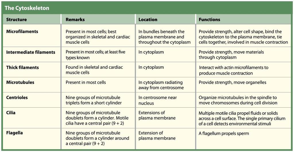
32 Module 3.4: The cytoskeleton plays both a structural and a functional role Cytoskeleton Functions as cell s skeleton Provides internal protein framework Gives cytoplasm strength and flexibility Components include: 1. Microfilaments 2. Intermediate filaments 3. Microtubules
33 Module 3.4: The cytoskeleton Microfilaments 6 nm in diameter (smallest cytoskeletal element) Typically composed of actin Commonly at periphery of cell
 Microvilli Finger-shaped extensions of cell membrane Have core of microfilaments to stiffen and")
34 Module 3.4: The cytoskeleton Microfilaments (continued) Microvilli Finger-shaped extensions of cell membrane Have core of microfilaments to stiffen and anchor Enhance surface area of cell for absorption Terminal web (microfilaments inside plasma membrane in cells forming a layer or lining)

35 Module 3.4: The cytoskeleton Intermediate filaments 7 11 nm in diameter Strongest and most durable cytoskeletal elements
36 Module 3.4: The cytoskeleton Microtubules ~25 nm in diameter Hollow tubes built from globular protein tubulin Largest components of cytoskeleton Extend outward from centrosome (near nucleus)
 Two in each centrosome Control movement")
37 Module 3.4: The cytoskeleton Centrioles Composed of microtubules (9 groups of triplets) Two in each centrosome Control movement of DNA strands during cell division Cells without centrioles cannot divide Red blood cells Skeletal muscle cells

38 Module 3.4: The cytoskeleton Cilia Long, slender plasma membrane extensions Motile cilia common in respiratory and reproductive tracts Microtubules surrounding a central pair Anchored to cell surface with basal body
39 Motile cilia beat rhythmically
40 Module 3.4: The cytoskeleton Cilia (continued) Primary cilium functions as sensor Flagella are longer than cilia and beat in a wavelike fashion
41
42 Module 3.4: Review A. List the three basic components of the cytoskeleton. B. Which cytoskeletal component is common to both centrioles and cilia? C. What is the function of motile cilia? D. Which cytoskeletal structure is found only in males? Learning Outcome: Differentiate among the structures and functions of the cytoskeleton.
43 Module 3.5: Ribosomes and endoplasmic reticulum Ribosomes Responsible for protein synthesis Two subunits (1 large, 1 small) containing special proteins and ribosomal RNA (rrna) Must join together before synthesis begins
44 Module 3.5: Ribosomes and endoplasmic reticulum Ribosomes (continued) Free ribosomes Throughout cytoplasm Manufactured proteins enter cytosol Bound or fixed ribosomes Attached to rough endoplasmic reticulum Synthesize proteins for export out of cell
 Network of intracellular membranes continuous with nuclear envelope, which surrounds nucleus")
45 Module 3.5: Ribosomes and endoplasmic reticulum Endoplasmic reticulum (ER) Network of intracellular membranes continuous with nuclear envelope, which surrounds nucleus Forms hollow tubes, sheets, and chambers (cisternae, singular, cisterna, reservoir for water) Synthesizes and stores proteins, lipids, and carbohydrates

 1.")
46 Module 3.5: Ribosomes and endoplasmic reticulum Two types of endoplasmic reticulum (ER) 1. Smooth (SER) Lacks ribosomes Cisternae are often tubular
 Has attached (fixed) ribosomes Modifies newly synthesized proteins Exports those proteins to Golgi apparatus Proportion of SER to")
47 Module 3.5: Ribosomes and endoplasmic reticulum Two types of endoplasmic reticulum (ER) (continued) 2. Rough (RER) Has attached (fixed) ribosomes Modifies newly synthesized proteins Exports those proteins to Golgi apparatus Proportion of SER to RER depends on the cell and its functions

48 Module 3.5: Ribosomes and endoplasmic reticulum Polypeptide formation in RER Polypeptide synthesized on attached ribosome Growing chain enters cisterna of RER Polypeptide assumes secondary/tertiary structures Completed protein may become enzyme or glycoprotein Products not destined for RER are packaged into transport vesicles Deliver products to Golgi apparatus
49 Module 3.5: Review A. Describe the immediate cellular destinations of newly synthesized proteins from free ribosomes and fixed ribosomes. B. Compare and contrast the structure of SER and RER. C. Why do certain cells in the ovaries and testes contain large amounts of SER? D. The ER is connected to and continuous with what other organelle in the cell? Learning Outcome: Describe the ribosome and smooth and rough endoplasmic reticula, and indicate their specific functions.
50 Module 3.6: The Golgi apparatus is a packaging center Golgi apparatus (Golgi complex) Functions 1. Renews or modifies plasma membrane 2. Modifies or packages secretions into secretory vesicles for release from cell (exocytosis) 3. Packages special enzymes within vesicles for use in cytosol (lysosomes) Typically consists of 5 6 flattened discs (cisternae) May be more than one apparatus in a cell Situated near nucleus
51 Module 3.6: Golgi apparatus Golgi apparatus process 1. Transport vesicles filled with proteins and/or glycoproteins from rough ER arrive at cis face ( receiving side ) of Golgi apparatus. 2. Transport vesicles fuse, forming new cisternae. Enzymes in Golgi apparatus modify arriving products. 3. Products modified and re-packaged as they move toward trans face ( shipping side ). 4. Finalized products packaged in secretory vesicles and released from trans face.

52 Golgi apparatus process
53 Module 3.6: Golgi apparatus Golgi apparatus products 1. Membrane renewal vesicles Add to plasma membrane Allow alteration of plasma membrane properties, changing sensitivity and functions of cells 2. Secretory vesicles Contain hormones or enzymes for extracellular release 3. Lysosomes Contain digestive enzymes for intracellular use
54 Module 3.6: Golgi apparatus Lysosomes Vesicles that isolate digestive processes from the rest of the cytoplasm Three basic functions 1. Fusion with another organelle and digestion of contents 2. Fusion with another vesicle containing fluid or solid extracellular materials and digestion of contents 3. Release of digestive enzymes within the cytoplasm when cell is injured or dying, resulting in autolysis (enzymes destroy cytoplasm) Leads to suicide packets name for lysosomes

55 Lysosomes
56 Module 3.6: Golgi apparatus Membrane flow Continuous movement and exchange of materials between organelles using vesicles Can replace parts of cell membrane to allow cell to grow, mature, or respond to changing environment In an actively secreting cell, the entire membrane surface can be replaced in 1 hour.
57 Module 3.6: Review A. List the three major functions of the Golgi apparatus. B. What do lysosomes contain? C. Describe three functions of lysosomes. Learning Outcome: Describe the Golgi apparatus, and indicate its specific functions.
58 Module 3.7: Mitochondria are the powerhouses of the cell Mitochondria (mitos, thread + chondrion, granule) Produce energy (ATP) for cells Vary in number per cell depending on cell s energy requirements (more energy needs = more mitochondria) Mitochondria account for 30 percent of the heart cardiac muscle cells Red blood cells have no mitochondria Contain their own DNA (mtdna) and ribosomes
59 Module 3.7: Mitochondria Mitochondrial double membrane Outer membrane surrounds organelle Inner membrane contains folds called cristae Inner membrane encloses liquid called matrix Cristae increase surface area exposed to matrix Metabolic enzymes in matrix catalyze reactions providing energy for cellular function

60 Cut-away view of mitochondrion organelle

61 Mitochondrion organelle
62 Module 3.7: Mitochondria Steps of ATP production 1. Glycolysis (glycos, sugar -lysis, a loosening) Occurs in cytosol 1 glucose 2 pyruvate Pyruvate absorbed into mitochondria 2. In mitochondrial matrix: CO 2 removed from pyruvate Enters citric acid (or TCA, tricarboxylic acid) cycle Systematically removes CO 2 and hydrogen atoms
63 Module 3.7: Mitochondria Steps of ATP production (continued) 3. Enzymes and coenzymes use hydrogen atoms to catalyze ATP from ADP Also forms H 2 O 4. ATP leaves mitochondrion

64 ATP Production
65 Module 3.7: Mitochondria Aerobic metabolism or cellular respiration ATP production that requires oxygen Occurs in the mitochondria Much more efficient than ATP production without oxygen (e.g., glycolysis) Produces about 95 percent of ATP needed by cell Remaining 5 percent produced by enzymatic reactions in the cytoplasm
66 Module 3.7: Review A. Describe the structure of a mitochondrion. B. Most of a cell s ATP is produced within its mitochondria. What gas do mitochondria require to produce ATP, and what gas results? C. What does the presence of many mitochondria imply about a cell s energy requirements? Learning Outcome: Describe the structure of a mitochondrion, and explain the significance of mitochondria to cellular function.
67 Section 2: Structure and Function of the Nucleus Learning Outcomes 3.8 Describe the role of the nucleus in maintaining homeostasis at the cellular level. 3.9 Describe the functions of the cell nucleus, and distinguish between chromatin and a chromosome Discuss the nature of the genetic code, and summarize the process of protein synthesis Summarize the process of transcription Summarize the process of translation.
68 Module 3.8: The nucleus is the control center for cellular homeostasis Nucleus Usually largest cellular structure Control center for cellular operations Can direct synthesis of >100,000 different proteins Genetic information coded in sequence of nucleotides Determines cell structure and function Usually only one per cell Exceptions Skeletal muscle cells have many Mature red blood cells have none o Because of no nucleus, they disintegrate within 3 4 months
 changes Short-term adjustments Enzyme activity changes Long-term")
69 Module 3.8: Role of the nucleus The nucleus directs cellular responses to environmental (ECF) changes Short-term adjustments Enzyme activity changes Long-term adjustments Changes in enzymes produced Changes in cell structure from changes in structural proteins Often occur as part of growth, development, and aging
70 Module 3.8: Review A. How is genetic information coded in the cell? B. How many nuclei do most body cells contain? C. Describe why the nucleus is said to be the control center for the cell. Learning Outcome: Describe the role of the nucleus in maintaining homeostasis at the cellular level.
71 Module 3.9: The nucleus contains DNA, RNA, organizing proteins, and enzymes Nuclear structures and functions Nuclear envelope Separates nucleus from cytoplasm Double membrane Perinuclear space (peri-, around) o Space between layers Nuclear pores Passageways that allow chemical communication between nucleus and cytoplasm Movement of ions and small molecules regulated by proteins at the pores Account for about 10% of the surface of the nucleus

72 Module 3.9: Contents of the cell nucleus Nucleoplasm Fluid contents of nucleus Contains network of fine filaments for structural support Also contains ions, enzymes, nucleotides, and small amounts of RNA and DNA
 Transient nuclear organelles Composed of RNA, enzymes, and proteins")
73 Module 3.9: Contents of the cell nucleus Nucleoli (singular, nucleolus) Transient nuclear organelles Composed of RNA, enzymes, and proteins (histones) Assemble RNA subunits Most prominent in cells manufacturing large amounts of proteins Examples: liver, nerve, muscle cells

74 Module 3.9: Contents of the cell nucleus DNA in the nucleus Stores instructions for protein synthesis Strands in nucleus coiled, allowing much to be packed in small space Wrap around histone molecules forming nucleosomes Loosely coiled (chromatin) in nondividing cells Tightly coiled (chromosomes) in dividing cells

75 Module 3.9: Contents of the cell nucleus DNA during cell division Starts by becoming tighter and more complex, forming chromosomes Two copies of each chromosome held together at centromere 23 paired chromosomes in somatic (general body) cells One each from mother/father Carry instructions for proteins and RNA Also some regulatory and unknown functions
76 Module 3.9: Review A. Describe the contents and the structure of the nucleus. B. What molecule in the nucleus contains instructions for making proteins? C. How many chromosomes are contained within a typical somatic cell? D. The total length of the DNA within a human cell nucleus is approximately 2 meters. How does the DNA fit into the relatively small space of a human nucleus, which ranges some 6 10 µm in diameter? Learning Outcome: Describe the functions of the cell nucleus, and distinguish between chromatin and a chromosome.

77 Module 3.10: Protein synthesis involves DNA, enzymes, and three types of RNA DNA Long parallel chains of nucleotides Chains held by hydrogen bonds between nitrogenous bases Four nitrogenous bases 1. Adenine (A) 2. Thymine (T) 3. Cytosine (C) 4. Guanine (G)
78 Module 3.10: The genetic code and protein synthesis DNA (continued) Genetic information stored in sequence of base pairs Known as the genetic code Triplet code Gene Sequence of three nitrogenous bases (triplet) Specifies single amino acid Functional unit of heredity Contains all the DNA nucleotides to produce a specific protein Size varies (~ nucleotides)

79 Module 3.10: The genetic code and protein synthesis

80 Module 3.10: The genetic code and protein synthesis Steps in protein synthesis 1. Gene activation Removal of histones and DNA uncoiling 2. DNA strands separate
 Complementary base pairing matches DNA nucleotide sequence with new mrna sequence (A-U; G-C) Series of")
81 Module 3.10: The genetic code and protein synthesis 3. Enzymes assemble nucleotides into a single strand of messenger RNA (mrna) Complementary base pairing matches DNA nucleotide sequence with new mrna sequence (A-U; G-C) Series of three RNA nucleotides called a codon Each codon codes for specific amino acid 4. mrna leaves nucleus through nuclear pores
 on transfer RNA (trna)")
82 Module 3.10: The genetic code and protein synthesis 5. At a ribosome in the cytoplasm, codons of mrna bind to anticodons (triplets of corresponding nucleotides) on transfer RNA (trna) 6. trna carries a specific amino acid (associated with specific anticodon)
 of the ribosome strings amino acids")
83 Module 3.10: The genetic code and protein synthesis 7. Ribosomal RNA (rrna) of the ribosome strings amino acids together
84 Protein synthesis

85 Module 3.10: The genetic code and protein synthesis
86 Module 3.10: Review A. What is a gene? B. Why is the genetic code described as a triplet code? C. List the three types of RNA involved in protein synthesis. D. Which type of RNA links the genetic information in the nucleus with the cytoplasmic sites of protein synthesis? Learning Outcome: Discuss the nature of the genetic code, and summarize the process of protein synthesis.
87 Module 3.11: Transcription encodes genetic instructions on a strand of RNA Transcription ( to copy or rewrite ) Takes place in the nucleus Production of RNA from DNA template All three types of RNA are formed
88 Module 3.11: Transcription Steps of transcription 1. Gene activation Occurs at control segment or promoter (1st segment of gene) Only template strand of DNA used to synthesize RNA
 binds to promoter Begins")
89 Module 3.11: Transcription Steps of transcription 2. Beginning of assembly RNA polymerase (enzyme) binds to promoter Begins assembly of mrna strand
90 Module 3.11: Transcription Steps of transcription (continued) 3. Continuation of mrna strand RNA polymerase promotes hydrogen bonding between nucleotides on DNA template strand and complementary RNA nucleotides in nucleoplasm Example: (DNA triplet TAC = mrna AUG) Nucleotides connected by covalent bonding
91 Module 3.11: Transcription Steps of transcription (continued) 4. Transcription ends Stop codon reached mrna detaches Complementary DNA strands reassociate (with hydrogen bonding between complementary base pairs)

92 Steps of transcription

93 Module 3.11: Transcription Final processing of mrna Initial strand of mrna called immature mrna or pre-mrna Before leaving nucleus, mrna requires additional processing Introns (noncoding sequences) removed Remaining coding segments (exons) spliced together Changing the editing can produce mrna for different proteins
94 Module 3.11: Review A. What is transcription? B. Define DNA template strand. C. Name the substrates and product in the enzymatic reaction catalyzed by RNA polymerase. D. What process would be affected if a cell could not synthesize the enzyme RNA polymerase? Learning Outcome: Summarize the process of transcription.
95 Module 3.12: Translation builds polypeptides as directed by an mrna strand Translation Formation of a linear chain of amino acids from an mrna strand Translates genetic information from nucleic acids to proteins Occurs in cytoplasm on ribosomes Three phases 1. Initiation 2. Elongation 3. Termination
96 Module 3.12: Translation Steps of translation 1. Initiation phase mrna binds to small ribosomal subunit near the P site trna binds to P site and to start codon on mrna strand Binding occurs between mrna codons and trna complementary anticodons Small and large ribosomal subunits interlock around mrna strand forming initiation complex Additional trna binds to A site More than 20 kinds of trna Each carries an amino acid
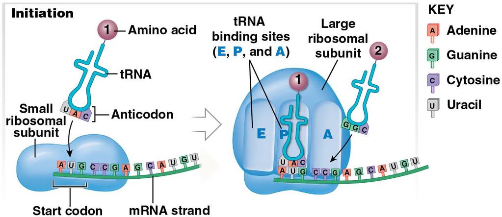
97 Initiation phase
98 Module 3.12: Translation Steps of translation (continued) 2. Elongation Ribosomal enzymes remove amino acid from trna at P site and attach it to trna in A site Ribosome links amino acids forming dipeptide Ribosome moves to next codon on mrna strand trna from P site moves to E site and is released This trna can go bind to another amino acid More trnas arrive, match codon to anticodon, and continue forming polypeptide

99 Elongation

100 Module 3.12: Translation Steps of translation (continued) 3. Termination Stop codon on mrna Recognized by protein releasing factor Ribosomal enzyme breaks bond between polypeptide and trna in P site Ribosomal subunits detach Leaves intact mrna and new polypeptide

101 Module 3.12: Translation
102 Module 3.12: Translation Translation Produces a typical protein in ~20 seconds mrna can interact with other ribosomes and produce more proteins Multiple ribosomes can attach to a single mrna strand to quickly produce many proteins

103 BioFlix: Protein Synthesis
104 Module 3.12: Review A. What is translation? B. The nucleotide sequence of three mrna codons is AUU-GCA-CUA. What is the complementary anticodon sequence for the second codon? C. During the process of transcription, a nucleotide was deleted from an mrna sequence that coded for a protein. What effect will this deletion have on the amino acid sequence of the protein? Learning Outcome: Summarize the process of translation.
105 Section 3: How Substances Enter and Leave the Cell Learning Outcomes 3.13 Contrast permeable, selectively permeable, and impermeable membranes Explain the process of diffusion, and identify its significance in the body Explain the process of osmosis, and identify its significance in the body.
106 Section 3: How Substances Enter and Leave the Cell Learning Outcomes (continued) 3.16 Describe carrier-mediated transport and its role in the absorption and removal of specific substances Describe vesicular transport as a mechanism for facilitating the absorption or removal of specific substances from cells.
107 Module 3.13: The plasma membrane is a selectively permeable membrane Permeability Property determining which substances can enter or leave cytoplasm Freely permeable Any substance can pass (not found in living cells) Selectively permeable Some substances cross Impermeable No substances can pass (not found in living cells) Plasma membrane must allow some movement in and out of cells to enable intercellular communication and coordination

108 Module 3.13: Permeability of membranes
109 Module 3.13: Permeability of membranes Selectively permeable membranes Permit free passage of some materials and restrict others 1. Characteristics of material to pass Size Molecular shape Lipid solubility Electrical charge Other factors 2. Characteristics of cell membrane What lipids and proteins present How components are arranged
 Diffusion Carrier-mediated transport 2.")
110 Module 3.13: Permeability of membranes Types of membrane transport 1. Passive (do not require ATP) Diffusion Carrier-mediated transport 2. Active (require ATP) Vesicular transport Carrier-mediated transport
111 Module 3.13: Review A. Define permeability. B. Identify three different types of membranes based on permeability. C. Distinguish between passive and active processes of membrane passage. D. What kinds of molecules are involved in both active and passive processes of membrane passage. Learning Outcome: Contrast permeable, selectively permeable, and impermeable membranes.
112 Module 3.14: Diffusion is passive movement driven by concentration differences Diffusion Net movement of a substance from higher concentration to lower concentration. Concentration gradient Concentration difference when molecules are not evenly distributed At an even distribution, molecular motion continues but no net movement
 Slow in air")
113 Module 3.14: Diffusion Diffusion (continued) Slow in air and water but important over small distances
114 Module 3.14: Diffusion Movement of water and solutes across plasma membrane: Selectively restricted diffusion Movement across lipid portion of membrane Examples: lipids, lipid-soluble molecules, soluble gases Movement through membrane channel Examples: water, small water-soluble molecules, ions Movement using carrier molecules Example: large molecules

115 Diffusion across a plasma membrane EXTRACELLULAR FLUID
116 Module 3.14: Diffusion Factors that influence diffusion rates Distance Shorter distance = faster diffusion Molecule or ion size Smaller size = faster diffusion Temperature Higher temperature = faster diffusion Concentration gradient Steeper gradient = faster diffusion Electrical forces Attraction of opposite charges (+, ) Repulsion of like charges (+,+ or, )
117 Module 3.14: Review A. Define diffusion. B. Describe the colliding molecules in the figure below (with the sugar cube in water). C. Identify factors that influence diffusion rates. D. How would a decrease in the oxygen concentration in the lungs affect oxygen diffusion into the blood? Learning Outcome: Explain the process of diffusion, and identify its significance in the body.
118 Module 3.15: Osmosis is the diffusion of water molecules across a selectively permeable membrane Osmosis (osmos, a push) Net diffusion of water across a membrane Maintains similar overall solute concentrations between the cytosol and extracellular fluid Osmotic flow Movement of water driven by osmosis
119 Module 3.15: Osmosis Osmosis (continued) Osmotic pressure Indication of force of pure water moving into a solution with higher solute concentration Hydrostatic pressure Fluid force Can be estimate of osmotic pressure when applied to stop osmotic flow

120 Water movement through a selectively permeable membrane
121 Module 3.15: Osmosis Osmolarity and tonicity Osmolarity (osmotic concentration) Total solute concentration in an aqueous solution Tonicity Effect of osmotic solutions on cell volume How a solution affects a cell
 Solution that does not cause")
122 Module 3.15: Osmosis Three effects of tonicity 1. Isotonic (iso-, same tonos, tension) Solution that does not cause osmotic flow across membrane

123 Module 3.15: Osmosis Three effects of tonicity (continued) 2. Hypotonic Causes osmotic flow into cell Example: swelling and hemolysis (hemo-, blood + lysis, loosening) of red blood cell

124 Module 3.15: Osmosis Three effects of tonicity (continued) 3. Hypertonic Causes osmotic flow out of cell Example: shriveling and crenation of RBCs

125 Effects of tonicity
126 Module 3.15: Osmosis Importance of tonicity vs. osmolarity Administering large fluid volumes to patients with blood loss or dehydration If administered solution has same osmolarity as ICF but higher concentrations of individual ions/molecules Diffusion of solutes may occur across cell membrane Water will follow through osmosis Cell volume increases Normal saline often administered in emergency 0.9 percent or 0.9 g/dl of NaCl Isotonic with blood
127 Module 3.15: Review A. Describe osmosis. B. Describe osmotic pressure, and state in which solution below it is greater. C. Contrast the effects of a hypotonic solution and a hypertonic solution on a red blood cell. D. Some pediatricians recommend using a 10 percent salt solution to relieve nasal congestion in infants. Explain the effects this treatment would have on the cells lining the nasal cavity. Would it be effective? Learning Outcome: Explain the process of osmosis, and identify its significance in the body.
128 Module 3.16: In carrier-mediated transport, integral proteins facilitate membrane passage Carrier proteins Transport hydrophilic or large molecules across cell membrane Many move specific molecules through the plasma membrane in only one direction Some move more than one substance in the same direction (cotransport) Some move more than one substance in opposite directions Process called countertransport Carrier called an exchange pump
 Carrier binds to molecule, then changes shape to move molecule across")
129 Module 3.16: Carrier-mediated transport 1. Facilitated diffusion Requires no ATP Passive transport (moves from high concentration to low concentration) Carrier binds to molecule, then changes shape to move molecule across membrane Rate of transport limited by number of available carrier proteins Once all carrier proteins saturated, no increase in rate of transport

130 Module 3.16: Carrier-mediated transport 2. Active transport Active process requiring energy molecule or ATP Independent of concentration gradient Examples: Ion pumps (Na +, K +, Ca 2+, and Mg 2+ ) Sodium potassium ATPase Exchanges 3 intracellular sodium ions for 2 extracellular potassium ions

131 Module 3.16: Carrier-mediated transport 3. Secondary active transport Transport mechanism itself does not require ATP Cell often needs ATP to maintain homeostasis associated with transport Movement for one of two substances follows concentration gradient Example: Sodium and glucose cotransporter

132 A&P Flix: Membrane Transport
133 Module 3.16: Review A. Describe the process of carrier-mediated transport. B. What two factors limit the rate of facilitated diffusion across a plasma membrane? C. What do the transport processes of facilitated diffusion and active transport have in common? D. During digestion, the concentration of hydrogen ions (H + ) in the stomach contents increases to many times that in cells lining the stomach. Which transport process could be responsible? Learning Outcome: Describe carrier-mediated transport and its role in the absorption and removal of specific substances.
134 Module 3.17: In vesicular transport, vesicles selectively carry materials into or out of cell Vesicular transport Materials move across cell membrane in small membranous sacs called vesicles Sacs form at or fuse with plasma membrane Two major types (both require ATP) 1. Endocytosis Importing extracellular substances into vesicles called endosomes 2. Exocytosis Movement of wastes or secretory products from intracellular vesicle to outside the cell
135 Module 3.17: Vesicular transport Receptor-mediated endocytosis Brings specific molecules into cell using receptor molecules on membrane surface a. Target molecule (ligand) binds to receptor
136 Module 3.17: Vesicular transport Receptor-mediated endocytosis (continued) b. Plasma membrane folds around receptors bound to ligands, forming pocket that pinches off c. Endosome called clathrin-coated vesicle forms

137 Module 3.17: Vesicular transport Receptor-mediated endocytosis (continued) d. Vesicle fuses with lysosomes
138 Module 3.17: Vesicular transport Receptor-mediated endocytosis (continued) e. Ligands freed from receptors and enter cytoplasm

139 Module 3.17: Vesicular transport Receptor-mediated endocytosis (continued) f. Lysosome detaches from vesicle
140 Module 3.17: Vesicular transport Receptor-mediated endocytosis (continued) g. Vesicle fuses with plasma membrane again
141 Receptor-mediated endocytosis
 Formation of endosomes with")
142 Module 3.17: Vesicular transport Pinocytosis ( cell drinking ) Formation of endosomes with ECF No receptor proteins involved Brings fluid and small molecules into cell
 Produces phagosomes containing solids No receptors involved Cytoplasmic")
143 Module 3.17: Vesicular transport Phagocytosis ( cell eating ) Produces phagosomes containing solids No receptors involved Cytoplasmic extensions (pseudopodia) surround object and bring it into cell Only specialized cells (phagocytes or macrophages) perform phagocytosis

144 Module 3.17: Vesicular transport Exocytosis functional opposite of endocytosis Vesicle contents are released to extracellular environment
145 Module 3.17: Review A. Describe endocytosis. B. Describe the three types of endocytosis. C. Describe exocytosis. D. Some white blood cells engulf bacteria and bring them into the cell. What is this process called? Learning Outcome: Describe vesicular transport as a mechanism for facilitating the absorption or removal of specific substances from cells.
146 Section 4: Cell Life Cycle Learning Outcomes 3.18 Distinguish between interphase and cell division in the cell cycle Describe interphase, and explain its significance Describe the process of mitosis and its role in the cell life cycle Clinical Module: Discuss the relationship between cell division and cancer.
147 Module 3.18: Interphase and cell division make up the life cycle of a cell Life starts as a single cell At maturity, roughly 75 trillion cells in the body Cell division form of cellular reproduction Responsible for initial increase in cell number Essential to continued development and survival Cells have varying life spans and abilities to divide Often genetically controlled death occurs (apoptosis) Cell life cycle ends when cell dies
148 Module 3.18: Cell life cycle Two types of cell division 1. Mitosis 2 daughter cells produced Each with 46 chromosomes 2. Meiosis Produces sex cells Each with only 23 chromosomes

149 Module 3.18: Cell life cycle Mitosis Form of cellular reproduction Division of single cell produces pair of daughter cells Half the size of parent cell Grow to size of original cell before dividing
150 Module 3.18: Cell life cycle Divisions of cell life cycle 1. Interphase (nondividing period) Cell performs normal activities
151 Module 3.18: Cell life cycle Divisions of cell life cycle (continued) 2. Cell division Begins with mitosis Distribution of identical copies of chromosomes to each daughter cell Ends with cytokinesis (division of the cytoplasm)
152 Module 3.18: Review A. Explain why cell division is important. B. Define apoptosis. C. When does cell division begin and end? Learning Outcome: Distinguish between interphase and cell division in the cell cycle.
153 Module 3.19: During interphase, the cell prepares for cell division Division of interphase Somatic (body) cells spend most of their lives in interphase For cells not preparing to divide, they stay in: G 0 phase Performing normal cell functions Examples: o Skeletal muscle cells and most neurons Stay in this phase forever o Stem cells Never enter G 0 Divide repeatedly

154 Module 3.19: Interphase For cells preparing to divide, interphase divided into: G 1 phase Normal cell functions, cell growth, duplication of organelles, protein synthesis S phase DNA replication, synthesis of histones and other proteins to allow duplication of chromosomes G 2 phase Last minute protein synthesis and centriole replication
155 Module 3.19: Interphase DNA replication process DNA helicase Unwinds DNA strands Disrupts hydrogen bonds between bases DNA polymerase Binds to exposed bases Promotes bonding between current DNA strand and complementary nucleotides in nucleoplasm Covalently links nucleotides together
156 Module 3.19: Interphase DNA replication process (continued) DNA polymerase (continued) Works only in one direction One polymerase works continuously along one strand toward zipper forming the leading strand
157 Module 3.19: Interphase DNA replication process (continued) DNA polymerase (continued) Works only in one direction One polymerase works away from zipper forming the lagging strand o As unzipping occurs, another polymerase binds closer point of unzipping o Two new DNA segments spliced together with DNA ligases Two identical DNA strands formed
158 DNA replication

159 A&P Flix: DNA Replication
160 Module 3.19: Review A. Describe interphase, and identify its stages. B. A cell is actively manufacturing enough organelles to serve two functional cells. This cell is probably in what phase of interphase? C. What enzymes must be present for DNA replication to proceed normally? D. DNA replication occurs during what two cellular processes? Learning Outcome: Describe interphase, and explain its significance.
161 Module 3.20: Mitosis distributes chromosomes before cytokinesis separates the daughter cells M phase of cell cycle Includes mitosis and cytokinesis Mitosis Division and duplication of the cell s nucleus Divided into four stages: 1. Prophase 2. Metaphase 3. Anaphase 4. Telophase Cytokinesis Division of cytoplasm


162 Module 3.20: Mitosis Interphase DNA replicated, DNA is loosely coiled and no visible chromosomes
 Nuclear envelope disintegrates Chromosomes")
 Spindle fibers")
163 Module 3.20: Mitosis Phases of mitosis 1. Prophase (pro-, before) Nuclear envelope disintegrates Chromosomes coil and become visible under light microscope Replicated centrioles move to poles Astral rays (extend from centrioles) Spindle fibers (interconnect centriole pairs)
 Each copy of chromosome called")
 at centromere attaches to spindle")
164 Module 3.20: Mitosis Phases of mitosis (continued) 1. Prophase (continued) Each copy of chromosome called chromatid Pair connected at centromere Raised region (kinetochore) at centromere attaches to spindle fibers

")
165 Module 3.20: Mitosis Phases of mitosis (continued) 2. Metaphase (meta, after) Chromosomes align at metaphase plate
 3.")

166 Module 3.20: Mitosis Phases of mitosis (continued) 3. Anaphase (ana-, apart) Centromere splits Chromatids separate Chromatids drawn toward opposite sides along spindle apparatus
 Cells prepare to enter interphase")

167 Module 3.20: Mitosis Phases of mitosis (continued) 4. Telophase (telo-, end) Cells prepare to enter interphase Cytoplasm constricts along metaphase plate (cleavage furrow) Nuclear membranes re-form Nuclei enlarge Chromosomes uncoil to chromatin


168 Module 3.20: Mitosis Cytokinesis (cyto-, cell + kinesis, motion) Begins with formation of cleavage furrow Continues through telophase Completion marks end of cell division

169 Module 3.20: Mitosis

170 A&P Flix: Mitosis
171 Module 3.20: Review A. Define mitosis, and list its four stages. B. What is a chromatid, and how many are present during normal mitosis in a human cell? C. What would happen if spindle fibers failed to form in a cell during mitosis? Learning Outcome: Describe the process of mitosis and its role in the cell life cycle.
172 Module 3.21: CLINICAL MODULE: Tumors and cancer are characterized by abnormal cell growth and division Cancer Illness that disrupts normal rates of cell division Characterized by permanent DNA sequence changes (mutations) Most common in tissues with actively dividing cells Examples: skin, intestinal lining Cancerous cells compete with normal cells for resources Usually begins with single abnormal cell
 Mass or swelling produced by abnormal cell growth and")
173 Module 3.21: CLINICAL MODULE: Tumors and cancer Tumor (neoplasm) Mass or swelling produced by abnormal cell growth and division 1. Benign tumor Cells remain within original tissue Seldom a threat Can be removed surgically if necessary
174 Module 3.21: CLINICAL MODULE: Tumors and cancer Malignant tumor Cells divide rapidly Released chemicals stimulate blood vessel growth (angiogenesis) to tumor area
175 Module 3.21: CLINICAL MODULE: Tumors and cancer Malignant tumor (continued) Accelerated growth due to blood vessel growth and supply to the area Tumor spreads to surrounding tissue by invasion Cells migrate to other areas and establish new tumors (called metastasis)

176 Module 3.21: CLINICAL MODULE: Tumors and cancer Malignant cells disrupt function No longer perform original functions or: Perform functions in an abnormal way Example: Malignant tumor of thyroid gland produces abnormal amounts of thyroid hormone Cancer cells compete with normal cells for space and nutrients
177 Module 3.21: Review A. Define cancer. B. What is a benign tumor? C. Define metastasis. D. How does angiogenesis aid tumor growth? Learning Outcome: Discuss the relationship between cell division and cancer.
CHAPTER 3. Cell Structure and Genetic Control. Chapter 3 Outline
 CHAPTER 3 Cell Structure and Genetic Control Chapter 3 Outline Plasma Membrane Cytoplasm and Its Organelles Cell Nucleus and Gene Expression Protein Synthesis and Secretion DNA Synthesis and Cell Division
Chapter 03. Lecture and Animation Outline
 Chapter 03 Lecture and Animation Outline To run the animations you must be in Slideshow View. Use the buttons on the animation to play, pause, and turn audio/text on or off. Please Note: Once you have
The Cellular Level of Organization
 C h a p t e r 3 The Cellular Level of Organization PowerPoint Lecture Slides prepared by Jason LaPres Lone Star College - North Harris Copyright 2009 Pearson Education, Inc., publishing as Pearson Benjamin
THE CELL 3/15/15 HUMAN ANATOMY AND PHYSIOLOGY I THE CELLULAR BASIS OF LIFE
 HUMAN ANATOMY AND PHYSIOLOGY I Lecture: M 6-9:30 Randall Visitor Center Lab: W 6-9:30 Swatek Anatomy Center, Centennial Complex Required Text: Marieb 9 th edition Dr. Trevor Lohman DPT (949) 246-5357 tlohman@llu.edu
Cells: The Living Units
 Golgi Apparatus Cells: The Living Units Chapter 3, PPT 2 Membrane Yes, stacked and flattened Description Stacked and flattened membranous sacs Modify, concentrate, and package proteins & lipids made at
Life of the Cell. Learning Objectives
 Life of the Cell Society on a micro-scale 1 Learning Objectives 1. What are the characteristics that distinguish prokaryotic and eukaryotic cells? Which type of cell is believed to be older (more primitive)?
The Cell. C h a p t e r. PowerPoint Lecture Slides prepared by Jason LaPres North Harris College Houston, Texas
 C h a p t e r 2 The Cell PowerPoint Lecture Slides prepared by Jason LaPres North Harris College Houston, Texas Copyright 2009 Pearson Education, Inc., publishing as Pearson Benjamin Cummings Introduction
Chapter 3: Cells and Their Functions. Copyright 2013 Wolters Kluwer Health Lippincott Williams & Wilkins
 Chapter 3: Cells and Their Functions Overview Key Terms active transport filtration mitochondria cancer gene mitosis carcinogen hemolysis mutation chromosome hypertonic nucleus cytology hypotonic organelle
Chapter 3: Structure and Function of the Cell
 Chapter 3: Structure and Function of the Cell I. Functions of the Cell A. List and describe the main functions of the cell: 1. 2. 3. 4. 5. II. How We See Cells A. Light microscopes allow us to B. Electron
Honors Biology-CW/HW Cell Biology 2018
 Class: Date: Honors Biology-CW/HW Cell Biology 2018 Multiple Choice Identify the choice that best completes the statement or answers the question. 1. Hooke s discovery of cells was made observing a. living
Chapter 3. Cells. Cells. Cells
 Chapter 3 Cells Cells Cytology The basic unit of life Humans have about 75 trillion cells Largest human cell is the egg While there are many varieties to cells there are many common characteristics. Cells
Cell Theory. The Cellular Level Of Organization. Cell Biology. The Typical Cell. The Diversity of Cells in the Human Body.
 Cell Theory The Cellular Level Of Organization Chapter 3 Developed from Robert Hooke s research Cells are the building blocks of all plants and animals All cells come from the division of preexisting cells
Hole s Human Anatomy and Physiology
 Hole s Human Anatomy and Physiology 1 Chapter 3 Cells vary in size possess distinctive shapes measured in micrometers 2 A Composite Cell hypothetical cell major parts nucleus cytoplasm cell membrane 3
Cell Structure and Cell Cycle
 E X E R C I S E 4 Cell Structure and Cell Cycle Materials model or diagram of a cell compound microscopes and lens paper prepared slides of human skeletal muscle cells, pseudostratified ciliated columnar
Passive. mechanisms. Active. mechanisms. Cell diffusion. Movement. Movement. Movement. Mechanisms that do not require cellular energy such as:
 Passive mechanisms Mechanisms that do not require cellular energy such as: Diffusion Facilitated diffusion Osmosis Filtration Active mechanisms Mechanisms that do require cellular energy such as: Active
Introduction to Cells
 Life Science Introduction to Cells All life forms on our planet are made up of cells. In ALL organisms, cells have the same basic structure. The scientist Robert Hooke was the first to see cells under
Chapter 3 Cells. Figure 3.1 Question: What are the three principal parts of a cell? a. b. c.
 Name Date Physiology & Anatomy Figure 3.1 Question: What are the three principal parts of a cell? Chapter 3 Cells a. b. c. As you go through the rest of this chapter, label the parts of the cell on the
O.k., Now Starts the Good Stuff (Part II) Eukaryotic Cell Structure and Function
 Eukaryotic Cell Structure and Function") O.k., Now Starts the Good Stuff (Part II) Eukaryotic Cell Structure and Function Eukaryotic Cells These cells have membrane-bound structures called organelles. Cell processes occur in these organelles.
O.k., Now Starts the Good Stuff (Part II) Eukaryotic Cell Structure and Function Eukaryotic Cells These cells have membrane-bound structures called organelles. Cell processes occur in these organelles.
Human biology Cells: The Basic Units of Life. Dr. Rawaa Salim Hameed
 Human biology Cells: The Basic Units of Life Dr. Rawaa Salim Hameed Reference Text book of human biology by John Kenneth Inglis 3 rd Ed (1985) Cells: The Basic Units of Life Cell theory Cell theory consists
Introduction to Cells
 Life Science Introduction to Cells All life forms on our planet are made up of cells. In ALL organisms, cells have the same basic structure. The scientist Robert Hooke was the first to see cells under
BIO 210 Chapter 4 Physiology of Cells. By Beth Wyatt, Jack Bagwell, & John McGill. Introduction
 BIO 210 Chapter 4 Physiology of Cells By Beth Wyatt, Jack Bagwell, & John McGill Introduction The living must exchange materials with the nonliving. How does this happen? Cell transport Two major types
Basic Structure of a Cell
 Basic Structure of a Cell Prokaryotic Cells No nucleus Archaea & Eubacteria One circular chromosome Extremely small Eukaryotic Cells Has a nucleus!!! Membrane-bound organelles Plants, Animals, Fungi, &
Overview of Cells. Prokaryotes vs Eukaryotes The Cell Organelles The Endosymbiotic Theory
 Overview of Cells Prokaryotes vs Eukaryotes The Cell Organelles The Endosymbiotic Theory Prokaryotic Cells Archaea Bacteria Come in many different shapes and sizes.5 µm 2 µm, up to 60 µm long Have large
Chapter 3 Part 1! 10 th ed.: pp ! 11 th ed.: pp !! Cellular Transport Mechanisms! The Cell Cycle!
 Chapter 3 Part 1! 10 th ed.: pp. 87 105! 11 th ed.: pp. 90 107!! Cellular Transport Mechanisms! The Cell Cycle! Transport Processes: Passive and Active (1 of 2)! 1. Passive transport! Does not use ATP!
Cells and Tissues PART B
 3 Cells and Tissues PART B PowerPoint Lecture Slide Presentation by Jerry L. Cook, Sam Houston University ESSENTIALS OF HUMAN ANATOMY & PHYSIOLOGY EIGHTH EDITION ELAINE N. MARIEB Cellular Physiology: Membrane
Objective: The Cell. The Cell Theory. Question of the day: The Plasma Membrane 11/2/09. The Generalized Cell: Parts and Organelles:
 Objective: The Cell Basic Unit of Life The student will become familiar with the structure and function of the basic cell :) The student will know the differences between and animal cell and a plant cell
Biology 1 Notebook. Review Answers Pages 17 -?
 Biology 1 Notebook Review Answers Pages 17 -? The History of Cell Studies 1. Robert Hook (1665) used a microscope to examine a thin slice of cork. The little boxes he observed reminded him of the small
Now starts the fun stuff Cell structure and function
 Now starts the fun stuff Cell structure and function Cell Theory The three statements of the cell theory are: All organisms are composed of one or more cells and the processes of life occur in these cells.
Class Work 31. Describe the function of the Golgi apparatus? 32. How do proteins travel from the E.R. to the Golgi apparatus? 33. After proteins are m
 Eukaryotes Class Work 1. What does the word eukaryote mean? 2. What is the one major difference between eukaryotes and prokaryotes? 3. List the different kingdoms of the eukaryote domain in the order in
2011 The Simple Homeschool Simple Days Unit Studies Cells
 1 We have a full line of high school biology units and courses at CurrClick and as online courses! Subscribe to our interactive unit study classroom and make science fun and exciting! 2 A cell is a small
Biology I Fall Semester Exam Review 2014
 Biology I Fall Semester Exam Review 2014 Biomolecules and Enzymes (Chapter 2) 8 questions Macromolecules, Biomolecules, Organic Compunds Elements *From the Periodic Table of Elements Subunits Monomers,
The Cell Notes 1 of 11
 The Cell The basic unit of structure and function in living things The smallest units in living things The smallest units in living things that show the characteristics of life Organisms can be made of
Cells. Every organism is made up of a cell or many cells Humans have ~ 100 TRILLION cells each!
 Cells Every organism is made up of a cell or many cells Humans have ~ 100 TRILLION cells each! History 1660 s Microscopes developed Allowed for the observation of cells for the first time Robert Hooke
Number of questions TEK (Learning Target) Biomolecules & Enzymes
 Biomolecules & Enzymes") Unit Biomolecules & Enzymes Number of questions TEK (Learning Target) on Exam 8 questions 9A I can compare and contrast the structure and function of biomolecules. 9C I know the role of enzymes and how
Unit Biomolecules & Enzymes Number of questions TEK (Learning Target) on Exam 8 questions 9A I can compare and contrast the structure and function of biomolecules. 9C I know the role of enzymes and how
Biology: Life on Earth
 Teresa Audesirk Gerald Audesirk Bruce E. Byers Biology: Life on Earth Eighth Edition Lecture for Chapter 4 Cell Structure and Function Copyright 2008 Pearson Prentice Hall, Inc. Chapter 4 Outline 4.1 What
8/25/ Opening Questions: Are all living things made of cells? What are at least five things you know about cells?
 Chapter 3 The Cell: Module Hyperlinks 3.1 Cells are the fundamental units of life 3.2 Plant vs. animal cells 3.3 Membranes: structure 3.4 Membranes: function 3.5 The nucleus 3.6 Organelles in protein production
CELL BIOLOGY. Which of the following cell structures does not have membranes? A. Ribosomes B. Mitochondria C. Chloroplasts D.
 1 CELL BIOLOGY PROKARYOTIC and EUKARYOTIC SP/1. SP/2. SP/4. Plant and animal cells both have A. ribosomes, cell walls and mitochondria. B. Golgi apparatus, chromosomes and mitochondria. C. Golgi apparatus,
Cell Theory Essential Questions
 Cells Vocab words 1. Cell 2. Cell theory 3. Nucleus 4. Eukaryote 5. Prokaryote 6. Organelle 7. Cytoplasm 8. Nuclear envelope 9. Chromatin 10. Chromosome 11. Nucleolus 12. Ribosome 13. Endoplasmic reticulum
3.1 Cell Theory. KEY CONCEPT Cells are the Basic unit of life.
 3.1 Cell Theory KEY CONCEPT Cells are the Basic unit of life. 3.1 Cell Theory The cell theory grew out of the work of many scientists and improvements in the microscope. Many scientists contributed to
Principles of Cellular Biology
 Principles of Cellular Biology آشنایی با مبانی اولیه سلول Biologists are interested in objects ranging in size from small molecules to the tallest trees: Cell Basic building blocks of life Understanding
Cell Types. Prokaryotes
 Cell Types Prokaryotes before nucleus no membrane-bound nucleus only organelle present is the ribosome all other reactions occur in the cytoplasm not very efficient Ex.: bacteria 1 Cell Types Eukaryotes
Chapter 3: Cells. Lectures by Mark Manteuffel, St. Louis Community College
 Chapter 3: Cells Lectures by Mark Manteuffel, St. Louis Community College Learning Objectives Be able to describe: what a cell is & two main classes of cells. structure & functions of cell membranes. how
To help you complete this review activity and to help you study for your test, you should read SC State Standards B
 Name: Test Date: PAGE: Biology I: Unit 3 Cell Structure Review for Unit Test Directions: You should use this as a guide to help you study for your test. You should also read through your notes, worksheets,
A. The Cell: The Basic Unit of Life. B. Prokaryotic Cells. D. Organelles that Process Information. E. Organelles that Process Energy
 The Organization of Cells A. The Cell: The Basic Unit of Life Lecture Series 4 The Organization of Cells B. Prokaryotic Cells C. Eukaryotic Cells D. Organelles that Process Information E. Organelles that
Chapter 3 Cell Structures and Their Functions Dividing Cells
 Chapter 3 Cell Structures and Their Functions Dividing Cells Copyright The McGraw-Hill Companies, Inc. Permission required for reproduction or display. Cell Organization Cell: basic structural & functional
Cell Organelles. a review of structure and function
 Cell Organelles a review of structure and function TEKS and Student Expectations (SE s) B.4 Science concepts. The student knows that cells are the basic structures of all living things with specialized
7-2 Eukaryotic Cell Structure
 1 of 49 Comparing the Cell to a Factory Eukaryotic Cell Structures Structures within a eukaryotic cell that perform important cellular functions are known as organelles. Cell biologists divide the eukaryotic
Chapter 6: A Tour of the Cell
 AP Biology Reading Guide Fred and Theresa Holtzclaw Chapter 6: A Tour of the Cell Name Period Chapter 6: A Tour of the Cell Concept 6.1 To study cells, biologists use microscopes and the tools of biochemistry
Chapter 6: A Tour of the Cell
 Chapter 6: A Tour of the Cell 1. The study of cells has been limited by their small size, and so they were not seen and described until 1665, when Robert Hooke first looked at dead cells from an oak tree.
Cell Cycle and Mitosis
 Cell Cycle and Mitosis THE CELL CYCLE The cell cycle, or cell-division cycle, is the series of events that take place in a eukaryotic cell between its formation and the moment it replicates itself. These
Introduction to Botany
 Introduction to Botany Alexey Shipunov Minot State University Lecture 13 Shipunov (MSU) Introduction to Botany Lecture 13 1 / 27 Outline 1 Questions and answers Quiz 2 Plant cell Cell boundaries Protein
Midterm Review Guide. Unit 1 : Biochemistry: 1. Give the ph values for an acid and a base. 2. What do buffers do? 3. Define monomer and polymer.
 Midterm Review Guide Name: Unit 1 : Biochemistry: 1. Give the ph values for an acid and a base. 2. What do buffers do? 3. Define monomer and polymer. 4. Fill in the Organic Compounds chart : Elements Monomer
Chapter 7: Membrane Structure and Function
 Chapter 7: Membrane Structure and Function 7.1 Cellular membranes are fluid mosaics of lipids and proteins 1. The large molecules of all living things fall into just four main classes. Name them. 2. Explain
CELLS STRUCTURE AND FUNCTION
 CELLS STRUCTURE AND FUNCTION Jhia Anjela D. Rivera Department of Biological Sciences School of Science and Technology Centro Escolar University DISCOVERY OF CELLS Robert Hooke (1665): Observed a thin slice
Life is Cellular Section 7.1
 Life is Cellular Section 7.1 Objectives Understand Cell theory Distinguish between prokaryotes and eukaryotes Understand different types of microscopy, and how they work in more detail What is a Cell?
The Cell. What is a cell?
 The Cell What is a cell? The Cell What is a cell? Structure which makes up living organisms. The Cell Theory l All living things are composed of cells. l Cells are the basic unit of life. l Cells come
THE CELL CYCLE & MITOSIS. Asexual Reproduction: Production of genetically identical offspring from a single parent.
 THE CELL CYCLE & MITOSIS Asexual Reproduction: Production of genetically identical offspring from a single parent. Sexual Reproduction: The fusion of two separate parent cells that produce offspring with
3.a.2- Cell Cycle and Meiosis
 Big Idea 3: Living systems store, retrieve, transmit and respond to information essential to life processes. 3.a.2- Cell Cycle and Meiosis EU 3.A: Heritable information provides for continuity of life.
REVIEW 2: CELLS & CELL DIVISION UNIT. A. Top 10 If you learned anything from this unit, you should have learned:
 Period Date REVIEW 2: CELLS & CELL DIVISION UNIT A. Top 10 If you learned anything from this unit, you should have learned: 1. Prokaryotes vs. eukaryotes No internal membranes vs. membrane-bound organelles
A. The Cell: The Basic Unit of Life. B. Prokaryotic Cells. C. Eukaryotic Cells. D. Organelles that Process Information
 The Organization of Cells A. The Cell: The Basic Unit of Life Lecture Series 4 The Organization of Cells B. Prokaryotic Cells C. Eukaryotic Cells D. Organelles that Process Information E. Organelles that
2:1 Chromosomes DNA Genes Chromatin Chromosomes CHROMATIN: nuclear material in non-dividing cell, composed of DNA/protein in thin uncoiled strands
 Human Heredity Chapter 2 Chromosomes, Mitosis, and Meiosis 2:1 Chromosomes DNA Genes Chromatin Chromosomes CHROMATIN: nuclear material in non-dividing cell, composed of DNA/protein in thin uncoiled strands
Biology: Life on Earth
 Biology: Life on Earth Eighth Edition Lecture for Chapter 11 The Continuity of Life: Cellular Reproduction Cellular Reproduction Intracellular activity between one cell division to the next is the cell
Mitosis and Meiosis Cell growth and division
 Mitosis and Meiosis Cell growth and division The larger the cell, the more trouble the cell has moving nutrients and waste across the cell membrane. 1. DNA/information overload As a cell increases in size,
Cells Cytology = the study of cells. Nonliving Levels. Organization Levels of Life. Living Levels 11/14/13. More Living Levels
 Cells Cytology = the study of cells What Are the Main Characteristics of organisms? 1. Made of CELLS 2. Require ENERGY (food) 3. REPRODUCE (species) 4. Maintain HOMEOSTASIS 5. ORGANIZED 6. RESPOND to environment
CHARACTERISTICS OF LIFE ORGANIZATION OF LIFE CELL THEORY TIMELINE
 CHARACTERISTICS OF LIFE 1. composed of cells either uni/multi 2. reproduce sexual and/or asexual 3. contain DNA in cells 4. grow and develop 5. use material/energy in metabolic reactions 6. respond to
9/8/2010. Chapter 4. Structures Internal to the Cell Wall. The Plasma Membrane. Functional Anatomy of Prokaryotic and Eukaryotic Cells
 Chapter 4 Functional Anatomy of Prokaryotic and Eukaryotic Cells Johana Meléndez Part II slides 39-87 Lectures prepared by Christine L. Case Structures Internal to the Cell Wall Learning Objectives 4-8
Chapter Outline. The Living Cell. The Cell Theory. The Nature and Variety of Cells. Cell theory. Observing Cells: The Microscope
 Chapter Outline The Living Cell Chapter 21 The Nature and Variety of Cells How Does a Cell Work? Metabolism: Energy and Life Cell Division Great Idea: Life is based on chemistry, and chemistry takes place
Cell (Learning Objectives)
") Cell (Learning Objectives) 1. Understand & describe the basic components necessary for a functional cell. 2. Review the order of appearance of cells on earth and explain the endosymbiotic theory. 3. Compare
Cell (Learning Objectives) 1. Understand & describe the basic components necessary for a functional cell. 2. Review the order of appearance of cells on earth and explain the endosymbiotic theory. 3. Compare
and their organelles
 and their organelles Discovery Video: Cells REVIEW!!!! The Cell Theory 1. Every living organism is made of one or more cells. 2. The cell is the basic unit of structure and function. It is the smallest
Cellular Neuroanatomy I The Prototypical Neuron: Soma. Reading: BCP Chapter 2
 Cellular Neuroanatomy I The Prototypical Neuron: Soma Reading: BCP Chapter 2 Functional Unit of the Nervous System The functional unit of the nervous system is the neuron. Neurons are cells specialized
Cells Dr. Gary Mumaugh
 Cells Dr. Gary Mumaugh Introduction The cell is the basic unit of biological organization Basic composition protoplasm cell membrane Organelles uman cells vary in size, shape and function From microscopic
Class IX: Biology Chapter 5: The fundamental unit of life. Chapter Notes. 1) In 1665, Robert Hooke first discovered and named the cells.
 In 1665, Robert Hooke first discovered and named the cells.") Class IX: Biology Chapter 5: The fundamental unit of life. Key learnings: Chapter Notes 1) In 1665, Robert Hooke first discovered and named the cells. 2) Cell is the structural and functional unit of all
Class IX: Biology Chapter 5: The fundamental unit of life. Key learnings: Chapter Notes 1) In 1665, Robert Hooke first discovered and named the cells. 2) Cell is the structural and functional unit of all
Chapter Life Is Cellular
 Chapter 7 7-1 Life Is Cellular The Discovery of the Cell Anton van Leeuwenhoek used a single-lens microscope to observe tiny little organisms in pond water. The Discovery of the Cell In 1665, Robert Hooke
122-Biology Guide-5thPass 12/06/14. Topic 1 An overview of the topic
 Topic 1 http://bioichiban.blogspot.com Cellular Functions 1.1 The eukaryotic cell* An overview of the topic Key idea 1: Cell Organelles Key idea 2: Plasma Membrane Key idea 3: Transport Across Membrane
2. Cellular and Molecular Biology
 2. Cellular and Molecular Biology 2.1 Cell Structure 2.2 Transport Across Cell Membranes 2.3 Cellular Metabolism 2.4 DNA Replication 2.5 Cell Division 2.6 Biosynthesis 2.1 Cell Structure What is a cell?
Zimmerman AP Biology CBHS South Name Chapter 7&8 Guided Reading Assignment 1) What is resolving power and why is it important in biology?
 What is resolving power and why is it important in biology?") Zimmerman AP Biology CBHS South Name Chapter 7&8 Guided Reading Assignment 1) What is resolving power and why is it important in biology? 2) How does an electron microscope work and what is the difference
Zimmerman AP Biology CBHS South Name Chapter 7&8 Guided Reading Assignment 1) What is resolving power and why is it important in biology? 2) How does an electron microscope work and what is the difference
CELL PART Expanded Definition Cell Structure Illustration Function Summary Location ALL CELLS DNA Common in Animals Uncommon in Plants Lysosome
 CELL PART Expanded Definition Cell Structure Illustration Function Summary Location is the material that contains the Carry genetic ALL CELLS information that determines material inherited characteristics.
Cell Is the basic structural, functional, and biological unit of all known living organisms. Cells are the smallest unit of life and are often called
 The Cell Cell Is the basic structural, functional, and biological unit of all known living organisms. Cells are the smallest unit of life and are often called the "building blocks of life". The study of
Components of a functional cell. Boundary-membrane Cytoplasm: Cytosol (soluble components) & particulates DNA-information Ribosomes-protein synthesis
 & particulates DNA-information Ribosomes-protein synthesis") Cell (Outline) - Components of a functional cell - Major Events in the History of Earth: abiotic and biotic phases; anaerobic and aerobic atmosphere - Prokaryotic cells impact on the biosphere - Origin
Cell (Outline) - Components of a functional cell - Major Events in the History of Earth: abiotic and biotic phases; anaerobic and aerobic atmosphere - Prokaryotic cells impact on the biosphere - Origin
5. The cells in the liver that detoxify poison substances contain lots of a. smooth ER b. rough ER c. Golgi apparatus d. lysosomes e.
 Chapter 7 practice 1. What scientist originally came up with the term "cell"? a. von Leeuwenhoek d. Watson b. Hooke e. Virchow c. van der Waals 2. When you wish to look at the coat of a virus on the surface
Chapter 4 Active Reading Guide A Tour of the Cell
 Name: AP Biology Mr. Croft Chapter 4 Active Reading Guide A Tour of the Cell Section 1 1. The study of cells has been limited by their small size, and so they were not seen and described until 1665, when
Mitochondria. Nickname: Batteries. bean shaped. Structure: Function: provides energy for the cell (powerhouse of the cell)
") The Cell #1 Mitochondria Mitochondria Nickname: Batteries Structure: bean shaped Function: provides energy for the cell (powerhouse of the cell) Mitochondria #2 Nucleus Nucleus Nickname: Structure: Function:
The Cell #1 Mitochondria Mitochondria Nickname: Batteries Structure: bean shaped Function: provides energy for the cell (powerhouse of the cell) Mitochondria #2 Nucleus Nucleus Nickname: Structure: Function:
AP Biology. Biology is the only subject in which multiplication is the same thing as division. The Cell Cycle: Cell Growth, Cell Division
 QuickTime and and a TIFF TIFF (Uncompressed) decompressor are are needed needed to to see see this this picture. picture. Biology is the only subject in which multiplication is the same thing as division
Class XI Chapter 8 Cell The Unit of Life Biology
 Question 1: Which of the following is not correct? (a) Robert Brown discovered the cell. (b) Schleiden and Schwann formulated the cell theory. (c) Virchow explained that cells are formed from pre-existing
02/02/ Living things are organized. Analyze the functional inter-relationship of cell structures. Learning Outcome B1
 Analyze the functional inter-relationship of cell structures Learning Outcome B1 Describe the following cell structures and their functions: Cell membrane Cell wall Chloroplast Cytoskeleton Cytoplasm Golgi
Cell Biology Review. The key components of cells that concern us are as follows: 1. Nucleus
 Cell Biology Review Development involves the collective behavior and activities of cells, working together in a coordinated manner to construct an organism. As such, the regulation of development is intimately
Cell Theory and Structure. Discoveries What are Cells? Cell Theory Cell Structures Organelles
 Cell Theory and Structure Discoveries What are Cells? Cell Theory Cell Structures Organelles Discoveries In 1665 Robert Hooke observed a thin slice of cork from an oak tree What he saw reminded him of
 Question 1: Which of the following is not correct? (a) Robert Brown discovered the cell. (b) Schleiden and Schwann formulated the cell theory. (c) Virchow explained that cells are formed from pre-existing
The Discovery of Cells
 The Discovery of Cells Microscope observations! General Cell & Organelle Discovery 1600s Observations made by scientists using more powerful microscopes in the 1800s led to the formation of the cell theory.
Module A Unit 4 Homeostasis and Transport. Mr. Mitcheltree
 Module A Unit 4 Homeostasis and Transport Mr. Mitcheltree Surface area to Volume Ratio in Plants Surface area to Volume Ratio in us There are a number of necessary conditions that allow effective gaseous
Cellular Reproduction = Cell Division. Passes on Genes from Cells to Cells Reproduction of Organisms
 Cellular Reproduction = Cell Division Passes on Genes from Cells to Cells Reproduction of Organisms Genes DNA Chromatin fiber Chromosomes Fig. 9.6 Genes, the segments of DNA, are part of chromatin fiber
KEY POINTS TO EMPHASIZE WHEN TEACHING THE CELL
 CHAPTER 2: THE CELL: BASIC UNIT OF STRUCTURE AND FUNCTION CHAPTER OVERVIEW This chapter presents the cell, the fundamental structure and functional unit of the human body (and all living things). In chapter
Lecture Series 3 The Organization of Cells
 Lecture Series 3 The Organization of Cells Reading Assignments Read Chapter 15 Endomembrane System Read Chapter 17 Cytoskeleton A. The Cell: The Basic Unit of Life Cell Theory: All cells come from preexisting
Reading Assignments. A. The Cell: The Basic Unit of Life. Lecture Series 3 The Organization of Cells
 Lecture Series 3 The Organization of Cells Reading Assignments Read Chapter 15 Endomembrane System Read Chapter 17 Cytoskeleton A. The Cell: The Basic Unit of Life Cell Theory: All cells come from preexisting
Biology 2018 Final Review. Miller and Levine
 Biology 2018 Final Review Miller and Levine bones blood cells elements All living things are made up of. cells If a cell of an organism contains a nucleus, the organism is a(n). eukaryote prokaryote plant
Division Ave. High School AP Biology
 Tour of the Cell 1 Types of cells Prokaryote bacteria cells - no organelles - organelles Eukaryote animal cells Eukaryote plant cells Why organelles? Specialized structures u specialized functions cilia
Outline. Cell Structure and Function. Cell Theory Cell Size Prokaryotic Cells Eukaryotic Cells Organelles. Chapter 4
 Cell Structure and Function Chapter 4 Cell Theory Cell Size Prokaryotic Cells Eukaryotic Cells Organelles! Nucleus Outline! Endomembrane System! Cytoskeleton! Centrioles, Cilia, and Flagella 1 2 Cell Theory
Biology Exam #1 Study Guide. True/False Indicate whether the statement is true or false. F 1. All living things are composed of many cells.
 Biology Exam #1 Study Guide True/False Indicate whether the statement is true or false. F 1. All living things are composed of many cells. T 2. Membranes are selectively permeable if they allow only certain
Chapter 1. DNA is made from the building blocks adenine, guanine, cytosine, and. Answer: d
 Chapter 1 1. Matching Questions DNA is made from the building blocks adenine, guanine, cytosine, and. Answer: d 2. Matching Questions : Unbranched polymer that, when folded into its three-dimensional shape,