Structure of EF-G ribosome complex in a pre-translocation state
|
|
|
- Elfrieda Lamb
- 6 years ago
- Views:
Transcription
1 Structure of EF-G ribosome complex in a pre-translocation state Yun Chen, Shu Feng, Veerendra Kumar, Rya Ero, Yong-Gui Gao
 and L11 rrna in the POST complex (EF-G trapped by fusidic acid in the ribosome) are colored gray 1.")
2 Supplementary Figure 1 Supplementary Figure 1 Conformational change of the stalk regions in the 50S subunit. (a) Global change of the stalks in the 50S subunit. The L1 stalk (L1 rrna and L1 protein) and L11 rrna in the POST complex (EF-G trapped by fusidic acid in the ribosome) are colored gray 1. The conformational changes of the L1 stalk and L11 rrna were indicated by arrows, while the structure of the PRE was aligned on that of
3 the POST on 23S rrna. The L11 region (L11 protein and L11 rrna) is an important component of the ribosome GTPase associated center, the rearrangement of EF-G positioning on the ribosome thereby appears to induce its conformational change. (b) A close-up view of the conformational change of the L1 rrna. H76 starts to diverge at the 23S rrna base pair G2094:C2195 shown as stick model. (c) Superposition of the present (PRE complex) L1 rrna interacting with P/E trna with that in the POST complex. Coupled to the fluctuation of L1 stalk, the P/E trna is likely placed into the classical E site position, with the CCA end inclined to act as an anchor by retaining the interactions with the E site. The classical E-site trna in the POST complex is colored blue. For clarity, only L1 rrna is shown.
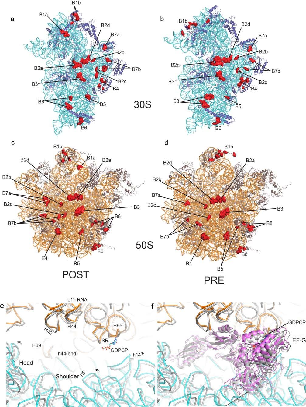
4 Supplementary Figure 2
5 Supplementary Figure 2 Intersubunit bridges and EF-G positioning in the PRE (ratcheted) and POST (classical) ribosome structures. The intersubunit contacts are highlighted as surface in red for both 30S and 50S subunits. (a) and (b) represent 30S subunits in POST and PRE ribosome structures, respectively. (c) and (d) represent 50S subunits, accordingly. Bridge numbering is according to the previous report by Yusupov and coworkers 2. (e) Comparison of the EF-G binding pocket in the PRE and POST (gray) complexes (aligned on 23S rrna). The RNA helices of 16S and 23S rrnas, which are involved in EF-G binding, are labeled by h and H followed by a number, respectively. The conformational change of the two complexes is indicated by arrow. (f) EF-G bound to the two complexes shown in the same view as (e).

6 Supplementary Figure 3 Supplementary Figure 3 Sequence alignment of E. coli and T. thermophilus EF-G. The switch I and II, and the G motifs (G1-G5) are indicated.
 and domain IV of EF-G (e). (a) Residues involved in the coordination of Mg 2+ ion are also presented.")
7 Supplementary Figure 4 Supplementary Figure 4 Electron density map showing Mg 2+ ion (a), the active site (b) and three universally conserved bases (c), as well as conformational change of SRL (d) and domain IV of EF-G (e). (a) Residues involved in the coordination of Mg 2+ ion are also presented. (b) Coupled to the opening of the hydrophobic gate
8 (indicated by arrows), GTP becomes accessible to the catalytic His87 repositioned into its activated conformation. (c) Three universally conserved bases A1913 (H69 of 23S rrna) and A1492 and A1493 (h44 of 16S rrna) are indicated. (d) Conformational change of the SRL. The two structures were superimposed by aligning the 23S rrnas. The SRL in the POST complex is colored gray. (e) The change of the positioning of domain IV of EF-G (domain IV in the POST complex is colored gray and Gly502 of EF-G in the two complexes are presented as spheres).
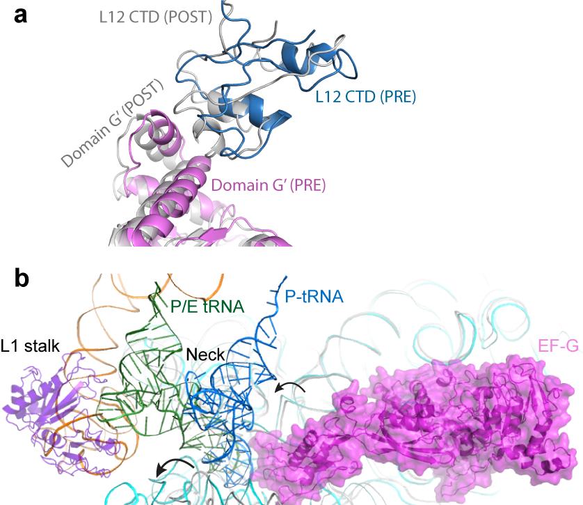
9 Supplementary Figure 5
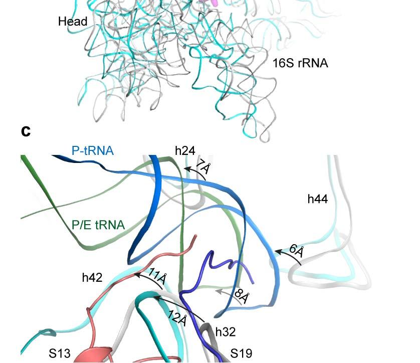
10 Supplementary Figure 5 Comparison of G domain of EF-G and the positioning of P/E trna in PRE and POST complexes. (a) Comparison of the G domain of EF-G with respect to C-terminal domain of L12 in PRE and POST complexes. The G domain of EF-G and the C-terminal domain of L12 in the PRE complex are colored violet and blue, respectively. The corresponding domains in the POST complex are colored gray. In both PRE and POST complexes, direct contact of EF-G (G domain) with the CTD of L12 were observed. (b) Superposing of the P/E trna with the classical P-site trna by aligning the 50S subunit. 16S rrna and P-site trna in the POST complex are colored gray and slate, respectively. The 30S head swiveling and body rotation are indicated by arrows. (c) A close-up view of the ASL binding pocket. Coupled to ribosome ratcheting (30S head swiveling and body rotation), the ASL of the P/E trna advances by 8.1 Å, whereas 16S rrna in the 30S head moves by Å, and 16S rrna in the neck region moves by 6-7 Å. Such unequal bilateral displacement of the classical P-site trna into the P/E positioning causes trna deformation.
11 Supplementary Table 1 Rearrangement of Intersubunit Bridges a Bridge Post-translocation b Pre-translocation c Change 30S 50S 30S 50S S13 B1a M82,D83, R93 H38 C Disrupted B1b S13 V7,G68 S13 L56,R57,R3 L5 D116,L139 L31 G17,E Disrupted S13 A72 S19 D13,L16,P42, E43, E64,V67 h33 P1012 L5 R115 L31 R48,V50,T52, A56,F59,R61 L31 K69 New B2a h44 r1407 H69 A1912 h44 r1408 H69 A1916 h44 r1409 H69 P1914 h44 r1409 H69 C h44 A1492 H69 A1913 New h44 r1494 H69 r1912 h44 r1494 Mg H69 P Mg lost - - h44 r1495 H69 r1919 New h45 rg1517 H69 r1919 B2b h24 r783 H68 P Disrupted - - h24 P784 H68 P1836 New B2c B2d B3 h24 r770...mg... H67 P1832 Mg retained h27 r899 H67 P Disrupted h24 U793...Mg... H69 P1920 h45 P1517 Mg H69 P1920 h44 A1418 H71 GC
12 B4 h44 A1483 h44 r1484 S15 S40,H53,L5 6 S15 R64,G89 H71 CG H71 r1960 h34 G715, h34 P715,P716 S15 S40,H53,L56, V60 h34 P714,P715,P 716 Similar Similar - - S15, K44 r716 New B5 h44 r1429 H62 r1703 h44 r1428 H62 r Disrupted - - h44 r1429 H62 P1704 New B6 h44 G1443 h44 r1443,r144 6 H101 P2864 L19 R118,D122 B7a h23 A702 H68 G Disrupted h23 A702 H68 A Disrupted - - h23 A702 H68 P1847 New B7b h23 P713 L2 Q166,R Disrupted B8 h24 P774 L2 K202 h24 H66 P773...Mg.. P Disrupted - - h23 r712 L2 Q164 New - - h24 P774 L2 K202 N203 New h14 h14 L19 R41 P345,P346 P345,P346 L19 E36,K35 h14 P339 L14 E9,N13 h14 L14 h14 L14 C345...Mg... S116,A118 C345 Mg V115,S h14 U343 Mg L14 S116 New - - h14 P338 L14 E9 New - - h14 P340 L14 T96 New - - h14 r345 L14 R107 New - - h14 P346 L14 R104,R107 New
13 aabbreviations:, no change (indicated interactions are maintained between post- and pre-translocation states); -, interaction absent; h and H indicates rrna 16S rrna and 23S rrna helixes, respectively; r, ribose 2'-OH interaction; P, phosphate non-bridging oxygen interaction;...mg..., interaction mediated by a magnesium bridge 3. bthe post-translocation structure refers to the T.thermophilus 70S ribosome bound with EF-G trapped in a post-translocation state (Gao et al, 2009) 1. cthe pre-translocation structure refers to the T.thermophilus 70S ribosome bound with EF-G in a GDPCP state presented in this paper.
14 Supplementary Table 2 Interactions of EF-G with Ribosome a Post-translocation b Pre-translocation c Change 23SRNA EF-G 23SRNA EF-G H95 G2656 Asp22(G) H95 G2656 Asp22(G) Asp22 Mg H89 C2658 Gln112(G) New H95 A2660 His20(G) H95 A2660 His20(G) - H95 A2660 Gln117(G) Disrupted H95 G2661 His20(G) H95 G2661 His20(G) - H95 G2661 Ile21(G) H95 G2661 Ile21(G) - H95 G2661 His87(G) New H95 A2662 Arg61(G) New H95 A2662 Ile63 (G) New H95 A2662 His87(G) New H95 G2663 Arg61(G) New H95 G2661 Glu456(III) H95 G2661 Glu456(III) - H95 G2661 Leu457(III) H95 G2661 Leu457(III) - H95 A2662 Glu456(III) New H95 A2662 Leu457(III) H95 A2662 Leu457(III) - H69 A1912 Arg499(IV) New H69 A1913 Arg499(IV) H69 A1913 Arg499(IV) - H69 A1913 Met580(IV) Disrupted H43 A1067 Asn625(V) New H43 A1067 Ile631(V) H43 A1067 Ile631(V) - H43 A1067 Leu632(V) H43 A1067 Leu632(V) - H43 A1067 Gly633(V) H43 A1067 Gly633(V) - H44 A1095 Glu614(V) New H44 A1095 Met617(V) H44 A1095 Met617(V) Stronger H44 A1095 Gly618(V) New H44 A1095 Met634(V) H44 A1095 Met634(V) - H44 A1096 Glu614(V) New H89 U2473 Gly622(V) H89 U2473 Gly622(V) - H89 U2473 Ala626(V) H89 U2473 Ala626(V) - H95 A2660 Thr657(V) H95 A2660 Thr657(V) - H95 A2660 Arg660(V) H95 A2660 Arg660(V) - H95 A2660 Ser661(V) H95 A2660 Ser661(V) - H95 A2660 Gln664(V) H95 A2660 Gln664(V) - 16SRNA EF-G 16SRNA EF-G h5 A55 Tyr321(II) h5 A55 Tyr321(II) - h5 A55 Arg354(II) New h5 G357 Arg354(II) New h5 U358 Val322(II) New h5 U358 Gly323(II) h5 U358 Gly323(II) - h5 U359 Arg324(II) h5 U359 Arg324(II) - h5 U359 Lys381(II) h5 U359 Lys381(II) - h15 U367 Tyr340(II) h15 U367 Tyr340(II) - h15 U367 Arg351(II) New h15 U368 Arg351(II) h15 U368 Arg351(II) - h15 U368 Ala353(II) New
15 h15 U368 Arg354(II) h15 U368 Arg354(II) - h15 U368 Glu365(II) New h15 C395 Tyr340(II) h15 C395 Tyr340(II) - h15 C395 Lys349(II) h15 C395 Lys349(II) - h15 G396 Lys349(II) h15 G396 Lys349(II) - h5 A360 Arg430(III) New h15 G361 Arg430(III) Disrupted h30 U955 Val533(IV) Disrupted h30 U955 Asp569(IV) Disrupted h30 U957 Val530(IV) Disrupted h31 A965 Glu574(IV) Disrupted h44 A1492 Glu579(IV) Disrupted h44 A1493 Ser578(IV) h44 A1493 Ser578(IV) - h44 A1493 Glu579(IV) Disrupted h44 A1493 Met580(IV) h44 A1493 Met580(IV) - h44 G1494 Arg499(IV) h44 G1494 Arg499(IV) - h44 G1494 Arg504(IV) New h44 U1495 Arg499(IV) h44 U1495 Arg499(IV) - h44 U1495 Thr501(IV) New h44 C1496 Thr501(IV) Disrupted S12 EF-G S12 EF-G Arg34 Gln426(III) New Arg59 Glu422(III) Disrupted Gln78 Pro444(III) New Glu79 Thr442(III) Disrupted Glu79 Thr449(III) Disrupted Glu79 Gln421(III) Disrupted His80 Thr422(III) His80 Thr442(III) - His80 Gln421(III) New His80 Ser425(III) Disrupted His80 Thr449(III) His80 Thr449(III) - S19 EF-G S19 EF-G No discussion Thr90 Glu574(IV) mrna EF-G mrna EF-G U17 Gly502(IV) Disrupted U17 Gly503(IV) Disrupted U18 Gly503(IV) Disrupted U19 Arg504(IV) Disrupted U19 Ser578(IV) Disrupted U19 Val575(IV) Disrupted U19 Ser577(IV) Disrupted trna(psite) EF-G trna(psite) EF-G A35 His573(IV) Disrupted U36 His573(IV) Disrupted U36 Gln500(IV) Disrupted U36 Thr501(IV) Disrupted U36 Gly502(IV) Disrupted U37 Thr501(IV) Disrupted
16 aabbreviations: -,interactions remaining; h, H indicate rrna helixes (h for 16SRNA and H for 23SRNA); Roman numerals in the parentheses indicate which domain the residue belongs to. bthe post-translocation structure refers to the T.thermophilus 70S ribosome bound with EF-G trapped in a post translocation state (Gao et al, 2009) 1. c The pre-translocation structure refers to the T.thermophilus 70S ribosome bound with EF-G in a GDPCP state presented in this paper. References 1. Gao, Y. G. et al. The structure of the ribosome with elongation factor G trapped in the posttranslocational state. Science 326, (2009). 2. Yusupov, M. M. et al. Crystal structure of the ribosome at 5.5 A resolution. Science 292, (2001). 3. Zhou, J., Lancaster, L., Trakhanov, S. & Noller, H. F. Crystal structure of release factor RF3 trapped in the GTP state on a rotated conformation of the ribosome. RNA 18, (2012).
Introduction to the Ribosome Overview of protein synthesis on the ribosome Prof. Anders Liljas
 Introduction to the Ribosome Molecular Biophysics Lund University 1 A B C D E F G H I J Genome Protein aa1 aa2 aa3 aa4 aa5 aa6 aa7 aa10 aa9 aa8 aa11 aa12 aa13 a a 14 How is a polypeptide synthesized? 2
Supplemental Information
 1 Supplemental Information Head swivel on the ribosome facilitates translocation via intra-subunit trna hybrid sites Andreas H. Ratje, Justus Loerke, Aleksandra Mikolajka, Matthias Brünner,Peter W. Hildebrand,
Coordinated conformational and compositional dynamics drive. ribosome translocation
 Coordinated conformational and compositional dynamics drive ribosome translocation Supplementary Information Jin Chen,2, Alexey Petrov 2, Albert Tsai,2, Seán E. O Leary 2, & Joseph D. Puglisi 2,3 Department
Advanced Topics in RNA and DNA. DNA Microarrays Aptamers
 Quiz 1 Advanced Topics in RNA and DNA DNA Microarrays Aptamers 2 Quantifying mrna levels to asses protein expression 3 The DNA Microarray Experiment 4 Application of DNA Microarrays 5 Some applications
Recognition of the amber UAG stop codon by release factor RF1
 Manuscript EMBO-2010-73984 Recognition of the amber UAG stop codon by release factor RF1 Andrei Korostelev, Jianyu Zhu, Haruichi Asahara and Harry F. Noller Corresponding author: Harry F. Noller, Univ.
Supplementary Figure 3 a. Structural comparison between the two determined structures for the IL 23:MA12 complex. The overall RMSD between the two
 Supplementary Figure 1. Biopanningg and clone enrichment of Alphabody binders against human IL 23. Positive clones in i phage ELISA with optical density (OD) 3 times higher than background are shown for
Supplementary Figure 1. Aligned sequences of yeast IDH1 (top) and IDH2 (bottom) with isocitrate
 and IDH2 (bottom) with isocitrate") SUPPLEMENTARY FIGURE LEGENDS Supplementary Figure 1. Aligned sequences of yeast IDH1 (top) and IDH2 (bottom) with isocitrate dehydrogenase from Escherichia coli [ICD, pdb 1PB1, Mesecar, A. D., and Koshland,
SUPPLEMENTARY FIGURE LEGENDS Supplementary Figure 1. Aligned sequences of yeast IDH1 (top) and IDH2 (bottom) with isocitrate dehydrogenase from Escherichia coli [ICD, pdb 1PB1, Mesecar, A. D., and Koshland,
Ranjit P. Bahadur Assistant Professor Department of Biotechnology Indian Institute of Technology Kharagpur, India. 1 st November, 2013
 Hydration of protein-rna recognition sites Ranjit P. Bahadur Assistant Professor Department of Biotechnology Indian Institute of Technology Kharagpur, India 1 st November, 2013 Central Dogma of life DNA
Nitrogenase MoFe protein from Clostridium pasteurianum at 1.08 Å resolution: comparison with the Azotobacter vinelandii MoFe protein
 Acta Cryst. (2015). D71, 274-282, doi:10.1107/s1399004714025243 Supporting information Volume 71 (2015) Supporting information for article: Nitrogenase MoFe protein from Clostridium pasteurianum at 1.08
SUPPLEMENTARY INFORMATION
 Supplementary Results DNA binding property of the SRA domain was examined by an electrophoresis mobility shift assay (EMSA) using synthesized 12-bp oligonucleotide duplexes containing unmodified, hemi-methylated,
Protein synthesis II Biochemistry 302. Bob Kelm February 25, 2004
 Protein synthesis II Biochemistry 302 Bob Kelm February 25, 2004 Two idealized views of the 70S ribosomal complex during translation 70S cavity Fig. 27.25 50S tunnel View with 30S subunit in front, 50S
Domain movements of elongation factor eef2 and the eukaryotic 80S ribosome facilitate trna translocation
 The EMBO Journal (2004) 23, 1008 1019 & 2004 European Molecular Biology Organization All Rights Reserved 0261-4189/04 www.embojournal.org Domain movements of elongation factor eef2 and the eukaryotic 80S
SUPPLEMENTARY FIGURES. Figure S1
 SUPPLEMENTARY FIGURES Figure S1 The substrate for DH domain (2R,3R,4R,6R,7S,8S,9R)-3,7,9-trihydroxy-5-oxo-2,4,6,8 tetramethylundecanoate) was docked as two separate fragments shown in magenta and blue
A conserved P-loop anchor limits the structural dynamics that mediate. nucleotide dissociation in EF-Tu.
 Supplemental Material for A conserved P-loop anchor limits the structural dynamics that mediate nucleotide dissociation in EF-Tu. Evan Mercier 1,2, Dylan Girodat 1, and Hans-Joachim Wieden 1 * 1 Alberta
Nature Structural & Molecular Biology: doi: /nsmb Supplementary Figure 1
 Supplementary Figure 1 Comparing rotated- and nonrotated-state lifetimes among mrna m 6 A modifications in different codon contexts. a. mrna sequences used for each experiments, as same as shown in Figure
Joachim Frank Wadsworth Center Empire State Plaza P.O. Box 509 Albany, New York Tel: (518)
") This material is provided for educational use only. The information in these slides including all data, images and related materials are the property of : Joachim Frank Wadsworth Center Empire State Plaza
This material is provided for educational use only. The information in these slides including all data, images and related materials are the property of : Joachim Frank Wadsworth Center Empire State Plaza
Supporting Information
 Supporting Information Allosteric communication disrupted by small molecule binding to the Imidazole glycerol phosphate synthase protein-protein interface. Ivan Rivalta*,#, George P. Lisi #, Ning-Shiuan
Nature Structural & Molecular Biology: doi: /nsmb Supplementary Figure 1
 Supplementary Figure 1 Cryo-EM structure and model of the C. thermophilum 90S preribosome. a, Gold standard FSC curve showing the average resolution of the 90S preribosome masked and unmasked (left). FSC
Structure and evolution of the spliceosomal peptidyl-prolyl cistrans isomerase Cwc27
 Acta Cryst. (2014). D70, doi:10.1107/s1399004714021695 Supporting information Volume 70 (2014) Supporting information for article: Structure and evolution of the spliceosomal peptidyl-prolyl cistrans isomerase
Nature Structural and Molecular Biology: doi: /nsmb Supplementary Figure 1. Experimental approach for enhancement of unbiased Fo Fc maps.
 Supplementary Figure 1 Experimental approach for enhancement of unbiased Fo Fc maps. a, c, Unbiased Fo-Fc maps of the Tth 70S post-catalysis complex at 2.55 Å resolution with (a) or without (c) bulk solvent
Gene regulation II Biochemistry 302. February 27, 2006
 Gene regulation II Biochemistry 302 February 27, 2006 Molecular basis of inhibition of RNAP by Lac repressor 35 promoter site 10 promoter site CRP/DNA complex 60 Lewis, M. et al. (1996) Science 271:1247
Protein synthesis I Biochemistry 302. February 17, 2006
 Protein synthesis I Biochemistry 302 February 17, 2006 Key features and components involved in protein biosynthesis High energy cost (essential metabolic activity of cell Consumes 90% of the chemical energy
Supporting information to: Time-resolved observation of protein allosteric communication. Sebastian Buchenberg, Florian Sittel and Gerhard Stock 1
 Supporting information to: Time-resolved observation of protein allosteric communication Sebastian Buchenberg, Florian Sittel and Gerhard Stock Biomolecular Dynamics, Institute of Physics, Albert Ludwigs
Structure, mechanism and ensemble formation of the Alkylhydroperoxide Reductase subunits. AhpC and AhpF from Escherichia coli
 Structure, mechanism and ensemble formation of the Alkylhydroperoxide Reductase subunits AhpC and AhpF from Escherichia coli Phat Vinh Dip 1,#, Neelagandan Kamariah 2,#, Malathy Sony Subramanian Manimekalai
Engineering an Mg 2 Site to Replace a Structurally Conserved Arginine in the Catalytic Center of Histidyl-tRNA Synthetase by Computer Experiments
 PROTEINS: Structure, Function, and Genetics 32:362 380 (1998) Engineering an Mg 2 Site to Replace a Structurally Conserved Arginine in the Catalytic Center of Histidyl-tRNA Synthetase by Computer Experiments
Structural characterization of NiV N 0 P in solution and in crystal.
 Supplementary Figure 1 Structural characterization of NiV N 0 P in solution and in crystal. (a) SAXS analysis of the N 32-383 0 -P 50 complex. The Guinier plot for complex concentrations of 0.55, 1.1,
Final Chem 4511/6501 Spring 2011 May 5, 2011 b Name
 Key 1) [10 points] In RNA, G commonly forms a wobble pair with U. a) Draw a G-U wobble base pair, include riboses and 5 phosphates. b) Label the major groove and the minor groove. c) Label the atoms of
TRANSLATION: How to make proteins?
 TRANSLATION: How to make proteins? EUKARYOTIC mrna CBP80 NUCLEUS SPLICEOSOME 5 UTR INTRON 3 UTR m 7 GpppG AUG UAA 5 ss 3 ss CBP20 PABP2 AAAAAAAAAAAAA 50-200 nts CYTOPLASM eif3 EJC PABP1 5 UTR 3 UTR m 7
SUPPLEMENTARY INFORMATION
 Supplementary Table 1: Data collection, phasing and refinement statistics ChbC/Ta 6 Br 12 Native ChbC Data collection Space group P4 3 2 1 2 P4 3 2 1 2 Cell dimensions a, c (Å) 132.75, 453.57 132.81, 452.95
Supplementary figure 1. Comparison of unbound ogm-csf and ogm-csf as captured in the GIF:GM-CSF complex. Alignment of two copies of unbound ovine
 Supplementary figure 1. Comparison of unbound and as captured in the GIF:GM-CSF complex. Alignment of two copies of unbound ovine GM-CSF (slate) with bound GM-CSF in the GIF:GM-CSF complex (GIF: green,
SUPPLEMENTARY INFORMATION
 doi:10.1038/nature11085 Supplementary Tables: Supplementary Table 1. Summary of crystallographic and structure refinement data Structure BRIL-NOP receptor Data collection Number of crystals 23 Space group
Supplemental Data SUPPLEMENTAL FIGURES
 Supplemental Data CRYSTAL STRUCTURE OF THE MG.ADP-INHIBITED STATE OF THE YEAST F 1 C 10 ATP SYNTHASE Alain Dautant*, Jean Velours and Marie-France Giraud* From Université Bordeaux 2, CNRS; Institut de
SUPPLEMENTARY INFORMATION
 doi:10.1038/nature12045 Supplementary Table 1 Data collection and refinement statistics. Native Pt-SAD X-ray source SSRF BL17U SPring-8 BL41XU Wavelength (Å) 0.97947 1.07171 Space group P2 1 2 1 2 1 P2
SUPPLEMENTARY INFORMATION
 SUPPLEMENTARY INFORMATION doi:10.1038/nature11524 Supplementary discussion Functional analysis of the sugar porter family (SP) signature motifs. As seen in Fig. 5c, single point mutation of the conserved
Insights to the Early Evolution of Earth Life From Studies of the Ribosome. Seven Pines XV-Origins of Life May 20, 2011
 Insights to the Early Evolution of Earth Life From Studies of the Ribosome George E. Fox from Dept. Biology & Biochemistry University of Houston Seven Pines XV-Origins of Life May 20, 2011 Modern Ribosomes
Supporting Information
 Supporting Information Wilson et al. 10.1073/pnas.0804276105 Fig. S1. Sites of oxazolidinone resistance mutations in bacteria and archaea. (a) Secondary structure of the peptidyltransferase ring of the
Bacterial protease uses distinct thermodynamic signatures for substrate recognition
 Bacterial protease uses distinct thermodynamic signatures for substrate recognition Gustavo Arruda Bezerra, Yuko Ohara-Nemoto, Irina Cornaciu, Sofiya Fedosyuk, Guillaume Hoffmann, Adam Round, José A. Márquez,
Lecture 25: Protein Synthesis Key learning goals: Be able to explain the main stuctural features of ribosomes, and know (roughly) how many DNA and
 how many DNA and") Lecture 25: Protein Synthesis Key learning goals: Be able to explain the main stuctural features of ribosomes, and know (roughly) how many DNA and protein subunits they contain. Understand the main functions
Lecture 25: Protein Synthesis Key learning goals: Be able to explain the main stuctural features of ribosomes, and know (roughly) how many DNA and protein subunits they contain. Understand the main functions
Information Content in Genetics:
 Information Content in Genetics: DNA, RNA and protein mrna translation into protein (protein synthesis) Francis Crick, 1958 [Crick, F. H. C. in Symp. Soc. Exp. Biol., The Biological Replication of Macromolecules,
What binds to Hb in addition to O 2?
 Reading: Ch5; 158-169, 162-166, 169-174 Problems: Ch5 (text); 3,7,8,10 Ch5 (study guide-facts); 1,2,3,4,5,8 Ch5 (study guide-apply); 2,3 Remember Today at 5:30 in CAS-522 is the second chance for the MB
Nature Structural & Molecular Biology: doi: /nsmb Supplementary Figure 1
 Supplementary Figure 1 Crystallization. a, Crystallization constructs of the ET B receptor are shown, with all of the modifications to the human wild-type the ET B receptor indicated. Residues interacting
Application examples of single particle 3D reconstruction. Ning Gao Tsinghua University
 Application examples of single particle 3D reconstruction Ning Gao Tsinghua University ninggao@tsinghua.edu.cn Electron Microscopes First electron microscope constructed by Ernst Ruska in 1930 s (1986
Genetics 304 Lecture 6
 Genetics 304 Lecture 6 00/01/27 Assigned Readings Busby, S. and R.H. Ebright (1994). Promoter structure, promoter recognition, and transcription activation in prokaryotes. Cell 79:743-746. Reed, W.L. and
Table 1. Crystallographic data collection, phasing and refinement statistics. Native Hg soaked Mn soaked 1 Mn soaked 2
 Table 1. Crystallographic data collection, phasing and refinement statistics Native Hg soaked Mn soaked 1 Mn soaked 2 Data collection Space group P2 1 2 1 2 1 P2 1 2 1 2 1 P2 1 2 1 2 1 P2 1 2 1 2 1 Cell
Protein synthesis I Biochemistry 302. Bob Kelm February 23, 2004
 Protein synthesis I Biochemistry 302 Bob Kelm February 23, 2004 Key features of protein synthesis Energy glutton Essential metabolic activity of the cell. Consumes 90% of the chemical energy (ATP,GTP).
SUPPLEMENTARY INFORMATION. doi: /nature07461
 Figure S1 Electrophysiology. a ph-activation of. Two-electrode voltage clamp recordings of Xenopus oocytes expressing in comparison to waterinjected oocytes. Currents were recorded at 40 mv. The ph of
The structure of a nucleolytic ribozyme that employs a catalytic metal ion. Yijin Liu, Timothy J. Wilson and David M.J. Lilley
 SUPPLEMENTARY INFORMATION The structure of a nucleolytic ribozyme that employs a catalytic metal ion Yijin Liu, Timothy J. Wilson and David M.J. Lilley Cancer Research UK Nucleic Acid Structure Research
ml. ph 7.5 ph 6.5 ph 5.5 ph 4.5. β 2 AR-Gs complex + GDP β 2 AR-Gs complex + GTPγS
 a UV28 absorption (mau) 9 8 7 5 3 β 2 AR-Gs complex β 2 AR-Gs complex + GDP β 2 AR-Gs complex + GTPγS β 2 AR-Gs complex dissociated complex excess nucleotides b 9 8 7 5 3 β 2 AR-Gs complex β 2 AR-Gs complex
eif5b employs a novel domain release mechanism to catalyze ribosomal subunit joining
 Article eif5b employs a novel domain release mechanism to catalyze ribosomal subunit joining Bernhard Kuhle * & Ralf Ficner Abstract eif5b is a eukaryal translational GTPase that catalyzes ribosomal subunit
SUPPLEMENTARY FIGURES
 SUPPLEMENTARY FIGURES Supplementary Figure 1 Protein sequence alignment of Vibrionaceae with either a 40-residue insertion or a 44-residue insertion. Identical residues are indicated by red background.
Lecture 9 Translation.
 1 Translation Summary of important events in translation. 2 Translation Reactions involved in peptide bond formation. Lecture 9 3 Genetic code Three types of RNA molecules perform different but complementary
BCH 4054 Spring 2001 Chapter 33 Lecture Notes
 BCH 4054 Spring 2001 Chapter 33 Lecture Notes Slide 1 The chapter covers degradation of proteins as well. We will not have time to get into that subject. Chapter 33 Protein Synthesis Slide 2 Prokaryotic
Gene regulation I Biochemistry 302. Bob Kelm February 25, 2005
 Gene regulation I Biochemistry 302 Bob Kelm February 25, 2005 Principles of gene regulation (cellular versus molecular level) Extracellular signals Chemical (e.g. hormones, growth factors) Environmental
The structure of a nucleolytic ribozyme that employs a catalytic metal ion Liu, Yijin; Wilson, Timothy; Lilley, David
 University of Dundee The structure of a nucleolytic ribozyme that employs a catalytic metal ion Liu, Yijin; Wilson, Timothy; Lilley, David Published in: Nature Chemical Biology DOI: 10.1038/nchembio.2333
Conformational Variability Experience with Ribosomes
 onformational Variability Experience with Ribosomes Exploration of reconstruction strategy High-resolution project Use small dataset (50,000) to optimize processing, with the idea to switch to larger dataset
Chapter
 Chapter 17 17.4-17.6 Molecular Components of Translation A cell interprets a genetic message and builds a polypeptide The message is a series of codons on mrna The interpreter is called transfer (trna)
The ribosome is the large molecular machine
 RESEARCH ARTICLES C. Schulze-Briese and A. Pauluhn for help with initial diffraction studies performed at the Swiss Light Source, and L. Ulisko for preparation of additional images. This work was supported
Multiperspective smfret reveals rate-determining late intermediates of ribosomal translocation
 Multiperspective sm reveals rate-determining late intermediates of ribosomal translocation Michael R Wasserman 1,3, Jose L Alejo 1,3, Roger B Altman 1 & Scott C Blanchard 1,2 216 Nature America, Inc. All
SUPPLEMENTARY INFORMATION
 SUPPLEMENTARY INFORMATION doi:10.1038/nature11744 Supplementary Table 1. Crystallographic data collection and refinement statistics. Wild-type Se-Met-BcsA-B SmCl 3 -soaked EMTS-soaked Data collection Space
TWO PARTNERS OF THE RIBOSOME, EF-TU AND LEPA EVELINA INES DE LAURENTIIS. B.Sc. University of Lethbridge, A Thesis
 TWO PARTNERS OF THE RIBOSOME, EF-TU AND LEPA EVELINA INES DE LAURENTIIS B.Sc. University of Lethbridge, 2007 A Thesis Submitted to the School of Graduate Studies of the University of Lethbridge in Partial
Supplementary Information. The protease GtgE from Salmonella exclusively targets. inactive Rab GTPases
 Supplementary Information The protease GtgE from Salmonella exclusively targets inactive Rab GTPases Table of Contents Supplementary Figures... 2 Supplementary Figure 1... 2 Supplementary Figure 2... 3
Biophysics 490M Project
 Biophysics 490M Project Dan Han Department of Biochemistry Structure Exploration of aa 3 -type Cytochrome c Oxidase from Rhodobacter sphaeroides I. Introduction: All organisms need energy to live. They
Section 7. Junaid Malek, M.D.
 Section 7 Junaid Malek, M.D. RNA Processing and Nomenclature For the purposes of this class, please do not refer to anything as mrna that has not been completely processed (spliced, capped, tailed) RNAs
Chapter 12. Genes: Expression and Regulation
 Chapter 12 Genes: Expression and Regulation 1 DNA Transcription or RNA Synthesis produces three types of RNA trna carries amino acids during protein synthesis rrna component of ribosomes mrna directs protein
Secondary Structure. Bioch/BIMS 503 Lecture 2. Structure and Function of Proteins. Further Reading. Φ, Ψ angles alone determine protein structure
 Bioch/BIMS 503 Lecture 2 Structure and Function of Proteins August 28, 2008 Robert Nakamoto rkn3c@virginia.edu 2-0279 Secondary Structure Φ Ψ angles determine protein structure Φ Ψ angles are restricted
Supporting Information
 Electronic Supplementary Material (ESI) for Physical Chemistry Chemical Physics. This journal is the Owner Societies 2016 Supporting Information Lipid molecules can induce an opening of membrane-facing
Translation. A ribosome, mrna, and trna.
 Translation The basic processes of translation are conserved among prokaryotes and eukaryotes. Prokaryotic Translation A ribosome, mrna, and trna. In the initiation of translation in prokaryotes, the Shine-Dalgarno
Supplementary Figure 1. Biochemical and sequence alignment analyses the
 Supplementary Figure 1. Biochemical and sequence alignment analyses the interaction of OPTN and TBK1. (a) Analytical gel filtration chromatography analysis of the interaction between TBK1 CTD and OPTN(1-119).
Physiochemical Properties of Residues
 Physiochemical Properties of Residues Various Sources C N Cα R Slide 1 Conformational Propensities Conformational Propensity is the frequency in which a residue adopts a given conformation (in a polypeptide)
Supplementary Information Energy barriers and driving forces of trna translocation through the ribosome
 Supplementary Information Energy barriers and driving forces of trna translocation through the ribosome Lars V. Bock, Christian Blau, Gunnar F. Schröder, Iakov I. Davydov, Niels Fischer, Holger Stark,
The ribosome is the macromolecular enzyme. The Crystal Structure of the Ribosome Bound to EF-Tu and Aminoacyl-tRNA RESEARCH ARTICLES
 RESEARCH ARTICLES 26. M. Bollig, Risk Management in a Hazardous Environment (Springer, New York, 2006). 27. P. Wiessner, Curr. Anthropol. 43, 233 (2002). 28. C. Boehm, Hierarchy in the Forest (Harvard
ومن أحياها Translation 2. Translation 2. DONE BY :Nisreen Obeidat
 Translation 2 DONE BY :Nisreen Obeidat Page 0 Prokaryotes - Shine-Dalgarno Sequence (2:18) What we're seeing here are different portions of sequences of mrna of different promoters from different bacterial
Time-dependence of key H-bond/electrostatic interaction distances in the sirna5-hago2 complexes... Page S14
 Supporting Information Probing the Binding Interactions between Chemically Modified sirnas and Human Argonaute 2 Using Microsecond Molecular Dynamics Simulations S. Harikrishna* and P. I. Pradeepkumar*
Structural and mechanistic insight into the substrate. binding from the conformational dynamics in apo. and substrate-bound DapE enzyme
 Electronic Supplementary Material (ESI) for Physical Chemistry Chemical Physics. This journal is the Owner Societies 215 Structural and mechanistic insight into the substrate binding from the conformational
Any protein that can be labelled by both procedures must be a transmembrane protein.
 1. What kind of experimental evidence would indicate that a protein crosses from one side of the membrane to the other? Regions of polypeptide part exposed on the outside of the membrane can be probed
SUPPLEMENTARY INFORMATION
 Supplementary Table 1: Amplitudes of three current levels. Level 0 (pa) Level 1 (pa) Level 2 (pa) TrkA- TrkH WT 200 K 0.01 ± 0.01 9.5 ± 0.01 18.7 ± 0.03 200 Na * 0.001 ± 0.01 3.9 ± 0.01 12.5 ± 0.03 200
SUPPLEMENTARY INFORMATION
 Fig. 1 Influences of crystal lattice contacts on Pol η structures. a. The dominant lattice contact between two hpol η molecules (silver and gold) in the type 1 crystals. b. A close-up view of the hydrophobic
Supersecondary Structures (structural motifs)
") Supersecondary Structures (structural motifs) Various Sources Slide 1 Supersecondary Structures (Motifs) Supersecondary Structures (Motifs): : Combinations of secondary structures in specific geometric
Supersecondary Structures (structural motifs) Various Sources Slide 1 Supersecondary Structures (Motifs) Supersecondary Structures (Motifs): : Combinations of secondary structures in specific geometric
SUPPLEMENTARY INFORMATION
 Supplementary materials Figure S1 Fusion protein of Sulfolobus solfataricus SRP54 and a signal peptide. a, Expression vector for the fusion protein. The signal peptide of yeast dipeptidyl aminopeptidase
SUPPLEMENTARY INFORMATION
 doi:10.1038/nature11054 Supplementary Fig. 1 Sequence alignment of Na v Rh with NaChBac, Na v Ab, and eukaryotic Na v and Ca v homologs. Secondary structural elements of Na v Rh are indicated above the
SUPPLEMENTARY INFORMATION
 SUPPLEMENTARY INFORMATION Structure of human carbamoyl phosphate synthetase: deciphering the on/off switch of human ureagenesis Sergio de Cima, Luis M. Polo, Carmen Díez-Fernández, Ana I. Martínez, Javier
NB-DNJ/GCase-pH 7.4 NB-DNJ+/GCase-pH 7.4 NB-DNJ+/GCase-pH 4.5
 SUPPLEMENTARY TABLES Suppl. Table 1. Protonation states at ph 7.4 and 4.5. Protonation states of titratable residues in GCase at ph 7.4 and 4.5. Histidine: HID, H at δ-nitrogen; HIE, H at ε-nitrogen; HIP,
protein synthesis and the ribosome
 protein synthesis and the ribosome Central dogma of biology DNA codes for DNA DNA codes for RNA RNA codes for proteins not surprisingly, many points for regulation of the process RNA codes for proteins
Crystal structure of the ribosome recycling factor bound to the ribosome
 Crystal structure of the ribosome recycling factor bound to the ribosome Albert Weixlbaumer 1, Sabine Petry 1,3, Christine M Dunham 1,3, Maria Selmer 1 3, Ann C Kelley 1 & V Ramakrishnan 1 In bacteria,
Viewing and Analyzing Proteins, Ligands and their Complexes 2
 2 Viewing and Analyzing Proteins, Ligands and their Complexes 2 Overview Viewing the accessible surface Analyzing the properties of proteins containing thousands of atoms is best accomplished by representing
Signal Transduction Phosphorylation Protein kinases. Misfolding diseases. Protein Engineering Lysozyme variants
 Signal Transduction Phosphorylation Protein kinases Misfolding diseases Protein Engineering Lysozyme variants Cells and Signals Regulation The cell must be able to respond to stimuli Cellular activities
Introduction to Comparative Protein Modeling. Chapter 4 Part I
 Introduction to Comparative Protein Modeling Chapter 4 Part I 1 Information on Proteins Each modeling study depends on the quality of the known experimental data. Basis of the model Search in the literature
Statics of the Ribosomal Exit Tunnel: Implications for Cotranslational Peptide Folding, Elongation Regulation, and Antibiotics Binding
 doi:10.1016/j.jmb.2009.01.037 J. Mol. Biol. (2009) 387, 502 517 Available online at www.sciencedirect.com Statics of the Ribosomal Exit Tunnel: Implications for Cotranslational Peptide Folding, Elongation
SUPPLEMENTARY INFORMATION
 Figure S1. Secondary structure of CAP (in the camp 2 -bound state) 10. α-helices are shown as cylinders and β- strands as arrows. Labeling of secondary structure is indicated. CDB, DBD and the hinge are
+ regulation. ribosomes
 central dogma + regulation rpl DNA tsx rrna trna mrna ribosomes tsl ribosomal proteins structural proteins transporters enzymes srna regulators RNAp DNAp tsx initiation control by transcription factors
Supporting Information
 Supporting Information Naganuma et al. 10.1073/pnas.0901572106 SI Text The Recognition of Ala-SA. Ala-SA is a nonhydrolyzable analog of alanyl-adenylate and is a potent inhibitor of AlaRS (1). The recognition
Structural Basis for Interaction of the Ribosome with the Switch Regions of GTP-Bound Elongation Factors
 Article Structural Basis for Interaction of the Ribosome with the Switch Regions of GTP-Bound Elongation Factors Sean R. Connell, 1,7 Chie Takemoto, 2,7 Daniel N. Wilson, 3,8 Hongfei Wang, 2 Kazutaka Murayama,
Model Mélange. Physical Models of Peptides and Proteins
 Model Mélange Physical Models of Peptides and Proteins In the Model Mélange activity, you will visit four different stations each featuring a variety of different physical models of peptides or proteins.
TRANSLATION: How to make proteins?
 TRANSLATION: How to make proteins? EUKARYOTIC mrna CBP80 NUCLEUS SPLICEOSOME 5 UTR INTRON 3 UTR m 7 GpppG AUG UAA 5 ss 3 ss CBP20 PABP2 AAAAAAAAAAAAA 50-200 nts CYTOPLASM eif3 EJC PABP1 5 UTR 3 UTR m 7
Three-dimensional structure of a viral genome-delivery portal vertex
 Three-dimensional structure of a viral genome-delivery portal vertex Adam S. Olia 1, Peter E. Prevelige Jr. 2, John E. Johnson 3 and Gino Cingolani 4 1 Department of Biological Sciences, Purdue University,
Recycling of eucaryotic ribosomes
 PRACE PRZEGL DOWE Recycling of eucaryotic ribosomes Agata Tyczewska 1, Kamilla B¹kowska- ywicka 2 1 Institute of Bioorganic Chemistry, Polish Academy of Sciences, Poznañ 2 Innsbruck Biocenter, Division
X-ray crystallography study on ribosome recycling: the mechanism of binding and action of RRF on the 50S ribosomal subunit
 The EMBO Journal (2005) 24, 251 260 & 2005 European Molecular Biology Organization All Rights Reserved 0261-4189/05 www.embojournal.org X-ray crystallography study on ribosome recycling: the mechanism
What makes a good graphene-binding peptide? Adsorption of amino acids and peptides at aqueous graphene interfaces: Electronic Supplementary
 Electronic Supplementary Material (ESI) for Journal of Materials Chemistry B. This journal is The Royal Society of Chemistry 21 What makes a good graphene-binding peptide? Adsorption of amino acids and
Degeneracy. Two types of degeneracy:
 Degeneracy The occurrence of more than one codon for an amino acid (AA). Most differ in only the 3 rd (3 ) base, with the 1 st and 2 nd being most important for distinguishing the AA. Two types of degeneracy:
B O C 4 H 2 O O. NOTE: The reaction proceeds with a carbonium ion stabilized on the C 1 of sugar A.
 hbcse 33 rd International Page 101 hemistry lympiad Preparatory 05/02/01 Problems d. In the hydrolysis of the glycosidic bond, the glycosidic bridge oxygen goes with 4 of the sugar B. n cleavage, 18 from